(2
E
)-1-(2-Methyl-4-phenylquinolin-3-yl)-
3-(3-methylthiophen-2-yl)prop-2-en-1-one
R. Prasath,aP. Bhavana,a‡ Seik Weng Ngb,cand Edward R. T. Tiekinkb*
aDepartment of Chemistry, BITS, Pilani–K. K. Birla Goa Campus, Goa 403 726, India,bDepartment of Chemistry, University of Malaya, 50603 Kuala Lumpur, Malaysia, andcChemistry Department, Faculty of Science, King Abdulaziz University, PO Box 80203 Jeddah, Saudi Arabia
Correspondence e-mail: [email protected]
Received 18 February 2013; accepted 18 February 2013
Key indicators: single-crystal X-ray study;T= 295 K; mean(C–C) = 0.003 A˚; Rfactor = 0.048;wRfactor = 0.135; data-to-parameter ratio = 18.2.
In the title compound, C24H19NOS, the quinoline residue
(r.m.s. deviation = 0.018 A˚ ) is essentially orthogonal to both the phenyl [dihedral angle = 88.95 (8)] and 2-thienyl [81.98 (9)] rings. The carbonyl O atom lies to one side of the quinoline plane, the carbonyl C atom is almost coplanar and the remaining atoms of the chalcone residue lies to the other side, so that overall the molecule has an L-shape. The conformation about the ethylene bond [1.340 (2) A˚ ] is E. In the crystal, a supramolecular chain with the shape of a square rod aligned along the b-axis direction is sustained by C— H interactions, the-systems being the heterocyclic rings.
Related literature
For background details and the biological application of quinoline and quinoline chalcones, see: Joshi et al. (2011); Prasath & Bhavana (2012); Kalanithiet al.(2012); Prasathet al. (2013). For the structure of the dimethyl-substituted quinolinyl compound without a methyl substituent on the 2-thienyl ring, see: Prasathet al.(2011).
Experimental
Crystal data
C24H19NOS
Mr= 369.46
Triclinic,P1 a= 10.0815 (7) A˚ b= 10.2956 (7) A˚ c= 10.5403 (7) A˚
= 71.013 (6)
= 78.697 (5)
= 70.412 (6)
V= 969.99 (11) A˚3
Z= 2
MoKradiation
= 0.18 mm1
T= 295 K
0.400.200.10 mm
Data collection
Agilent SuperNova Dual diffractometer with an Atlas detector
Absorption correction: multi-scan (CrysAlis PRO; Agilent, 2011) Tmin= 0.780,Tmax= 1.000
8430 measured reflections 4473 independent reflections 3484 reflections withI> 2(I) Rint= 0.025
Refinement
R[F2> 2(F2)] = 0.048
wR(F2) = 0.135
S= 1.03 4473 reflections
246 parameters
H-atom parameters constrained
max= 0.28 e A˚
3 min=0.26 e A˚
[image:1.610.315.565.384.423.2]3
Table 1
Hydrogen-bond geometry (A˚ ,).
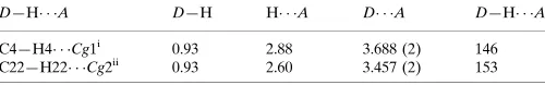
Cg1 andCg2 are the centroids of the S1,C20–C23 and N1,C1,C6–C9 rings, respectively.
D—H A D—H H A D A D—H A C4—H4 Cg1i
0.93 2.88 3.688 (2) 146 C22—H22 Cg2ii
0.93 2.60 3.457 (2) 153
Symmetry codes: (i)xþ1;yþ1;zþ1; (ii)x;yþ1;z.
Data collection: CrysAlis PRO(Agilent, 2011); cell refinement: CrysAlis PRO; data reduction: CrysAlis PRO; program(s) used to solve structure: SHELXS97(Sheldrick, 2008); program(s) used to refine structure:SHELXL97(Sheldrick, 2008); molecular graphics: ORTEP-3 for Windows (Farrugia, 2012) and DIAMOND (Bran-denburg, 2006); software used to prepare material for publication: publCIF(Westrip, 2010).
PB and RP gratefully acknowledge the Council of Scientific and Industrial Research (CSIR), India, for research grant 02 (0076)/12/EMR-II and Senior Research Fellowship (09/919/ (0014)/2012 EMR-I), respectively. We also thank the Ministry of Higher Education (Malaysia) for funding structural studies through the High-Impact Research scheme (UM.C/HIR-MOHE/SC/12).
Supplementary data and figures for this paper are available from the IUCr electronic archives (Reference: HB7043).
References
Agilent (2011).CrysAlis PRO. Agilent Technologies, Yarnton, England. Brandenburg, K. (2006).DIAMOND. Crystal Impact GbR, Bonn, Germany. Farrugia, L. J. (2012).J. Appl. Cryst.45, 849–854.
Joshi, R. S., Mandhane, P. G., Khan, W. & Gill, C. H. (2011).J. Heterocycl. Chem.48, 872–876.
Kalanithi, M., Rajarajan, M., Tharmaraj, P. & Sheela, C. D. (2012).
Acta Crystallographica Section E Structure Reports Online
Prasath, R. & Bhavana, P. (2012).Heteroat. Chem.23, 525–530.
Prasath, R., Bhavana, P., Ng, S. W. & Tiekink, E. R. T. (2011).Acta Cryst.E67, o2283–o2284.
Prasath, R., Bhavana, P., Ng, S. W. & Tiekink, E. R. T. (2013).J. Organomet. Chem.726, 62–70.
Sheldrick, G. M. (2008).Acta Cryst.A64, 112–122. Westrip, S. P. (2010).J. Appl. Cryst.43, 920–925.
organic compounds
Acta Cryst.(2013). E69, o426–o427 Prasathet al. C
supporting information
Acta Cryst. (2013). E69, o426–o427 [doi:10.1107/S1600536813004753]
(2
E
)-1-(2-Methyl-4-phenylquinolin-3-yl)-3-(3-methylthiophen-2-yl)prop-2-en-1-one
R. Prasath, P. Bhavana, Seik Weng Ng and Edward R. T. Tiekink
S1. Comment
In addition to their being valuable intermediates in organic synthesis (Prasath & Bhavana, 2012; Joshi et al., 2011), quinoline and heterocyclic analogues, such as chalcones, exhibit a variety of biological activities, e.g. anti-plasmodial, anti-microbial and anticancer activities (Prasath et al., 2013; Kalanithi et al., 2012). The title quinolinyl/chalcone bearing a thienyl substituent, (I), was investigated in the context of the above.
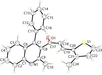
In (I), Fig. 1, the phenyl ring is perpendicular to the quinolinyl residue (r.m.s. deviation = 0.018 Å), forming a dihedral angle of 88.95 (8)°. The 3-thienyl ring also occupies a position approximately orthogonal to the quinolinyl residue with a dihedral angle of 81.98 (9)°. With respect to the plane through the quinolinyl residue, the carbonyl-O1 atom lies to one side, the carbonyl-C17 atom is almost co-planar and the remaining chalcone residue lies to the other side so that the molecule has an L-shape. The conformation about the ethylene bond [1.340 (2) Å] is E. A similar conformation and displacement of atoms was found in the most closely related structure, namely that of the recently reported (2E )-1-(2,4-dimethylquinolin-3-yl)-3-(thiophen-2-yl)prop-2-en-1-one (Prasath et al., 2011).
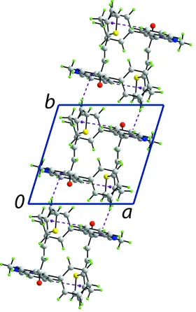
The most notable feature of the crystal packing is the formation of supramolecular chains along the b axis and sustained by C—H···π interactions between quinolinyl-C6—H4 and the 3-thienyl ring, and between 3-thienyl-H22 and the pyridyl
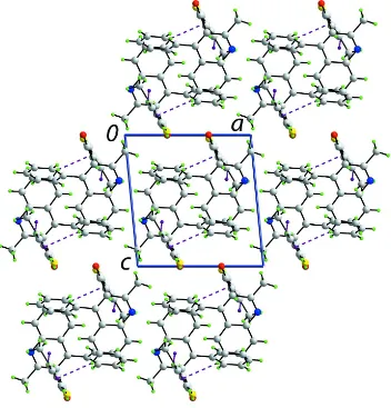
ring, Fig. 2 and Table 1. Owing to the L-shape of the molecule, the chain has the shape of a square rod. Chains stack with no specific interactions between them, Fig. 3.
S2. Experimental
A mixture of 3-acetyl-2-methyl-4-phenylquinoline (1.3 g, 0.005 M), 3-methylthiophene-2-carbaldehyde (630 mg, 0.005
M) and KOH (0.5 g) in distilled ethanol (50 ml) was stirred for 12 h at room temperature. The resulting mixture was neutralized with dilute acetic acid. The deposited solid was filtered, dried and purified by column chromatography using a 1:1 mixture of ethyl acetate and hexane. Re-crystallization was by slow evaporation of an acetone solution of (I), which yielded colourless prisms in 76% yield; M.pt: 453–455 K.
S3. Refinement
The C-bound H atoms were geometrically placed (C—H = 0.95–0.96 Å) and refined as riding with Uiso(H) = 1.2–
supporting information
[image:4.610.130.481.70.329.2]sup-2
Acta Cryst. (2013). E69, o426–o427Figure 1
Figure 2
supporting information
[image:6.610.130.482.72.439.2]sup-4
Acta Cryst. (2013). E69, o426–o427Figure 3
A view in projection down the b axis of the unit-cell content for (I). The C—H···π interactions are shown as purple dashed lines.
(2E)-1-(2-Methyl-4-phenylquinolin-3-yl)-3-(3-methylthiophen-2-yl)prop-2-en-1-one
Crystal data
C24H19NOS
Mr = 369.46
Triclinic, P1 Hall symbol: -P 1
a = 10.0815 (7) Å
b = 10.2956 (7) Å
c = 10.5403 (7) Å
α = 71.013 (6)°
β = 78.697 (5)°
γ = 70.412 (6)°
V = 969.99 (11) Å3
Z = 2
F(000) = 388
Dx = 1.265 Mg m−3
Mo Kα radiation, λ = 0.71073 Å Cell parameters from 2781 reflections
θ = 3.1–27.5°
µ = 0.18 mm−1
Data collection
Agilent SuperNova Dual
diffractometer with an Atlas detector Radiation source: SuperNova (Mo) X-ray
Source
Mirror monochromator
Detector resolution: 10.4041 pixels mm-1
ω scan
Absorption correction: multi-scan
(CrysAlis PRO; Agilent, 2011)
Tmin = 0.780, Tmax = 1.000
8430 measured reflections 4473 independent reflections 3484 reflections with I > 2σ(I)
Rint = 0.025
θmax = 27.6°, θmin = 3.1°
h = −12→13
k = −13→13
l = −12→13
Refinement
Refinement on F2
Least-squares matrix: full
R[F2 > 2σ(F2)] = 0.048
wR(F2) = 0.135
S = 1.03 4473 reflections 246 parameters 0 restraints
Primary atom site location: structure-invariant direct methods
Secondary atom site location: difference Fourier map
Hydrogen site location: inferred from neighbouring sites
H-atom parameters constrained
w = 1/[σ2(F
o2) + (0.0573P)2 + 0.2365P]
where P = (Fo2 + 2Fc2)/3
(Δ/σ)max < 0.001
Δρmax = 0.28 e Å−3
Δρmin = −0.26 e Å−3
Special details
Geometry. All e.s.d.'s (except the e.s.d. in the dihedral angle between two l.s. planes) are estimated using the full covariance matrix. The cell e.s.d.'s are taken into account individually in the estimation of e.s.d.'s in distances, angles and torsion angles; correlations between e.s.d.'s in cell parameters are only used when they are defined by crystal symmetry. An approximate (isotropic) treatment of cell e.s.d.'s is used for estimating e.s.d.'s involving l.s. planes.
Refinement. Refinement of F2 against ALL reflections. The weighted R-factor wR and goodness of fit S are based on F2,
conventional R-factors R are based on F, with F set to zero for negative F2. The threshold expression of F2 > σ(F2) is used
only for calculating R-factors(gt) etc. and is not relevant to the choice of reflections for refinement. R-factors based on F2
are statistically about twice as large as those based on F, and R- factors based on ALL data will be even larger.
Fractional atomic coordinates and isotropic or equivalent isotropic displacement parameters (Å2)
x y z Uiso*/Ueq
S1 0.33532 (6) 0.72592 (6) 0.97283 (5) 0.05153 (17) O1 0.33453 (18) 0.20079 (15) 1.00072 (14) 0.0629 (4) N1 0.08222 (15) 0.34043 (16) 0.65204 (16) 0.0454 (4) C1 0.18240 (18) 0.31032 (18) 0.54916 (17) 0.0399 (4) C2 0.1367 (2) 0.3073 (2) 0.4323 (2) 0.0527 (5)
H2 0.0406 0.3285 0.4262 0.063*
C3 0.2316 (3) 0.2738 (2) 0.3286 (2) 0.0602 (5)
H3 0.2002 0.2718 0.2522 0.072*
C4 0.3757 (2) 0.2425 (3) 0.3361 (2) 0.0608 (6)
H4 0.4397 0.2196 0.2645 0.073*
C5 0.4241 (2) 0.2449 (2) 0.44696 (19) 0.0511 (5)
H5 0.5208 0.2241 0.4502 0.061*
supporting information
sup-6
Acta Cryst. (2013). E69, o426–o427C10 0.0110 (2) 0.3675 (3) 0.8747 (2) 0.0672 (6)
H10A −0.0802 0.3905 0.8445 0.101*
H10B 0.0164 0.4466 0.9008 0.101*
H10C 0.0244 0.2833 0.9505 0.101*
C11 0.52458 (17) 0.24958 (18) 0.69071 (16) 0.0377 (4) C12 0.6053 (2) 0.1100 (2) 0.7428 (2) 0.0514 (5)
H12 0.5634 0.0361 0.7717 0.062*
C13 0.7480 (2) 0.0793 (2) 0.7525 (2) 0.0561 (5)
H13 0.8016 −0.0151 0.7874 0.067*
C14 0.8112 (2) 0.1878 (2) 0.71062 (19) 0.0520 (5)
H14 0.9074 0.1666 0.7162 0.062*
C15 0.7321 (2) 0.3266 (2) 0.6608 (2) 0.0526 (5)
H15 0.7745 0.4002 0.6334 0.063*
C16 0.5892 (2) 0.3582 (2) 0.65095 (19) 0.0467 (4)
H16 0.5360 0.4531 0.6174 0.056*
C17 0.30768 (19) 0.31202 (19) 0.90990 (18) 0.0415 (4) C18 0.30830 (19) 0.44686 (19) 0.92575 (18) 0.0416 (4)
H18 0.3353 0.4453 1.0059 0.050*
C19 0.27227 (18) 0.57334 (18) 0.83162 (17) 0.0391 (4)
H19 0.2463 0.5721 0.7522 0.047*
C20 0.26953 (18) 0.71087 (18) 0.84036 (17) 0.0385 (4) C21 0.2191 (2) 0.84246 (19) 0.75107 (18) 0.0454 (4) C22 0.2355 (2) 0.9534 (2) 0.7922 (2) 0.0590 (5)
H22 0.2071 1.0497 0.7435 0.071*
C23 0.2960 (3) 0.9070 (2) 0.9081 (2) 0.0606 (6)
H23 0.3141 0.9670 0.9480 0.073*
C24 0.1539 (3) 0.8692 (3) 0.6258 (2) 0.0666 (6)
H24A 0.2032 0.7936 0.5843 0.100*
H24B 0.1604 0.9595 0.5644 0.100*
H24C 0.0563 0.8715 0.6484 0.100*
Atomic displacement parameters (Å2)
U11 U22 U33 U12 U13 U23
C12 0.0469 (10) 0.0448 (10) 0.0613 (12) −0.0176 (9) −0.0012 (9) −0.0114 (9) C13 0.0451 (11) 0.0522 (12) 0.0639 (13) −0.0066 (9) −0.0087 (9) −0.0128 (10) C14 0.0379 (9) 0.0735 (14) 0.0508 (11) −0.0182 (10) −0.0040 (8) −0.0239 (10) C15 0.0509 (11) 0.0640 (13) 0.0551 (11) −0.0318 (10) −0.0013 (9) −0.0194 (10) C16 0.0476 (10) 0.0449 (10) 0.0523 (11) −0.0189 (8) −0.0074 (8) −0.0129 (8) C17 0.0421 (9) 0.0416 (9) 0.0419 (9) −0.0136 (8) 0.0001 (7) −0.0142 (8) C18 0.0449 (9) 0.0446 (10) 0.0395 (9) −0.0130 (8) −0.0047 (7) −0.0171 (8) C19 0.0414 (9) 0.0427 (9) 0.0380 (9) −0.0134 (8) −0.0026 (7) −0.0172 (7) C20 0.0397 (9) 0.0426 (9) 0.0374 (8) −0.0139 (7) 0.0003 (7) −0.0170 (7) C21 0.0489 (10) 0.0430 (10) 0.0456 (10) −0.0133 (8) −0.0019 (8) −0.0157 (8) C22 0.0767 (14) 0.0408 (10) 0.0626 (13) −0.0196 (10) −0.0046 (11) −0.0173 (9) C23 0.0802 (15) 0.0572 (12) 0.0616 (13) −0.0354 (12) 0.0030 (11) −0.0293 (11) C24 0.0771 (15) 0.0589 (13) 0.0609 (13) −0.0090 (12) −0.0250 (12) −0.0138 (11)
Geometric parameters (Å, º)
S1—C23 1.698 (2) C11—C16 1.389 (2)
S1—C20 1.7281 (17) C12—C13 1.382 (3)
O1—C17 1.217 (2) C12—H12 0.9300
N1—C9 1.310 (2) C13—C14 1.376 (3)
N1—C1 1.368 (2) C13—H13 0.9300
C1—C6 1.413 (2) C14—C15 1.366 (3)
C1—C2 1.410 (3) C14—H14 0.9300
C2—C3 1.358 (3) C15—C16 1.384 (3)
C2—H2 0.9300 C15—H15 0.9300
C3—C4 1.390 (3) C16—H16 0.9300
C3—H3 0.9300 C17—C18 1.453 (2)
C4—C5 1.362 (3) C18—C19 1.340 (2)
C4—H4 0.9300 C18—H18 0.9300
C5—C6 1.412 (2) C19—C20 1.439 (2)
C5—H5 0.9300 C19—H19 0.9300
C6—C7 1.426 (2) C20—C21 1.370 (3)
C7—C8 1.370 (2) C21—C22 1.413 (3)
C7—C11 1.495 (2) C21—C24 1.494 (3)
C8—C9 1.423 (2) C22—C23 1.345 (3)
C8—C17 1.509 (2) C22—H22 0.9300
C9—C10 1.505 (2) C23—H23 0.9300
C10—H10A 0.9600 C24—H24A 0.9600
C10—H10B 0.9600 C24—H24B 0.9600
C10—H10C 0.9600 C24—H24C 0.9600
C11—C12 1.381 (3)
supporting information
sup-8
Acta Cryst. (2013). E69, o426–o427C3—C2—H2 119.7 C13—C14—H14 120.1
C1—C2—H2 119.7 C14—C15—C16 120.21 (17)
C2—C3—C4 120.30 (19) C14—C15—H15 119.9
C2—C3—H3 119.9 C16—C15—H15 119.9
C4—C3—H3 119.9 C15—C16—C11 120.58 (18)
C5—C4—C3 120.85 (19) C15—C16—H16 119.7
C5—C4—H4 119.6 C11—C16—H16 119.7
C3—C4—H4 119.6 O1—C17—C18 121.50 (17)
C4—C5—C6 120.50 (18) O1—C17—C8 119.87 (15)
C4—C5—H5 119.8 C18—C17—C8 118.60 (15)
C6—C5—H5 119.8 C19—C18—C17 123.79 (16)
C1—C6—C5 118.52 (16) C19—C18—H18 118.1 C1—C6—C7 117.62 (15) C17—C18—H18 118.1 C5—C6—C7 123.85 (16) C18—C19—C20 126.93 (16) C8—C7—C6 118.51 (15) C18—C19—H19 116.5 C8—C7—C11 121.58 (15) C20—C19—H19 116.5 C6—C7—C11 119.90 (14) C21—C20—C19 127.31 (16) C7—C8—C9 119.67 (16) C21—C20—S1 111.30 (13) C7—C8—C17 120.94 (15) C19—C20—S1 121.39 (13) C9—C8—C17 119.35 (15) C20—C21—C22 111.32 (18) N1—C9—C8 123.07 (15) C20—C21—C24 125.56 (17) N1—C9—C10 116.42 (16) C22—C21—C24 123.12 (18) C8—C9—C10 120.50 (17) C23—C22—C21 113.90 (19)
C9—C10—H10A 109.5 C23—C22—H22 123.1
C9—C10—H10B 109.5 C21—C22—H22 123.1
H10A—C10—H10B 109.5 C22—C23—S1 111.86 (15)
C9—C10—H10C 109.5 C22—C23—H23 124.1
H10A—C10—H10C 109.5 S1—C23—H23 124.1
H10B—C10—H10C 109.5 C21—C24—H24A 109.5 C12—C11—C16 118.55 (16) C21—C24—H24B 109.5 C12—C11—C7 120.36 (15) H24A—C24—H24B 109.5 C16—C11—C7 121.08 (16) C21—C24—H24C 109.5 C11—C12—C13 120.52 (17) H24A—C24—H24C 109.5 C11—C12—H12 119.7 H24B—C24—H24C 109.5
C1—C6—C7—C8 1.2 (2) C9—C8—C17—C18 −88.2 (2) C5—C6—C7—C8 −178.19 (16) O1—C17—C18—C19 −176.33 (18) C1—C6—C7—C11 −179.93 (15) C8—C17—C18—C19 2.0 (3) C5—C6—C7—C11 0.7 (2) C17—C18—C19—C20 179.62 (16) C6—C7—C8—C9 0.1 (2) C18—C19—C20—C21 −172.65 (18) C11—C7—C8—C9 −178.75 (15) C18—C19—C20—S1 7.9 (3) C6—C7—C8—C17 177.80 (14) C23—S1—C20—C21 −0.25 (14) C11—C7—C8—C17 −1.1 (2) C23—S1—C20—C19 179.28 (15) C1—N1—C9—C8 1.5 (3) C19—C20—C21—C22 −179.31 (17) C1—N1—C9—C10 −177.62 (17) S1—C20—C21—C22 0.2 (2) C7—C8—C9—N1 −1.6 (3) C19—C20—C21—C24 1.0 (3) C17—C8—C9—N1 −179.27 (15) S1—C20—C21—C24 −179.51 (16) C7—C8—C9—C10 177.54 (18) C20—C21—C22—C23 0.0 (3) C17—C8—C9—C10 −0.2 (3) C24—C21—C22—C23 179.70 (19) C8—C7—C11—C12 90.2 (2) C21—C22—C23—S1 −0.2 (3) C6—C7—C11—C12 −88.7 (2) C20—S1—C23—C22 0.24 (17)
Hydrogen-bond geometry (Å, º)
Cg1 and Cg2 are the centroids of the S1,C20–C23 and N1,C1,C6–C9 rings, respectively.
D—H···A D—H H···A D···A D—H···A
C4—H4···Cg1i 0.93 2.88 3.688 (2) 146
C22—H22···Cg2ii 0.93 2.60 3.457 (2) 153