Exploring ultra-fast charge transfer and vibronic coupling with N 1s RIXS maps of an aromatic molecule coupled to a semiconductor
James N. O’Shea,1,a) Karsten Handrup,2 Robert H. Temperton,1 Andrew J. Gibson,1 Alessandro Nicolaou,3 and Nicolas Jaouen3
1)School of Physics, University of Nottingham, NG7 2RD,
UK
2)Synchrotron Radiation Research, Department of Physics, Lund University,
Box 118, SE-221 00 Lund, Sweden
3)Synchrotron SOLEIL, Saint-Aubin, BP 48, 91192 Gif-sur-Yvette,
France
We present for the first time two-dimensional resonant inelastic x-ray scattering
(RIXS) maps of multilayer and monolayer biisonicotinic acid adsorbed on the rutile
TiO2(110) single crystal surface. This enables the elastic channel to be followed over
the lowest unoccupied molecular orbitals resonantly excited at the N 1s absorption
edge. The data also reveals ultra-fast intramolecular vibronic coupling, particularly
during excitation into the LUMO-derived resonance. Both elastic scattering and
the vibronic coupling loss features are expected to contain the channel in which the
originally excited electron is directly involved in the core-hole decay process. This
allows RIXS data for a molecule coupled to a wide bandgap semiconductor to be
considered in the same way as the core-hole clock implementation of resonant
pho-toemission spectroscopy (RPES). However, contrary to RPES measurements, we find
no evidence for depletion of the participator channel under the conditions of
ultra-fast charge transfer from the molecule to the substrate densities of states, on the
timescale of the core-hole lifetime. These results suggest that the radiative core-hole
decay processes in RIXS are not significantly modified by charge transfer on the
femtosecond timescale in this system.
I. INTRODUCTION
Ultra-fast electron transfer between a molecule and a surface to which it is coupled plays
a key role in light-harvesting devices such as dye-sensitised solar cells1 and water-splitting
photoelectrochemical cells2- to name just a couple of examples. A particularly elegant way to
probe these charge transfer processes that can often occur on the low femtosecond timescale,
is the use of resonant core-level spectroscopy - the most widely applied implementation being
resonant photoemission (RPES).
In a solar cell, a light-harvesting molecule absorbs a photon of visible light through the
excitation of an electron from the highest-occupied molecular orbital (HOMO) to the lowest
unoccupied molecular orbital (LUMO). In RPES, the LUMO (and higher energy molecular
orbitals) is instead populated by the resonant excitation of a core-electron through the
absorption of a soft x-ray photon tuned to the specific energy of that resonance. This
excitation is atom-specific because the transition probability is proportional to the overlap
of the unoccupied molecular orbital with the specific core-level being probed. Moreover,
the core-hole has an intrinsically short lifetime,3 opening up the possibility to probe what
happens to the excited electron on the low femtosecond timescale.4 If the electron transfers
away from the molecular orbital on the timescale of the core-hole lifetime then the core-hole
decay channel in which this electron is a direct participant will be depleted.
In RPES this participator (participant)5 channel is an Auger-like transition in which the
originally excited electron fills the core-hole and the energy is released non-radiatively by
the emission of an electron from one of the highest occupied molecular orbitals, leaving the
molecule in a one-hole final state identical to direct photoemission of that occupied orbital.
This core-hole clock implementation of resonant photoemission has been reviewed in detail
by Br¨uhwiler et al4 and led to the experimental observation of sub-3 fs charge transfer
from the aromatic molecule biisonicotinic acid to the conduction band of a single crystal
rutile TiO2(110) substrate.6 This is an ideal model for a dye-sensitised light harvesting
surface since it functions as the anchor ligand of many organometallic dye molecules.1 It is
has been shown to bond to the TiO2(110) surface in a 2M-bidentate fashion through the
deprotonation of the carboxyl groups7 as shown in Fig. 1. The strong chemical coupling
between the molecule and the surface provides an efficient pathway for charge injection into
N
H
C
O
[image:3.612.208.406.71.239.2]Ti
FIG. 1. Geometry of the biisonicotinic acid molecule chemisorbed in a 2M-bidentate fashion
through the deprotonation of both carboxylic groups. The titanium and oxygen atoms in the
rutile TiO2(110) surface are represented by red and dark gray spheres, respectively. The nitrogen,
oxygen, carbon and hydrogen atoms of the molecule are represented by blue, red, light gray, and
white spheres, respectively.
organometallic complexes to the dyes themselves to probe the charge transfer dynamics on
the femtosecond timescale.8–10
As pointed out by Br¨uhwiler et al,4 resonant inelastic x-ray scattering (RIXS) offers a
similar potential for observing the charge transfer dynamics of adsorbed molecules as RPES.
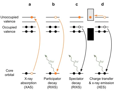
A schematic illustration of the electronic transitions involved is shown in Fig. 2. Following
the resonant excitation of a core-electron to an unoccupied valence state through x-ray
absorption (Fig. 2a), the core-hole can decay via two channels. In the participator channel
(Fig. 2b) the core-hole is filled by the originally excited electron from the unoccupied valence
state, resulting in the emission of an x-ray photon of the same energy as the x-ray absorption.
Participator decay therefore results in elastic scattering. Alternatively, in the the spectator
channel (Fig. 2c) the core-hole is filled by an electron in one of the occupied valence states,
resulting in the emission of an x-ray photon of lower energy than the one absorbed. Spectator
decay therefore results in inelastic scattering.
If we now couple the molecule to a surface such that ultra-fast charge transfer can occur
to the conduction band of the substrate on the timescale of the core-hole lifetime, both the
participator and spectator channels will be depleted in favour of ’normal’ x-ray emission
Core orbital Occupied
valence Unoccupied
valence
X-ray absorption
(XAS)
Participator decay (RIXS)
Spectator decay (RIXS)
Charge transfer & x-ray emission
(XES)
hv
d c
b a
[image:4.612.205.404.68.228.2]hv hv
FIG. 2. Relevant processes. a XAS resonant excitation of a core electron to an unoccupied state
(molecular orbital). b Participator decay in which the core-hole is filled by the originally excited
electron resulting in elastic scattering and c spectator decay in which the core-hole is filled by a
different valence electron resulting in inelastic scattering. dIf the molecular orbital couples to the
substrate empty states (conduction band) charge transfer of the electron can take place, which
competes with RIXS, resulting in XES.
used in the same way as in RPES to provide information on the degree of localisation of
the core electron excited to different unoccupied molecular orbitals – on the timescale of the
core-hole lifetime.11
In this paper we present for the first time two-dimensional N 1s RIXS maps of multilayer
and monolayer biisonicotinic acid adsorbed on the rutile TiO2(110) single crystal surface,
allowing the participator channel to be followed over the lowest unoccupied molecular
or-bitals. The data also reveals ultra-fast vibronic coupling on the timescale of the core-hole
lifetime, leading to an strong inelastic component of the participator channel.
II. METHOD
RIXS measurements were performed at the AERHA spectrometer12 at the SEXTANTS
beamline at Synchrotron SOLEIL.13 Multilayer samples were prepared in a separate UHV
system in our home laboratory equipped with a Scienta R3000 analyser and dual anode (Al
kα, Mg kα) x-ray source. Substrates were rutile TiO2(110) single crystals (Pi-Kem, UK)
cleaned by repeated cycles of sputtering at 2kV Ar+ followed by 1 kV Ar+, and annealing in
(4,4-dicarboxy-2,2-bipyridine fromAlfa Aesar), were deposited onto the prepared substrates
held at room temperature by sublimation from a Knudsen-type cell evaporation source until
no Ti 2p signal was detected in the XPS. This procedure produces atomically flat surfaces
suitable for RIXS measurements with low/negligible contribution of the diffuse scattering
to the elastic line intenisty. The multilayer samples were then transferred into the analysis
chamber of the AERHA spectrometer where the base pressure was 1×10⁻⁹ mbar. For RIXS
measurements on the multilayers, samples were used as prepared. For RIXS measurements
of the monolayers, these were prepared by annealing fresh multilayer samples in-situ to 500 K
to desorb the physisorbed multilayer to leave just the chemisorbed monolayer (see Fig. 1).7
The thickness of the multilayer films is estimated from the XPS to be around 10 nm. Due to
the increased penetration depth of x-rays compared to photoelectrons, there will be a small
contribution of the first monolayer in the multilayer RIXS measurements. However, this is
estimated to be at most a few percent of the overall signal and there are no features in the
multilayer RIXS data presented here that can be clearly attributed to this contribution.
The incident beam at SEXTANTS is focused to a spot size of 2(v) × 100(h) µm. To
prevent beam damage of the molecules, the sample was continuously moved during the
mea-surements at a rate that was shown to not give rise to changes between successive fast x-ray
absorption spectroscopy (XAS) scans. Due to the very low x-ray emission signal monolayer
N 1s RIXS maps were accumulated over more than one sample and the total x-ray emission
intensity detected at each absorption energy normalized to the corresponding
fluorescence-yield XAS intensity measured using an MCP detector. The photon energy scale of the
RIXS spectrometer was calibrated by measuring the elastic peak at 5 eV increments across
the whole detector range using linear vertical polarization of the incoming beam to
max-imise the elastic (Rayleigh) scattering intensity. For the RIXS maps and XAS over the N 1s
absorption edge the beam was linearly polarized in the horizontal plane and impinging on
the sample with an incidence angle of 35° with respect to the surface. The spatial
distribu-tion of the three lowest unoccupied molecular orbitals in the core-excited state for both the
isolated and adsorbed biisonicotinic acid molecules have been calculated by DFT and
pub-lished previously.14 These π∗ orbitals will have a significant absorption cross-section when
the polarisation of the incoming beam cuts through the plane of the molecule, which is the
case for the ordered monolayer (Fig. 1) at the chosen experimental geometry in addition to
(com-LUMO
LUMO+1
vibronic participator
[image:6.612.178.428.74.325.2]elastic participator
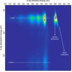
FIG. 3. N 1s RIXS map for a biisonicotinic acid multilayer (deposited to a thickness where
pho-tomemission from the underlying TiO2(110) surface is not visible in XPS). The features observed
below 396 eV emission energy are attributed to spectator decay (inelastic scattering) and those
above 397 eV emission energy to participator decay (elastic scattering and small inelastic losses
due to vibronic coupling). These features are observed at the LUMO and LUMO+1 resonances at
398.7 eV and 402.6 eV, respectively. Total acquisition time for the map was approximately 9 hours.
pared to the optimum resolution of 110 meV) as a compromise between energy resolution
and spectrometer transmission to keep the molecule below the radiation damage threshold.
For the multilayer RIXS map, each line is an x-ray emission spectrum accumulated for 10
mins at 0.1 eV intervals of absorption energy. For the monolayer RIXS map each line is the
average of between 3 and 12 spectra, each accumulated for 10 mins, at 0.2 eV intervals.
III. RESULTS AND DISCUSSION
The RIXS map measured over the N 1s absorption edge of multilayer biisonicotinic acid is
shown in Fig. 3. The horizontal axis of the map represents the x-ray emission energy, while
the vertical axis represents the x-ray excitation energy (absorption energy). Two main
LUMO
LUMO+2
vibronic participator
[image:7.612.178.428.72.326.2]elastic participator LUMO+1
FIG. 4. N 1s RIXS map for a biisonicotinic acid monolayer adsorbed on the rutile TiO2(110) single
crystal surface. The features observed below 396 eV emission energy are attributed to inelastic
scattering of the x-rays and those above 397 eV emission energy to elastic scattering and small
inelastic losses due to vibronic coupling. These features are observed at the LUMO, LUMO+1 and
LUMO+2 resonances at 398.4 eV, 400.2 eV and 402.3 eV, respectively. Total acquisition time for
the map was approximately 24 hours.
These correspond to the LUMO and LUMO+1 resonances of the molecule, respectively.
The features within these bands below around 396 eV emission energy are attributed to
inelastic x-ray scattering arising from the N 1s core-hole being filled by valence electrons
from the occupied molecular orbitals. This is the projection of the occupied densities of
states of the molecule onto the nitrogen core-level within the context of the dipole selection
rule. These features are therefore a measure of the occupied π orbitals derived from the
nitrogen valence electrons. Since the molecules are decoupled from the underlying oxide
surface in thick physisorbed multilayer, the core-excited electrons will be localised in the
unoccupied molecular orbital many orders longer than the timescale of the core-hole lifetime
as there are no empty states available to tunnel into. The inelastic scattering features below
396 eV are therefore attributed to the spectator decay process illustrated in Fig. 2c.
equivalent absorption and emission energy and therefore attributed to elastic scattering.
Diffuse reflection of the incoming beam will inevitably contribute to some extent to the
elastic peak, however the experiment has been configured to minimise this by avoiding the
specular reflection angle and using horizontally polarised light. Moreover, this contribution
will be constant over the absorption energy range probed so that changes in the intensity of
the elastic peak can be attributed to changes in the participator decay process illustrated in
Fig. 2b.
A decrease in the intensity of the participator channel can arise for two reasons that are
essential to disentangle. The first is charge transfer of the participator electron out of the
molecular orbital on the timescale of the core-hole lifetime. The second is a decrease in the
probability for participator decay relative to spectator decay for the higher-energy molecular
orbitals. Since ultra-fast charge transfer is not possible for molecules decoupled from the
substrate densities of states, we can assess the orbital-specific participator probability in the
absence of charge transfer from the multilayer RIXS.
Such a quantification requires identification of all the x-ray photons emitted from a
par-ticipator decay process, including those that are not strictly elastic. This is relevant in the
context of the multilayer RIXS data shown in Fig. 3 as considerable intensity is observed to
the low energy side of the elastic line observed over the LUMO resonance. This feature has
an intensity centred around an emission energy of 398 eV. There are however, no occupied
molecular orbitals at this energy since this corresponds to the onset of the LUMO resonance.
This feature is instead attributed to intra-molecular vibronic coupling and interpreted in a
Franck-Condon scheme where the excited electron rapidly couples to the lowest vibrational
level of the LUMO-derived resonance. The electron then decays to fill the core-hole, with the
emission of a photon energy corresponding to the difference between this lowest vibrational
state and the N 1s ground state.
It follows that this intra-molecular vibronic coupling must occur on on the same order or
faster than the timescale of the core-hole lifetime of a few fs. Ultra-fast vibronic dynamics
have previously been observed in the C 1s RPES15,16 and RIXS15 of C
60 multilayers and
also in N 1s RIXS maps of aqueous NH3.17 It also follows that since the emission associated
with the vibronic coupling arises from a transition involving the originally excited electron,
that these photons must be considered part of the participator decay channel.
on the rutile TiO2(110) single crystal surface is shown in Fig. 4. Here, three main bands
of emission features are observed at absorption energies of 398.4 eV, 400.2 eV and 402.3 eV.
These correspond to the LUMO, LUMO+1 and LUMO+2 resonances of the molecule,
re-spectively. The LUMO and LUMO+2 of the chemisorbed molecules correspond to the
LUMO and LUMO+1 observed for the physisorbed multilayer. The LUMO+1 observed at
400.2 eV for the monolayer is derived from the adsorption bond and has previously been
observed both experimentally and computationally for a biisonicotinic acid monolayer on
the rutile TiO2(110) surface.7 The interpretation of the monolayer RIXS is largely the same
as for the multilayer, with features below 396 eV emission energy attributed to inelastic
scattering, and those above to the elastic scattering. The broad intensity around the elastic
line at the LUMO resonance is again attributed to vibronic coupling within the molecule in
the core-excited state.
The key difference between the monolayer and multilayer is that in the monolayer some
molecular orbitals can couple to empty densities of states in the oxide substrate, opening up
the possibility of molecule-surface charge transfer. From combined XAS and photoemission
studies6,18,19of biisonicotinic acid on TiO
2(110) we know that the LUMO of the core-excited
molecule lies below the conduction band edge of the oxide and therefore overlaps with the
bandgap. For the LUMO state then we still expect to observe an intense participator channel
since an electron excited to this state cannot tunnel away into empty states in the surface.
The vibronic coupling feature at the LUMO resonance is consistent with this localisation
on the timescale of the core-hole lifetime. The LUMO+1 and LUMO+2 states however,
lie well above the conduction band edge of the TiO2(110) surface and have been shown
through RPES to undergo ultra-fast charge transfer into the surface on the low femtosecond
timescale.6,18
Integrating over the participator region of the RIXS for both the multilayer and monolayer
as a function of absorption energy, we compare the relative intensities of the participator
channel for each unoccupied molecular orbital compared to the x-ray absorption (XAS).
Since charge transfer out of the LUMO is not allowed for either the multilayer (as the
molecules are decoupled from the substrate densities of states) or the monolayer (as it lies
energetically below the conduction band) the participator RIXS of both surfaces can be
normalised to the intensity of the LUMO in the XAS. These spectra are shown in Fig. 5.
LUMO
LUMO
LUMO+2
LUMO+1
LUMO+1
ImultiXAS
RIXS
Imulti
mono
IXAS
mono
IRIXS
Intensity
403 402 401 400 399 398
X-ray absorption energy (eV)
Multilayer XAS RIXS VC
Intensity
403 402 401 400 399 398
Monolayer XAS RIXS VC
[image:10.612.189.420.66.503.2]X-ray absorption energy (eV)
FIG. 5. Integrations over the participator region of the N 1sRIXS for multilayer (a) and monolayer
(b) biisonicotinic acid on the TiO2(110) surface, compared to XAS. The integration window for the
RIXS at each absorption energy was 2 eV wide starting from the high emission energy side of the
elastic line, and 1.7 eV wide for the vibronic coupling feature (VC) starting from the low emission
energy side of the elastic line. All spectra have been normalised to the height of the LUMO which
lies energetically below the conduction band and therefore cannot participate in charge transfer
into states in the underlying oxide surface.
transfer timeτCT for the LUMO+2 state of the coupled molecule from the core-hole lifetime
τCT =τC
IRIXSmono Imono XAS
Imulti RIXS
Imulti XAS
− IRIXSmono
Imono XAS
(1)
where IRIXSmulti
Imulti XAS
is the ratio of participator signal in the RIXS to the total x-ray absorption
measured in the fluorescence yield XAS for the multilayer, and IRIXSmono
Imono XAS
is the corresponding
ratio for the monolayer. Replacing the intensity ratios in Eqn. 1 with the values extracted
from the integrations in Fig. 5, and setting to τC = 6 fs,3 the charge transfer timescale for
LUMO+2 could, in principle, be estimated. However, the ratios extracted from an
integra-tion of the RIXS over a 2 eV range covering both the elastic peak and the vibronic coupling
are essentially the same within experimental uncertainty (0.2±0.1). For completeness, Fig. 5
also shows the integration performed over just the vibronic coupling region on its own (not
including the elastic line) with no change in the result. This corresponds to a localisation
of the electron in the LUMO+2 on a timescale orders of magnitude longer that the
core-hole lifetime, in direct contradiction to previous experimental evidence for ultra-fast charge
transfer based on the RPES participator channel.
Charge transfer on the sub-3 fs timescale from the LUMO+2 of the biisonicotinic acid
molecule into the conduction band of the underlying TiO2(110) conduction band has been
experimentally measured using RPES and is supported by sub-5 fs timescales measured
for isonicotinic acid on the same surface.6,18 However, in our measurements there is no
evidence of the channels identified as participator decay processes (elastic scattering and
vibronic coupling) being depleated for the monolayer for which fs charge transfer out of the
LUMO+2 has been shown to occur. Enhancements of the Rayleigh (elastic) line previously
observed in the RIXS of isolated molecules are typically understood in terms of core-excitonic
states leading to an enhanced probability of the originally excited electron filling the
core-hole, resulting in elastic participator decay.16,17 Such enhancements as observed here in the
multilayer data would be expected to vanish if the originally excited electron tunnels away
on a fast timescale in the monolayer.
Since the intermediate state in RIXS and RPES are the same (resonant excitation of the
core-electron to the unoccupied valence state) we do not expect differences in the timescales
of electron delocalization in the core-excited state between the two techniques. If the
radia-tive core-exciton decay process occurs on a faster timescale it would of course not be depleted
with the core-hole lifetime of around 6 fs.3 This suggests instead a challenge for the
two-step model of RIXS that is implicit in the core-hole clock approach, instead requiring a full
quantum mechanical description of RIXS as a coherent one-step process. It is also possible
that the measurement statistics in this experiment are insufficient to detect the depletion of
the participator channel in the RIXS. Further experiments are required to explore whether
the participator channel behaves differently to RPES in other molecule-surface systems.
One candidate is C60 on Au(111) for which the RPES data has already been measured.20
This system has the advantage of substantially more probed atoms per molecule, and more
intense LUMO+1, 2 and 3 resonances relative to the LUMO. Moreover, the core-excited
LUMO straddles the Fermi level of the metal surface opening up the possibility for charge
transfer out of those vibrational states that overlap with empty densities of states in the
surface and modification of the vibronic coupling15 contribution to the participator channel.
IV. CONCLUSIONS
Resonant inelastic x-ray scattering has been used to study the excited-state charge
trans-fer dynamics of an aromatic molecule, biisonicotinic acid, coupled to a rutile TiO2(110)
single crystal surface. By measuring, for the first time, full RIXS maps for both multilayer
and monolayer coverages of the molecules, the participator channel can be clearly identified
and quantified as a function of excitation into different unoccupied molecular orbitals.
In-cluded in this quantification is the elastic line and the inelastic scattering due to ultra-fast
vibronic coupling on the timescale of the core-hole lifetime. The orbital-specific participator
channel has been probed in the absence of charge transfer in the physisobred multilayer,
and compared to the coupled molecules in the chemisorbed monolayer. A core-hole clock
analysis of the RIXS data in order to extract a charge transfer time for the LUMO+2
ex-cited state that lies above the conduction band edge has been performed. However, there is
no compelling evidence in the data to show that participator channel is depleted under the
conditions of ultra-fast charge transfer of the excited electron into the substrate. The data
instead suggests that the processes that give rise to both the elastic scattering and the
in-tramolecular vibronic coupling, both of which are participator processes are not modified on
the femtosecond timescale on which charge transfer out of the LUMO+2 to the conduction
V. ACKNOWLEDGEMENTS
Funding was provided by RCUK — Engineering and Physical Sciences Research Council
(EPSRC) and Molecularspray Ltd through a DTG/CASE conversion studentship.
REFERENCES
1M. Gr¨atzel, Journal of Photochemistry and Photobiology C: Photochemistry Reviews 4,
145 (2003).
2J. J. Concepcion, J. W. Jurss, M. K. Brennaman, P. G. Hoertz, A. O. T. Patrocinio, N. Y.
Murakami Iha, J. L. Templeton, and T. J. Meyer, Acc. Chem. Res. 42, 1954 (2009).
3B. Kempgens, A. Kivim¨aki, M. Neeb, H. M. K¨oppe, A. M. Bradshaw, and J. Feldhaus,
Journal of Physical Chemistry B 29, 5389 (1996).
4P. A. Br¨uhwiler, O. Karis, and N. M˚artensson, Reviews of Modern Physics74, 703 (2002).
5Participator has become the accepted term in the literature to describe the core-hole decay
process in which the originally excited electron is a direct participant, resulting in resonant
photoemission in RPES and inelastic scattering in RIXS. In Ref. 4 by P. A. Br¨uhwiler et
al, the grammatically correct term participant is used instead. Both terms refer to the
same processes.
6J. Schnadt, P. A. Br¨uhwiler, L. Patthey, J. N. O’Shea, S. S¨odergren, M. Odelius, R. Ahuja,
O. Karis, M. B¨assler, P. Persson, H. Siegbahn, S. Lunell, and N. M˚artensson, Nature418,
620 (2002).
7L. Patthey, H. Rensmo, P. Persson, K. Westermark, L. Vayssieres, A. Stashans, ˚A.
Peters-son, P. A. Br¨uhwiler, H. Siegbahn, S. Lunell, and N. M¨artensson, J. Chem. Phys. 110,
5913 (1999).
8L. C. Mayor, J. B. Taylor, G. Magnano, A. Rienzo, C. J. Satterley, J. N. O’Shea, and
J. Schnadt, J. Chem. Phys. 129, 114701 (2008).
9M. Weston, A. J. Britton, and J. N. O’Shea, J. Chem. Phys 134, 054705 (2011).
10M. Weston, K. Handrup, T. J. Reade, N. R. Champness, and J. N. O’Shea, J. Chem.
Phys. 137, 224706 (2012).
12S. G. Chiuzbaian, C. F. Hague, A. Avila, R. Delaunay, N. Jaouen, M. Sacchi, F. Polack,
M. Thomasset, B. Lagarde, A. Nicolaou, S. Brignolo, C. Baumier, J. Luening, and J.-M.
Mariot, Rev. Sci. Instrum. 85, 043108 (2014).
13M. Sacchi, N. Jaouen, H. Popescu, R. Gaudemer, J. M. Tonnerre, S. G. Chiuzbaian, C. F.
Hague, A. Delmotte, J. M. Dubuisson, G. Cauchon, B. Lagarde, and F. Polack, in 11th
International Conference on Synchrotron Radiation Instrumentation (SRI 2012), J. Phys.
Conf. Ser., Vol. 425, edited by Susini, J and Dumas, P (2013) p. 072018.
14P. Persson, S. Lunell, P. Bruhwiler, J. Schnadt, S. Sodergren, J. O’Shea, O. Karis, H.
Sieg-bahn, N. Martensson, M. Bassler, and L. Patthey, J. Chem. Phys. 112, 3945 (2000).
15L. Kjeldgaard, T. Kaambre, J. Schiessling, I. Marenne, J. O’Shea, J. Schnadt, C. Glover,
M. Nagasono, D. Nordlund, M. Garnier, L. Qian, J. Rubensson, P. Rudolf, N. Martensson,
J. Nordgren, and P. Bruhwiler, Phys. Rev. B 72, 205414 (2005).
16L. Weinhardt, O. Fuchs, D. Batchelor, M. Baer, M. Blum, J. D. Denlinger, W. Yang,
A. Schoell, F. Reinert, E. Umbach, and C. Heske, J. Chem. Phys. 135, 104705 (2011).
17L. Weinhardt, E. Ertan, M. Iannuzzi, M. Weigand, O. Fuchs, M. Bar, M. Blum, J. D.
Denlinger, W. Yang, E. Umbach, M. Odelius, and C. Heske, Phys. Chem. Chem. Phys.
17, 27145 (2015).
18J. Schnadt, J. N. O’Shea, L. Patthey, L. Kjeldgaard, J. ˚Ahlund, K. Nilson, J. Schiessling,
J. Krempask´y, M. Shi, O. Karis, C. Glover, H. Siegbahn, N. M˚artensson, and P. A.
Br¨uhwiler, J. Chem. Phys. 119, 12462 (2003).
19J. Schnadt, J. O’Shea, L. Patthey, J. Krempasky, N. Martensson, and P. Bruhwiler, Phys.
Rev. B 67, 235420 (2003).