ISSN Online: 2162-2019 ISSN Print: 2162-2000
DOI: 10.4236/wjns.2018.82024 May 22, 2018 303 World Journal of Neuroscience
Identification and Physiochemical Analysis
of ERK Interacting Proteins Using
Bio-Computational Tools
Khuleshwari Kurrey, Vijay Paramanik
*Cellular and Molecular Neurobiology & Drug Targeting Laboratory, Department of Zoology, Indira Gandhi National Tribal University, Amarkantak, Madhya Pradesh, India
Abstract
ERK (Extracellular Signal Regulated Kinase) or MAP kinase is an intracellular signaling molecule. ERK is involved in regulation of various functions i.e. cell proliferation, cell migrations, cell survival and many more. It gets activated in response of various stimuli like growth factors, cytokines, virus, second mes-sengers, transforming agents and carcinogens. While transferring signals from cell surface receptors to cell nucleus, ERK interacts with a numbers of pro-teins. Physiochemical and functional characterization of these proteins is little known. Thus, we attempted to study physiochemical and functional proper-ties of ERK interacting proteins using bio-computational tools. ExPASy and SOSUI server suggested 22 ERK interacting proteins. Physical and chemical parameters of these ERK interacting partners indicated higher percentage of hydrophobic amino acid and leucine as major constituent. Moreover, the in-stability index indicated that four proteins are stable in over wide range tem-perature in vitro, and remaining eighteen proteins were found unstable. In addition, SOSUI server showed that fifteen proteins were soluble and six are trans-membrane in nature.
Keywords
Extracellular Signal Regulated Kinase (ERK), GRAVY, SOSUI, STRING, STITCH, ExPASy
1. Background
ERK (Extracellular Signal Regulated Kinase) or MAP kinase is an intracellular signaling molecule. It is associated to regulate various cell functions, namely cell proliferation, cell migrations, cell survival and many other biological functions.
How to cite this paper: Kurrey, K. and Paramanik, V. (2018) Identification and Physiochemical Analysis of ERK Interact-ing Proteins UsInteract-ing Bio-Computational Tools. World Journal of Neuroscience, 8, 303-313.
https://doi.org/10.4236/wjns.2018.82024
Received: March 28, 2017 Accepted: May 19, 2018 Published: May 22, 2018
Copyright © 2018 by authors and Scientific Research Publishing Inc. This work is licensed under the Creative Commons Attribution International License (CC BY 4.0).
http://creativecommons.org/licenses/by/4.0/
DOI: 10.4236/wjns.2018.82024 304 World Journal of Neuroscience ERK gets activated in response to the response of various stimuli such as differ-ent growth factors, cytokines, virus, second messengers, transforming agdiffer-ents and carcinogens. ERK helps to transfer signals from cell surface receptors to nucleus of the cells. However, it’s still undefined how single ERK pathway regulates veri-ty of specific and different cellular functions [1]. ERK interacts with wide range of proteins to perform specific function. It is important to line up the action of ERK with its interactive proteins at molecular level. Thus the physiochemical and functional properties are needed.
With increasing knowledge in bioinformatics, the computational server is used to determine physical, chemical and functional properties of proteins. These services are useful to facilitate researchers to frame numerous experiments at molecular level. Although physiochemical and functional studies of various proteins and drugs have been done by many workers using bioinformatics tools, [2] computational study on ERK interactive proteins is little studied. Thus, we focused to study physiochemical and functional characterization of ERK interac-tive proteins using computational tools. Such studies are helpful to understand ERK dependent signal transduction.
2. Methodology
The methodology was followed in this manuscript as under:
2.1. Identification of Putative ERK Interactive Proteins
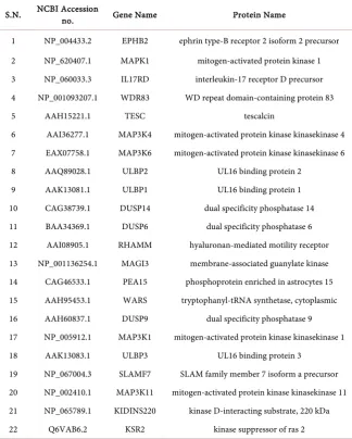
ERK interactive proteins were retrieved by using STITCH 4., which is a hugear-chives of protein-chemical interaction (http://stitch.embl.de/) [3] and STRING 10.0 (http://string-db.org/), is a huge archives of protein-protein interactions [4] [5]. These tools predict 42ERK interactive proteins. Physiochemical analysis of 22 commonly predicted proteins by both servers were done (Table 1).
2.2. Sequence Analysis
DOI: 10.4236/wjns.2018.82024 305 World Journal of Neuroscience
Table 1. Interacting proteins of ERK retrieved from STITCH and STRING.
S.N. NCBI Accession no. Gene Name Protein Name
1 NP_004433.2 EPHB2 ephrin type-B receptor 2 isoform 2 precursor 2 NP_620407.1 MAPK1 mitogen-activated protein kinase 1 3 NP_060033.3 IL17RD interleukin-17 receptor D precursor 4 NP_001093207.1 WDR83 WD repeat domain-containing protein 83 5 AAH15221.1 TESC tescalcin
6 AAI36277.1 MAP3K4 mitogen-activated protein kinase kinasekinase 4 7 EAX07758.1 MAP3K6 mitogen-activated protein kinase kinasekinase 6 8 AAQ89028.1 ULBP2 UL16 binding protein 2
9 AAK13081.1 ULBP1 UL16 binding protein 1 10 CAG38739.1 DUSP14 dual specificity phosphatase 14 11 BAA34369.1 DUSP6 dual specificity phosphatase 6 12 AAI08905.1 RHAMM hyaluronan-mediated motility receptor 13 NP_001136254.1 MAGI3 membrane-associated guanylate kinase 14 CAG46533.1 PEA15 phosphoprotein enriched in astrocytes 15 15 AAH95453.1 WARS tryptophanyl-tRNA synthetase, cytoplasmic 16 AAH60837.1 DUSP9 dual specificity phosphatase 9 17 NP_005912.1 MAP3K1 mitogen-activated protein kinase kinasekinase 1 18 AAK13083.1 ULBP3 UL16 binding protein 3
19 NP_067004.3 SLAMF7 SLAM family member 7 isoform a precursor 20 NP_002410.1 MAP3K11 mitogen-activated protein kinase kinasekinase 11 21 NP_065789.1 KIDINS220 kinase D-interacting substrate, 220 kDa 22 Q6VAB6.2 KSR2 kinase suppressor of ras 2
number (http://www.ncbi.nlm.nih.gov/protein) [6][7][8][9] from NCBI.
2.3. Physiochemical Characterization of Interacting Proteins
Using ExPASy ProtParam (http://web.expasy.org/protparam/) computational server, primary structure and physiochemical characters of proteins, such as amino acids number (AA), Molecular weight of protein, Theoretical pI, Amino acids compositions of protein, total number of negatively charged residues (Asp + Glu), total number of positively charged residues (Arg + Lys), extinction coef-ficient, instability index, aliphatic index and Grand Average Hydropathicity (GRAVY) were observed.2.4. Functional Characterization
DOI: 10.4236/wjns.2018.82024 306 World Journal of Neuroscience tools was utilized to analyze them [10].
3. Results
3.1. Physiochemical Characterization
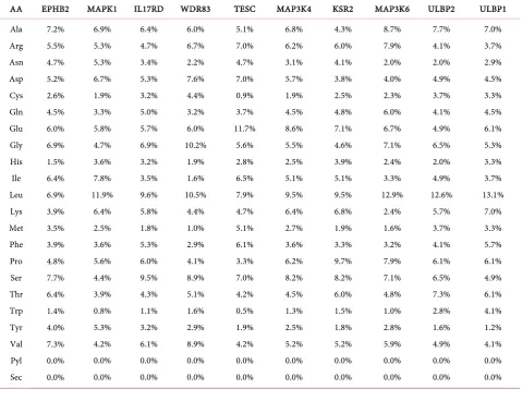
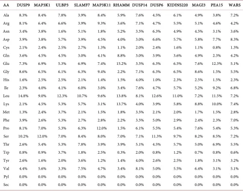
ERK interactive protein’s amino acids (AA) composition was presented in Table 2 and Table 3. Most of the ERK interacting proteins are hydrophilic in nature. AA composition indicates that higher percentage of hydrophobic amino acid leucine. This is due to presence of all the hydrophobic proteins inside the cell membrane and hydrophilic amino acids on the outside. Sequence length of pro-tein represents basic nature of propro-tein [6][7][8].
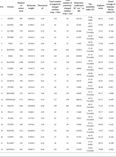
[image:4.595.59.539.366.734.2]Theoretical pI (isoelectric point) (Table 4) value of 17 proteins i.e. EPHB2, MAPK1, IL17RD, WDR83, TESC, MAP3K4, MAP3K6, ULBP2, ULBP1, DUSP6, RHAMM, KIDINS220, PEA15, WARS, DUSP9, MAP3K1, SLAMF7 is less than 7. Hence, these all are acidic in nature and other 5 proteins i.e. KSR2, DUSP14, MAGI3, ULBP3 and MAP3K11 pI value is greater than 7. Hence, these all are basic in nature. pI value may be utilized for making buffer system when these proteins are to be purified in solution by isoelectric focusing method [6][7][8].
Table 2. Amino Acids (AA) Composition (in %) of ERK Interacting Proteins Followed ExPASy server.
DOI: 10.4236/wjns.2018.82024 307 World Journal of Neuroscience
Table 3. Amino Acids (AA) Composition (in %) of ERK Interactive Proteins Followed ExPASy server.
AA DUSP9 MAP3K1 ULBP3 SLAMF7 MAP3K11 RHAMM DUSP14 DUSP6 KIDINS220 MAGI3 PEA15 WARS
Ala Arg Asn Asp Cys Gln Glu Gly His Ile Leu Lys Met Phe Pro Ser Thr Trp Tyr Val Pyl Sec 8.3% 8.1% 3.4% 3.9% 2.1% 3.6% 7.3% 8.6% 1.6% 2.3% 14.8% 2.1% 1.3% 3.9% 8.1% 10.2% 2.6% 0.8% 2.6% 4.4% 0.0% 0.0% 8.4% 6.4% 3.8% 3.8% 2.4% 4.5% 6.9% 6.5% 2.5% 4.0% 9.0% 4.5% 2.4% 2.6% 7.0% 12.0% 5.4% 0.9% 1.6% 5.6% 0.0% 0.0% 7.8% 6.6% 1.6% 5.7% 2.5% 4.5% 5.3% 6.1% 2.5% 4.1% 12.3% 5.3% 3.7% 5.3% 5.3% 7.0% 5.3% 3.7% 2.0% 3.3% 0.0% 0.0% 3.9% 3.9% 5.1% 3.9% 2.7% 3.0% 6.9% 6.3% 2.1% 6.0% 10.7% 5.7% 2.1% 2.7% 6.3% 8.4% 7.8% 1.8% 3.6% 7.5% 0.0% 0.0% 8.4% 9.3% 1.8% 4.5% 1.3% 4.1% 7.4% 9.4% 1.4% 3.0% 9.6% 3.1% 1.5% 2.8% 12.0% 8.0% 3.9% 2.5% 1.2% 4.7% 0.0% 0.0% 5.9% 3.6% 5.2% 4.0% 1.1% 8.8% 15.2% 2.2% 1.5% 3.4% 13.8% 13.7% 1.8% 2.2% 1.5% 7.0% 3.9% 0.3% 1.4% 3.4% 0.0% 0.0% 7.6% 7.1% 3.5% 3.0% 2.0% 3.0% 3.5% 7.1% 4.0% 7.6% 8.1% 4.0% 3.5% 3.5% 6.1% 7.1% 5.1% 2.0% 4.0% 8.1% 0.0% 0.0% 4.5% 4.7% 6.3% 6.6% 2.4% 3.9% 6.3% 6.3% 1.0% 4.7% 12.6% 3.9% 2.1% 5.0% 5.5% 11.3% 4.5% 0.8% 2.6% 5.0% 0.0% 0.0% 6.1% 5.5% 4.9% 5.7% 1.6% 3.6% 6.5% 6.5% 2.3% 5.7% 11.0% 5.8% 2.0% 2.9% 5.4% 9.7% 5.7% 1.2% 2.5% 5.5% 0.0% 0.0% 4.9% 5.1% 4.5% 5.8% 1.1% 4.9% 7.6% 8.6% 2.5% 5.2% 7.2% 8.8% 1.7% 2.4% 7.6% 8.2% 5.0% 0.7% 1.8% 6.4% 0.0% 0.0% 3.8% 4.6% 3.1% 7.7% 0.8% 2.3% 12.3% 1.5% 1.5% 9.2% 11.5% 10.0% 1.5% 2.3% 5.4% 8.5% 6.9% 0.8% 3.1% 3.1% 0.0% 0.0% 7.2% 4.2% 3.6% 8.3% 1.3% 4.2% 5.1% 5.5% 2.3% 6.8% 7.2% 7.4% 2.8% 7.0% 5.3% 7.2% 5.5% 0.6% 3.2% 5.1% 0.0% 0.0%
3.2. Extinction Coefficients (EC)
The extinction coefficient of ERK interacting proteins at 280nm ranged from 11460 M−1∙cm−1 to 188310 M−1∙cm−1 in comparison to the concentration of Cys, Trp and Tyr (Table 4). Extinction coefficients of KIDINS220, MAP3K4, EPHB2, MAP3K11, MAP3K6, MAP3K, KSR2 were very higher indicating presence of high concentration of aromatic amino acid. Extinction coefficient of PEA15 was as lower 11460 M−1∙cm−1 indicating that the presence of aromatic amino acids is low. The EC values are useful in the quantitative study of protein-protein and protein-ligand interactions in solution [6][7][8].
3.3. The Instability Index (II)
DOI: 10.4236/wjns.2018.82024 308 World Journal of Neuroscience
Table 4. Physiochemical properties of Interacting Proteins of ERK.
S.N. Protein
Number of amino
acids
Molecular
weight Theoretical pI
Total number of negatively charged residues (Asp + Glu)
Total number of
positively charged residues (Arg + Lys)
Extinction coefficients M−1∙cm−1, at
280
The instability index (II)
Aliphatic index
Grand average of
hydropa-thicity (GRAVY)
1 EPHB2 987 110030.2 5.49 110 92 136735 stable 37.88 80.11 −0.202
2 MAPK1 360 41389.7 6.50 45 42 45185 stable 39.71 95.94 −0.287
3 IL17RD 739 82410.5 6.78 81 78 81260 Unstable 56.54 75.21 −0.346
4 WDR83 315 34342.6 5.36 43 35 41785 Unstable 42.56 78.86 −0.305
5 TESC 214 24749.8 4.84 40 25 11585 Unstable 49.58 73.83 −0.568
6 MAP3K4 1608 181691.7 5.92 230 203 176975 Unstable 51.45 78.81 −0.509
7 KSR2 950 107632.3 8.95 103 122 103830 Unstable 61.10 75.93 −0.600
8 MAP3K6 1288 142596.0 6.70 133 138 127015 Unstable 58.54 88.79 −0.254
9 ULBP2 246 27367.9 6.93 24 24 44960 Unstable 49.34 90.04 0.019
10 ULBP1 244 27996.5 7.07 26 26 59970 Unstable 40.06 84.39 −0.162
11 DUSP14 198 42333.7 9.62 13 22 34170 Stable 37.79 92.07 −0.012
12 DUSP6 381 22254.9 4.75 49 33 31900 Unstable 52.25 86.48 0.265
13 RHAMM 725 84175.5 5.68 139 125 26400 Unstable 53.72 83.17 −0.988
14 KIDINS220 1771 196542.1 6.19 217 199 188310 Unstable 44.54 87.17 −0.369
15 MAGI3 1481 162948.8 8.26 199 205 96230 Unstable 48.35 71.65 −0.746
16 PEA15 130 15068.1 4.93 26 19 11460 Unstable 64.55 93.77 −0.625
17 WARS 471 53178.5 5.83 63 55 39225 Unstable 40.34 76.65 −0.345
18 DUSP9 384 41934.6 5.96 43 39 31900 Unstable 67.23 88.20 −0.245
19 MAP3K1 1512 164469.9 7.93 161 165 115010 Unstable 63.95 75.07 −0.425
20 ULBP3 244 27949.3 8.20 27 29 57325 Unstable 46.78 81.23 −0.236
21 SLAMF7 335 37420.9 6.02 36 32 51380 Stable 37.55 90.72 −0.188
DOI: 10.4236/wjns.2018.82024 309 World Journal of Neuroscience
3.4. Aliphatic Index
Aliphatic index of a protein is considered as the relative volume captured by aliphatic side chains (alanine, valine, isoleucine, and leucine). It is considered as a positive factor for the increase of thermal stability of annular proteins. Ali-phatic index of ERK interactive proteins limit from 62.34 to 107.02. Both MAP3K11 and MAGI3 have lower thermal stability representing their more flexible structure as compared to other proteins (Table 4). The high aliphatic index of other proteins indicates their stability in high range of temperature [6] [7][8].
3.5. A GRAVY (Grand Average of Hydropathy)
GRAVY is understood as the total hydropathy values of all the amino acids of a protein, divided by the number of amino acids residues in the sequence. ExPASy Protparam server for GRAVY indicated that ERK interactive proteins are hy-drophilic in nature except ULBP2. Lowest GRAVY value of DUSP14 suggests its better communication with water [6][7][8].
3.6. Functional Characterization
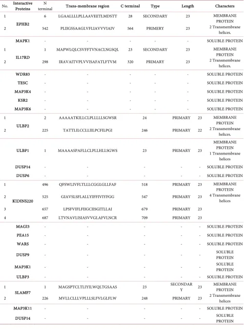
The SOSUI server data expressed ERK interacting proteins namely EPHB2 (2 trans membrane), IL17RD (2 trans membrane), ULBP2 (2 trans membrane), ULBP1 (1 trans membrane), KIDINS220 (4 trans membrane) and SLAMF7 (2 trans membrane) are membrane protein. However, MAPK1, WDR83, TESC, MAP3K4, KSR2, MAP3K6, DUSP6, DUSP14, MAGI3, PEA15, WARS, DUSP9, MAP3K1, ULBP3, MAP3K11 and DUSP4 are classified as a soluble protein (Table 5).
4. Discussion
There are several genes responsible for the long term synaptic plasticity. ERK is one of these gene involved in long term learning and memory. During this process, a cascade of regulatory immediate early genes is recruited to particular gene including ERK [11] [12]. These genes are required for long-term synaptic plasticity and long-term memory formation. For gene expression, chromatin remodeling is crucial event. Studies from several researchers have established that chromatin remodeling is involved in learning and memory, long-term neu-ronal responses, drug addiction, stress, epilepsy and depression [13] [14]. For performing any functions ERK interacts with a host of proteins. Hence the ERK interacting proteins are crucial and involved in almost all brain related disorders and diseases.
DOI: 10.4236/wjns.2018.82024 310 World Journal of Neuroscience
Table 5. Functional characterization of ERK interactive proteins.
No. Interactive Proteins terminal N Trans-membrane region C terminal Type Length Characters
1
EPHB2
6 LGAALLLLPLLAAVEETLMDSTT 28 SECONDARY 23 MEMBRANE PROTEIN 2 Transmembrane
helices. 2 542 PLIIGSSAAGLVFLIAVVVIAIV 564 PRIMERY 23
MAPK1 - - - SOLUBLE PROTEIN
1
IL17RD
1 MAPWLQLCSVFFTVNACLNGSQL 23 SECONDARY 23 MEMBRANE PROTEIN 2 Transmembrane
helices. 2 298 IRAVAITVPLVVISAFATLFTVM 320 PRIMARY 23
WDR83 - - - SOLUBLE PROTEIN
TESC - - - SOLUBLE PROTEIN
MAP3K4 - - - SOLUBLE PROTEIN
KSR2 - - - SOLUBLE PROTEIN
MAP3K6 - - - SOLUBLE PROTEIN
1
ULBP2
2 AAAAATKILLCLPLLLLLSGWSR 24 PRIMARY 23 MEMBRANE PROTEIN 2 Transmembrane
helices. 2 225 TATTLILCCLLIILPCFILPGI 246 PRIMARY 22
ULBP1 1 MAAAASPAFLLCLPLLHLLSGWS 23 PRIMARY 23
MEMBRANE PROTEIN 1 Transmembrane
helices
DUSP14 - - - SOLUBLE PROTEIN
DUSP6 - - - SOLUBLE PROTEIN
1
KIDINS220
496 QFSWLIVFLTLLLCGGLGLLFAF 518 PRIMARY 23 MEMBRANE PROTEIN 4 Transmembrane
helices 2 525 GIAVSLSFLALLYIFFIVIYFGG 547 PRIMARY 23
3 657 LPSFVIFLFIIGCIISGITLLAI 679 PRIMARY 23 4 687 LTVNAVLISIASVVGLAFVLNCR 709 PRIMARY 23
MAGI3 - - - SOLUBLE PROTEIN
PEA15 - - - SOLUBLE PROTEIN
WARS - - - SOLUBLE PROTEIN
DUSP9 - - - SOLUBLE PROTEIN
MAP3K1 - - - SOLUBLE PROTEIN
ULBP3 - - - SOLUBLE PROTEIN
1
SLAMF7 1 MAGSPTCLTLIYILWQLTGSAAS 23
SECONDAR
Y 23 MEMBRANE PROTEIN 2 Transmembrane
helices 2 226 MVLLCLLLVPLLLSLFVLGLFLW 248 PRIMARY 23
MAP3K11 - - - SOLUBLE PROTEIN
DOI: 10.4236/wjns.2018.82024 311 World Journal of Neuroscience reverse manner and removes the acetyl groups from lysine residues. Actually, acetylation neutralizes the basic histone tails by weakening of the histone-DNA interaction and promoting the accessibility of transcription factors. The present study revealed that all ERK interacting proteins are rich in lysine residues i.e. ba-sic in nature. Further ERK interacting proteins are hydrophobic in nature which favors gene expression at transcriptional level. So these proteins may be involved in histone modification needed for chromatin dynamics and gene transcription. Furthermore, there are several kinases which are decisively involved in histone phosphorylation affecting long-term memory formation nemely ERK1/2, p38 MAP kinase, and ribosomal 6 kinase (RSK) [14]. The aforementioned findings favor our result as ERK interacted with various proteins as shown in this paper.
In addition, calsyntenins (Cst) comprise a family of trans-membrane proteins with the unique potential to link extracellular proteolytic activity with intracel-lular calcium signaling. It is molecular class of calcium binding protein, which can bind calcium ion and being involved in cell signaling and cell-cell commu-nication. It contains three members, calsyntenin-1, calsyntenin-2 and calsynte-nin-3, which are postsynaptic membrane proteins and predominantly expressed in brain neurons. Each of the three calsyntenins exhibits a distinct neuronal mRNA expression pattern. The calsyntenin-1 is located in the postsynaptic membrane of CNS and a proteolytically processed protein of the postsynaptic membrane contains calcium-binding cytoplasmic domain. Calsyntenin-1 is a dynamic modulator of postsynaptic calcium by extracellular proteolysis. It may modulate Ca2+ transients locally either beneath the postsynaptic membrane or around intracellular Ca2+ stores, such as LTP and LTD. Recent studies showed that impairment of the coordinated metabolic regulation of APP, calsyntenins and the consequent loss of the reciprocal regulation by APP and calsyntenins in the gene transactivation in AD [15]. Almost all ERK interacting proteins are trans-membrane in nature. Thus ERK interacting proteins may be involved in metabolic regulation of APP and calsyntenins mediated pathway of AD.
DOI: 10.4236/wjns.2018.82024 312 World Journal of Neuroscience [20]. In the process of dimerization, parallel leucine zipper motifs interact through a coiled coil hydrophobic interface that juxtaposes 2 adjacent basic re-gions [21]. Further, once bound to DNA, these adjacent basic regions undergo key changes in conformation [22]. As almost ERK interacting proteins contain leucine so it may be assumed that these are involved in ERK mediated functions including learning and memory. In addition, leucine rich food could be good for cognition and healthy brain functions. Thus such findings regarding ERK inte-racting proteins are instrumental to understand learning and memory via ERK. Moreover, it may be helpful to target these interacting proteins during learning and memory deficit and neurodegenerative diseases.
Acknowledgements
VP acknowledges Science and Engineering Research Board (SERB), Government of India for providing financial support (Registration No.SERB/LS-485/2013). KK acknowledges SERB for Junior Research Fellowship.
References
[1] Ebisuya, M., Kondoh, K. and Nishida, E. (2005) The Duration, Magnitude and Compartmentalization of ERK MAP Kinase Activity: Mechanisms for Providing Signaling Specificity. Journal of Cell Science,118, 2997-3002.
https://doi.org/10.1242/jcs.02505
[2] Vidhya, V.G., Upgade, A., Bhaskar, A. and Deb, D. (2012) In Silico Characterization of Bovine (Bostaurus) Antiapoptotic Proteins. Journal of Proteins and Proteomics, 3, 187-196.
[3] Kuhn, M., Mering, V.C., Campillos, M., Jensen, J.L. and Bork, P. (2015) STITCH: Interaction Networks of Chemicals and Proteins. Nucleic Acids Research, 36, D684-D688. https://doi.org/10.1093/nar/gkm795
[4] Szklarczyk, D., Franceschini, A., Wyder, S., Forslund, K., Heller, D., Huerta. C.J., Simonovic, M., Alexander, R., Santos, A., Tsafou, P.K., Kuhn, M., Bork, P., Jensen, J.L. and Mering, V.C. (2015) STRING v10: Protein-Protein Interaction Networks, Integrated over the Tree of Life. Nucleic Acids Research,43, D447-D452.
https://doi.org/10.1093/nar/gku1003
[5] Bidkar, A.P., Thakur, K.K., Bolshette, NB., Dutta, J. and Gogoi, R. (2014) In-Silico Structural and Functional Analysis of Hypothetical Proteins of Leptospira Interro-gans. Biochemistry & Pharmacology, 3, 3.
https://doi.org/10.4172/2167-0501.1000136
[6] Jabalia, N., Bansal, H., Mishra, P.C. and Chaudhary, N. (2015) In-Silico Compara-tive Analysis of Papain Family Cysteine Protease Using Computational Tools and Servers. International Journal of Basic and Applied Biology,2, 310-314.
[7] Mahalakshmi, K. (2015) Insilico Analysis of Proteins of Curcuma aromatica Salisb.
International Journal of PharmTech Research, 8, 51-56.
[8] Vishwanath, K.V., Pattabhiramaiah, M. and Keerthi, R. (2016) Bio Computational Analysis of Protein Sequence of Sickle Cell Anemia. International Journal of Engi-neering Research and General Science,1, 63-73.
Compu-DOI: 10.4236/wjns.2018.82024 313 World Journal of Neuroscience
tational Approach. Biosciences, 76, 28414-28421.
[10] Hirokawa, T (1998) SOSUI: Classification and Secondary Structure Prediction Sys-tem for Membrane Proteins. Bioinformatics, 14, 378-379.
https://doi.org/10.1093/bioinformatics/14.4.378
[11] Goelet, P., Castellucci, V.F., Schacher, S. and Kandel E.R. (1986) The Long and the Short of Long-Term Memory—A Molecular Framework. Nature,322, 419-422. https://doi.org/10.1038/322419a0
[12] Taubenfeld, S.M., Milekic, M.H., Monti, B. and Alberini, C.M. (2001) The Consoli-dation of New but Not Reactivated Memory Requires Hippocampal C/EBPβ. Na-ture Neuroscience, 4, 813-818.https://doi.org/10.1038/90520
[13] McClung, C.A. and Nestler, E.J. (2008) Neuroplasticity Mediated by Altered Gene Expression. Neuropsychopharmacology, 33, 3-17.
https://doi.org/10.1038/sj.npp.1301544
[14] Taniura, H., Sng, J.C. and Yoneda, Y. (2008) Histone Modifications in the Brain.
Neurochemistry International,51, 85-91.
https://doi.org/10.1016/j.neuint.2007.04.018
[15] Cheng, X.R., Zhou, W.X. and Zhang, Y.X. (2006) The Family of Calsyntenins: Learning and Memory Related Genes. Progress in Physiology,37, 205-210.
[16] McCann, J.C. and Ames, B.N. (2005) Is Docosahexaenoic Acid, an N-3 Long-Chain Polyunsaturated Fatty Acid, Required for Development of Normal Brain Function? An Overview of Evidence from Cognitive and Behavioral Tests in Humans and Animals.The American Journal of Clinical Nutrition,82, 281-295.
https://doi.org/10.1093/ajcn/82.2.281
[17] Wu, A., Ying, Z. and Gomez-Pinilla, F. (2007) Omega-3 Fatty Acids Supplementa-tion Restores Mechanisms That Maintain Brain Homeostasis in Traumatic Brain Injury. Journal of Neurotrauma,24, 587-1595.
https://doi.org/10.1089/neu.2007.0313
[18] Greenwood, C.E. and Winocur, G. (2005) High-Fat Diets, Insulin Resistance and Declining Cognitive Function. Neurobiology of Aging, 26, 42-45.
https://doi.org/10.1016/j.neurobiolaging.2005.08.017
[19] Molteni, R., Barnard, J.R., Ying, Z., Roberts, C.K. and Gomez-Pinilla, F. (2002) A High-Fat, Refined Sugar Diet Reduces Hippocampal Brain-Derived Neurotrophic Factor, Neuronal Plasticity and Learning. Neuroscience, 112, 803-814.
https://doi.org/10.1016/S0306-4522(02)00123-9
[20] Landschulz, W.H., Johnson, P.F. and McKnight, S.L. (1988) The Leucine Zipper: A Hypothetical Structure Common to a New Class of DNA Binding Proteins. Science, 240, 1759-1764. https://doi.org/10.1126/science.3289117
[21] Ellenberger, T.E., Brandl, C.J., Struhl, K. and Harrison, S.C. (1992) The GCN4 Basic Region Leucine Zipper Binds DNA as a Dimer of Uninterrupted α Helices: Crystal Structure of the Protein-DNA Complex. Cell,71, 1223-1237.
https://doi.org/10.1016/S0092-8674(05)80070-4