Does pH of tyrode solution modify glucose and
electrolyte jejunal absorption in rats?
Elizabeth Lage Borges*, Marcelo de Pinho Viana
Departamento de Fisiologia e Biofísica, Instituto de Ciências Biológicas, Universidade Federal de Minas Gerais, Belo Horizonte, Brazil; *
Corresponding Author: [email protected]
Received 14 December 2011; revised 29 January 2012; accepted 10 February 2012
ABSTRACT
Studies have shown that glucose is able to de- crease the pH of the surface epithelium jejunal preparations when added in vitro and the exis- tence of a high concentration of protons in the immediate area of the mucosa could be of con- siderable significance for absorption of electro- lytes. The aim of this study is to assess whether the change in pH of Tyrode (solution used for perfusion of the jejunum) interferes with the absorption of glucose and electrolytes. Male Wis- tar rats weighing 200 to 220 g (n = 6) were util-ized. Jejunal absorption of glucose and electro-lytes was investigated in rats. A Tyrode solution containing twice glucose, sodium and potas-sium concentration (pH 7.0, 7.4, 8.0 and 8.5) was infused through the jejunal loops during 40 min- utes. The glucose absorption was not signifi-cantly affected by Tyrode. However, there was significantly decrease in sodium absorption at pH 7.0 and 8.5 (41.13 ± 2.79 and 41.37 ± 1.71, respectively, P < 0.05) when compared with the uptake at pH 7.4 and 8.0 (61.06 ± 6.50 and 56.28 ± 7.03, respectively, P < 0.05). Moreover, potas- sium absorption increased at pH 8.0 (1.04 ± 0.07) when compared with the uptake at pH 7.0 (0.59 ± 0.04), 7.4 (0.78 ± 0.08) and 8.5 (0.54 ± 0.05) (P < 0.05). These data indicate that the pH of Tyrode has no influence on glucose absorption. How- ever, the major potassium uptake occurs at pH 8.0, while the absorption of sodium is impaired at pH 7.0 and 8.5.
Keywords:Absorption; pH; Glucose; Sodium; Potassium; Jejunum
1. INTRODUCTION
Studies have shown that glucose is able to decrease the pH of the surface epithelium of duodenal and jejunal preparations when added in vitro [1,2], although the aci-
dification is not caused by glucose transport alone [3]. This effect is reproduced when the substrate is adminis- tered in vivo [1]. It is plausible that the generation of protons, which are secreted into this region, is closely re- lated to the intracellular metabolism of glucose and lac- tate production. The existence of a high concentration of protons in the immediate surface of the mucosa could be of considerable significance for the absorption of elec- trolytes [1].
Recent studies suggest that changes in extracellular pH affect the ionization state of membrane proteins include- ing transporters and ion channels with specific functions in transporting epithelia [4]. The surface pH of rat intes- tine had been measured in vivo, confirming the pheno- menon of acidification in the jejunum followed by an alkalinization process in the ileum [5]. However, no dif- ference in the pH of the intestinal surface was detected when the bicarbonate buffer was used or when glucose was included in the buffer. Several factors can affect the intestinal absorption of glucose, electrolytes and water, such as nutritional deficiency [6] or absorption modula- tion by hormones [7] and peptides [8-12] via the auto- nomic nervous system. However, the different experi- mental models used in absorption studies have employed different physiological solutions for infusion, such as Ringer’s solution [13], Krebs’ solution [14], Tyrode solu- tion [8-11,14,15] and phosphate buffer [16,17], with a broad range of variation in the pH of these solutions (pH 7 to pH 8).
The purpose of the present study was to determine whether a change in the pH of Tyrode solution (used for jejunal perfusion) affects the absorption of glucose and electrolytes. These significant new data concerning the dependence of glucose or electrolytes absorption on the pH of Tyrode solution may help to improve methodo- logical approach in future studies of intestinal transport.
2. MATERIAL AND METHODS
2.1. AnimalsThe rats were anesthetized with thiopental (Cristália, Brazil) (40 mg/kg i.p.). Following the procedures of me- dian xypho-pubic laparotomy, a 20-cm segment of jeju- num after the duodenojejunal ligament was isolated, pre- serving the nerves and the vascular pedicle. Two cannu- lae were then inserted into the extremities of the jejunal loop—one for perfusion and the other for fluid drainage. The abdominal wall was then closed in order to prevent tissue dehydration. Both cannulae were exteriorized throu- gh the extremities of the abdominal suture. Tyrode solu- tion (137 mM NaCl, 2.7 mM KCl, 1.36 mM CaCl2, 0.49 mM MgCl2, 11.9 mM NaHCO3, and 5 mM D-glucose) was maintained in a bottle connected to the catheter in- fusion pump at 37˚C in a bath. Tyrode solution pH 8.0 (buffered by 3 ) was perfused at a rate of 0.5 ml·msin–1 for 15 min in order to equilibrate the fluids to reach steady state within the jejunal lumen [8].
HCO
The rats were divided into four groups: Group 1 (n = 6) received Tyrode solution pH 7.0; Group 2 (n = 6) received Tyrode solution pH 7.4; Group 3 (n = 6) received Tyrode solution pH 8.0; Group 4 (n = 6) received Tyrode solu- tion pH 8.5 The rats in all four groups were submitted to the infusion of a Tyrode solution containing twice the usual concentrations of glucose, sodium and potassium during the 40-min experiment, under the same conditions described above for Tyrode solution. The effluents were collected separately in test tubes at 10-min intervals and maintained in ice, then stored in a freezer at –20˚C until the biochemical analysis.
2.3. Biochemical Determinations
The glucose concentration of the effluent was deter- mined by an enzymatic method based on glucose oxidase (Glucose PAP Liquiform, Labtest, Brazil), estimated from a standard glucose concentration curve. Effluent potassium and sodium ion concentrations were measured by flame photometry. The results were expressed by the difference between influx and efflux.
2.4. Statistical Analysis
Split-plot analysis of variance (ANOVA), followed by the Student-Newman-Keuls method, was used for the statistical analysis, with the level of significance set at
with uptake at pH 7.4 and 8.0 (P < 0.05). Tyrode pH 7.4 increased sodium absorption. Moreover, no statistically significant difference was found between Tyrode pH 8.0 and Tyrode pH 7.4.
A significant decrease in sodium absorption at pH 7.0 and 8.5 as compared with uptake at pH 7.4 and 8.0 pro- bably is a result of adverse conditions of extreme acido- sis or alkalosis. However, the physiological pH favored sodium absorption that was not different from pH 8.0.
(a)
[image:2.595.310.539.330.666.2](b)
(a)
[image:3.595.308.535.76.422.2](b)
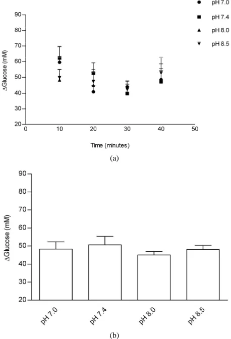
Figure 2. Effects of Tyrode pH on jejunal sodium absorption; (a) represents the time course of the effect of Tyrode pH; (b) represents mean ± SEM for all time points of each pH. The results were expressed by the difference between influx and efflux. Sodium absorption in the group perfused with Tyrode solution pH 7.4 was higher that of the other groups (*P < 0.05). Sodium absorption increased at pH 8.0 when compared with Tyrode pH 8.5 and pH 7.0 (*P < 0.05). *Denotes significant (P < 0.05) difference in relation to the other groups.
Figure3 shows the effect of Tyrode pH on jejunal po-tassium absorption. Absorption in the group that received Tyrode solution pH 8.0 was greater than that in the groups that received solutions with pH 8.5 and 7.4 (P < 0.05). It seems that the absorption of potassium is strongly de- pendent on pH. Moreover, there was a decrease in potas- sium absorption at pH 7.0, indicating that a high concen- tration of protons in the Tyrode solution could be of con- siderable significance for impair potassium jejunal absor- ption. This impairment maybe due to conformational chan- ges in potassium channels since the activation of these channels occurs in extracellular alkalinization [4,18-20].
The novel findings of the present study are that the pH of Tyrode solution in jejunal perfusion did not affect glu- cose absorption, whereas greater potassium absorption occurred with Tyrode pH 8.0 and greater sodium absorp- tion occurred with Tyrode pH 7.4.
(a)
[image:3.595.58.290.92.429.2](b)
Figure 3. Effects of Tyrode pH on jejunal potassium absorption; (a) represents the time course of the effect o Tyrode pH; (b) represents mean ± SEM for all time points of each pH. The results were expressed by the difference between influx and efflux. Potassium absorption in the group perfused with Tyrode solution pH 8.0 increased when compared with uptake at pH 7.0, 7.4 and 8.5 (*P < 0.05). *Denotes significant (P < 0.05) difference in relation to the other groups.
dition (Tyrode pH 7.4) or alkalinization (Tyrode pH 8.5) reduced potassium absorption when compared to Tyrode pH 8.0. The mechanism proposed for potassium absorp-tion in the small intestine is passive diffusion through either an intercellular or paracellular pathway, favoring a potential transmembrane gradient due to water absorp-tion secondary to the active transport of sodium or other solutes that concentrate potassium within the intestinal lumen [26]. It has also been suggested that potassium may be absorbed by an active transport mechanism oper-ating through the cellular pathway [26]. Interestingly, potassium is the most abundant ion in intracellular fluid and plays an important role in acid-base homeostasis. Moreover, the results of the present study suggest that potassium absorption mechanisms seem to be dependent on pH. The active transport mechanism is likely to be influenced by pH.
4. CONCLUSION
In conclusion, Tyrode pH did not affect jejunal glucose absorption, but did affect jejunal electrolyte absorption. The optimal value for jejunal potassium absorption was pH 8.0 and the optimal value for sodium absorption was 7.4, which did not differ significantly from the result obtained with pH 8.0. These results suggest that a Tyrode solution with pH 8.0 is the best choice in studies involv-ing jejunal perfusion and glucose and electrolyte absorp-tion.
REFERENCES
[1] Daniel, H. and Gertrud, R. (1986) Effect of metabolizable
sugars on the mucosal surface pH of rat intestine. Journal
of Nutrition, 116, 768-777.
[2] Lucas, M.L. Lei, F.H. and Blair, J.A. (1980) The influ-
ence of buffer pH, glucose and sodium ion concentration on the acid microclimate in rat proximal jejunum in vitro.
Pflüegers Archives, 385, 137-142.
doi:10.1007/BF00588693
[3] Blair, J.A., Lucas, M.L. and Matty, A.J. (1975) Acidifica-
tion in the rat proximal jejunum. Journal of Physiology,
245, 333-350.
[4] Sandoval, M., Burgos, J., Sepúlveda, F.V. and Pablo, L.
(2011) Extracellular pH in restricted domains as a gating signal for ion channels involvement in transepithelial trans-
[7] Hubel, K.A. (1976) Intestinal ion transport: Effect of
norepinephrine, pilocarpine, and atropine. The American
Journal of Physiology, 231, 252-257.
[8] Borges, E.L., Machado, A.D.C.V., Haibara, A.S. and Pe-
troianu, A. (2003) Effects of vasoactive intestinal polypep- tide microinjected into the nucleus tractus solitarius on je-
junal glucose absorption in rats. Autonomic Neuroscience:
Basic and Clinical, 107, 111-113.
doi:10.1016/S1566-0702(03)00074-2
[9] Machado, D.C.V., Haibara, A.S., Petroianu, A. and Borges,
E.L. (2005) Effects of vasoactive intestinal polypeptide microinjected into the nucleus tractus solitarius on jejunal electrolytes absorption in rats. Neuropeptides, 39, 15-19.
doi:10.1016/j.npep.2004.10.001
[10] Nogueira, M.C., Haibara, A.S. and Borges, E.L. (2010)
Effect of L-NAME microinjected into the nucleus tractus solitarius on jejunal glucose and electrolyte absorption in anesthetized rats. Brain Research, 1359, 107-115.
doi:10.1016/j.brainres.2010.08.079
[11] Vaz, G.C., Xavier, C.H., Coimbra, C.C., Fontes, M.A.P.
and Borges, E.L. (2011) Increased Jejunal Absorption of Glucose in Rats Submitted to Blockade of GABAA
Re-ceptors in the Hypothalamic Paraventricular Nucleus. The
Open Neuroendocrinology Journal, 4, 120-126.
doi:10.2174/1876528901104010120
[12] Cox, H.M. (2007) Neuropeptide Y receptors;
antisecre-tory control of intestinal epithelial function. Autonomic
Neuroscience, 133, 76-85.
doi:10.1016/j.autneu.2006.10.005
[13] Tanaka, K., Morita, H., Suwaki, H., Hosokawa, K. and
Hosomi, H. (1994) Effects of microinjection of kainic acid into the nucleus tractus solitarius on fluid and NaCl
absorption across the jejunum. Journal of the Autonomic
Nervous System, 48, 97-104.
doi:10.1016/0165-1838(94)90025-6
[14] Kim, M.H., Hardin, J.A. and Gall, D.G. (1996) The role
of nitric oxide in the regulation of macromolecular trans- port in rat jejunum. Journal of Physiology, 490, 243-248.
[15] Schirgi-Degen, A. and Beubler, E. (1995) Significance of
nitric oxide in the stimulation of intestinal fluid absorp-
tion in the rat jejunum in vivo. British Journal of Phar-
macology, 114, 13-18.
[16] Fetih, G., Habib, F., Katsumi, H., Okada, N., Fujita, T.,
Attia, M. and Yamamoto, A. (2006) Excellent absorption enhancing characteristics of NO donors for improving the intestinal absorption of poorly absorbable compound com-
pared with conventional absorption enhancers. Drug Me-
doi:10.2133/dmpk.21.222
[17] Varma, M.V.S. and Panchagnula, R. (2005). pH-Depen-
dent functional activity of P-glycoprotein in limiting in- testinal absorption of protic drugs: Kinetic analysis of
quinidine efflux in situ. Journal of Pharmaceutical Sci-
ences, 94, 2632-2643. doi:10.1002/jps.20489
[18] Goldstein, S.A., Bockenhauer, D., O’Kelly, I. and
Zilber-berg, N. (2001) Potassium leak channels and the KCNK
family of two-P-domain subunits. Nature Reviews
Neu-roscience, 2, 175-184. doi:10.1038/35058574
[19] Lotshaw, D.P. (2007) Biophysical, pharmacological, and
functional characteristics of cloned and native
mammal-ian two-pore domain K+ channels. Cell Biochemistry and
Biophysics, 47, 209-256. doi:10.1007/s12013-007-0007-8
[20] Zúñiga, L., Márquez, V., González-Nilo, F.D., Chipot, C.,
Cid, L.P., Sepúlveda, F.V. and Niemeyer, M.I. (2011)
Gating of a pH-sensitive K2P potassium channel by an
electrostatic effect of basic sensor residues on the selec-tivity filter. PLoS One, 6, e16141.
doi:10.1371/journal.pone.0016141
[21] Zahedi, A.S.L. and Alipour, M. (2007) The effects of
insu-lin on glucose and fluid transport in the isolated small in-
testine of normal rats. Life Sciences, 81, 26-30.
doi:10.1016/j.lfs.2007.04.021
[22] Kato, A. and Romero, M.F. (2011) Regulation of elec-
troneutral NaCl absorption by small intestine. Annual Re-
view of Physiology, 73, 261-281.
doi:10.1146/annurev-physiol-012110-142244
[23] Zachos, N.C., Tse, M. and Donowitz M. (2005) Molecu-
lar physiology of intestinal Na+/H+ exchange. Annual Re- view of Physiology, 67, 411-443.
doi:10.1146/annurev.physiol.67.031103.153004
[24] Orlowski, J. and Grinstein, S. (2004) Diversity of the
mammalian sodium proton exchanger SLC9 gene family.
Pflüegers Archives, 447, 549-565.
doi:10.1007/s00424-003-1110-3
[25] Therien, A.G. and Blostein, R. (2000) Mechanisms of
sodium pump regulation. American Physiologial Society,
279, 541-566.
[26] Inagaki, E., Kawamata, K. and Suzuki, Y. (2002) In vitro
potassium transport in the mouse small intestine.
Japa-nese Journal of Physiology, 52, 515-520.