Original Article
Response gene to complement-32 mediates
TGF-β-induced epithelial-mesenchymal transition
via Erk-/p38-MAPK signaling pathways in human
pancreatic cancer cell line BxPC-3
Liang Zhu1*, Ying Ding2*, Qiu Zhao3
1Department of Gastroenterology, The First Affiliated Hospital of Nanchang University, Nanchang, Jiangxi, China; 2Department of Plastic and Cosmetic Surgery, The Second Affiliated Hospital of Nanchang University, Nanchang,
Jiangxi, China; 3Department of Gastroenterology, Zhongnan Hospital of Wuhan University, Wuhan, Hubei, China. *Equal contributors.
Received March 8, 2017; Accepted May 3, 2017; Epub June 1, 2017; Published June 15, 2017
Abstract: Pancreatic cancer (PC) is a highly life-destructive human digestive system carcinoma, the five-year survival
of which is less than 6%. The PC progression was believed to be associated with epithelial-mesenchymal transition
(EMT) which could be stimulated by TGF-β. We previously found that response gene to complement-32 (RGC-32) mediated TGF-β-induced EMT. However, the underlying mechanism remained unclear. In the present study, BxPC-3 cells were used and treated with 10 ng/mL TGF-β1 alone or in combination with 10 µM U0126/SB20BxPC-3580/
LY294002. The mRNA and protein expressions of RGC-32, E-cadherin and vimentin were analyzed by qRT-PCR and
Western blot. The siRGC-32 was introduced to measure the expression of Snail and Zeb1 in BxPC-3 cells treated with or without TGF-β1. The results showed that U0126/SB203580 combined with TGF-β1 inhibited EMT develop -ment, decreased the mRNA and protein expressions of RGC-32 and vimentin, and increased those of E-cadherin
compared with the cells treated with TGF-β1 alone. After combined use of siRGC-32 and TGF-β1, the EMT occur -rence and the levels of RGC-32, snail and Zeb1 were all inhibited compared with the cells treated with siCtrl alone
or siCtrl + TGF-β1. The present study demonstrated that the regulation of RGC-32 on TGF-β-induced EMT might be
associated with non-Smad pathways, and Erk-MAPK and p38-MAPK pathways seemed to play more important roles than PI3K-Akt pathway in PC progression.
Keywords: RGC-32, EMT, TGF-β, pancreatic cancer, signaling pathways
Introduction
Pancreatic cancer (PC) is a highly life-destruc-tive human digeslife-destruc-tive system carcinoma charac-terized by the evident parallel between its inci-dence and mortality. The overall five-year sur-vival was reported to be less than 6% in PC patients in USA even after the therapeutic inter-ventions alone or in combination (such as sur-gery, radiation, chemotherapy) [1]. Although PC occurs usually in the elderly population, family history, smoke, chronic pancreatitis, obesity and diabetes mellitus are believed to be the main risk factors for PC occurrence and devel-opment [2].
Epithelial-mesenchymal transition (EMT) is a critically biological process that participates in
lopment and fibrosis, wound healing, tissue regeneration as well as cancer progression [3]. Mounting evidences demonstrated that EMT played an essential role in a variety of in vitro
progres-[14]. TGF-β pathway was reported to be either Smad-dependent or -independent [15].
Response gene to complement 32 (RGC-32), a gene first cloned by Badea et al. in 1998, was induced by complement involving in cell cycle activation [16]. RGC-32 can be found compre-hensively in heart, brain, liver, skeletal muscle, placenta, kidney, and pancreas [17]. Overexpre- ssion of RGC-32 has been observed in colon cancer and many other tumors [18]. In a pre- vious work, we demonstrated that RGC-32 could mediate TGF-β-induced EMT in human PC cells [7]. However, the underlying mecha-nism remained largely unknown. In the present study, the involvements of three non-Smad sig-naling pathways, i.e. PI3K-Akt, Erk-MAPK and p38-MAPK in TGF-β-induced EMT mediated by RGC-32 were further explored.
Materials and methods
Cell culture and treatments
The BxPC-3 human pancreatic cancer cell line was provided by the Institute of Liver Disease, Tongji Hospital, Tongji Medical College, Hua- zhong University of Science and Technology, Wuhan, Hubei, China. BxPC-3 cells were cul-tured in RPMI-1640 (GIBCO, NY, United States) culture medium containing 10% fetal bovine serum (FBS, Gibco), 300 mg/L glutamine, 100 U/mL penicillin and 100 μg/mL streptomycin at 37°C in a humidified atmosphere of 95% air and 5% CO2. The cells were seeded in a 6-well plate at a density of 3 × 10⁵ cells/well. After reaching 70% of confluency, the media was removed and the cells were washed with PBS twice. Then, BxPC-3 cells were incubated in serum-free RPMI-1640 supplemented with 10 ng/mL TGF-β1 and 10 µM U0126/SB203- 580/LY294002 (Beyotime, Shanghai, China) for another 72 h. The cells treated with TGF-β1
restriction sites. The cloned cDNA was sequ- enced for verification. The shRGC-32 sequen- ce was CAGAUUCACUUUAUAGGAAUUCCUAUA- AAGUGAAUCUG. The scrambled shRNA (shCtrl) sequence was CGCCTCTCTCTTAGTGAGATTTC- AAGAGAATCTCACTAAGAGAGAGGCCT and used as the negative control. Both shRNA sequen- ces were synthesized by Ribobio (Guangzhou, China). After plasmid construction, BxPC-3 cells were seeded in a 6-well plate at a density of 3 × 10⁵ cells/well and incubated to reach 95% of confluency. Then, the cells were tran-siently transfected with lipofectamine 2000 (Invitrogen, USA) according to the manufactur-er’s recommendations. Freshly prepared plas-mid DNA (= 4.0 μg) and lipofectamine 2000 (= 10 μl) diluted separately in Opti-MEM I medium (Gibco, USA) were co-incubated for 30 min at 37°C. Then, the solution was added into each well and mixed with the cells for another 5 h at 37°C. Subsequently, the medium was replaced with RPMI-1640 medium containing 10% FBS and incubated for another 72 h. For shRGC-32 transfection, BxPC-3 cells were seeded in a 6-well plate at a density of 1 × 10⁵ cells/well and incubated to reach 50% of confluency. The cells were transfected with 50 nM shRGC-32/ shCtrl using lipofectamine 2000 at 37°C in a 5% CO2 incubator (Thermo, USA) according to the manufacturer’s instructions. After 6 h of transfection, the cells were transferred to fre- shly prepared RPMI-1640 for another 72 h.
Quantitative reverse transcription polymerase chain reaction (qRT-PCR)
total RNA (= 2 μg) was transcribed into cDNA followed by incubations with 25 μL of reverse transcription reaction mixture containing 5 μL of 5 × RT Reaction Buffer, 3 μL of dNTPs (10 mmol/L), 1 μL of Oligo (dT), 1 μL of M-MLV (Promega, USA), 1 μL of Rnasin (Fermentas, USA) and indicated amount of DEPC water. The conditions of reverse transcription reaction were 70°C for 5 min, 37°C for 1 h and 85°C for 10 min. The primers of RGC-32, E-cadherin, vimentin and GAPDH were synthesized by Sangon (Shanghai, China) and the sequences were as follows: RGC-32 forward: 5’-AGAAG- GCTGGGGCTCATTTG-3’, reverse: 5’-AGGGGCC- ATCCACAGTCTTC-3’ E-cadherin forward: 5’-AA- TGCCGCCATCGCTTAC-3’, reverse: 5’-CCACCA- GGGTATACGTAGGGA-3’; Vimentin forward: 5’- CCGCTTCGCCAACTACATC-3’, reverse: 5’-GGTT-
AGCTGGTCCACCTGCC-3’; GAPDH forward: 5’- AGAAGGCTGGGGCTCATTTG-3’, reverse: 5’-AG- GGGCCATCCACAGTCTTC-3’. GAPDH was used as the reference gene. The qPCR was per-formed with StepOne real-time PCR systems (ABI, USA) in a reaction volume of 20 μL con-taining 2 μL of cDNA, 0.8 μL of forward primer (10 nM), 0.8 μL of reverse primer (10 nM), 10 μL of SYBR Green Real-time PCR Master Mix (Toyobo, Japan) and 6.4 μL of ddH2O. The pro-cedures was followed by 95°C for 60 s, 40 cycles of 95°C for 15 s and 60°C for 30 s. The analysis of qPCR was carried out using the 2-ΔΔCt method as described before [19].
Western blot
Western blot assay was performed as descri- bed previously [7]. Briefly, Total proteins were extracted from BxPC-3 cells using EpiQuik Total Histone Extraction Kit (Epigentek, Farmingdale, NY, USA). The protein was quantified using a BCA Protein Quantification Kit (Vazyma, Nan- jing, Jiangsu, China) according to the manufac-turer’s instructions. Then, 80 μg of cell protein was separated on a 12% SDS-PAGE and trans-ferred to nitrocellulose membranes (Millipore, Billerica, MA, USA). The nitrocellulose mem-branes were blocked at room temperature for 2 h in blocking buffer (5% skim milk in TBST) and then respectively incubated with RGC-32 anti-body (1:200), E-cadherin antianti-body (1:400) and vimentin antibody (1:1000, ProteinTech Group, Inc., USA) overnight at 4°C. β-actin antibody (1:1000, ProteinTech Group, Inc., USA) was set as the control. After three times of washing with TBST, nitrocellulose membranes were incubat-ed in HRP-conjugatincubat-ed goat anti-rabbit
second-Figure 1. Representative images of EMT after the treatments of TGF-β1, TGF-β1 + U0126, TGF-β1 + SB203580 and TGF-β1 + LY294002 in BxPC-3 cells. U0126, SB203580 and LY294002 are three specific inhibitors of Erk-MAPK, p38-MAPK and PI3K-Akt pathways. The cells treated with TGF-β1 was set as the control.
Figure 2. qRT-PCR results revealed the effects of
TGF-β1, TGF-β1 + U0126, TGF-β1 + SB203580 and TGF-β1 + LY294002 on relative mRNA expressions of RGC-32, E-cadherin and Vimentin after 72-h of incubations in BxPC-3 cells. GAPDH was set as the
reference gene. *P < 0.05, compared with the cells
[image:3.612.90.525.73.224.2] [image:3.612.91.286.286.410.2]ary antibody (1:3000, Boster, Wuhan, China) for 1 h at room temperature. After three times of washing with TBST, the protein samples were visualized by Super Signal West Pico Chemilu- minescent Substrate (Thermo Fisher Scientific Inc, USA) according to the manufacturer’s in- structions. The blots were scanned and densi-tometric analysis was performed by Image J software (National Institutes of Health, USA).
Statistical analysis
All experiments were performed triplicate in three independent occasions. The data were presented as mean ± standard deviation. The data between two groups were analyzed by the two-tailed Student’s t-test, while the data among groups were analyzed by one-way ana- lysis of variance (ANOVA). Statistical analyses were conducted by SPSS 17.0 (SPSS, Chicago, IL, USA). A value of P < 0.05 was considered statistically significant.
Results
Signaling pathway inhibitors interfered with EMT
Compared with the control treated with TGF-β1, the BxPC-3 cells co-incubated with TGF-β1 and signaling pathway inhibitors (U0126, SB203- 580 and LY294002) displayed evident EMT reversion in cell morphology from a mesen- chymal-like spindle-cell shape with increased
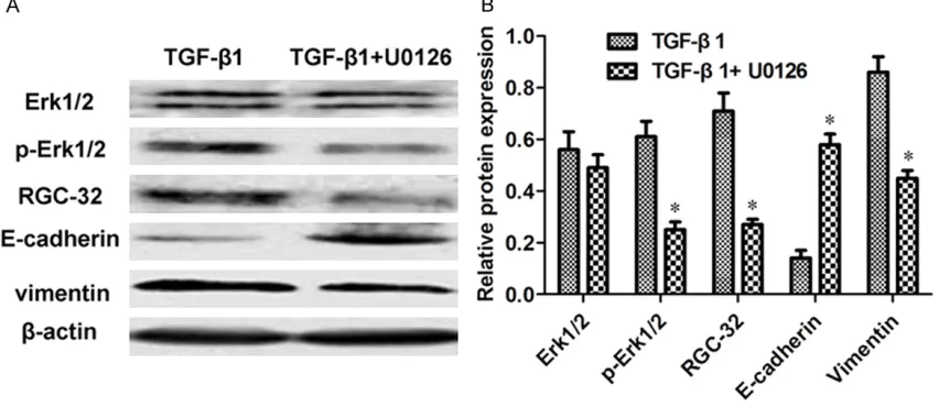
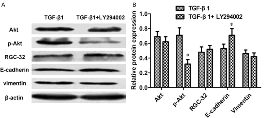
intercellular space to a more epithelial-like appearance with close cell-cell connection (Figure 1). The PCR results showed that the mRNA expressions of RGC-32 and Vimentin were downregulated by 1.41-1.96 fold (P < 0.05) except the cells treated with TGF-β1 + LY294002 with a decrease of 1.04-fold. The mRNA expressions of E-cadherin were upregu-lated by 1.70-1.85 fold (P < 0.05, Figure 2). Compared with the cells treated with TGF-β1 alone, the protein expressions of phosphory-lated Erk1/2 (p-Erk1/2), RGC-32 and Vimentin decreased by 2.44-, 2.63- and 1.91-fold (P < 0.05), while that of E-cadherin increased by 4.14-fold (P < 0.05) after the combination of TGF-β1 and U0126 (Figure 3A and 3B). As for TGF-β1 + SB203580, the protein levels of phos-phorylated p38 (p-p38), RGC-32, Vimentin and E-cadherin experienced 3.44-, 1.66- and 1.81-fold of decreases and 2.15-1.81-fold of increase re- spectively (P < 0.05, Figure 4A and 4B). Follow- ing the combination of TGF-β1 and LY294002, the protein level of phosphorylated Akt (p-Akt) had a decrease of 2.22-fold, while that of E- cadherin owned an increase of 1.34-fold (P < 0.05, Figure 5A and 5B).
Silenced RGC-32 inhibited EMT and the ex-pressions of snail and Zeb1 proteins
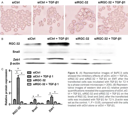
[image:4.612.94.520.79.264.2]The technique of siRNA was employed to sil- ence the expression of RGC-32. It could be observed that siCtrl + TGF-β1 promoted EMT
development, while siRGC-32 + TGF-β1 inhibit-ed EMT progression comparinhibit-ed with the control transfected with siCtrl (Figure 6A). Compared with siCtrl alone, the protein levels of RGC-32, snail and Zeb1 acquired 5.88-, 2.67- and 2.17-fold increases (P < 0.05) after co-treatment of siCtrl + TGF-β1. When siRGC-32 was used in combination with TGF-β1, the RGC-32, snail and Zeb1 levels obtained 47-, 4.44- and 8.13-fold of decreases (P < 0.05) compared with siCtrl + TGF-β1 (Figure 6B and 6C).
Discussion
During the past decade, growing evidence indi-cated that RGC-32 could induce and promote EMT in lung cancer cells, renal tubular cells, renal proximal tubular cells, etc. [17, 20, 21]. In a previous study, RGC-32 was observed to enhance TGF-β-induced EMT in a human PC cell line BxPC3 which was deficient in Smad4 gene [7]. In canonical Smad signaling pathway, Smad2 and Smad3 proteins activated by
phos-Figure 4. The analysis of proteins related with p38-MAPK pathway. (A) Representative images of western blot and
[image:5.612.94.522.75.256.2](B) relative protein quantifications displayed the effects of TGF-β1 and TGF-β1 + SB203580 on the levels of p38, p-p38, RGC-32, E-cadherin and Vimentin after the BxPC-3 cells was incubated with TGF-β1 for 72 h. β-actin was set as the control. *, P < 0.05, compared with the cells treated with TGF-β1 alone.
[image:5.612.94.523.333.525.2]phorylated TGF-βR1 will form a complex with Smad4 protein to regulate EMT-related genes. Smad4, a tumor suppressor gene, is in charge of suppressing epithelial cell growth via TGF-β signaling pathway [22]. Although it is functional in multiple cancers, Smad4 is believed to lose activity in more than 50% PC cases resulting in poor prognosis [23]. Therefore, it was suggest-ed that RGC-32 might msuggest-ediatsuggest-ed TGF-β-inducsuggest-ed EMT via non-Smad pathways. Deciphering the roles of non-Smad pathways in EMT progres-sion during PC processes will provide new in- sights into the plasticity of cellular phenotypes and possible therapeutic interventions.
It is a common strategy to employ inhibitors to investigate relevant signaling pathways. U0126, SB203580 and LY294002 have been widely used to block Erk-MAPK, p38-MAPK and PI3K-Akt pathways which are the three
exten-sively studied non-Smad signaling pathways in cancer development [24-26]. Compared with the control treated with TGF-β, the three inhibi-tors could hinder the EMT development induc- ed by TGF-β in treated PC cells. Subsequently, we analyzed the expressions of several critical genes and proteins related with the three path-ways to further evaluate their roles in TGF-β-induced EMT mediated by RGC-32.
Erk-MAPK is one of the most clarified MAPK pathways and has been reported to be related with cell proliferation, differentiation, migra-tion, senescence and apoptosis [27]. Erk pro-teins are a family of serine/threonine kinases including ERK 1 (MW 44 KD) and ERK2 (MW 42 KD) and can be activated by MEK1/2 [28]. Erk-MAPK pathway was believed to be involved in the regulations of prostate cancer cell prolif-eration and migration by chloride intracellular
Figure 6. (A) Representative images of BxPC-3 cells showed the inhibitory effects of siCtrl, siCtrl + TGF-β1, siRGC-32 and siRGC-32 + TGF-β1 on EMT after the transfected cells was incubated with TGF-β1 for 72 h by a phase contrast microscope (× 200). (B) Represen -tative images of western blot and (C) relative protein
quantifications revealed the suppressions of siCtrl, siC
-trl + TGF-β1, siRGC-32 and siRGC-32 + TGF-β1 on the
levels of RGC-32, Snail and Zeb1 after the transfected
cells was incubated with TGF-β1 for 72 h. β-actin was
set as the control. *, P < 0.05, compared with the cells
[image:6.612.89.522.73.461.2]channel 1, the proliferations of renal-cell noma by β-Elemene and hepatocellular carci-noma cell by lysyl oxidase propeptide [29-31]. In this study, the levels of phosphorylated Erk1/2 (p-Erk1/2), RGC-32, E-cadherin (epithe-lial markers) and vimentin (mesenchymal mark-ers) were altered significantly, indicating an in- volvement of Erk-MAPK pathway in TGF-β-in- duced EMT mediated by RGC-32.
P38 proteins are also a group of serine/th- reonine kinases including four members, i.e. p38α, p38β, p38γ and p38δ [32]. p38-MAPK is another important MAPK pathway and induces a wide variety of biological responses including cell cycle, apoptosis, embryonic development and cancer progression [33]. p38-MAPK path-way could regulate TGF-β-induced EMT in mam-mary epithelial cells [34], adult rat hepatocytes [35], etc. In present study, except the expres-sion of p38, the significant changes of phos-phorylated p38 (p-p38), RGC-32, E-cadherin and vimentin implied a participation of p38-MAPK pathway in TGF-β-induced EMT mediated by RGC-32.
PI3K ubiquitous in the cytoplasm has a re- gulatory subunit (p85) and a catalytic subunit (p110). PI3K-Akt signaling pathway critical for cell proliferation and EMT possesses the activi-ties of both phosphatidylinositol kinase and serine/threonine protein kinase [36]. Increas- ing evidence revealed that PI3K-Akt pathway was closely associated with TGF-β-induced EMT [37-39]. To our surprise, only the levels of p-Akt and E-cadherin changed significantly. These results suggested that PI3K-Akt path- way seemed not to be implicated in TGF-β-induced EMT mediated by RGC-32.
Snail and Zeb1 proteins belong to zinc finger transcriptional factors and act as inducers of ETM by downregulating E-cadherin and upre- gulating Vimentin [40]. Both proteins support tumor growth and progression in different can-cer types via Erk-MAPK/p38-MAPK/PI3K-Akt pathway [41, 42]. After silencing RGC-32, Snail and Zeb1 levels decreased significantly, infer-ring that Snail and Zeb1 proteins might be downstream targets of RGC-32.
Of note, TGF-β has been demonstrated to acti-vate autophagy in a manner dependent on both Smad and non-Smad pathways [43]. We also noticed that Smad3 and Smad2 were assumed
to form transcriptional complexes with other transcriptional factors in favor of tumor ini- tiation and progression without Smad4 [44]. Therefore, both Smad and non-Smad pathways might be associated with TGF-β-induced EMT mediated by RGC-32.
In conclusion, we demonstrated that Erk-MA- PK and p38-MAPK pathways might play more important roles than PI3K-Akt pathway in TGF-β-induced EMT mediated by RGC-32. The re- sults in the present study could expand our insight in the molecular mechanisms of TGF-β-induced EMT mediated by RGC-32 and provide new therapeutic targets in PC.
Acknowledgements
This study was supported by National Natural Science Foundation of China (No. 81302152), Project of Health and Family Planning Com- mission of Jiangxi Province (No. 20155207) and 2017 Natural Science Foundation of Jiangxi Province (Project title: The research on the effect of RGC-32 in hypoxia-induced EMT in pancreatic cancer and the underlying mechanism).
Disclosure of conflict of interest
None.
Address correspondence to: Dr. Liang Zhu, De-
partment of Gastroenterology, The First Affiliated Hospital of Nanchang University, 17 Yongwaizheng
Street, Nanchang 330006, Jiangxi, China. Tel: +86-791-8869-4228; Fax: +86-+86-791-8869-4228; E-mail: [email protected]
References
[1] Kamisawa T, Wood LD, Itoi T and Takaori K.
Pancreatic cancer. Lancet 2016; 388: 73-85. [2] Yabar CS and Winter JM. Pancreatic cancer: a
review. Gastroenterol Clin North Am 2016; 45: 429-45.
[3] Rasheed ZA, Yang J, Wang Q, Kowalski J, Freed I, Murter C, Hong SM, Koorstra JB, Rajeshku
-mar NV, He X, Goggins M, Iacobuzio-Donahue C, Berman DM, Laheru D, Jimeno A, Hidalgo M, Maitra A and Matsui W. Prognostic significance
of tumorigenic cells with mesenchymal fea-tures in pancreatic adenocarcinoma. J Natl Cancer Inst 2010; 102: 340-51.
-Sci 2016; 17: 1285.
[7] Zhu L, Qin H, Li PY, Xu SN, Pang HF, Zhao HZ, Li
DM and Zhao Q. Response gene to comple-ment-32 enhances metastatic phenotype by mediating transforming growth factor beta-in-duced epithelial-mesenchymal transition in
human pancreatic cancer cell line BxPC-3. J
Exp Clin Cancer Res 2012; 31: 29.
[8] Kalluri R and Weinberg RA. The basics of epi -thelial-mesenchymal transition. J Clin Invest 2009; 119: 1420-8.
[9] Su Y, Li J, Witkiewicz AK, Brennan D, Neill T,
Talarico J and Radice GL. N-cadherin
haploin-sufficiency increases survival in a mouse mod -el of pancreatic cancer. Oncogene 2012; 31: 4484-9.
[10] Javle MM, Gibbs JF, Iwata KK, Pak Y, Rutledge
P, Yu J, Black JD, Tan D and Khoury T.
Epithelial-mesenchymal transition (EMT) and activated extracellular signal-regulated kinase (p-Erk) in surgically resected pancreatic cancer. Ann Surg Oncol 2007; 14: 3527-33.
[11] Thiery JP, Acloque H, Huang RY and Nieto MA.
Epithelial-mesenchymal transitions in develop-ment and disease. Cell 2009; 139: 871-90. [12] Jones S, Zhang X, Parsons DW, Lin JC, Leary
RJ, Angenendt P, Mankoo P, Carter H, Kamiya
-ma H, Jimeno A, Hong SM, Fu B, Lin MT, Cal
-houn ES, Kamiyama M, Walter K, Nikolskaya T, Nikolsky Y, Hartigan J, Smith DR, Hidalgo M,
Leach SD, Klein AP, Jaffee EM, Goggins M, Mai-tra A, Iacobuzio-Donahue C, Eshleman JR,
Kern SE, Hruban RH, Karchin R, Papadopoulos N, Parmigiani G, Vogelstein B, Velculescu VE and Kinzler KW. Core signaling pathways in hu -man pancreatic cancers revealed by global ge-nomic analyses. Science 2008; 321: 1801-6. [13] Truty MJ and Urrutia R. Basics of TGF-beta and
pancreatic cancer. Pancreatology 2007; 7: 423-35.
[14] Mu Y, Gudey SK and Landstrom M. Non-Smad signaling pathways. Cell Tissue Res 2012; 347: 11-20.
[15] Derynck R, Muthusamy BP and Saeteurn KY.
Signaling pathway cooperation in
TGF-beta-in-Overexpression of RGC-32 in colon cancer and other tumors. Exp Mol Pathol 2005; 78: 116-22.
[19] Livak KJ and Schmittgen TD. Analysis of rela-tive gene expression data using real-time
quantitative PCR and the 2-ΔΔCT method.
Methods 2001; 25: 402-08.
[20] Sun Q, Yao X, Ning Y, Zhang W, Zhou G and
Dong Y. Overexpression of response gene to complement 32 (RGC32) promotes cell inva-sion and induces epithelial-mesenchymal
tran-sition in lung cancer cells via the NF-kappaB signaling pathway. Tumour Biol 2013; 34:
2995-3002.
[21] Guo X, Jose PA and Chen SY. Response gene to
complement 32 interacts with Smad3 to pro-mote epithelial-mesenchymal transition of hu-man renal tubular cells. Am J Physiol Cell Physiol 2011; 300: C1415-21.
[22] Miyaki M and Kuroki T. Role of Smad4 (DPC4)
inactivation in human cancer. Biochem Bio -phys Res Commun 2003; 306: 799-804. [23] Singh P, Wig JD and Srinivasan R. The Smad
family and its role in pancreatic cancer. Indian J Cancer 2011; 48: 351-60.
[24] Montagut C and Settleman J. Targeting the RAF-MEK-ERK pathway in cancer therapy. Can-cer Lett 2009; 283: 125-34.
[25] Yong HY, Koh MS and Moon A. The p38 MAPK inhibitors for the treatment of inflammatory
diseases and cancer. Expert Opin Investig Drugs 2009; 18: 1893-905.
[26] Dienstmann R, Rodon J, Serra V and Tabernero
J. Picking the point of inhibition: a comparative review of PI3K/AKT/mTOR pathway inhibitors. Mol Cancer Ther 2014; 13: 1021-31.
[27] Sun Y, Liu WZ, Liu T, Feng X, Yang N and Zhou HF. Signaling pathway of MAPK/ERK in cell
proliferation, differentiation, migration, senes-cence and apoptosis. J Recept Signal Transduct Res 2015; 35: 600-4.
[28] McCubrey JA, Steelman LS, Chappell WH, Abrams SL, Wong EW, Chang F, Lehmann B,
Terrian DM, Milella M, Tafuri A, Stivala F, Libra
Franklin RA. Roles of the Raf/MEK/ERK path-way in cell growth, malignant transformation
and drug resistance. Biochim Biophys Acta
2007; 1773: 1263-84.
[29] Tian Y, Guan Y, Jia Y, Meng Q and Yang J. Chloride intracellular channel 1 regulates prostate cancer cell proliferation and migra-tion through the MAPK/ERK pathway. Cancer
Biother Radiopharm 2014; 29: 339-44.
[30] Zhan YH, Liu J, Qu XJ, Hou KZ, Wang KF, Liu YP and Wu B. beta-Elemene induces apoptosis in
human renal-cell carcinoma 786-0 cells through inhibition of MAPK/ERK and PI3K/ Akt/ mTOR signalling pathways. Asian Pac J Cancer Prev 2012; 13: 2739-44.
[31] Zheng Y, Wang X, Wang H, Yan W, Zhang Q and Chang X. Expression of the lysyl oxidase pro -peptide in hepatocellular carcinoma and its clinical relevance. Oncol Rep 2014; 31: 1669-76.
[32] Cuadrado A and Nebreda AR. Mechanisms
and functions of p38 MAPK signalling. Bio -chem J 2010; 429: 403-17.
[33] Bradham C and McClay DR. p38 MAPK in de -velopment and cancer. Cell Cycle 2006; 5: 824-8.
[34] Gal A, Sjoblom T, Fedorova L, Imreh S, Beug H
and Moustakas A. Sustained TGF beta expo-sure suppresses Smad and non-Smad signal-ling in mammary epithelial cells, leading to EMT and inhibition of growth arrest and apop-tosis. Oncogene 2008; 27: 1218-30.
[35] Kojima T, Takano K, Yamamoto T, Murata M,
Son S, Imamura M, Yamaguchi H, Osanai M, Chiba H, Himi T and Sawada N. Transforming
growth factor-beta induces epithelial to mes-enchymal transition by down-regulation of claudin-1 expression and the fence function in adult rat hepatocytes. Liver Int 2008; 28: 534-45.
[36] Xu W, Yang Z and Lu N. A new role for the PI3K/
Akt signaling pathway in the epithelial-mesen-chymal transition. Cell Adh Migr 2015; 9: 317-24.
[37] Baek SH, Ko JH, Lee JH, Kim C, Lee H, Nam D, Lee J, Lee SG, Yang WM, Um JY, Sethi G and
Ahn KS. Ginkgolic acid inhibits invasion and migration and TGF-beta-induced EMT of lung cancer cells through PI3K/Akt/mTOR inactiva-tion. J Cell Physiol 2017; 232: 346-354.
[38] Yeh YH, Wang SW, Yeh YC, Hsiao HF and Li TK.
Rhapontigenin inhibits TGF-beta-mediated epi-thelialmesenchymal transition via the PI3K/ AKT/mTOR pathway and is not associated with
HIF-1alpha degradation. Oncol Rep 2016; 35:
2887-95.
[39] Chen WC, Lai YA, Lin YC, Ma JW, Huang LF, Yang NS, Ho CT, Kuo SC and Way TD. Curcumin
suppresses doxorubicin-induced epithelial-mesenchymal transition via the inhibition of TGF-beta and PI3K/AKT signaling pathways in triple-negative breast cancer cells. J Agric Food Chem 2013; 61: 11817-24.
[40] Peinado H, Olmeda D and Cano A. Snail, Zeb and bHLH factors in tumour progression: an al -liance against the epithelial phenotype? Nat Rev Cancer 2007; 7: 415-28.
[41] Hong KO, Kim JH, Hong JS, Yoon HJ, Lee JI, Hong SP and Hong SD. Inhibition of Akt activity
induces the mesenchymal-to-epithelial revert-ing transition with restorrevert-ing E-cadherin
expres-sion in KB and KOSCC-25B oral squamous cell
carcinoma cells. J Exp Clin Cancer Res 2009; 28: 28.
[42] Ho MY, Liang CM and Liang SM. MIG-7 and
phosphorylated prohibitin coordinately regu-late lung cancer invasion/metastasis. Oncotar-get 2015; 6: 381-93.
[43] Kiyono K, Suzuki HI, Matsuyama H, Morishita
Y, Komuro A, Kano MR, Sugimoto K and Miya-zono K. Autophagy is activated by TGF-beta and potentiates TGF-beta-mediated growth in-hibition in human hepatocellular carcinoma cells. Cancer Res 2009; 69: 8844-52.
[44] Pessah M, Marais J, Prunier C, Ferrand N,
Lallemand F, Mauviel A and Atfi A. c-Jun associ -ates with the oncoprotein Ski and suppresses
Smad2 transcriptional activity. J Biol Chem