METHODOLOGIC PERSPECTIVES
A New Aneurysm Occlusion Classification after the Impact of
Flow Modification
H.S. Cekirge and I. Saatci
ABSTRACT
SUMMARY: A new classification is proposed for cerebral aneurysms treated with any endovascular technique, for example, coiling with or without adjunctive devices, flow diversion, intrasaccular flow modifiers, or any combination of the above. Raymond-Roy Occlusion Classification is expanded with novel subgroups such as class 1 represents complete occlusion and is subdivided if a branch is integrated to, or originated from, the aneurysm sac; class 2 represents neck filling; class 3 represents incomplete occlusion with aneurysm filling as in the previous classification; and class 4 describes the immediate postoperative status after extra- or intrasaccular flow modification treatment. A new concept, “stable remodeling,” is included as class 5, which represents filling in the neck region that stays unchanged or reduced, as shown with at least 2 consecutive control angiographies, at least 6 months apart, for not⬍1 year, or the remodeled appearance of a dilated and/or tortuous vessel in continuation with the parent artery without sac filling.
ABBREVIATION:FM⫽flow modifier
A
lthough endovascular cerebral aneurysm treatment has al-ready been established,1,2there is still controversy in regardto the possibility of not always providing complete occlusion or showing recanalization. However, it has also been shown that only a small group of incomplete occlusions or aneurysm remnants have clinical relevance.3,4However, the Cerebral Aneurysm
Re-rupture After Treatment study5reported that the degree of
aneu-rysm occlusion after treatment was strongly associated with risk of rerupture. Aneurysm re-treatment may or may not carry a higher risk than the stable incomplete occlusion or recanaliza-tion.6,7Unfortunately, the relevant information mostly originates
from coiled aneurysms and, therefore, may not be generalized to the entire population of cerebral aneurysms, particularly those aneurysms treated with new devices that modify flow from inside or outside of the aneurysm sac.
Flow modifiers (FMs) (dedicated extrasaccular flow divert-ers, multiple stent-in-stent applications, and intrasaccular flow disrupters) have been introduced as a new concept for
treatment of cerebral aneurysms; FMs cause, over time, cura-tive reconstruction of the aneurysm neck. The reconstruction after extrasaccular FMs (ie, flow diverters) starts immediately after the construct is in place and then evolves over a period of weeks to months.8Intrasaccular FMs (ie, flow disrupters) may
result in instantaneous occlusion after placement or may re-quire some time for final effect.9The delay in aneurysm
occlu-sion with FMs occurs consistently if the flow through the sac continues due to flow demand through a branch coming off or integrated into the sac. Hence, in most cases, the initial result immediately after treatment does not represent the ultimate goal, and reconstruction proceeds after surgery over time.8,9
Overall, it is evident that evolution of aneurysm management necessitates development of unified terminology to describe both initial and delayed procedural efficacies, regardless of treatment technique.
In the literature, there have been several classifications pub-lished to describe the appearance of aneurysm and/or remnant filling. The Raymond-Roy Occlusion Classification, also known as the Montreal scale10has been the most widely used, and it
classifies the results after aneurysm coiling, which can be applied immediately after the treatment as well as during the follow-up.10,11In the literature, there exist some articles about the
impli-cations on future management of aneurysm and/or neck remnants that state class 3 aneurysm remnants, according to Ray-mond-Roy Occlusion Classification, are more likely to be re-treated than class 2 neck remnants, which are most often followed up.12,13
Received March 9, 2015; accepted after revision June 8.
From the Department of Radiology (H.S.C), Bayindir Hospital, Ankara, Turkey; and Department of Radiology (I.S.), Yu¨ksek Ihtisas University, Koru Hospital, Ankara, Turkey.
Please address correspondence to Isil Saatci, MD, Incek Bulvari Havuzlubag Evleri No: 11, Incek, Golbasi, Ankara, 06830, Turkey; e-mail: [email protected]
Indicates open access to non-subscribers at www.ajnr.org Indicates article with supplemental on-line photo.
The introduction of extrasaccular flow diverters created a need for different classifications, not only to describe the initial results but also to anticipate the outcome, including the risk of infrequent but severe complications of postoperative rup-ture.14-16However, these classifications are exclusively for the
extrasaccular FMs, and, yet, none has gained common acceptance.
Subsequently, with the use of intrasaccular FMs, the control angiographic findings become even more controversial. Lubicz et al17described 4 patterns in the follow-up of intrasaccular FM
treatment, namely, complete occlusion, filling of the proximal recess of the device, neck remnant, and aneurysm remnant. Based on their follow-up experience, the investigators suggested a dif-ferent definition, that is, “adequate occlusion,” which includes the first 3 patterns.
So far in the literature, none of the classifications address the entire spectrum of current endovascular aneurysm treatment, ir-respective to treatment technique. To solve this problem of am-biguity and to exclude the need for the use of different classifica-tions for different modalities, we propose a new classification system, by expanding on the widely used Raymond-Roy Occlu-sion Classification scale, that describes results with current and evolving treatment techniques.
MATERIALS AND METHODS
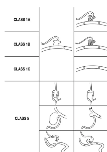
The proposed classification is presented in theTable, and the schematic drawing (Fig 1) highlights the newly added class 1 A, B, and C as well as class 5 in different case settings. Class 1 calls for complete occlusion of an aneurysm when a branch is not directly involved, and the subgroups of class 1—A, B, C—are to be used when a branch is originating directly from a sidewall aneurysm (Figs 2and3) or filling from a bifurcation aneurysm (Fig 4). To assign class 5 (Figs 4Band5–7; On-line Figs 1 and 2), at least 2 angiographic controls, at least 6 months apart, and expanding for a period of not⬍1 year are required to demonstrate the stability. DSA is the criterion standard at the moment, although CTA may serve the purpose for the aneurysms treated with FMs. However, with the evolving technology, noninvasive angiographic imaging may replace this in the future. The remodeling concept18was first
defined in a more restricted concept that referred to 1 as “in-fundibulum like” enlargement of the branch coming off the an-eurysm sac after the shrinkage of the anan-eurysm after flow diverter treatment (Fig 5) and 2, as a tortuous course of the branch (which is coming off the aneurysm sac) at its proximal segment after flow diverter treatment of the sac and the sac not filling (Figs 4and
7).19These 2 situations, in which the branch that originates from
Classification of angiographic results after endovascular treatment with any technique Classification
Class 1: Complete occlusion of the aneurysm sac. When there is a branch integrated with the aneurysm sac, ie, coming off the aneurysm, at any point of the sac, further analysis is carried out with subgroups
1A: Complete occlusion with the full patency of the integrated branch 1B: Complete occlusion with the branch reduced in caliber
1C: Complete occlusion with no antegrade filling of the branch Class 2: Neck filling
Class 3: Incomplete occlusion with aneurysm filling
Class 4: Aneurysm filling. This class is reserved for an immediate postoperative result based on end-of-treatment DSA; after extra-and/or intrasaccular flow modification treatment
4A: With contrast stagnation—contrast stagnation is referred to when there happens to be any change in the duration of the contrast stay within
the aneurysm sac after treatment 4B: Without contrast stagnation
Class 5: Stable remodeling with flow modification. Filling in the neck region, which stays unchanged or reduced; to be included in this group, there have to be at least 2 consecutive control angiographies, by definition, at least 6 months apart, and expanding for a period of not⬍1 year; exceptionally, 1 control angiography could be sufficient for definition of class 5, only in selected cases of contrast filling the branch coming off the sac, with an appearance of a different vessel course than the original, eg, tortuous or dilated, given that it is in continuation with the parent artery with no sac filling
[image:2.594.310.522.258.559.2]the aneurysm sac is in continuation with the parent artery with no filling of the sac any more, are the only exceptions for which we do not require a second angiography to grade as class 5 occlusion
(On-line Fig 1 for class 5 after intrasaccular FM). In the other situations, remodeling is called for after confirmation of the sta-bility (as described above) when 3, the bifurcation appears enlarged after the intrasaccular FM obliterates the sac, which stays stable in the controls (Fig 6), and 4, when there is an unchanged focal bulging at the neck region (On-line Fig 2). Therefore, in such cases, the first control result would be clas-sified as class 2 (neck remnant) by definition; then, after con-firmation of stability, it can be changed to class 5 (Fig 6and On-line Fig 2).
DISCUSSION
The healing process or so-called reconstruction of the vessel wall at the aneurysm neck after FM and/or flow diverter treatment is different from that after endosaccular coiling,18,20-22and
aneu-rysms may behave differently when treated with flow modifica-tion treatments versus endosaccular coiling.23,24Therefore, the
previous classifications, particularly Raymond-Roy Occlusion Classification, described for coiled aneurysms could not address the results of flow modification treatments. Several new classifi-cations14,17have been introduced. In these previous
classifica-tions for extrasaccular flow diverter treatment, flow stagnation is defined as a determining feature.14-16However, the flow
stagna-tion does not necessarily have a direct implicastagna-tion regarding the future or final treatment result. That is, flow stagnation within the aneurysm may or may not result in total occlusion of the aneu-rysm sac. On the contrary, an aneuaneu-rysm that shows no or little, if any, postoperative contrast stagnation may end up in complete occlusion in the follow-up. Yet, the presumed importance of the contrast stagnation is whether or not it has any predictive value in regard to postoperative rupture of the index (treated) aneurysm. This is a controversial issue, with no proven data to date. Our classification system does not emphasize the degree of contrast filling (eg, the pattern, timing) immediately after the FM treat-ment, not only for these reasons but also because it may be rela-tively subjective or may vary in regard to technical parameters, such as contrast injection power, acquisition parameters, and so forth. Contrast stagnation is referred to in the immediate postoperative DSA findings (class 4) after FM treatment to differentiate that grade from the aneurysm filling after aneu-rysm packing (class 3).
In some grading scales previously described exclusively for use after flow diverter treatment,15,16parent artery size is taken into
FIG 2. Class 1A.AandB, Preoperative images show the ICA aneurysm in which the anterior choroidal artery (arrow) is originating from the aneurysm at the neck.C, Six-month control angiography after single Pipeline device (Covidien, Irvine, California) placement demonstrates total occlusion of the aneurysm with the anterior choroidal artery preserved (arrow). Reprinted from Saatci et al.18
[image:3.594.58.530.48.170.2] [image:3.594.65.272.217.626.2]consideration. Parent artery occlusion may occur, at least in some cases, related to the inefficiency of antiaggregating medication, but, the rate of in-stent stenosis is reported to be not any higher
than that of conventional stents18,22and in-stent stenosis is not a
part of this classification.
The patency of the branch that originates from, or is integrated to, the aneurysm is assessed in this classification, which has not been included in previous classifications. The flow demand through the relevant branch is important in its patency after the treatment.18-20Although the aneurysm is getting occluded, the
branch originating from the aneurysm may remain the same, re-duce in caliber, or may not be filling antegrade. This classification can apply to the results of flow diverter treatment not only in sidewall aneurysms but also in bifurcation aneurysms19; the series
of exclusive MCA bifurcation aneurysms treated with flow di-verter placement demonstrated 84% of class 1 occlusions with a variety of occlusion patterns, including branch occlusion in 12%. Another 8% showed class 5 occlusion. In addition, transition be-tween classes is also possible (Fig 4) in the group of aneurysms with integrated branches, whether located at the side wall or the bifurcation.
This classification is also well grounded for the intrasaccu-lar FM treatments in that it takes not only the involving branches into consideration but also the interval change in regard to stability. With these features, this classification is differentiated from the classification for intrasaccular FMs previously described.17
Another advantage of this proposed classification is that it can be applied by single angiographic images, in several planes, and does not necessitate seeing the entire series of the angiographic run to evaluate the flow pattern within the aneurysm. Therefore, objective findings on the angiographic image are enough for clas-sification, with no further subjective interpretation. However, ro-tational angiography may be a requisite, particularly in the defi-nition of class 5 in some cases.
CONCLUSIONS
With preventing rupture as the ultimate goal of cerebral aneu-rysm treatment, if the treatment serves this purpose and the an-eurysm is secured, then the treatment can be called “successful” and the angiographic appearance of the vascular reconstruction can be considered irrelevant. Long-term stability of remodeling is to be determined and not free of concern until that time.
How-FIG 4. Class 5 evolving into class 1B eventually.A, Preoperative angiography shows right MCA aneurysm with a branch coming off from the sac.
B, A 3D image from a 6-month control angiography after treatment with single Pipeline device shows remodeling of the flow with a tortuous appearance of the branch proximally at its direct continuation with the parent artery, and no sac filling. This appearance is referred to as class 5.C, An 18-month control angiography shows complete occlusion of the aneurysm with the originating branch in reduced caliber, that is, class 1B. Reprinted from Yavuz et al.19
[image:4.594.58.530.47.186.2] [image:4.594.65.271.248.614.2]ever, in complex aneurysms where “perfect” anatomic results may not be possible or at least have increased risks, then, “re-modeling” may be acceptable, given that it is stable. This clas-sification not only addresses the new concept of remodeling that emerged after the use of extra- and intrasaccular flow
modifications but also covers the entire spectrum of result possibilities with any current technique.
We acknowledge that this classification should be validated in regard to the interobserver agreement to test its reproducibility. In addition, we also plan to extract a random sample of credible size among our entire aneurysm population to review the stability in long-term follow-up (eg, 5 years) to investigate the outcome of “designated” classes. This classification may then be ready to es-tablish a clinical algorithm for follow-up, including the risk of bleeding after endovascular treatment.
Disclosures: H. Saruhan Cekirge—UNRELATED:Consultancy: Covidien/Medtronic, MicroVention, Sequent. Isil Saatci—UNRELATED: Consultancy: Covidien, Medtronic.
REFERENCES
1. Molyneux A, Kerr R, Stratton I, et al; International Subarachnoid Aneurysm Trial (ISAT) Collaborative Group.International Sub-arachnoid Aneurysm Trial (ISAT) of neurosurgical clipping versus endovascular coiling in 2143 patients with ruptured intracranial aneurysms: a randomised trial.Lancet2002;360:1267–74CrossRef Medline
2. McDougall CG, Spetzler RF, Zabramski JM, et al.The Barrow Rup-tured Aneurysm Trial. J Neurosurg 2012;116:135– 44 CrossRef Medline
3. Campi A, Ramzi N, Molyneux AJ, et al.Retreatment of ruptured cerebral aneurysms in patients randomized by coiling or clipping in the International Subarachnoid Aneurysm Trial (ISAT).Stroke
2007;38:1538 – 44CrossRef Medline
4. Molyneux AJ, Birks J, Clarke A, et al.The durability of endovascular coiling versus neurosurgical clipping of ruptured cerebral aneurysms: 18 year follow-up of the UK cohort of the International Subarachnoid Aneurysm Trial (ISAT). Lancet 2015;385:691–97
CrossRef Medline
5. Johnston SC, Dowd CF, Higashida RT, et al; CARAT Investigators.
Predictors of rehemorrhage after treatment of ruptured intracra-nial aneurysms: the Cerebral Aneurysm Rerupture After Treat-ment (CARAT) study.Stroke2008;39:120 –25CrossRef Medline
6. Molyneux AJ, Kerr RS, Birks J, et al; ISAT Collaborators.Risk of recurrent subarachnoid haemorrhage, death, or dependence and standardised mortality ratios after clipping or coiling of an intra-cranial aneurysm in the International Subarachnoid Aneurysm Trial (ISAT): long-term follow-up.Lancet Neurol 2009;8:427–33
CrossRef Medline
7. Crobeddu E, Lanzino G, Kallmes DF, et al.Review of 2 decades of aneurysm-recurrence literature, part 2: managing recurrence after
FIG 6. Class 5.A, Preoperative angiography shows a large left MCA aneurysm at the trifurcation, with the branches incorporated in the sac.B, Six months after the treatment with a WEB device (Sequent Medical, Aliso Viejo, California), control angiography shows neck filling at the trifurcation, with the branches patent, and this result is classified as class 2.C, An 18-month control angiography shows an unchanged appearance of the MCA trifurcation, and this result is classified as class 5, with the apparently “stable” remodeling.
[image:5.594.56.530.47.191.2] [image:5.594.66.270.245.583.2]endovascular coiling. AJNR Am J Neuroradiol 2013;34:481– 85
CrossRef Medline
8. Fiorella D, Lylyk P, Szikora I, et al.Curative cerebrovascular recon-struction with the Pipeline embolization device: the emergence of definitive endovascular therapy for intracranial aneurysms.J Neu-rointerv Surg2009;1:56 – 65CrossRef Medline
9. Ding YH, Lewis DA, Kadirvel R, et al.The Woven EndoBridge: a new aneurysm occlusion device.AJNR Am J Neuroradiol2011;32:607–11
CrossRef Medline
10. Roy D, Milot G, Raymond J.Endovascular treatment of unruptured aneurysms.Stroke2001;32:1998 –2004CrossRef Medline
11. Raymond J, Guilbert F, Weill A, et al.Long-term angiographic re-currences after selective endovascular treatment of aneurysms with detachable coils.Stroke2003;34:1398 – 403CrossRef Medline
12. Mascitelli JR, Moyle H, Oermann EK, et al.An update to the Ray-mond-Roy Occlusion Classification of intracranial aneurysms treated with coil embolization.J Neurointerv Surg2015;7:496 –502
CrossRef Medline
13. Mascitelli JR, Oermann EK, De Leacy RA, et al.Angiographic out-come of intracranial aneurysms with neck remnant following coil embolization.J Neurointerv Surg2015;7:484 – 89CrossRef Medline
14. O’Kelly CJ, Krings T, Fiorella D, et al.A novel grading scale for the angiographic assessment of intracranial aneurysms treated using flow diverting stents.Interv Neuroradiol2010;16:133–37CrossRef Medline
15. Kamran M, Yarnold J, Grunwald IQ, et al.Assessment of angio-graphic outcomes after flow diversion treatment of intracranial aneurysms: a new grading schema.Neuroradiology2011;53:501– 08
CrossRef Medline
16. Grunwald IQ, Kamran M, Corkill RA, et al.Simple measurement of aneurysm residual after treatment: the SMART scale for evaluation
of intracranial aneurysms treated with flow diverters.Acta Neuro-chir (Wien)2012;154:21–26; discussion 26CrossRef Medline
17. Lubicz B, Klisch J, Gauvrit JY, et al.WEB-DL endovascular treat-ment of wide-neck bifurcation aneurysms: short- and midterm re-sults in a European study.AJNR Am J Neuroradiol2014;35:432–38
CrossRef Medline
18. Saatci I, Yavuz K, Ozer C, et al.Treatment of intracranial aneurysms using the Pipeline flow-diverter embolization device: a single-cen-ter experience with long-single-cen-term follow-up results.AJNR Am J Neuro-radiol2012;33:1436 – 46CrossRef Medline
19. Yavuz K, Geyik S, Saatci I, et al.Endovascular treatment of middle cerebral artery aneurysms with flow modification with the use of the Pipeline embolization device.AJNR Am J Neuroradiol2014;35: 529 –35CrossRef Medline
20. Dai D, Ding YH, Kadirvel R, et al.Patency of branches after coverage with multiple telescoping flow-diverter devices: an in vivo study in rabbits.AJNR Am J Neuroradiol2012;33:171–74CrossRef Medline
21. Pierot L, Liebig T, Sychra V, et al.Intrasaccular flow-disruption treatment of intracranial aneurysms: preliminary results of a mul-ticenter clinical study. AJNR Am J Neuroradiol2012;33:1232–38
CrossRef Medline
22. Becske T, Kallmes DF, Saatci I, et al.Pipeline for uncoilable or failed aneurysms: results from a multicenter clinical trial.Radiology2013; 267:858 – 68CrossRef Medline
23. Kadirvel R, Ding YH, Dai D, et al.Cellular mechanisms of aneurysm occlusion after treatment with a flow diverter.Radiology2014;270: 394 –99CrossRef Medline