ORIGINAL RESEARCH
EXTRACRANIAL VASCULAR
Transient Ischemic Attack and Carotid Web
XH. Hu,XX. Zhang,XJ. Zhao,XY. Li, andX Y. Zhao
ABSTRACT
BACKGROUND AND PURPOSE: Carotid web was recognized as a cause of ischemic stroke. We sought to determine the clinical and imaging profiles of patients with a carotid web as well as its association with TIA.
MATERIALS AND METHODS: A retrospective review of carotid CT angiography studies and brain MR imaging in patients with TIA during the past 3 years (n⫽135) was performed to determine the presence of carotid webs by 2 experienced neuroradiologists according to previously published criteria. Demographics and clinical and imaging characteristics are shown by descriptive statistics for patients with an identified carotid web. The agreement in the detection of carotid webs between 2 neuroradiologists was examined usingstatistics.
RESULTS: There were 12 (8.9%) carotid webs at the symptomatic bifurcation and 1 carotid web (0.7%) at the asymptomatic bifurca-tion, and no hyperintensity was seen on DWI of these 12 patients. Eight of these 12 (75%) patients with a carotid web were women. None of the 12 patients with a carotid web had major risk factors or other causes of TIA. Fair-to-good interobserver agreement (⫽ 0.87) was seen for diagnosing a carotid web with CT angiography. The rate (10/12, 83.3%) of short-term recurrent episodes of TIA in patients with TIA with a carotid web was significantly higher than that of patients without a carotid web (15/123, 12.2%) (P⬍.001).
CONCLUSIONS: The incidence of carotid web in patients with TIA was 8.9%. There is an association between carotid web and patients with TIA without other identified risks. Carotid web may be an underestimated risk factor for TIA.
T
ransient ischemic attack is a common ischemic cerebrovascu-lar disease. According to the guidelines proposed by the American Heart Association and American Stroke Association in 2009,1TIA is defined as a transient episode of neurologic dysfunc-tion caused by focal brain, spinal cord, or retinal ischemia, with-out acute infarction. The probability of stroke occurring at 2, 7, 30, and 90 days after TIA onset was approximately 2%– 4%, 6%,4%–10%, and 10%–20% in previous studies.2,3If the patient is treated properly, the risk of stroke at 90 days after TIA could be reduced to 1%–3%.4Therefore, TIA is a stroke-warning event requiring urgent intervention, and its occurrence is also a good time for secondary prevention.
Decisions about TIA treatment and secondary prevention rely on identification of the main cause of the disease. However, in one-third of patients with TIA, the cause cannot be identified.5 In several recent case series, carotid webs were identified as a possible cause of ischemic stroke in a predominantly female population with no otherwise known reason for stroke.6-9A carotid web is defined as a thin intraluminal filling defect along the posterior wall of the carotid bulb in oblique sagittal refor-mats, and, most important, with a septum evident on the axial section on CTA.6-9However, there are few reports on the rela-tionship between a carotid web and TIA. Is carotid web one of the causes of TIA?
Therefore, we hypothesized that a carotid web may be one of the risk factors for TIA without identifiable cause. We retrospectively analyzed carotid CTA data to explore the incidence of carotid webs in patients with TIA and to determine the clinical and imaging profiles of patients with carotid webs as well as their association with TIA.
Received August 25, 2018; accepted after revision December 3.
From the Institute of Diagnostic and Interventional Radiology (H.H., X.Z., J.Z., Y.L.), and Department of Neurology (Y.Z.), Shanghai Jiao Tong University Affiliated Sixth People’s Hospital, Shanghai, People’s Republic of China.
This work was supported by National Natural Science Foundation of China No. 81871329; National Natural Science Foundation of China No. 81471656; Shanghai Talent Development Fund, No. 201555; Shanghai Key Discipline of Medical Imaging grant No. 2017ZZ02005; Shanghai Municipal Education Commission-Gaofeng Clini-cal Medicine Grant Support No. 2016427; CliniClini-cal Science and Technology Innova-tion Project of Shanghai Shen Kang Hospital Development Center No. SHDC22015038; and Shanghai Municipal Science and Technology Commission Medical Guide Project No. 16411968900.
Please address correspondence to Yuehua Li, MD, Institute of Diagnostic and In-terventional Radiology, Shanghai Jiao Tong University Affiliated Sixth People’s Hos-pital, 600, Yishan Rd, Shanghai, 200233, China; e-mail: [email protected]
Indicates open access to non-subscribers at www.ajnr.org
MATERIALS AND METHODS
PatientsThis retrospective study was approved by the ethics committee of the Sixth People’s Hospital of Shanghai Jiao Tong University; the need for informed consent was waived. A chart review of all pa-tients (n⫽819) presenting to our hospital with TIA from January 1, 2015, to December 31, 2017, was undertaken.Figure 1provides details of patient selection for the study.
Clinical Information
Data on age, sex, history of hypertension, diabetes mellitus, car-diac insufficiency, atrial fibrillation, and current smoking were
retrospectively collected. All patients were treated with antiplatelet therapy. Data on routine follow-up within 3 months after discharge were also col-lected. A short-term recurrent episode was considered if TIA relapse occurred during the first hospitalization or within 3 months, and short-term progression of acute cerebral infarction was consid-ered if an ischemic stroke was confirmed on MR imaging.
Image Evaluation
Carotid CTA was performed on a 128 – multidetector row CT scanner with 1-mm thick sections and was acquired from the aortic arch through the circle of Willis. Scanning parameters included the following: gantry rotation time, 0.5 seconds; pitch, 1; voltage, 120 kV(peak); and current, 360 mAs. Intravenous con-trast material (80 mL of iohexol, 300 mg/mL, Omnipaque; GE Healthcare, Piscataway, New Jersey) and a 50-mL saline flush were administered at a rate of 4 mL/s with a power injector. Images were reconstructed with axial, coronal, sagittal, and 3D volume reconstructions.
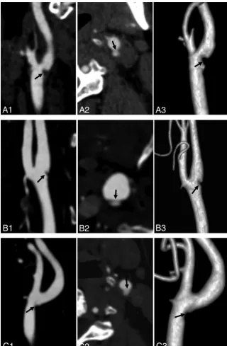
An independent review of these studies was then performed by 2 experienced neuroradiologists with 6 – 8 years of experience in ca-rotid CTA imaging and analysis. The readers were blinded to patient age and sex. The presence of a carotid web was evaluated by each reader at the baseline CTA. A carotid web was identified when there was a thin, smooth, membrane-like intraluminal filling defect along the posterior wall of the carotid bulb on oblique sagittal images and a corresponding thin septum on axial images (Fig 2).5-8Then, there was a consensus reading of all webs identified by the 2 radiologists independently. A carotid web was diagnosed only if both radiologists agreed regarding the presence of a web on sagittal and axial images. The degree of carotid stenosis and the length of the carotid web were measured according to NASCET criteria10and the study of Haussen et al.11The differential diagnosis was performed, as shown inFig 3, when calcified atherosclerotic plaques, carotid artery embolism, ca-rotid artery dissection, or typical fibromuscular dysplasia at 3 cm above and below the carotid bifurcation was detected. FLAIR and DWI imaging were used to assess the presence of TIA-induced brain tissue damage. Follow-up brain MR imaging (DWI and FLAIR) was assessed for the presence of stroke.
Statistical Analysis
Demographic data and clinical information were presented as medians. For patients both with and without a carotid web on CTA, group differences in age were assessed using the Mann-WhitneyUtest. For other baseline values (demographics and risk factors), group differences were assessed using the Fisher exact test. The proportion of carotid webs at the symptomatic and asymptomatic bifurcations was estimated using conditional logis-FIG 1. Flow chart of inclusion criteria.
[image:2.594.58.387.48.339.2] [image:2.594.54.285.365.541.2]tic regression, in which odds ratios were calculated with 95% con-fidence intervals. The Cohenstatistic was used to calculate the interobserver agreement for identifying carotid webs for all pa-tients. Statistical significance was defined asP⬍ .05. Analyses were performed with SPSS 22.0 (IBM, Armonk, New York).
RESULTS
Of the 135 patients with TIA with complete carotid CTA data included in this study, both readers agreed on 12 cases of carotid web (Cohen coefficient, 0.87; 95% CI, 0.55–1). Disagreement was encountered in 2 patients. Furthermore, the 2 cases were both rejected as webs because the 2 readers could not reach an agree-ment. Radiologically, the typical appearance of a carotid web on CTA is a thin intraluminal filling defect arising from the posterior wall of the carotid bulb on oblique sagittal images and a corre-sponding thin septum on axial images (Fig 4). There were 12 (8.9%; 95% CI, 4%–13.8%) carotid webs at the symptomatic bi-furcation and 1 (0.7%; 95% CI, 0%–2.2%) at the asymptomatic bifurcation (odds ratio, 3.4; 95% CI, 1.9 – 6.26;P⬍ .001). As shown inTable 1, the median age (58 years) of patients with a carotid web at the symptomatic carotid bifurcation differed (P⫽ .026) from those (64 years) without a carotid web at the symp-tomatic carotid bifurcation. We found that carotid webs in the symptomatic carotid artery were more often observed in female patients (n⫽9) than in male patients (n⫽3;P⬍.015). However, there were no differences in symptomatic carotid artery location between the right and left sides (P⫽.548). Among 12 patients with a carotid web at the symptomatic side, 1 had a history of smoking (case 2), one had a history of diabetes (case 3), and 10 had no risk factors for TIA such as hypertension, diabetes, atrial fibrillation, and myocardial infarction.
All 135 patients with TIA underwent brain MR imaging within 24 hours after admission. Carotid CTA was performed within 1 week. There was no hyperintensity detected on DWI, in both pa-tients with symptomatic carotid webs and those without carotid webs. The rate (10/12, 83.3%) of short-term recurrent episodes of TIA in patients with TIA with a carotid artery web was signifi-cantly higher than that of patients without a carotid artery web (15/123, 12.2%) (P⬍.00).
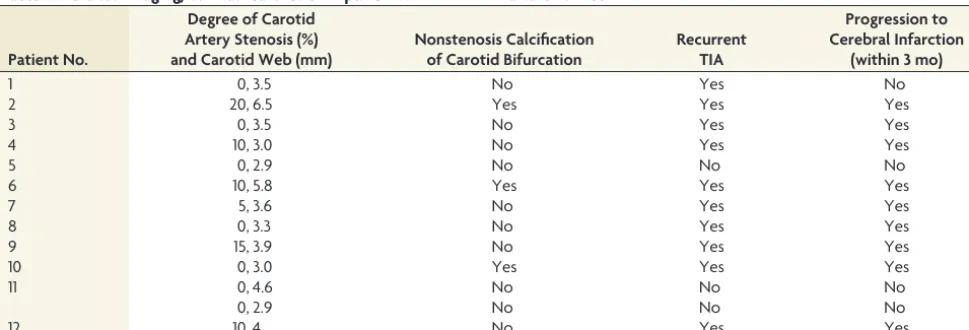
As shown inTable 2, the degree of carotid artery stenosis (NASCET criteria)10was⬍50% (range, 0%–20%), and the
aver-age length of the carotid web was 3.9⫾ 1.1 mm (range, 2.9 – 6.5 mm) in all 12 patients with carotid webs. Further-more, the average size (3.5⫾0.8 mm) of the carotid artery web in patients with a carotid web without recurrent TIA and progressive ischemic stroke within 3 months was smaller than that (4.1⫾1.2 mm) in patients with carotid web with recurrent TIA and deteriorating isch-emic stroke within 3 months (P ⫽ .416). All 12 patients with a carotid web had no typical medial fibromus-cular dysplasia. Three of 12 patients had nonstenotic calcification at the ca-rotid bifurcation.
DISCUSSION
Our findings showed a prevalence of 8.9% of symptomatic carotid webs at the carotid bifurcation in patients with TIA due to carotid system disease. Other notable major risk factors for TIA could not be identified in most patients with a symptomatic carotid web. These findings support our hypothesis that a carotid web may be an important risk factor for ischemic TIA when no other cause is found.
We found a higher prevalence (8.9%) of carotid webs in pa-tients with TIA than in previously reported data (1.2%6 and 2.5%9) according to a hospital-based sample of patients suspected of having stroke and intracranial large-vessel occlusion, respec-tively. TIA, a precursor of ischemic stroke, carries a high risk of recurrent stroke within 90 days from the event,12which could explain the higher prevalence. In addition, the previously re-ported prevalence of a carotid web was 9.4%, 21.2%, and 37%, respectively, in patients with cryptogenic ischemic stroke7,8,13and 23%13in all patients with carotid ischemic stroke. Our result was similar to the earlier reported prevalence (9.4%) of carotid webs in patients with cryptogenic ischemic stroke, but lower than the prevalence of 21.2%, 37%, and 23% in patients with cryptogenic ischemic stroke and all patients with carotid ischemic stroke, re-spectively. Differences in methodology and population character-istics may explain discrepancies in these findings because the prevalence of a carotid web is thought to be increased in African American and Afro-Caribbean compared with white and Asian populations.8,13
In our data, 2 patients with TIA and a carotid web each had a history of smoking and diabetes. While smoking and diabetes were independent risk factors for TIA,14-16 no atheromatous changes were seen on CTA. Thus, a carotid web was the main identifiable cause of our patients’ TIAs. Existing literature about the relation of carotid web and TIA is scarce. Morgenlander and Goldstein17reported that a young female patient with a carotid web but without other identifiable risk factors for stroke pro-gressed to acute ischemic stroke after a recurrent TIA. Thus, our findings are in line with those in the literature.
In our study, carotid webs were more frequently found at the ipsilateral symptomatic bifurcation and in female patients with-out cardiovascular risk factors or other major risk factors for TIA. FIG 3. The differential diagnosis includes calcified atherosclerotic plaques (A), carotid artery
[image:3.594.56.376.47.203.2]This result was consistent with literature reports about patients with stroke with carotid webs.6-9,13However, the median age (58 years) of patients with TIA with a carotid web in our study was higher than that previously reported for patients with stroke with a carotid web,6-9,13which may be related to an older age of onset in patients with TIA.
In this study, the rate of short-term recurrent episodes of TIA within 3 months in patients with TIA with carotid webs was 83.3%, higher than that of patients with TIA without a carotid artery web (12.2%) (P⬍.001) and higher than the average re-ported in the literature.2,3Moreover, the degree of carotid artery
stenosis (NASCET criteria)10of all 12 patients with carotid webs was ⬍50% (range, 0%–20%). Generally, the degree of carotid artery stenosis⬍50% is not considered a risk factor for TIA or isch-emic stroke.7Thus, studies have postu-lated that carotid web–induced ischemic stroke may be mediated by an embolic mechanism, with sluggish/turbulent blood flow produced by the filling defect increasing the thrombogenicity and risk of artery-to-artery embolism.6-9,13,18 Therefore, we speculated that the pres-ence of a carotid web might lead to changes of carotid hemodynamics, which was related to recurrence of TIA and progression to acute ischemic stroke within 3 months in patients with TIA with carotid webs.
However, because of the limitation of small samples and lack of fluid dy-namics data, the relation between the thrombogenicity of webs and the size of the “shelf” of the web was not clear in this study. Although the average size of a carotid web (4.1⫾1.2 mm) in patients with recurrence of TIA and progression to stroke within 3 months was larger than that (3.5⫾ 0.8 mm) in patients without recurrence of TIA and progres-sion to stroke within 3 months, there were no statistically significant differ-ences between the 2 groups (P⫽.416). Hence, the relation between the change of carotid hemodynamics and the size of the shelf of the carotid web in patients with TIA deserves further study.
In our study, the agreement between the 2 neuroradiologists on the CTA-based diagnosis of carotid webs was good (⫽0.87). The carotid web had characteristics on CTA, and it can be definitely diagnosed. Carotid webs could be distinguished from dissection and atherosclerotic plaques by their characteristic location and appearance, being thin and focal in nature and exclu-sively located in the posterior wall of the carotid bulb. In con-trast, dissection is typically located beyond the carotid bulb and may be related to intramural hematomas and/or pseudoa-neurysms. Soft carotid plaques often, but not necessarily, are seen as focal areas of vessel wall thickening, confined to the posterior wall of the carotid bulb.9Histopathologically, ca-rotid web has been referred to as an atypical variant of muscular dysplasia, though without actual evidence of fibro-muscular dysplasia in other vessels, which is different from the typical presentation of fibromuscular dysplasia.6,13 Typical myofibroblastic dysplasia mainly involves the arterial media, FIG 4. Carotid artery web in 3 patients with TIA (A–C). Sagittal oblique CTA (A1,B1, andC1) shows
[image:4.594.55.376.46.536.2]with a classic “string of beads” imaging appearance, and is not associated with ischemic stroke.6-9,13
Proper detection of carotid webs could help optimize the man-agement of TIA and stroke; in particular, for patients with TIA and acute ischemic stroke without other clear risk factors. Several treatment options have been proposed, such as carotid endarter-ectomy and stent placement.10,11,19,20However, optimal treat-ment strategies need to be investigated in clinical trials.
Our research had several limitations. First, the study design was a retrospective analysis with a small sample size. Second, all patients with carotid webs were identified on the basis of carotid CTA, without histologic observation. Third, although small pro-truding lesions of a carotid bifurcation were considered a risk factor in previous studies,6,9they were not included in our study because of lower interobserver agreement of assessment during carotid CTA.
CONCLUSIONS
Our study showed that the incidence of a carotid web in patients with TIA was 8.9% and the rate of progression to ischemic stroke in the short term was high. Therefore, carotid webs should be considered in patients with TIA who do not have other major risk factors.
REFERENCES
1. Easton JD, Saver JL, Albers GW, et al; American Heart Association, American Stroke Association Stroke Council, Council on Cardiovas-cular Surgery and Anesthesia, Council on CardiovasCardiovas-cular Radiology and Intervention, Council on Cardiovascular Nursing, Interdisciplin-ary Council on Peripheral Vascular Disease.Definition and evalua-tion of transient ischemic attack: a scientific statement for health-care professionals from the American Heart Association/American Stroke Association Stroke Council; Council on Cardiovascular Sur-gery and Anesthesia; Council on Cardiovascular Radiology and Intervention; Council on Cardiovascular Nursing; and the Inter-disciplinary Council on Peripheral Vascular Disease—The Ameri-can Academy of Neurology affirms the value of this statement as an educational tool for neurologists.Stroke2009;40:2276 –93CrossRef Medline
2. Wu CM, McLaughlin K, Lorenzetti DL, et al.Early risk of stroke after transient ischemic attack: a systematic review and meta-analysis. Arch Intern Med2007;167:2417–22CrossRef Medline
3. Giles MF, Rothwell PM.Risk of stroke early after transient isch-aemic attack: a systematic review and meta-analysis.Lancet Neurol 2007;6:1063–72CrossRef Medline
4. Sanders LM, Srikanth VK, Jolley DJ, et al.Monash transient ischemic attack triaging treatment: safety of a transient ischemic attack mechanism-based outpatient model of care. Stroke 2012;43: 2936 – 41CrossRef Medline
[image:5.594.54.536.56.241.2]5. Li L, Yiin GS, Geraghty OC, et al.Incidence, outcome, risk factors, and long-term prognosis of cryptogenic transient ischaemic attack
Table 1: Demographics and clinical characteristics of patients in our study
Parameter
Patients with TIA without Carotid Web
(n= 123)
Patients with TIA with Carotid Web
(n= 12) PValue
Median age (yr) 64 58 .028
Sex (male) 77 (62.6%) 3 (25%) .015
Ethnicity (No.) (%)
Chinese 123 (100%) 12 (100%)
Other 0 0
Right-hemisphere stroke 56 (45.5%) 4 (33.3%) .548
Risk factors
Hypertension 26 (21.1%) 0 .122
Diabetes 16 (13.0%) 1 (8.3%) 1.0
Atrial fibrillation 9 (7.3%) 0 1.0
Myocardial infarction 2 (1.6%) 0 1.0
Smoking 20 (16.3%) 1 (8.3%) .691
Alcohol abuse 10 (8.1%) 0 .599
MRI (DWI hyperintensity) 0 0
Recurrent episode within 3 mo 15 (12.2%) 10 (83.3%) .00
Note:—Unless otherwise indicated, data are absolute values with percentages in parentheses.
Table 2: Detailed imaging/clinical features of 12 patients with TIA with a carotid web
Patient No.
Degree of Carotid Artery Stenosis (%) and Carotid Web (mm)
Nonstenosis Calcification of Carotid Bifurcation
Recurrent TIA
Progression to Cerebral Infarction
(within 3 mo)
1 0, 3.5 No Yes No
2 20, 6.5 Yes Yes Yes
3 0, 3.5 No Yes Yes
4 10, 3.0 No Yes Yes
5 0, 2.9 No No No
6 10, 5.8 Yes Yes Yes
7 5, 3.6 No Yes Yes
8 0, 3.3 No Yes Yes
9 15, 3.9 No Yes Yes
10 0, 3.0 Yes Yes Yes
11 0, 4.6 No No No
0, 2.9 No No No
[image:5.594.54.539.272.437.2]and ischaemic stroke: a population-based study. Lancet Neurol 2015;14:903–13CrossRef Medline
6. Choi PM, Singh D, Trivedi A, et al.Carotid webs and recurrent isch-emic strokes in the era of CT angiography.AJNR Am J Neuroradiol 2015;36:2134 –39CrossRef Medline
7. Coutinho JM, Derkatch S, Potvin AR, et al.Carotid artery web and ischemic stroke: a case-control study.Neurology 2017;88:65– 69 CrossRef Medline
8. Sajedi PI, Gonzalez JN.Carotid bulb webs as a cause of “crypto-genic” ischemic stroke.AJNR Am J Neuroradiol2017;38:1399 – 404 CrossRef Medline
9. Compagne KCJ, van Es AC, Berkhemer OA, et al; MR CLEAN Trial Investigators.Prevalence of carotid web in patients with acute intra-cranial stroke due to intraintra-cranial large vessel occlusion.Radiology 2018;286:1000 – 07CrossRef Medline
10. Barnett HJ, Taylor DW, Eliasziw M, et al.Benefit of carotid endarterec-tomy in patients with symptomatic moderate or severe stenosis: North American Symptomatic Carotid Endarterectomy Trial Collaborators. N Engl J Med1998;339:1415–25CrossRef Medline
11. Haussen DC, Grossberg JA, Bouslama M, et al.Carotid web (intimal fibromuscular dysplasia) has high stroke recurrence risk and is amenable to stenting.Stroke2017;48:3134 –37CrossRef Medline 12. Cruz-Flores S.Acute stroke and transient ischemic attack in the
outpatient clinic.Med Clin North Am2017;101:479 –94CrossRef Medline
13. Joux J, Boulanger M, Jeannin S, et al.Association between carotid
bulb diaphragm and ischemic stroke in young Afro-Caribbean patients: a population-based case-control study. Stroke2016;47: 2641– 44CrossRef Medline
14. Wong SW, McGrath NN.Screening, prevalence and ethnic varia-tion of diabetes mellitus in people with acute stroke and transient ischaemic attack: a cross-sectional study in Northland, New Zea-land.N Z Med J2016;129:62– 66Medline
15. Daubail B, Durier J, Jacquin A, et al.Factors associated with early recurrence at the first evaluation of patients with transient isch-emic attack.J Clin Neurosci2014;21:1940 – 44CrossRef Medline 16. Strom JO, Tavosian A, Appelros P.Cardiovascular risk factors and
TIA characteristics in 19,872 Swedish TIA patients.Acta Neurol Scand2016;134:427–33CrossRef Medline
17. Morgenlander JC, Goldstein LB.Recurrent transient ischemic at-tacks and stroke in association with an internal carotid artery web. Stroke1991;22:94 –98CrossRef Medline
18. Kim SJ, Nogueira RG, Haussen DC.Current understanding and gaps in research of carotid webs in ischemic strokes: a review.JAMA Neurol2018 Nov 5. [Epub ahead of print]CrossRef Medline 19. Lenck S, Labeyrie MA, Saint-Maurice JP, et al.Diaphragms of the
carotid and vertebral arteries: an under-diagnosed cause of isch-aemic stroke.Eur J Neurol2014;21:586 –93CrossRef Medline 20. Elmokadem AH, Ansari SA, Sangha R, et al.Neurointerventional