© Indian Journal of Medical Research and Pharmaceutical Sciences http://www.ijmprs.com/
[41]
MORPHOMETRIC STUDY OF RENAL VEIN AND ITS VARIATIONS
USING CT
Kumaresan. M*, Sankaran. PK, Gunapriya .R, Karthikeyan. G, Priyadarshini. A
*Tutor, Dept. Of Anatomy Saveetha Medical College,
Associate Professor, Dept. Of Anatomy Saveetha Medical College, Professor, Dept. Of Anatomy Saveetha Medical College,
Assistant Professor, Dept. Of Anatomy Saveetha Medical College, Tutor, Dept. Of Anatomy Saveetha Medical College
Abstract
Keyword:
RENAL VEIN, CT
Purpose: Venous anatomy of the kidney is considered important and subjected to many variations. The commonest venous variation is presence of multiple renal veins and retroaortic left renal vein. In this study we evaluated the morphometry of renal vein and its variations.
Methods: Prospective study was conducted on 100 kidney donors who underwent CT
angiograms. Renal angiographic images were analysed for length, width of renal vein and its variations. The length was measured from hilum upto its termination and width measured near hilum of kidney and near termination.
Results: The mean length of right renal vein was 3.01cm and left renal vein was 7.01cm. The mean width of right renal vein near termination was 1.10cm and near hilum was 1.03cm. The mean width of left renal vein near termination was 0.88cm and near hilum was0.99cm. out of 100 kidney donors the variation of renal vein was found on 23 individual. In this 19 had accessory renal vein and 4 individual had retroaortic left renal vein.
Conclusion: Analysing CT Images demonstrated the morphometry of renal vein and
its variations. Presence of these variations can be susceptible to trauma and threat to abdominal surgical procedure especially during kidney transplantation and retroperitoneal surgery.
Introduction
© Indian Journal of Medical Research and Pharmaceutical Sciences http://www.ijmprs.com/
[42]
renal transplantation 11,12. Since there are detailed studies done on morphometric analysis and variations of renal veins, this study was done to measure length, diameter and frequency of variations in renal veins.
Materials and methods
This prospective study was conducted on 100 kidney donors of age group between 25-60 years after obtaining Human ethical clearance (IHEC:MSc26/SU139/2011). The CT angiographic images of those individuals willing to donate kidney were collected from specialized scan centre in Chennai during the period of November 2014 to June 2015.Individuals with any history of abdominal trauma and surgery, elevated creatinine levels and hypertension were excluded from this study. Renal angiographic images were collected and analyzed for the length, width and variations in course and number. The length of renal veins was measured from the hilum upto its termination in inferior vena cava. The width was measured near the termination and near the hilum of kidney. The data obtained was entered in MS excel sheet and analyzed using SPSS software for calculating mean and standard deviation. Also the renal veins were analysed for any variations like accessory vein and retro aortic course.
Results
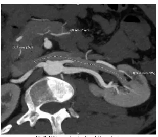
The CT images obtained were measured for length and width of renal veins (fig: 1).
Morphometric analysis of renal veins:
The mean length of right renal vein was 3.01cm and left renal vein was 7.01cm (Table 1). The length of right renal vein varied from 1.2 to 5.2cm and left renal vein length varied from 3.5 to 10.1 cm (fig 2). The length of right renal vein in 45% of individuals varied from 2.1cm to 3.0cm and 37% of individuals varied from 3.1cm to 4cm (Fig.5) (Table:2). The length of left renal vein in 35% of individuals varied from 7.1 to 8.0cm and 34% of individuals varied from 6.1 to 7.0 cm(Fig:6) (Table:3). The mean width of right renal vein near termination was 1.10cm and near hilum was 1.03cm. Similarly the mean width of left renal vein near termination was 0.88cm and near hilum was 0.99cm (Table 4).
Variations in the renal vein:
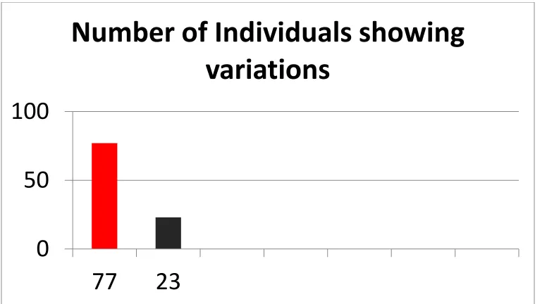
Out of 100 kidney donors the variations in renal vein was found in 23 individuals (Table 5) (Fig: 7) in which 19 individuals had right accessory renal vein (fig.3) and 4 individuals had retroaortic left renal vein (fig.4) (Table 6).
Table 1: Mean Length Of Renal Vein
S.No Renal vein length Mean ± SD (cm)
1. Right Renal vein length
3.01 ± 0.95 2. Left Renal vein
length
© Indian Journal of Medical Research and Pharmaceutical Sciences http://www.ijmprs.com/
[image:3.612.118.503.93.644.2][43]
[image:3.612.189.425.130.210.2]Table 2: Length Of Right Renal Vein
Table 3: Length Of Left Renal Vein
Table 4: Mean Width Of Renal Vein
S.No Renal vein width Mean ± SD(cm)
1. Right Renal vein width near termination 1.10 ± 0.27 2. Left Renal vein width near termination 0.99 ± 0.74 3. Right Renal vein width near hilum 1.03 ± 0.27 4. Left Renal vein width near hilum 0.88 ± 0.22
Table 5: Number Of Variations In Renal Vein
S.No Normal & variations of renal vein Number of individuals
1. Normal 77
2. Variations 23
Table 6: Variations In Renal Vein
S.No Variations of renal vein Number of individuals
1. Accessory renal vein 19 (all on right side) 2. Retro aortic left renal vein 4 (all on left side)
S.No Range
cm
Number of Individuals
1 1.2 – 2.0 11 2 2.1 – 3.0 45 3 3.1 – 4.0 37 4 4.1 – 5.2 7
S.No Range
cm
Number of Individuals
© Indian Journal of Medical Research and Pharmaceutical Sciences http://www.ijmprs.com/
[image:4.612.145.470.114.429.2][44]
Figure .1: CT image showing measurement of renal vein.
[image:4.612.153.462.433.701.2]© Indian Journal of Medical Research and Pharmaceutical Sciences http://www.ijmprs.com/
[45]
Fig.3: CT image showing right accessory renal vein
© Indian Journal of Medical Research and Pharmaceutical Sciences http://www.ijmprs.com/
[46]
[image:6.612.92.525.101.640.2]Figure 5: showing number of individuals in each range in length of the right renal vein
Figure 6: showing number of individuals in each range in length of the left renal vein
0
10
20
30
40
50
1.0 to
2.0 cm
2.1 to
3.0
3.1 to
4.0
4.1 to
5.0
5.1 to
6.0
Number of Individuals
Number of
Individuals
X axis - Length of
right renal vein
Y axis- Number of
individuals
0
5
10
15
20
25
30
35
40
3.5
to
4.0
4.1
to
5.0
5.1
to
6.0
6.1
to
7.0
7.1
to
8.0
8.1
to
9.0
9.1
to
10.1
Number of Individuals
Number of
Individuals
© Indian Journal of Medical Research and Pharmaceutical Sciences http://www.ijmprs.com/
[image:7.612.116.504.112.328.2][47]
Figure 7: showing number individuals showing normal pattern and variations in renal veins:
Discussion
Morphometric analysis of renal vein:
In this study the mean length of right renal vein was 3.01 ± 0.95 cm and mean length of left renal vein was 7.01 ± 1.14 cm. One of the previous study showed that the mean right renal vein length was 2.36±0.821 cm, mean left renal vein length was 5.65±1.27 cm and the right renal vein range from 2.4-2.6 cm, left renal vein range from 5.4–5.9 cm [13]. Another study on right renal vein showed that the length varied from 21 to 71 mm which was similar to the current study [14]. The studies on left renal vein also showed similar results that the length varied from 2.6cm to 8.0 cm [15, 16].
The mean width of right renal renal vein near the entry into inferior vena cava was 1.10 ± 0.27cm which was similar to previous study which showed the variation from 1.09±0.156 cm 13 but slightly smaller than reported by sathyapal 2003 [17] and, Janscheck et al. (2004) 18 were they reported 1.2 cm. The mean width of left renal vein was 0.99 ± 0.74 which was slightly smaller as compared to other studies where they reported 1.23 cm [13] [6]. The differences in length and width of renal veins in the literature reported can be explained by the size of the subjects analysed and the diversity in the population with their respective phenotypic expressions.
Variations in renal veins:
This study showed 23% of individuals had renal vein variations out of which 19 were accessory renal veins and 4 were retro aortic left renal vein. The occurrence of accessory right renal vein was a common variation (19%) which was less compared to other studies like Satyapal 1999 [5] and Okada et al 2010[19] where they showed 31% and 33% respectively. The prevalence of right and left accessory renal veins done by various studies were tabulated(table:7)
0
50
100
77
23
© Indian Journal of Medical Research and Pharmaceutical Sciences http://www.ijmprs.com/
[48]
Table 7: Prevalence of accessory renal vein
Clinical implications
The multiple renal veins can be susceptible to trauma and threat to abdominal surgical procedure especially during kidney transplantation and retroperitoneal region. Also the use of kidney with accessory renal vessels has been discouraged due to increased risk to donor because of multiple vessels anastomosis, prolonged ischemia time and poorly controlled hypertension from segmental infarcts of allograft (24). Direct venous extension of renal cell carcinoma via the renal vein is of normal occurrence and the presence of extra renal veins might prove an extra route for spread of metastasis. This can enhance the spread of primary renal cell carcinoma to multiple sites in the body.
Retro aortic left renal vein:
In this study 4% of individuals had retro aortic left renal veins when compared to other studies in which it ranged from 0.5% to 6.6%. This retro aortic renal vein can be compressed between aorta and lumbar spine leading to posterior nutcrackers phenomenon manifesting as abdominal pain with or without hematuria. Also this compression can reflux venous blood to gonadal veins resulting in varicocele and varicose veins in lower limb. This retroaortic left renal vein can be associated with pelvic congestion syndrome in females. The retroaortic vein can be obstructed by peritoneal growths leading to nephritis if it is prolonged [25]. The prevalence of retroaortic left renal vein were tabulated in table 8.
Table 8: prevalence of retroaortic left renal vein
Author Retroaortic left renal vein Sathyapal et al 1999[5] 0.5%
Holden et al 2005[23] 3% Anupma et al 2011 [22]. 6.6%
Current study 4%
Embryological basis of accessory renal vein:
The cardinal veins form the main venous drainage system of the embryo. During fourth week this system consist of anterior and posterior cardinal veins joining together to form common cardinal veins. Around 7-8 weeks bilateral symmetrical venous system converts into unilateral right side inferior vena cava. Two or more renal veins are present on each side of inferior vena cava draining kidney, one on anterior side and other on posterior side. On the right side these anterior and posterior veins fuse together and form single renal vein, persistence of which can lead to accessory renal vein. Due to the right shift of venous system the retention of accessory veins on left side is very rare, So the accessory right renal veins are common [22]
References
1. Cooley DA, Wukasch DC, eds. Techniques in vascular surgery. Philadelphia: Saunders, 1978:282.
2. Anson BJ, Cauldwell EW, Pick JW, Beaton LE. The anatomy of the pararenal system of veins, with comments on the renal arteries. J UroI1948;60:714-37.
3. Baptista-Silva JCC, Cal RGR, Castro MJ, Veríss!mo MJM, Câmara ALG. Estudoanatômico das veiasrenais e suasvariações, observaçãodurante as nefrectomiasemdoadoresvivos. Rev Col Bras Cir 1995;22(2): 18. 4. Anson BJ, Daseler EH. Common v~riations in the renal blood supply. SurgGynecolObstet
1961;112:439-49.
5. Satyapal KS, Kalideen JM, Haffejee AA, Sing B, Robbs JV, Left renal vein variations, SurgRadiolAnat, Authors Right accessory renal vein Left accessory renal vein
© Indian Journal of Medical Research and Pharmaceutical Sciences http://www.ijmprs.com/
[49]
1999, 21(1):77– 81.
6. Satyapal KS, Rambiritch V, Pillai G, Additional renal veins: incidence and morphometry, ClinAnat, 1995, 8(1):51–55.
7. Duques P, Rodrigues JR, Da Silva Neto FB, Neto EMVS, De Tolêdo ES, Estudoanatômico da veia renal esquerda de cadávereshumanosbrasileiros (Anatomical study of the left vein of human Brazilians cadavers), Medicina (RibeirãoPreto), 2002, 35(2):184–191.
8. Thomas VT, Surgical implications of retroaortic left renal vein, Arch Surg, 1970, 100(6):738–741.
9. Mathews R, Smith PA, Fishman EK, Marshall FF, Anomalies of the inferior vena cava and renal veins: embryologic and surgical considerations, Urology, 1999, 53(5):873–880.
10. Kramer B, The incidence of the renal venous collar in South African blacks, S Afr Med J, 1980, 57(21):875–876.
11. Hoeltl W, Hruby W, Aharinejad S, Renal vein anatomy and its implications for retroperitoneal surgery, J Urol, 1990, 143(6): 1108–1114.
12. Nam JK, Park SW, Lee SD, Chung MK, The clinical significance of a retroaortic left renal vein, Korean J Urol, 2010, 51(4): 276–280.
13. Luis ernesto Ballesteros, Vladimir Saldarriaga, Luis Miguel Ramirez. Morphologic evaluation of the renal veins: a study with autopsy material from Colombian subjects Rom J Morphol Embryol 2014, 55(1):77–81 14. Janschek EC1, Rothe AU, Hölzenbein TJ, Langer F, Brugger PC, Pokorny H, Domenig CM, Rasoul-Rockenschaub S, Mühlbacher F. Anatomic basis of right renal vein extension for cadaveric kidney transplantation. Urology. 2004 Apr;63(4):660-4
15. Duques P, Rodrigues JR, Da Silva Neto FB, Neto EMVS, De Tolêdo ES, Estudo anatômico da veia renal esquerda de cadáveres humanos brasileiros (Anatomical study of the left vein of human Brazilians cadavers), Medicina (Ribeirão Preto), 2002, 35(2):184–191.
16. Gillot C, La veine rénale gauche. Etude anatomique, aspects angiographiques, abord chirurgical (The left renal vein. Anatomical study, angiographic aspects and surgical approach), Anatomia Clinica, 1978, 1(2):135–156.
17. Satyapal KS, The renal veins: a review, Eur J Anat Suppl, 2003, 7(Suppl 1):43–52.
18. Beckmann CF, Abrams HL, Renal venography: anatomy, technique, applications, analysis of 132 venograms, and a review of the literature, Cardiovasc Intervent Radiol, 1980, 3(1):45–70.
19. Y. Okada, Y. Kayashima, N. Nishi, W. Mizukoshi, T. Watadani, Y. Matsuo, E. Kozawa, F. Kimura; Hidaka, Saitama/JP. Anatomic variation of the right renal vein: MDCT evaluation with interactive 3D reformation. 10.1594/ecr2010/C-1305
20. Bregman RA, Afifi AK and Miyauchi R. Virtual Hospital: Illustrated encyclopedia of human renal arteries: 2000; 15-8.
21. Dhar P. and Ajmani ML. Major anomalies of left renal vein and inferior vena cava. Int Med J. 2004; (2): Dec.
22. Anupma gupta, Raman gupta, Rikki singal . Congenital Variations of Renal Veins: Embryological Background And Clinical Implications. Journal of Clinical and Diagnostic Research. 2011 November (Suppl-1), Vol-5(6): 1140-1143.
23. Holden A, Smith A, Dukes P, Pilmore H, Yasutomi M. Assessment of 100 live potential renal donors for laparoscopic nephrectomy with multi-detector row helical CT. Radiology 2005;237:973-980
24. Mandal AK, Cohen C, Montgomery RA, Kavoussi LR, Ratner LE. Transplantation 660-4. 2001. p. 1 25. Singla RK, Sharma T, Gupta R. Retro-aortic left renal vein with left suprarenal vein draining into inferior