DISTRIBUTION OF INTERSEGMENTAL INTERNEURONES
THAT CAN RESET THE RESPIRATORY RHYTHM OF THE
LOCUST
BY J. M. RAMIREZ AND K. G. PEARSON
Department of Physiology, University of Alberta, Edmonton, Canada, T6G 2H7
Accepted 27 July 1988
Summary
Interneurones in the respiratory rhythm generator of the locust were identified by means of intracellular recording and staining techniques. A description is made of the properties and structures of nine intersegmental neurones which reset the respiratory rhythm when injected with current pulses. All but one of these neurones discharged in phase with expiration. The injection of constant depolariz-ing current into these interneurones altered the respiratory rate (increase for six, decrease for three). The respiratory rhythm generator extends more posteriorly within the ventral nerve cord than the metathoracic ganglion. In the first fused abdominal ganglion, four individual interneurones were identified descending into the unfused abdominal ganglia. In the first unfused abdominal ganglion an intemeurone which reset the respiratory rhythm was found ascending into the metathoracic ganglion. The respiratory rhythm generator also extends more anteriorly within the ventral nerve cord than the metathoracic ganglion. Two interneurones influencing the respiratory rhythm send their axons from the first fused abdominal ganglion into the meta- and mesothoracic ganglia. One of these directly excited a mesothoracic intemeurone which also influenced the respiratory rhythm when injected with current. In the suboesophageal ganglion another intemeurone was found which, although capable of resetting the respiratory rhythm, was not alway active during respiration. We conclude that the respiratory rhythm generator is distributed over abdominal, thoracic and suboesophageal ganglia. At least one part of the respiratory rhythm generator (in the suboeso-phageal ganglion) is not always active and can be recruited during vigorous respiration. Thus the number of active components in the respiratory rhythm generator is variable and additional elements can be recruited depending on the behavioural situation.
Introduction
Considerable information has been obtained about the neuronal mechanisms underlying certain rhythmic behaviour in insects, such as flight and walking. Surprisingly, a far simpler rhythmic behaviour, respiration, has received relatively
little attention, although respiratory movements are exhibited in highly dissected preparations so that the neuronal basis of the behaviour can be studied by means of intracellular recordings. The rhythm has been found to be generated at the interneuronal level, and two sets of interneurones have been proposed to drive closer and opener spiracle motoneurones (Burrows, 1974, 1975a,b). The first interneurone in the respiratory rhythm-generating system, a neurone capable of resetting the respiratory rhythm, has been identified in the first fused abdominal ganglion (Pearson, 1980). This interneurone descends in the abdominal nerve cord, is active in phase with expiration and probably has a role in coordinating the respiratory rhythm in abdominal ganglia. Another interneurone which coordi-nates the activity in the thoracic spiracles has also been described (Burrows, 1982). This interneurone ascends from the metathoracic ganglion and inhibits spiracle motoneurones in the mesothoracic ganglion. However, current injection into this neurone does not influence the respiratory rhythm. In the past six years, no further information has been published on the role of interneurones in the respiratory system. This can in part be attributed to the technical difficulty of recording from respiratory interneurones for a sufficient time to investigate their properties. The processes of these interneurones appear to be smaller than those involved in flight (Robertson & Pearson, 1985) and jumping (Gynther & Pearson, 1986).
Some obvious gaps in our knowledge are the exact distribution of the respiratory rhythm generator within the nerve cord, the pattern of interconnec-tions between sensory organs, interneurones and motoneurones, the mechanisms for generating the rhythm and the function of peripheral feedback. In this study we will concentrate only on one problem, the extent to which the respiratory oscillator is distributed within the ventral nerve cord. It is generally assumed that in locusts a primary oscillator is located in the metathoracic ganglion and that this oscillator controls secondary oscillators in the abdominal ganglia (Miller, 1966; Lewis et al. 1973). Only one neuronal element of the primary oscillator has been identified (Pearson, 1980) and no neurones in the secondary abdominal oscillators have been identified. There is uncertainty about whether the rhythm generator contains elements rostral to the metathoracic ganglion. Some muscles in the thorax and the neck are involved in respiration but respiratory movements in these muscles cease after cutting the anterior connectives of the metathoracic ganglion. From these results it has been concluded that there is no respiratory rhythmic centre anterior to the metathoracic ganglion (Miller, 1960i>). However, it is conceivable that neurones in the rhythm generator are located rostral to the metathoracic ganglion even though these ganglia might not be autorhythmic. Changes in abdominal pumping following transection of the meso- and meta-thoracic connectives (Lewis et al. 1973) may be due to removal of rostrally located elements of the oscillator.
Materials and methods Animals
Adult male and female Locusta migratoria were obtained from a colony at the University of Alberta. All experiments were performed at room temperature (22-25 °C).
Preparation
Following the removal of the wings and legs the animals were mounted dorsal side up on a cork board. To expose the mesothoracic ganglion, a dorsal incision was made in the thorax, and the gut and small muscles overlying the ganglion were removed. Abdominal ganglia were exposed in a similar manner, but the dorsal incision was extended more posteriorly. The suboesophageal ganglion was exposed by opening the head capsule and removing the mandibular and dorsal neck muscles as well as the tentorium. The ganglion from which recordings were obtained was supported on a stainless-steel platform and kept covered with locust saline. Previous studies have shown that expiratory activity occurs almost synchronously in all abdominal segments (Lewis et al. 1973), so it was usually monitored by placing a hook electrode on one of nerves 8, 9 or 10 of the metathoracic ganglion. These nerves contain the axons of motoneurones which discharge in phase with expiration (Lewis et al. 1973). In some cases electromyo-gram recordings were obtained from muscle 177 (Lewis et al. 1973) which is active in phase with inspiration.
Recording and staining
Intracellular recordings were obtained from neuropile processes of neurones which were rhythmically active in phase with the respiratory rhythm. The recording electrodes were filled with a 5 % solution of Lucifer Yellow in distilled water, which was injected following recording to allow the identification of the recorded neurone. Electrode resistance was in the range 50-200MQ. Only the ganglia from which recordings were obtained were processed, as described by Robertson & Pearson (1982). Thus, no information exists on the terminals of stained neurones in other ganglia. The interneurones were identified and numbered according to the three-digit numbering scheme of Robertson & Pearson (1983). The first digit indicates the path of the axon and its position relative to the soraa. The two following numerals have been assigned arbitrarily.
Data analysis Pattern of activity
normalized to 1 and divided into 50 bins. The average number of spikes occurring in each bin was then determined.
Effect of constant-current injection
Both positive and negative currents ( ± 2 - 6 nA) were injected into a neurone to examine whether it had an accelerating or decelerating effect on the respiratory rhythm. The effect was assessed by averaging the duration of four respiratory cycles immediately before, during and after the period of increased activity in the neurone. The average of 4-5 of these stimulation experiments and the deviation about the mean were then represented in a histogram.
Resetting of the respiratory rhythm
To examine whether the neurone had a resetting effect on the rhythm, short current pulses were injected (duration 100-300 ms, in one case 1000 ms; current magnitude 2-5 nA). The effect on the respiratory rhythm was calculated by subtracting the duration of the cycle in which the current pulse was applied (t2) from the duration of the undisturbed cycle immediately preceding this cycle (tl) and dividing this difference by t l ; i.e. (t2—tl)/tl. The effect of stimulation on the first (t3) and the second cycle (t4) following the cycle in which the stimulus was applied (t2) was calculated in a similar way, i.e. (t3-tl)/tl or (t4—tl)/tl. The phase of application of the stimulus within a respiratory cycle was calculated by dividing the interval between the onset of the cycle in which the stimulus was applied and the onset of the stimulus pulse by the duration of the preceding undisturbed cycle t l . For expiratory interneurones a respiratory cycle was taken as the time between the onset of two consecutive bursts of activity in the expiratory motor nerve. For the only inspiratory neurone examined a respiratory cycle was measured as the time between the onset of two consecutive bursts of activity in the inspiratory neurone.
It should be noted that in this study phase 0 is the onset of activity in the expiratory motor nerve, whereas in the study of Pearson (1980) phase 0 is the end of the burst in the stimulated interneurone. This difference causes a shift in phase values but does not affect the result.
Comments on analysis
been due to our recording in different sites in the neurones or to the different levels of spontaneous activity in the neurones. The variable excitation levels of a neurone due to its rhythmic modulation may also cause distortions of reset curves even in the same animal. A pulse given during the phase in which the neurone was depolarized resulted in a much higher spike frequency than a pulse applied during the neurone's inhibitory phase. We did not attempt to control spike numbers at different phases. Therefore, all diagrams in this study can give only qualitative information on the effect of current injection on the respiratory rhythm.
Results
The aim of this study was to identify, in different ganglia, intemeurones which are elements of the respiratory rhythm-generating system. In accordance with previous studies in other motor systems (Friesen et al. 1978; Weeks, 1981), a neurone was considered to be an element of the rhythm generator if (1) its cell membrane was rhythmically polarized in phase with the activity in a respiratory motor nerve and (2) current injection into this neurone shifted the phase of the respiratory rhythm (i.e. reset the rhythm). A cell's ability to shift the phase of the respiratory rhythm was always accompanied by its ability either to speed or to slow the respiratory rhythm and this was therefore consistently taken as a third criterion. Only nine intemeurones which met these criteria will be described in this study. However, 21 intemeurones were found which met the first but failed the second and third criteria, and which may also be elements of the respiratory rhythm generator.
Intemeurones in the first fused abdominal ganglion
found in the third fused abdominal ganglion. The only interneurones we found that were able to affect the respiratory rhythm had their somata located in the first fused abdominal ganglion. This finding suggests that a major part of the primary respiratory rhythm generator is located within this ganglion. However, it cannot be excluded that there are also undiscovered neurones in other ganglia.
Interneurones 326, 327, 328, 329
Interneurones have been described in the first fused abdominal ganglion which cause an increase in the respiratory rate when injected with constant depolarizing currents (Pearson, 1980). Injection of short pulses of depolarizing currents resets the rhythm. Extracellular recordings from the connectives indicate the presence of more than one pair of these neurones (Pearson, 1980). In this section we will describe four such neurones which are anatomically and physiologically distinct. Each of these neurones was found in at least two different animals and could be re-identified by its shape and physiology. The structure of these interneurones (326, 327, 328, 329) has some common features. The somata of all the neurones are located near the dorsal surface in the lateral region of the first fused abdominal ganglion and their axons leave the ganglion in the abdominal connective contralateral to their somata. The primary neurites run from the somata in an anterior medial direction. Some features, however, allow distinctions to be made between these neurones. 326 has a prominent secondary process arising ipsilateral to the midline and running towards the midline before projecting anteriorly (Fig. 1A). 327 has two prominent secondary processes running anteriorly, ipsi-and contralateral to the midline (Fig. IB). 328 has two prominent secondary branches arising near the soma and running parallel to the main neurite (Fig. 1C). One of these branches nearly overlaps the main neurite, the other runs more anteriorly to the main neurite. 329 has a prominent secondary branch arising contralateral to the midline and running towards the midline before projecting anteriorly. It also has a characteristic secondary neurite running posteriorly from the midline (Fig. ID).
c
326 328
D
327 329
500 ms
[image:7.451.54.388.74.545.2]20 mV
3 0 1 326
[image:8.451.63.377.62.299.2]Phase
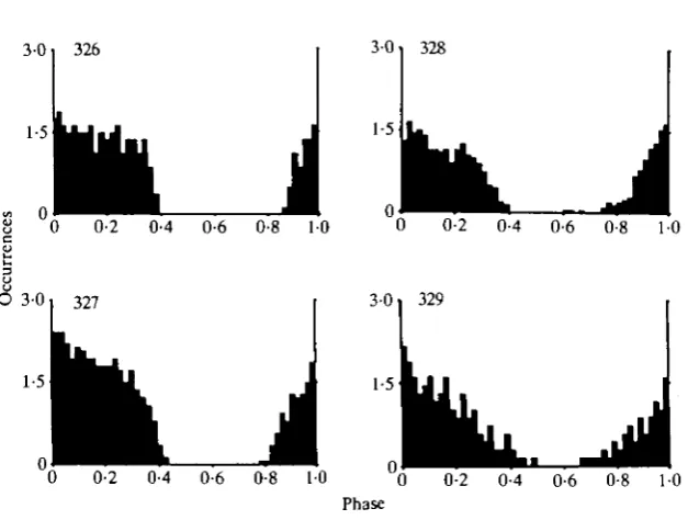
Fig. 2. Phase histograms of the four descending expiratory interneurones. 326: number of cycles ( ^ = 8, average period (AP) = 528 ms, average bin width (AB) = 10-6ms. 327: N=14, AP = 804ms, AB = 16ms. 328: N = 7O, AP = 655ms, AB = 13ms. 329: N= 7, AP = 839 ms, AB = 17 ms.
produced even at the end of the expiratory phase (see, for example, Fig. 4D). Just after the termination of activity in the expiratory motor nerve, all neurones repolarized rapidly and, in the absence of current injection, spikes were usually not produced after the termination of expiratory activity (Fig. IE). The spike distribution of the interneurones in the absence of current injection is shown in the phase histograms of Fig. 2. Spike activity of all neurones started before the onset of expiratory activity (phase value 0) at a value around 0-8, and activity ceased with the termination of activity in the expiratory nerve at a value around 0-4. These phase histograms also show that the spike activity of these neurones was not uniform during the expiratory phase. For example, it is obvious that spike activity declined in 329 but was steadier in 326.
0-9 326
i
i
328 1-8
0-9
-d.c. 0 - d . c . -d.c. 0 - d . c .
I I
I 1-8
Q.
0-9 327
I
i
1-8
0-9
I 1
0 +d.c. 0 0 +d.c. 0-d.c. -d.c.
[image:9.451.109.348.64.449.2]1000 ms 20mV
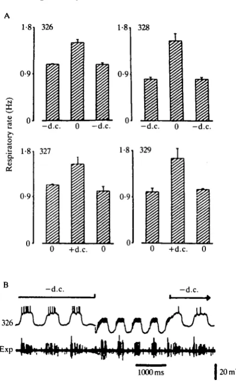
Fig. 3. Activity increase in the descending interneurones causes an increase in the respiratory rate. (A) Each histogram is the average of four stimulation experiments. For each stimulation experiment we calculated the respiratory rate from four subsequent respiratory cycles before, during and after activity increase in the neurone. Activity increase in a neurone was obtained either by release from negative or by injection of positive current (d.c). The average spike activity of the neurones for each histogram was: 326, -d.c. = 10 Hz, no d.c. = 100 Hz; 327, no d.c. = 72Hz, +d.c. = 143Hz; 328, -d.c. = 3Hz, no d.c. = 100Hz; 329, no d.c. =60Hz, +d.c. = 120Hz. (B) Stimulation experiment. Release of inhibition in 326 (upper trace) resulted in an increase in the respiratory rate. Lower trace (Exp), EMG recording from nerve 9 of the metathoracic ganglion.
A 0-8 326 Prolong 0-6
0-4
_ 0-2
-0-2
-0-4
Phase
B 0-2
-0-2
0-2 0-4 0-6 0-ILX 1 0 C 0-2
Shorten
•2 0
-0-2
• 0-2 *0-4 0-6' 0-8 1-0 Phase
Fig. 4. Resetting of the respiratory rhythm by injecting short current pulses into the descending expiratory interneurone 326. (A) Phase-response curve for the cycle in which the stimulus was applied (t2). The dotted area indicates the time in the respiratory cycle in which 326 was active. (B) Phase-response curve for the cycle immediately following the cycle in which the stimulus was applied (t3). (C) Phase-response curve for the second cycle (t4) following the cycle in which the stimulus was applied. Note that the stimulus effect is not compensated in subsequent cycles. Duration of the stimulus pulses = 300 ms, magnitude of current = 3 nA. (D) Example of a stimulation experiment. The 326 recording in D was obtained in a different animal from that used for A-C. Upper two traces, activity of 326 (upper trace) recorded simultaneously with an EMG of nerve 9 of the metathoracic ganglion (Exp, lower trace). 326 was constantly hyperpolarized (—2nA). Lower two traces, the current pulse (d.c.) (+3nA, 150ms) injected into 326 (upper trace) reset the respiratory rhythm.
A 0-4
0-2
-0-2
-0-4
- 0 - 6 327
Prolong
,Q-2 \ 0-4
B 0-4
0-2
0-6 0-8 . 1 0
Shorten
,•* Phase
-0-2
-0-4 328
. ' " *
0-4 ,0-6 0-8 ty\Q
Phase
0-6
0-4
0-2
- 0
-0-2
-0-4
- 0 - 6 329 Prolong
i
0-2 0-41 0-6 »
\ I
0-8 / 1-0 Phase
Shorten
Fig. 5. Phase-response curves of the descending expiratory interneurones 327, 328, 329. (A) 327, duration of pulse = 200ms, magnitude of current = 5nA; (B) 328, duration of pulse = 200 ms, magnitude of current = 4 nA; (C) 329, duration of pulse = 300 ms, magnitude of current = 5 nA. In this and all subsequent reset curves dotted lines are drawn if only few values with a high scatter were obtained and solid lines are drawn if many values with a relatively low scatter were obtained. At the bottom of all phase-response curves the dotted areas indicate the average times in the cycle in which the neurones were active.
slight shortening of cycle duration was visible at a phase around 0-8 (Fig. 4B). This weak shortening effect in the second cycle seems to be compensated in the third cycle but, because of the high scatter, this compensation might be not significant (Fig. 4C).
intemeurone described by Pearson (1980) also has a shortening effect at a similar phase range. Differences between the neurones 327, 328 and 329 were found in the phase range 0-0-6. Since the values in this phase range were relatively highly scattered and for reasons mentioned in Materials and methods, it is not possible to determine whether these differences are significant.
Intemeurone 516
Of six inspiratory interneurones found in the metathoracic ganglion only one, interneurone 516, was able to reset the respiratory rhythm. The cell body of intemeurone 516 is located in the same dorsolateral region of the first fused abdominal ganglion (Fig. 6A). The primary neurite leaves the soma in an anterior medial direction. Before the main neurite crosses the midline one secondary branch projects anteriorly and another posteriorly. From these branches several processes project towards the lateral neuropile, ipsilateral to the soma. At the midline, a thick secondary branch projects posteriorly from which several smaller processes run into both ipsilateral and contralateral neuropile regions. After crossing the midline, further secondary processes arise anteriorly and posteriorly from the main neurite. These secondary processes and the branches which arise from the main neurite before it crosses the midline are symmetrical. The axon of 516 ascends contralateral to the cell body and projects at least as far as the prothoracic ganglion. Several thin branches project from the axon into the lateral and medial neuropile regions of the metathoracic ganglion.
516 had its maximum activity just after activity had ceased in the expiratory nerve (Fig. 6B). Injection of depolarizing currents into 516 considerably increased the respiratory rate (Fig. 6C,D) and the injection of short depolarizing pulses reset the respiratory rhythm (Fig. 6E,F). A shortening of the respiratory cycle was evoked when the stimulus was applied in the phase range 0-0-8 (Fig. 6E,F). A prolongation of the respiratory cycle was evoked only when the stimulus pulse was applied in a relatively narrow phase range between 0-85 and 1-0 (Fig. 6F).
Interneurone 606
Intemeurone 606 has its cell body in a dorsolateral region in the first fused abdominal ganglion (Fig. 7A). The axon ascends ipsilateral to the cell body. In the first and second abdominal segments, as well as in the metathoracic segment, several secondary processes cross the midline and project into the contralateral neuropile. Smaller branches arise from these secondary processes near the midline and project posteriorly. Branches which run posteriorly are also found in the lateral regions ipsilateral to the cell body.
516
516
JUULX
500 ms 15 mV
c
516 E x p « « *
+d.c.
1000 ms
+d.c.
tmm* V
500 ms 15 mV
1.6
£• 0-8 o
i
+d.c.I
F 0-4 0-2
u
2 -0-2
-0-4 -0-6 -0-8
Prolong 0-2 0-4
Shorten
t •—**""•
mm
0-6 0 - 8 / l;0 •/ Phase
1
B
606
606
500 ms 20 mV
1-0
£> 0-5 o
I
i
1
-d.c. 0 - d . c . 0-2 0-4 0-6 0-8
Phase
1-0
Fig. 7. Characterization of the ascending expiratory interneurone 606. (A) Structure of the interneurone 606. (B) Activity of 606 (upper trace) recorded with an EMG from nerve 8 of the metathoracic ganglion (Exp) (lower trace). (C) Histogram showing that activity increase in 606 causes a decrease in the respiratory rate. The histogram was evaluated as described in Fig. 3. The average activity of 606 was: —d.c. = 2 Hz, no d.c. = 55 Hz. (D) Phase histogram of 606: N=5, average period = 1168 ms, average bin width = 23 ms.
the spike activity in 606 by injecting hyperpolarizing currents increased the respiratory rate (Fig. 7C), and increasing the spike activity in 606 decreased the respiratory rate. Thus current injection into 606 had opposite effects on the respiratory rate from those described for interneurones 326-329. The recordings of 606 were not stable enough to investigate its resetting properties. However, since 606 strongly excited an interneurone which was able to reset the respiratory rhythm we assume that 606 also has the ability to reset this rhythm. This excitatory connection will be described in the following section.
Interneurones in abdominal, mesothoracic and suboesophageal ganglia
606
200 ms 4OmV
20mV
7201
100 ms
110 mV 2-5 ms llOmV
1 0
£> 0-5 o
E
i
I
-d.c. 0 -d.c.
0-4
0-2
0
-0-2
Prolong
0-2 Shorten
0-4 0 - 6 / 01! 1-0 Phase
mmm
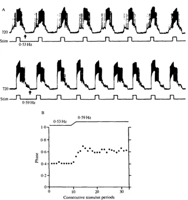
Fig. 8. Characterization of the mesothoracic interneurone 720. (A) Structure of 720. (B) Simultaneous intracellular recording of 720 (upper trace) and 606 (lower trace) during respiration. (C) Intracellular recording of 720. Note the occurrence of many IPSPs during the interburst interval. (D) Each spike in 606 (lower trace) produces an EPSP in 720 (upper trace). The constant latency following of the EPSPs in 720 is displayed by multiple oscilloscope sweeps triggered by the spike in 606. (E) Histogram showing that activity increase in 720 induced by release from negative current (d.c.) (—5nA) causes a decrease in the respiratory rate. The average activity of 720 was: —d.c. = 6Hz, no d.c. = 42 Hz. (F) Injection of short current pulses into 720 reset the respiratory rhythm. Duration of current pulse = 200 ms, magnitude of current = 4 nA.
Interneurone 720
process arises from the main neurite and projects posteriorly, where several smaller branches run into the ipsi- and contralateral neuropile regions. The secondary neurite reaches the posterior edge of the mesothoracic ganglion where it ramifies into a dense bundle of fine branches which spread ipsi- and contralater-ally from the branching point. Several branches arise from the descending axon. These relatively fine branches project laterally and medially from the neurone but never reach the midline.
Spike activity in 720 overlapped the activity of interneurone 606 (Fig. 8B) from which it received direct excitatory input. Each spike in 606 was followed 1:1 by an EPSP in 720 (Fig. 8D). In addition to this excitatory input during expiration, 720 also received strong inhibitory input in the interburst interval (Fig. 8C). This inhibitory input was very characteristic of 720 and was not found for the metathoracic expiratory interneurones. Intracellular injection of constant depolar-izing current into 720 decreased the respiratory rate, as was also observed for 606 (Fig. 8E). Short depolarizing current pulses (of 200 ms) into 720 reset the rhythm (Fig. 8F). Consistent with the inhibitory effect on the respiratory rhythm, the cycle duration was prolonged if the current pulses were applied in a phase in which 720 was usually active (phase 0-0-4 and phase 0-8-1-0). Current pulses had only a weak shortening effect when applied at a phase in which 720 was usually strongly inhibited (phase 0-5). Thus, for this interneurone the reset curve can explain its inhibitory effect on the respiratory rhythm.
In one animal the entrainment of the respiratory oscillator was examined by injecting current pulses into 720 at different constant stimulus frequencies for 25-30 respiratory cycles (Fig. 9). The respiratory oscillator followed in a 1:1 manner with a low scatter (r = 0-97-0-99, see also Horsmann et al. 1983) in a stimulus frequency range from 0-52 to 0-74 FIz (absolute coordination). Changes in the stimulus frequency caused (1) a change in the frequency of the respiratory oscillator and (2) a shift in the phase at which the oscillator locked to the stimulus (Fig. 9). A decrease in the stimulus frequency caused the respiratory oscillator to advance and an increase in the stimulus frequency was immediately followed by a retardation of the respiratory oscillator relative to the stimulus period (Fig. 9A,B).
Interneurone 377
Interneurone 377 has its cell body in the suboesophageal ganglion at the ventral midline of the anterior labial neuromere (Fig. 10A). Its axon descends in the connectives contralateral to the cell body at least as far as the metathoracic ganglion. The processes of 377 are symmetrically distributed in all the neuromeres of the suboesophageal ganglion. Ipsi- and contralateral to the midline two relatively thick secondary processes project posteriorly into the labial neuromere. Two secondary processes arise from the crossing neurite at the midline and project anteriorly into the mandibular neuromere. Further branches arise from the descending axon.
0-53 Hz
0-59 Hz
0-53 Hz
0-59 Hz
10-i
0-8- 0-6-
0-4-
0-2-10 20 Consecutive stimulus periods
[image:17.451.41.415.70.467.2]30
A
377
Insp
Fig. 10
strength of this activity was variable and it could even fail completely (Fig. 10B). No synaptic modulation was visible during the absence of rhythmic activity in 377, indicating an absence of rhythmic input to it. That this variation was caused by a variable rhythmic input and was not due to individual differences among 377 interneurones in different animals is indicated by the observation of variation during the same 377 recording (Fig. 10B-D). A reproducible alteration of the strength of synaptic input to 377 was obtained in all animals by eliciting rhythmic activity in flight motoneurones by application of a wind stream to the head. The strength of the 377 modulation was considerably increased after this treatment (Fig. 10C,D). During increased activity of 377 a high initial input frequency, which was not present before flight rhythmic activity (Fig. IOC), was visible at the onset of expiration (Fig. 10D). The amplitude of the depolarization was also changed (Fig. 10C,D).
Despite the occasional complete absence of respiratory rhythmic input to 377, intracellular injection of depolarizing currents into 377 always accelerated the respiratory rhythm (Fig. 11A,B). The effect of injections of current pulses on the respiratory rhythm was phase-dependent. The injection of depolarizing current pulses at the end of the inspiratory phase and during some of the expiratory phase caused a shortening of the respiratory cycle (Fig. 11 A,C). The reset curve of 377 was qualitatively similar to that described for expiratory interneurones 326-329 in the metathoracic ganglion (Fig. 11C).
Interneurone 725
Interneurone 725 has its soma in the ventral posterior midline of the first unfused abdominal ganglion (Fig. 12A). The primary neurite projects anteriorly, crosses the midline and bifurcates to send ascending and descending axons into the contralateral connectives. The secondary processes ipsilateral to the soma arise from the primary neurite whereas all contralateral processes arise from the axons.
Interneurone 725 was rhythmically active in phase with expiration (Fig. 12B). The activity of 725 started after the onset of expiratory activity in the metathorax (phase 0-01) and was maximally active in phase 0-2-0-4 (Fig. 12C). Thus, 725 was clearly activated later than the metathoracic expiratory interneurones. Another difference was that fewer spikes were produced in 725 in each respiratory cycle. Intracellular injection of constant depolarizing currents slowed the respiratory rhythm (Fig. 12E). The effects of injections of current pulses were
A
377«
Insp
+d.c.
1000 ms | l 5 m V
g- 0-9
i
+d.c.
i
C 0-6
0-4
0-2
-0-2
-0-4
- 0 - 6
Prolong
/ V
0-2 0-4\ 0-6
Phase 0-8 10
[image:20.451.48.413.47.436.2]Shorten
Fig. 11. The influence of 377 on the respiratory rhythm. (A) Injection of a current pulse (3 nA, 1000 ms) causes a shortening of the respiratory cycle. The recording in A is a.c. filtered. (B) Injection of constant depolarizing current (+3nA) increased the respiratory rate. Average activity of 377: nod.c. = 0 , +d.c. = 40 Hz. (C) Reset curve of 377. Pulse duration = 1000 ms, magnitude of current pulse = 3nA. The dotted area at the bottom of C indicates the activity distribution of 377, were 377 to have been active. In this stimulation experiment 377 was recorded with an inspiratory myogram, but the reset curve was normalized to an expiratory cycle by shifting the ordinate.
dependent and the injections prolonged the cycle period at the phases examined (phase 0-2-0-8) (Fig. 12D,F).
Discussion
Interneurones in the metathoracic ganglion
B
725
500 ms 15 mV
+d.c. D
0-5
WLJ
0 0-2 0-4 0-6 0-8 1-0 Phase
500 ms 15 mV
1-6
0-8
a.
I
i
i
- d . c . 0
0-8
0-6
•Z 0-4
0-2
0
Prolong
J 0-2 0-4 0-6
Phase
0-8 1-0
larvae (Fitch & Kammer, 1982), adult cockroaches (Farley et al. 1967) and adult locusts (Miller, 1960a) have revealed that the respiratory rhythm is generated in more than one ganglion. Some of these ganglia are able to generate a rhythm in respiratory motoneurones when isolated from the rest of the nervous system. However, lesion experiments have shown that the relative importance of these ganglia in the generation of the respiratory rhythm is not uniform. One ganglion usually leads the respiratory rhythm (primary respiratory oscillator) and coordi-nates the rhythmic activity in the other ganglia (secondary oscillators). In different insect species, the primary respiratory oscillator is located in different ganglia. In dragonfly larvae it is located in the most posterior abdominal ganglion (the eighth) (Komatsu, 1982) and in dobsonfly larvae in the third abdominal ganglion (Fitch & Kammer, 1982). In adult locusts (Miller, 1966), crickets (Huber, 1960) and cockroaches (Farley, 1967) the primary respiratory oscillator is located somewhere in the metathoracic ganglion, although it is not known whether it is in the 'true' metathoracic ganglion or in one of the three abdominal ganglia which are fused to it.
It is assumed that intersegmental interneurones located in or associated with the primary oscillator coordinate the activity in the secondary oscillators. In the dragonfly a pair of ascending interneurones has been described which might be responsible for such coordination (Komatsu, 1984). In crickets, cockroaches and locusts it has been demonstrated that burst activity related to expiration is present in abdominal connectives (Huber, 1960; Farley et al. 1967; Miller, 1966) and it is assumed that this activity is caused by interneurones descending from the metathoracic ganglion and coordinating the secondary oscillators which are located in the abdominal ganglia (Lewis et al. 1973). By means of intracellular recordings, descending interneurones have been identified in the locust meta-thoracic ganglion which discharge rhythmically in phase with expiration and which also affect the respiratory rhythm when stimulated (Pearson, 1980). The proper-ties of these interneurones are consistent with the properproper-ties proposed for the coordinating interneurones by Lewis (1973). However, the exact number of these coordinating interneurones has not been established.
et al. (1973) are caused by these neurones. This would imply that the primary
respiratory rhythm generator of locusts is not located in the 'true' metathoracic ganglion but in the first fused abdominal ganglion. Consistent with this suggestion is the finding that 606 and 516, which both have an effect on the respiratory rhythm, were also located in the first fused abdominal ganglion.
Ascending influence from the abdominal ganglia to the metathoracic ganglion
There is general agreement that the primary respiratory oscillator controls the secondary respiratory oscillators in the abdominal ganglia (Miller, 1960a; Lewis et
al. 1973; Pearson, 1980). However, it is not known whether the secondary
oscillators also have an effect on the primary oscillator. The existence of interactions between the secondary oscillators and the primary oscillator is indicated by the discovery of interneurone 725. This interneurone is located in the first unfused abdominal ganglion and strongly affects the respiratory rhythm in the first fused abdominal ganglion (primary oscillator). Activity in 725 started after the onset of activity in the descending interneurones located in the first fused abdominal ganglion as well as after the onset of activity in the expiratory motor nerve of this ganglion. Therefore, 725 activity was too late to have an immediate effect on the onset of the expiratory phase in the primary oscillator. However, 725 could act through the neuronal elements of the primary oscillator on the next expiratory cycle. This is very likely, since stimulation of 725 clearly prolonged the onset of the next expiratory phase in the primary oscillator.
What might be the functional relevance of feedback from the secondary to the primary oscillator? It is known that the respiratory rhythm depends on the animal's fat supplies and egg loads and that experimental loading of the abdomen can strongly affect the overall respiratory motor pattern (Giszter, 1986). Such an adaptation of the rhythm to loads in the abdomen might be caused by direct feedback from sensory receptors to the primary oscillator but might also be caused by more indirect feedback via the secondary oscillator. Neurone 725 might be a good candidate for mediating feedback from the secondary to the primary oscillator. The number of interneurones in the segmental abdominal ganglia which have an effect on the overall respiratory rhythm is unknown. Of six different respiratory interneurones which were identified in the segmental abdominal ganglia only interneurone 725 had an effect on the respiratory rhythm of the first fused abdominal ganglion. Neurone 725 also has a descending axon and therefore may also affect oscillators in more posterior abdominal ganglia.
Anterior distribution of the respiratory rhythm generator into the thoracic and suboesophageal ganglia
(1960a) has stated that there appears to be no rhythmic centre anterior to the metathoracic ganglion. Nevertheless, it is known that, in the intact animal, muscles in the thorax and the neck contract rhythmically during respiration (see Miller, 1960a). This rhythmic activity could be derived through at least three possible pathways: (1) through interneurones ascending from the primary oscil-lator which are not elements of the respiratory rhythm-generating system; (2) through interneurones ascending from the primary oscillator which are elements of the respiratory rhythm-generating system; and (3) through interneurones located in the thoracic ganglia and suboesophageal ganglion which are elements of the respiratory rhythm-generating system.
Until now there has been evidence only for the first possibility. One inspiratory interneurone has been found which ascends from the metathoracic ganglion into the meso- and prothoracic ganglia and affects several thoracic spiracles without having any effect on the respiratory rhythm itself (Burrows, 1982). Some clues for the two other possibilities were presented in this study. Consistent with the second possibility, we found two interneurones which have their somata in the first fused abdominal ganglion and which ascend into the thoracic ganglia. These neurones, the inspiratory interneurone 516 and the expiratory interneurone 606, both have an effect on the respiratory rhythm. The third possibility, which was the most unlikely considering the results of the lesion experiments (Miller, 1960a), became more likely as a result of the findings presented in this study. We showed that 606 excited interneurone 720 which is located in the mesothoracic ganglion. Intracellu-lar stimulation of 720 had a strong effect on the respiratory rhythm. Constant depolarizing currents slowed the respiratory rhythm and injection of short depolarizing pulses reset and entrained the rhythm. The coupling of the respirat-ory oscillator to 720 stimulation was very strong and therefore there is no doubt that the respiratory rhythm generator has elements in the mesothoracic ganglion.
change, and in some animals cease completely, is unusual since it demonstrates that the number of neuronal elements in the respiratory rhythm-generating system may vary. The additional recruitment of interneurones such as 377 during situations when the oxygen consumption is high seems to be a mechanism to accelerate the respiratory rhythm generator, and might also be a mechanism to activate additional muscles. Indeed, it is known that the muscles of the thorax and the neck are only activated during vigorous respiration, for example after flight, when 377 is strongly active (Miller, 1960a). At this time the respiratory rate is also increased. An additional recruitment of neuronal elements also shows that the number of active components in a rhythm generator may not be fixed but can be adapted to the behavioural situation.
We thank I. C. Gynther and B. Hedwig for their valuable comments on the manuscript and Y. Tang for her excellent technical assistance. This study was supported by a grant from the Medical Research Council of Canada.
References
BURROWS, M. (1974). Modes of activation of motoneurones controlling ventilatory movements of the locust abdomen. Phil. Trans. R. Soc. Ser. B 269, 29-48.
BURROWS, M. (1975a). Co-ordinating interneurones of the locust which convey two patterns of motor commands: their connexions with flight motoneurones. 7. exp. Biol. 63, 713-733. BURROWS, M. (19756). Co-ordinating interneurones of the locust which convey two patterns of
motor commands: their connexions with ventilatory motoneurones. 7. exp. Biol. 63, 735-753. BURROWS, M. (1982). Interneurones co-ordinating the ventilatory movements of the thoracic
spiracles in the locust. 7. exp. Biol. 97, 385-400.
FARLEY, R. D., CASE, J. F. & ROEDER, K. D. (1967). Pacemaker for tracheal ventilation in the cockroach Periplaneta americana (L.). 7. Insect Physiol. 13, 1713-1728.
FITCH, G. K. & KAMMER, A. E. (1982). Modulation of the ventilatory rhythm of the hellgrammite Corydalus comutus by mechanosensory input. 7. comp. Physiol. 149, 423—434. FRIESEN, W. O., POON, M. & STENT, G. S. (1978). Neuronal control of swimming in the medicinal leech. IV. Identification of a network of oscillatory interneurones. 7. exp. Biol. 75, 25-44.
GISZTER, S. F. (1986). Breathing in fat and thin locusts: Adaptations by ventilation motor pattern generators to altered mechanical properties of the abdomen. Soc. Neurosci. Abstr. 330,5.
GYNTHER, I. C. & PEARSON, K. G. (1986). Intracellular recordings from interneurones and motoneurones during bilateral kicks in the locust: implications for mechanisms controlling the jump. 7. exp. Biol. 122, 323-343.
HORSMANN, U., HEINZEL, H.-G. & WENDLER, G. (1983). The phasic influence of self-generated air current modulations on the locust flight motor. J. comp. Physiol. 150, 427-438.
HOYLE, G. (1959). Action of carbon dioxide on an insect spiracular muscle. 7. Insect Physiol. 3, 378-394.
HUBER, F. (1960). Experimentelle Untersuchungen zur nervosen Atmungsregulation der Orthopteren (Saltatoria: Gryllidae). Z. vergl. Physiol. 44, 60-132.
KOMATSU, A. (1982). Respiratory nerve activity in the isolated nerve cord of the larval dragonfly, and location of the respiratory oscillator. Physiol. Entomol. 7,183-191.
KOMATSU, A. (1984). Ascending interneurones that convey a respiratory signal in the central nervous system of the dragonfly larva. 7. comp. Physiol. 154, 331-340.
LEWIS, G. W., MILLER, P. L. & MILLS, P. S. (1973). Neuro-muscular mechanisms of abdominal pumping in the locust. 7. exp. Biol. 59, 149-168.
MILLER, P. L. (1960a). Respiration in the desert locust. I. The control of ventilation. /. exp.
Biol. 37, 224-236.
MILLER, P. L. (1960b). Respiration in the desert locust. II. The control of the spiracles. /. exp.
Biol. 37, 237-263.
MILLER, P. L. (1966). The regulation of breathing in insects. Adv. Insect Physiol. 3, 279-354.
PEARSON, K. G. (1980). Burst generation in coordinating interneurons of the ventilatory system of the locust. J. comp. Physiol. 137, 305-313.
RAMIREZ, J.-M. (1986). Interneuronal control of locust flight. Ph.D. thesis, University of Regensburg, FRG.
ROBERTSON, R. M. & PEARSON, K. G. (1982). A preparation for the intracellular analysis of neuronal activity during flight in the locust. J. comp. Physiol. 146, 311-320.
ROBERTSON, R. M. & PEARSON, K. G. (1983). Interneurons in flight system of the locust: distribution, connections and resetting properties. /. comp. Neurol. 215, 33-50.
ROBERTSON, R. M. & PEARSON, K. G. (1985). Neural circuits in the flight system of the locust.
J. Neurophysiol. 53, 110-128.