Differentiating between Scar and Herniated Disk
Canh M. Nguyen, Khang-Cheng Ho, Howard An, Lee H. Riley III, Xu Rongming, and Victor M. Haughton
PURPOSE: To compare the MR contrast enhancement produced by ionic and nonionic
paramag-netic contrast media in herniated disk fragments with that in epidural scar tissue. METHODS: A recurrent herniated disk was modeled in canines by using laminectomy to place a fragment of disk cartilage in the epidural space. The dogs were studied 88 and 90 days after laminectomy with MR imaging enhanced with an ionic or a nonionic paramagnetic contrast medium. Contrast enhance-ment of the epidural scar tissue and the epidural disk fragenhance-ment was measured at 2, 22, and 45 minutes after the injection. RESULTS: Contrast enhancement was consistently and significantly higher in scar tissue than in disk fragments, although the difference decreased between 2 and 45 minutes after administration of contrast medium. Enhancement of disk fragments was less with the ionic than with the nonionic contrast medium. Contrast between the disk fragments and scar was also greater with the ionic than with the nonionic medium. CONCLUSIONS: The contrast between recurrent disk fragments and scar tissue is affected by the timing of the scan and the choice of contrast medium. Scans obtained immediately after the injection of contrast medium show more contrast between disk fragment and scar than do delayed scans. Recurrent herniated disk frag-ments are more effectively shown by ionic than by nonionic media.
Index terms: Magnetic resonance, contrast enhancement; Spine, intervertebral disks, herniation;
Spine, magnetic resonance; Spine, scar tissue; Animal studies
AJNR Am J Neuroradiol17:501–505, March 1996
Magnetic resonance (MR) imaging is the pri-mary imaging technique for differentiating re-current herniated disks from postoperative scarring. Contrast enhancement is used rou-tinely to increase the contrast between the scar tissue and the disk fragments. Disk fragments usually show less enhancement than scar tissue does. However, the enhancement of scar tissue and disk fragments is variable. In previous ex-perimental studies (1, 2), the enhancement of scar tissue varied depending on the time
be-tween injection of contrast medium and imag-ing, the age of the scar tissue, and the dose of contrast medium used. Cartilage in the interver-tebral disk also shows enhancement after intra-venous injection of paramagnetic contrast me-dium (3, 4). The rate and the magnitude of contrast enhancement in the disk differs from that in the scar primarily because the contrast medium reaches the cartilaginous disk by diffu-sion rather than perfudiffu-sion (5–20). Enhance-ment of cartilage caused by diffusion of the contrast medium is a function of the dose of contrast medium injected intravenously and the charge on the molecule (4). Ionic media diffuse into negatively charged cartilage more slowly than do nonionic media. Theoretically, para-magnetic contrast media diffuse into the carti-lage in herniated disk fragments as into normal intervertebral disk cartilage. We studied the dif-ferential enhancement of disk fragments and scar tissue after intravenous injection of ionic and nonionic contrast media in an experimental model of recurrent disk herniation (2).
Received June 19, 1995; accepted after revision September 27. Supported by a grant-in-aid from Berlex Laboratories, Seacaucus, NJ. From the Departments of Radiology (C.M.N., V.M.H.), Pathology (K-C.H.), and Orthopedic Surgery (H.A., L.H.R., X.R.), Medical College of Wisconsin, Milwaukee.
Address reprint requests to Victor M. Haughton, MD, Department of Radiology, Doyne Clinic/Box 151, 8700 W Wisconsin Ave, Milwaukee, WI 53226.
AJNR 17:501–505, Mar 1996 0195-6108/96/1703–0501
qAmerican Society of Neuroradiology
Materials and Methods
Eleven mongrel dogs (weight range, 17 to 23 kg) un-derwent laminectomy and placement of a disk fragment in the epidural space followed by MR imaging with ionic and nonionic contrast media. Each animal was sedated with acepromazine (1 mg/kg) and atropine (0.05 mg/kg) in-tramuscularly and phenobarbital (0.05 mg/kg) intrave-nously. The animal was intubated, ventilated with a me-chanical respirator, and placed prone on an operating table. The skin over the lumbosacral area and tail was prepped and draped for surgery. A left hemilaminectomy was performed (2). The skin and the subcutaneous tissue were incised longitudinally at L3– 4. The paraspinal mus-cles were dissected and retracted to expose the spinous processes and lamina. With a dental drill (Microdrill 5033-001; Hall Surgical, Santa Barbara, Calif) and cutting burr bits, a 7-mm-wide, 17-mm-long defect was created in the L3– 4 lamina. The ligamentum flavum was removed, and the nerve root was exposed. The L3– 4 disk space was located and incised by a #15 scalpel blade until the nu-cleus pulposus escaped from the disk space. Bleeding was controlled with gel foam and light compression. The tail was then resected, and the proximal wound was repaired. One intervertebral disk was removed aseptically from the distal portion of the tail. A fragment of fibrocartilage (height, 1.5 mm; diameter, 7 mm) was removed from the disk and inserted into the epidural space through the lam-inectomy defect anterior to the thecal sac. The laminec-tomy defect was closed in layers after hemostasis was achieved.
The dogs were allowed to recover from anesthesia in a humidified, warmed environment for 24 hours and then returned to their cages. After surgery, cefazolin sodium was given (1 g/d for 5 days) prophylactically and bu-prenorphine hydrochloride (1 ampule, intramuscularly) was given as necessary as an analgesic.
In a crossover study design, MR imaging with either gadopentetate dimeglumine or gadoteridol was performed on days 88 and 90 after surgery. For MR imaging, the animal was sedated with acepromazine and atropine and anesthetized with intravenous phenobarbital as was done for the laminectomy. The animal was placed supine with the L3– 4 disk space centered on a 3-in (7.5-cm) solenoid coil in the 1.5-T imager. Localizer images were obtained with conventional spin-echo techniques. Either gado-pentetate dimeglumine or gadoteridol was injected intra-venously at a dose of 0.3 mmol/kg. MR images were obtained in the axial projection immediately before injec-tion and at 2, 22, and 45 minutes after injecinjec-tion of the contrast medium. The parameters used for each of the acquisitions included 3-mm section thickness and 600/ 25/2 (repetition time/echo time/excitations). The type of contrast medium used was alternated in each dog and between dogs systematically.
Preenhancement and postenhancement images were compared, and the disk fragment and the scar were iden-tified in the epidural space. Signal intensity in the disk fragment and scar tissue in the preenhancement and
postenhancement images was measured with a cursor and a resident computer program. Contrast enhancement was calculated as the change in intensity from baseline divided by the baseline signal intensity. Measurements were en-tered into a spreadsheet program (Quattro, Borland Inter-national, Scotts Valley, Calif). Enhancement for each time period after the injection and enhancement for each con-trast medium were recorded and averaged. Concon-trast en-hancement was plotted as a function of time after the injection of contrast medium. Contrast enhancement of disk fragments was compared with that of scar tissue. Differences between enhancement in disk fragment and scar tissue were calculated for each contrast medium and each time period after the injection. The variances of the enhancement for each contrast medium were compared by using Scheffe’sFtest to measure the significance of the differences.
The animals were killed 90 days after surgery. The lumbar spine was removed carefully en bloc and fixed in 10% buffered formalin, decalcified, embedded in paraffin, sectioned axially, and stained with hematoxylin-eosin. The stained sections were examined by the neuropathologist. The location of the disk fragments in the MR images was verified, and the histologic appearance of each disk frag-ment was characterized.
Results
Of the 11 animals enrolled in the study, 10 had surgery and MR imaging without any com-plications. One animal developed hindquarter paralysis, was killed 2 days after surgery, and was replaced by another animal. In the other animals, normal activity was seen within 1 week after surgery.
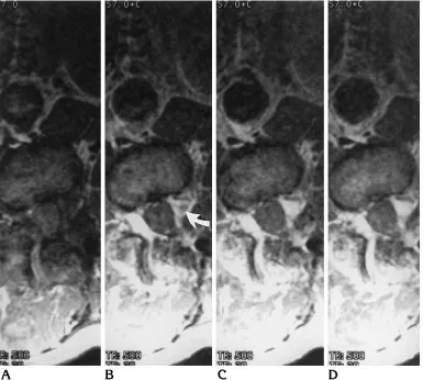
At 88 to 90 days after the surgery, the ani-mals underwent MR imaging with each contrast medium. The disk fragment location in the MR image (confirmed subsequently by histologic section) appeared as a region of decreased en-hancement within the epidural scar, regardless of which contrast medium was used (Fig 1).
fragments for both contrast media at 2, 22, and 45 minutes.
The differences in signal intensity between the disk fragment and the scar tissue for 45 minutes after injection of gadopentetate dime-glumine or gadoteridol is shown in Figure 3. The difference between the signal intensity of scar tissue and that of the disk fragment was greater at 2 minutes than at 22 or 45 minutes. The difference between the signal intensity of the scar tissue and that of the disk fragment was significantly greater for the ionic than the non-ionic medium at 2 and 22 minutes (P , .01, Student’s t test).
Disk fragments in the epidural space were characterized histologically by fibrocartilagi-nous tissue and variable amounts of fibrosis. All the disk fragments showed infiltrating blood vessels to a marked degree. Inflammatory cell
infiltration was seen in all but 1 of the speci-mens. In 8 of the 10 animals, histologic exam-ination showed that the fragment was adherent to the dura.
Discussion
[image:3.612.115.500.87.433.2]ment of the fragments for the 45-minute period. The scar tissue probably shows enhancement more rapidly than the disk because the blood supply of the scar tissue has a fenestrated cap-illary endothelium (22). Our study results indi-cate that disk fragments show less enhance-ment with the ionic medium than with the nonionic medium and that the contrast between disk fragments and scar is greater after the use of ionic rather than nonionic contrast medium. The animal model simulates clinical recurrent herniated disks, but there are some differences between the model and the clinical situation. The recurrent herniated disk and scar in hu-mans are not likely to be of the same age. The cartilage in recurrent herniated disk fragments in humans may be larger and more degenerated than in our model. Intervertebral disks from ca-nine tails normally have cartilage similar to that in lumbar intervertebral disks (23). Partial vol-ume averaging is probably greater in measuring enhancement in the small disk fragments in the epidural space in our animals than it is in hu-mans; therefore, the differences are probably underestimated in the experimental animal. The different rates of enhancement for disk and scar tissue cannot be explained on the basis of par-tial volume errors. The magnitude of enhance-ment in this study exceeds that in the typical clinical experience because the dose of contrast medium (0.3 mmol/kg) exceeds the usual clin-ical dose.
The measurements in scar tissue in this study are consistent with previous experimental re-sults. The peaking of enhancement in scar tis-sue soon after intravenous injection of contrast medium has been described previously (1). The enhancement of cartilage by the process of dif-fusion has also been reported (3). The degree of enhancement in scar tissue has been shown to correlate with the amount of contrast medium administered (2). Enhancement of the interver-tebral disk is affected by the composition of the
disk. Ionic media diffuse more slowly through cartilage (4) because the fixed negative charges in cartilage, which make it hygroscopic, impede the diffusion of charged particles, such as those in ionic contrast media. The ionic and nonionic contrast media used in the study have similar molecular weights.
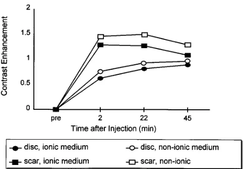
[image:4.612.315.556.84.251.2] [image:4.612.315.554.559.710.2]We conclude that for optimal enhancement in imaging recurrent herniated disk fragments and scar tissue, it is important to choose the most effective dose and type of contrast medium and the most favorable timing of the imaging. The-oretically, the detection of recurrent herniated disks may be improved by increasing the dose of contrast medium, choosing a contrast me-dium that diffuses more slowly into cartilage, and obtaining images rapidly after injection of the contrast medium. Our results suggest that Fig 2. Graph of average contrast enhancement in disk frag-ments and epidural scar surrounding the disk fragment as a func-tion of time after the injecfunc-tion of 0.3 mmol/kg of gadopentetate dimeglumine or gadoteridol. The data were acquired 88 or 90 days after placement of the disk fragment in the epidural space.
Fig 3. Difference in signal intensity in scar and disk tissue after injection of gadoteridol or gadopentetate dimeglumine.
Contrast enhancement in disk fragment and scar tissue after intra-venous injection of an ionic or nonionic contrast medium
Location
Type of Contrast Medium
Enhancement at Time after Injection, min
2 22 45
Disk Ionic 0.6260.1 0.8160.2 0.8960.1 Nonionic 0.7660.2 0.9360.2 0.9760.2 Scar Ionic 1.2960.3 1.2760.4 1.0860.3 Nonionic 1.4660.2 1.5060.2 1.2960.2
ionic media diffuse more slowly into disk frag-ments than do nonionic media. Further work is needed to determine whether use of an ionic medium or larger doses of contrast medium are effective clinically.
Acknowledgment
We thank Debbie Bauer, who facilitated the preparation of the manuscript.
References
1. Nguyen CM, Haughton VM, Ho KC, et al. MR contrast enhance-ment: experimental study in postlaminectomy epidural fibrosis. AJNR Am J Neuroradiol1993;14:997–1000
2. Nguyen C, An H, Ho KC, Haughton VM, Hasegawa T. Utility of high-dose contrast enhancement for detecting recurrent herniated intervertebral disks.AJNR Am J Neuroradiol1994;15:1291–1295 3. Ibrahim MA, Jesmanowicz A, Hyde J, Estkowski L, Haughton VM. Contrast enhancement of normal intervertebral disks: time and dose dependence.AJNR Am J Neuroradiol1994;15:419 – 424 4. Ibrahim MA, Haughton VM, Hyde JS. Enhancement of
interverte-bral disks with gadolinium complexes: comparison of an ionic and a nonionic medium in an animal model.AJNR Am J Neuroradiol 1994;15:1907–1910
5. Katz MM, Hargens AR, Garfin SR. Intervertebral disc nutrition.Clin Orthop1986;210:243–245
6. Ogata K, Whiteside LA. Nutritional pathways of the intervertebral disc: an experimental study using hydrogen washout technique. Spine1981;6:211–216
7. Brown M, Tsaltas T. Studies on the permeability of the interverte-bral disc during skeletal maturation.Spine1976;1:240 –244 8. Maroudas A. Nutrition and metabolism of the intervertebral disc.
In: Ghosh P, ed.The Biology of the Intervertebral Disc.Boca Raton, Fla: CRC Press, 1988:1–38
9. Brown MD, Tsaltas TT. Studies on the permeability of the inter-vertebral disc during skeletal maturation.Spine1976;1:240 –244
10. Maroudas A. Transport of solutes through cartilage: permeability to large molecules.J Anat1976;122:335–347
11. Urban JPG, Maroudas A, Nachemson A. Nutrition of the interver-tebral disk: an in vivo study of solute transport. Clin Orthop 1977;129:101–114
12. Maroudas A, Bullough P, Swanson SAV, Freeman MAR. The per-meability of articular cartilage.J Bone Joint Surg [Br] 1968;50-B:166 –177
13. Maroudas A. Physicochemical properties of cartilage in the light of ion exchange theory.Biophys J1968;8:575–595
14. Riley LH III, Banovac K, Martinez OV, Eismont FJ. The qualitative distribution of antibiotics within the rabbit disc. 1994;19:2619 15. Eismont FJ, Weisel SW, Brighton CT, Rothman RH. Antibiotic
penetration into rabbit nucleus pulposus. Spine 1987;12:254 – 256
16. Brower TD, Akahoshi Y, Orlic P. The diffusion of dyes through articular cartilage in vivo.J Bone Joint Surg [Am]1962;44-A: 456 – 463
17. Hansen H-J, Ullberg S. Uptake of S35 in the intervertebral discs after injection of S35 sulphate: an autoradiographic study.Acta Orthop Scand1966;30:84 –90
18. Nachemson A, Lewin T, Maroudas A, Freeman MA. In vitro diffu-sion of dye through the endplates and the annulus fibrosus of human inter-vertebral discs.Acta Orthop Scand1970;41:589 – 607
19. Brodin H. Paths of nutrition in articular cartilage and intervertebral discs.Acta Orthop Scand1955;24:177–183
20. Kantori TG, Schubert M. The differences in permeability of carti-lage to cationic and anionic dyes.J Histochem Cytochem1957; 5:28 –32
21. Ross JS, Delamarter R, Hueftle MG, et al. Gadolinium-DTPA-enhanced MR imaging of the postoperative spine: time course and mechanism of enhancement.AJNR Am J Neuroradiol1989;10: 37– 41
22. Bundschuh CV, Modic MT, Ross JS, et al. Epidural fibrosis and recurrent disk herniation in the lumbar spine.AJNR Am J Neuro-radiol1988;9:169 –173