Ionic versus Nonionic Paramagnetic Contrast
Media in Differentiating between Postoperative
Scar and Recurrent Disk
Jui-Sheng Hsu, Gin-Chung Liu, Shih-Hsien Chen, Twei-Shiun Jaw, Wun-Jer Shen, and Chiao-Yun Chen
BACKGROUND AND PURPOSE:In theory, ionic solutes diffuse more slowly in cartilage than do nonionic solutes. We tested the hypothesis that the contrast ratio between scar and recurrent disk fragment on MR images is greater after IV administration of an ionic rather than a nonionic contrast medium when a clinical dose is used.
METHODS:Twenty patients who had recurrent lumbar disk herniation were enrolled in this study. The enhancement of epidural scar and recurrent disk fragment was measured at 5, 25, 40, and 50 min after IV injection of ionic and nonionic contrast media (0.1 mmol/kg)
RESULTS: The enhancement was consistently and significantly higher for scar than for recurrent disk fragment, although the contrast ratio between scar and recurrent disk fragment decreased between 5 and 50 min after the administration of each contrast medium. No significant difference was shown between ionic and nonionic contrast media in the enhancement of recurrent disk fragment at 5, 25, 40, and 50 min after injection. The contrast ratio between scar and recurrent disk fragment was not a significant difference at 5, 25, and 40 min after administration of both contrast media. At 50 min, the contrast ratio between scar and recurrent disk fragment was 1.32 ⴞ 0.41 with ionic contrast medium and 1.20 ⴞ 0.56 with nonionic contrast medium. The difference was significant.
CONCLUSION:The contrast ratio between scar and recurrent disk fragment is affected by the timing of the imaging. Images obtained immediately after the injection of each contrast medium showed a greater contrast ratio than did delayed images. In addition, with the ionic medium, this difference was greater than with nonionic medium at 5, 25, 40, and 50 min after injection and that difference reached statistical significance at 50 min.
In theory, nonionized solutes diffuse more quickly into cartilage than do ionized solutes. This principle of diffusion explains the greater efficacy of un-charged, nonionizing antibiotics in the treatment of diskitis than charged ionizing antibiotics that pene-trate poorly into disk cartilage. Therefore, in princi-ple, paramagnetic contrast media that have a charge should diffuse less readily in cartilage than contrast media lacking a charge.
The diffusion of the ionic and nonionic contrast media into cartilage have been widely studied with
isotope techniques (1–5) and MR imaging (6–14). Nonionic paramagnetic contrast media diffuse more rapidly into cartilage and disk than do ionic con-trast media (6–7). Diffusion of paramagnetic concon-trast media into immature disks is also greater than into mature disks (14). Nguyen et al (8) found that the implanted epidural disk fragments showed less en-hancement with the ionic medium than with the non-ionic medium, and the tissue contrast between disk fragment and scar was greater after the use of ionic rather than nonionic contrast medium. Therefore, we designed a study to test the hypothesis that with human MR imaging, greater enhancement of recur-rent disk fragment occurs with nonionic rather than ionic media. We compared the enhancement of scar and recurrent disk fragment in patients undergoing lumbar MR imaging with an ionic and a nonionic medium. To improve the power of the study, the enhancement of the scar and the recurrent disk frag-ment was measured at four time points after the administration of contrast medium.
Received March 11, 2003; accepted after revision November 6. From the Department of Radiology (J.S.H., G.C.L., T.S.J., C.Y.C), Kaohsiung Medical University, and the Po-Cheng Ortho-pedic Institute (W.J.S.), Kaohsiung, Taiwan, R.O.C.
Address reprint requests to Chiao-Yun Chen, Department of Medical Imaging, Kaohsiung Medical University, 100 Shih-Chuan 1st Road, Kaohsiung, 807, Taiwan, R.O.C.
©American Society of Neuroradiology
Methods
With the approval of the institutional review board, study patients were recruited from consecutive patients referred for contrast-enhanced MR imaging of the lumbar spine because of suspected recurrent lumbar disk herniation. The recurrent lum-bar disk herniation was typically defined as disk herniation on the basis of MR findings (15–21) obtained at the same level, regardless of ipsilateral or contralateral herniation, with a pain-free interval⬎6 months after lumbar disk surgery.
These patients were studied with serial imaging performed on a 1.5-T superconductive imaging unit. T1-weighted (450/16 [TR/TE]) and T2-weighted (3405/150) sagittal view spin-echo images with a section thickness of 3 mm were acquired with a 256 ⫻ 256 matrix and a 24-cm field of view. T1-weighted (475/18) and T2-weighted (3900/150) axial view spin-echo im-ages with 4-mm section thickness were acquired with a 256⫻
256 matrix and a 16-cm field of view. Contrast-enhanced MR
imaging, started immediately after the administration of 0.1 mmol/kg ionic gadopentetate dimeglumine, consisted of a sag-ittal view fat-saturated T1-weighted sequence (400/12) for ac-cessory evaluation of the presence and localization of the re-current disk and scar. The imaging matrix was 256⫻256, with a section of thickness of 4 mm. In addition, T1-weighted axial images were obtained 5 min after injection of contrast medium, with the same parameters as were used for the unenhanced axial view images. If the images suggested recurrent disk, the additional axial view T1-weighted MR imaging series was ob-tained at 25, 40, and 50 min after injection. Two radiologists reviewed the images to verify that a probable recurrent herni-ated disk was present. They also reviewed the medical records to verify that the clinical signs and symptoms were referable to the probable disk herniation. The patient was then contacted by phone or referred clinician and was offered the opportunity to participate in the research project in which subsequent MR
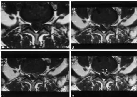
FIG 1. Axial view images of a patient with surgically confirmed recurrent herniated disk.
A, Obtained before the administration of gadopentetate dimeglumine (0.1 mmol/kg).
B, Obtained 5 min after the administration of gadopentetate dimeglumine (0.1 mmol/kg).
C, Obtained 50 min after the administration of gadopentetate dimeglumine (0.1 mmol/kg).
[image:2.603.60.527.56.386.2]D, Obtained 5 min after the administration of contrast medium. Placement of cursor to measure recurrent disk (1) and scar (2) enhancement. Contrast ratio between scar and recurrent disk fragment is greater at 5 min than at 50 min.
TABLE 1: Contrast enhancement in recurrent disk fragment and scar after IV injection of ionic and nonionic contrast media
Location Type of ContrastMedium
Enhancement at Time after Injection (min)
5 25 40 50
Ionic 0.60⫾0.10 0.68⫾0.07 0.72⫾0.07 0.70⫾0.07
Disk Nonionic 0.58⫾0.09 0.66⫾0.08 0.71⫾0.08 0.70⫾0.08
Ionic 1.37⫾0.26 1.15⫾0.18 0.98⫾0.17 0.90⫾0.15
Scar Nonionic 1.04⫾0.21 0.88⫾0.15 0.83⫾0.12 0.78⫾0.11
[image:2.603.52.540.463.543.2]imaging would be performed with a different contrast medium. If the patient accepted, informed consent was obtained and secondary MR imaging was performed with identical technical factors and imaging timing after injection with nonionic ga-dodiamide (0.1 mmol/kg) instead of ionic gadopentetate dime-glumine. The interval permitted between imaging sessions for this protocol was 3 to 5 days.
Twenty patients entered the pilot study group and com-prised seven women and 13 men ranging in age from 35 to 65 years (mean, 46.6 years). The time from previous surgery to imaging study varied from 7 months to 6 years (mean, 2.75 years).
The unenhanced and contrast-enhanced images were eval-uated together for the presence of recurrent disk. A diagnosis of disk material was made when aberrant soft tissue produced mass effect contiguous to the parent disk and did not enhance on the early contrast-enhanced images. Early linear or band-like enhancement surrounding irregularities of the peripheral parent disk margin without evidence of mass effect was inter-preted as a scar (5–10). After MR imaging, two of the 20 patients underwent surgical re-exploration at the site of abnor-mality noted on the MR images. The operative findings, in-cluding the location and the nature of the abnormal tissue, were consistent with the MR imaging findings.
Data Analysis
The measurement of the recurrent disk and scar was per-formed by a radiologist who did not know what contrast me-dium was injected when he measured the signal intensity. Sig-nal intensity in the recurrent disk fragments and scar was measured on the T1-weighted axial view MR images with a interest cursor and resident software. A region-of-interest cursor was placed within the disk fragment and another one in the enhancing scar tissue surrounding the disk fragment on the 5-min image, and signal intensity was recorded (Fig 1). The same cursor locations were used for the baseline, 25-min, 40-min, and 50-min images, and signal intensity was recorded for each of these time points. The enhancement and contrast ratio were calculated as enhancement⫽(SIpost⫺SIpre) / SIpre
and contrast ratio⫽SIscar/ SIdisk, where SIpostand SIprewere
the signal intensities of the targets at contrast-enhanced and unenhanced statuses, respectively. The SIscarand SIdisk
repre-sented the signal intensity of scar and recurrent disk fragment at the same time point of unenhanced or contrast-enhanced status, respectively. Differences in the enhancement and con-trast ratio were tested for significance with a repeated mea-sures analysis of variance test. If a significant difference was present, the posterior comparison test was tested by means of Bonferronittest.P⬍.05 was considered statistically significant.
Results
Average enhancement in scar and in recurrent disk fragment after injection of gadopentetate
dimeglu-mine or gadodiamide is summarized in Table 1 and in Figure 2. In the MR imaging studies, the scar and recurrent disk fragment showed different rates of enhancement. The scar showed greater enhancement at 5 min than at 25, 40, and 50 min after injection of either contrast medium, whereas the disk fragment tended to increase in enhancement for 50 min after injection of either contrast medium. Average en-hancement of scar was greater than that of recurrent disk fragment for both contrast media at 5, 25, 40, and 50 min (P⬍ .05) (Fig. 3). Average enhancement of scar was less with gadodiamide than with gado-pentetate dimeglumine at 5, 25, 40, and 50 min (P⬍ .05). No significant difference was shown in the en-hancement of recurrent disk fragment after the ad-ministration of either contrast medium at 5, 25, 40, and 50 min (P⬎ .05).
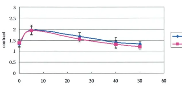
The contrast ratio between scar and recurrent disk fragment after injection of gadopentetate dimeglu-mine and gadodiamide is summarized in Table 2 and in Figure 4. The contrast ratio between scar and recurrent disk fragment was greater at 5 min than at 25, 40, and 50 min. No significant difference was shown between gadopentetate dimeglumine and ga-dodiamide in the contrast ratio between scar and recurrent disk fragment at 5, 25, and 40 min (P⬎.05). At 50 min, the average contrast ratio was 1.32⫾0.41 with gadopentetate dimeglumine and 1.20 ⫾ 0.56 with gadodiamide. The difference was significant (P⬍.05).
Discussion
Once a molecule is injected into the blood stream, it encounters the following barriers before reaching the interstitial space: distribution through vascular space, transport across the microvascular wall, and transport through the interstitial space. The intra-and extravascular exchange of fluid intra-and solute mole-cule in a tissue is determined by two mechanisms: diffusion and convection (22). The factors involved in the molecular transport included the transluminal concentration and pressure gradients, the surface area available for exchange, and three transport pa-rameters: vascular permeability (related to diffusion), hydraulic conductivity (related to hydrostatic convec-tion), and reflection coefficient (related to osmotic FIG 2. Graph of the average enhancement
[image:3.603.218.534.57.205.2]FIG 3. MR images of a patient with recurrent herniated disk
A, Sagittal view unenhanced T2-weighted MR image.
B, Axial view image obtained before the IV administration of ionic contrast medium.
C, Axial view image obtained 5 min after the IV administration of ionic contrast medium.
D, Axial view image obtained 50 min after the IV administration of ionic contrast medium. Contrast ratio is greater after administration of ionic rather than nonionic contrast media at 50 min (seepanel G).
E, Axial view image obtained before the IV administration of nonionic contrast medium.
F, Axial view image obtained 5 min after the IV administration of nonionic contrast medium.
convection). These three transport parameters are governed by the number and the width of the endo-thelial junctions on the vessel wall for a given size of molecule. Once the molecules are transported into the interstitial space, their distribution in the intersti-tium is again governed by molecular diffusion and possible convection due to pressure heterogeneity within the interstitium (23). For small molecular weight hydrophilic and lipophilic solute molecules, diffusion is the primary mechanism in the cartilage (3). Small molecules such as glucose and sulfate with molecular weight greater than 1000 diffuse readily through the proteoglycans gel (24, 25), which is the major constituent of cartilage, the nucleus pulposus, and inner annulus fibrosis and in diarthrotic joints. Gadopentetate dimeglumine (molecular weight 938) and gadodiamide (molecular weight 547) may diffuse into intervertebral disk and recurrent disk fragment (1–5).
Our results showed that the enhancement mea-sured with gadopentetate dimeglumine and gadodia-mide in clinical dose (0.1 mmol/kg) agrees with pre-viously obtained results (8). Both recurrent disk fragment and scar in postoperative spines normally show enhancement on MR images. The enhancement of scar is more than that of disk fragment, and the disk fragment tends to increase in enhancement with time after IV injection of either ionic or nonionic contrast medium. The scar shows apparent enhance-ment because the blood supply of scar has a capillary fenestrate endothelium, which allows contrast media to enter the interstitial space rapidly (26). Gradual diffusion of contrast medium into the disk fragment probably explains the enhancement mechanism of the disk fragment (6–14). In our study, abrupt change was noted in the enhancement during the first 5 min; gradual increased enhancement was then noted at 25,
40, and 50 min after injection of either ionic or non-ionic contrast medium. This confounding effect of a jump in the enhancement during the first 5 min may result from the partial volume effects of scar and recurrent disk in the region-of-interest cursor. In an animal study of mongrel dogs, Nguyen et al (8) showed that the epidural implanted disk fragment tended to gradually increase in signal intensity and showed less enhancement with gadopentetate dime-glumine (0.3 mmol/kg) than with gadoteridol (0.3 mmol/kg). This occurred because the charges on the ionic medium slow its entry into cartilage, which con-tains a high concentration of fixed negative charges. The difference between the enhancement of im-planted disk fragment and that of scar was therefore thought to be greater after the use of ionic rather than nonionic contrast medium. Our study, however, showed no significant difference between ionic and nonionic contrast media in the enhancement of re-current disk fragment when a clinical dose was used. In addition, the ionic contrast medium showed greater contrast ratio than did the nonionic one at 5, 25, 40, and 50 min after injection and the difference reached statistical significance at 50 min. The differ-ence between the results of our study and those of the study presented by Nguyen et al (8) could be ex-plained by the dose difference of the contrast media. Diffusion is the primary mechanism that determines enhancement of disk fragment. The higher dose may produce higher concentration gradients that increase the permeation of more nonionic contrast media into recurrent disk than that of ionic ones because nonionic solutes diffuse more quickly in disk than do ionic sol-utes. Our study, however, showed that this phenomenon is less prominent when a clinical dose is used.
[image:5.603.52.536.71.130.2]The study conducted by Haughton et al (27) showed that contrast between disk fragment and scar
TABLE 2: Contrast between the scar and recurrent disk fragment after IV injection of ionic and nonionic contrast media
Type of Contrast Medium
Contrast (SI scar/SI disk) at Time after Injection (min)
0 5 25 40 50
Ionic 1.32⫾0.42 1.95⫾0.80 1.66⫾0.67 1.40⫾0.42 1.32⫾0.41 Nonionic 1.37⫾0.61 1.93⫾0.78 1.54⫾0.44 1.30⫾0.44 1.20⫾0.56
Note.—SI indicates signal intensity.
* Contrast between scar and recurrent disc fragment is expressed as arithmetic mean⫾SD (n⫽20).
FIG 4. Graph of the contrast ratio
[image:5.603.219.536.161.313.2]tissue was greater after the use of an ionic contrast medium than a nonionic one with clinical dose in eight patients. The sample size was small. In our study, the ionic contrast medium provided relatively greater contrast ratio between the scar and the recur-rent disk than did the nonionic one at 5, 25, 40, and 50 min after injection and the difference reached statis-tical significance at 50 min after injection in 20 pa-tients. The difference between our results and those reported by Haughton et al may be attributable to the relatively small sample size in the other study. Addi-tional studies with larger sample sizes may be neces-sary for evaluating the difference of ionic and non-ionic contrast media–induced differentiation between the scar and recurrent disk in clinical practice. In addition, the study presented by Haughton et al also showed that the enhancement of scar tended to be greater with gadodiamide than with gadopentetate dimeglumine (27). Our study showed contrary results. Gadopentetate dimeglumine and gadodiamide are nonspecific extracellular agents and have similar pharmacokinetics and biodistribution (28). The blood supply of the scar includes capillary fenestrate endo-thelium that allows contrast media to enter interstitial space rapidly (26). In our experience, some variation exists in the enhancement of the scar. These differing results may be caused by different stages of the scar or the other unknown mechanisms.
One limitation of our study was the relatively small sample size. Another potential source of error was the placement of the region-of-interest cursor. To sample the signal intensity in disk fragment or scar, relatively small region-of-interest cursors are re-quired. Partial volume effects, inhomogeneity of scar and disk tissue, and motion between the baseline and the subsequent two series may have affected the ac-curacy of enhancement calculations. Imprecision in the timing of the images obtained after injection of contrast medium may have skewed the enhancement calculations; however, these sources of error are likely to produce random rather than systematic errors.
Conclusion
For optimal enhancement in imaging recurrent disk fragments and scar, it is important to choose the most favorable timing. The ionic medium showed greater contrast ratios than did the nonionic medium at 5, 25, 40, and 50 minutes after injection, and the difference reached statistical significance at 50 min-utes when a clinical dose was used.
References
1. Maroudas A, Bullough P, Swanson SA, Freeman MA.The perme-ability of articular cartilage.J Bone Joint Surg Br.1968 Feb;50:166– 177
2. Holm S, Maroudas A, Urban JP, Selstam G, Nachemson A. Nutri-tion of the intervertebral disc: solute transport and metabolism. Connect Tissue Res.1981;8:101–119
3. Urban JP, Holm S, Maroudas A.Diffusion of small solutes into the intervertebral disk: an in vivo study.Biorheology1978;15:203–221
4. Urban JP, Holm S, Maroudas A, Nachemson A.Nutrition of the intervertebral disk: effect of fluid on solute transport.Clin Orthop 1982;170:296–302
5. Stairmand JW, Holm S, Urban JP.Factors influencing oxygen concentration gradients in the intervertebral disk: a theoretical analysis.Spine1991;16:444–449
6. Ibrahim MA, Jesmanowicz A, Hyde JS, Estkowski L, Haughton VM.Contrast enhancement of normal intervertebral discs: time and dose dependence.AJNR Am J Neuroradiol1994;15:419–423 7. Ibrahim MA, Haughton VM, Hyde JS.Enhancement of
interver-tebral disks with gadolinium complexes: comparison of an ionic and a nonionic medium in an animal model.AJNR Am J Neurora-diol1994;15:1907–1910
8. Nguyen CM, Ho KC, An H, Riley LH III, Rongming X, Haughton VM.Ionic versus nonionic paramagnetic contrast media in differ-entiating between scar and herniated disc.AJNR Am J Neuroradiol 1996;17:501–505
9. Nguyen-minh C, Haughton VM, Papke RA, An H Censky SC. Measuring diffusion of solutes into intervertebral disks with MR imaging and paramagnetic contrast medium.AJNR Am J Neuro-radiol1998;19:1781–1784
10. Nguyen-minh C, Haughton VM, An H, You JW, Wook S, Ho KC. Contrast media of high and lower molecular weights in the detec-tion of recurrent herniated disks.AJNR Am J Neuroradiol1998;19: 889–893
11. Akansel G, Haughton VM, Papke RA, Censky S.Diffusion into human intervertebral disk studies with MR and gadoteridol.AJNR Am J Neuroradiol1997;18:443–445
12. Nguyen-minh C, Riley L III, Ho KC, Xu R, An H, Haughton VM. Effect of degeneration of the intervertebral disk on the process of diffusion.AJNR Am J Neuroradiol1997;18:435–442
13. Perlewitz TJ, Haughton VM, Riley LH III, Nguyen-Minh C, George V.Effect of molecular weight on the diffusion of contrast media into cartilage.Spine1997;22:2707–2710
14. Ibrahim MA Haughton VM, Hyde JS.Effect of disk maturation on diffusion of low-molecular-weight gadolinium complexes: an experi-mental study in rabbits.AJNR Am J Neuroradiol1995;16:1307–1311 15. Babar S, Saifuddin A.MRI of the post-discectomy lumbar spine.
Clin Radiol2002 Nov;57:969–981
16. Ross JS.MR imaging of the postoperative lumbar spine.Magon Reson Imaging Clin N Am1999;513–523
17. Hueftle MG, Modic MT, Ross JS, et al.Lumbar spine: postoper-ative MR imaging with Gd-DTPA.Radiology1988;167:817–824 18. Ross JS, Masaryk TJ, Schrader M, Gentili A, Bohlman H, Modic
MT.MR imaging of the postoperative lumbar spine: assessment with gadopentetate dimeglumine.AJR Am J Roentgenol1990;155: 867–872
19. Cavanagh S, Stevens J, Johnson JR.High-resolution MRI in the investigation of recurrent pain after lumbar discectomy.J Bone Joint Surg Br1993;75:524–528
20. Jinkins JR, Van Goethem JW.The postsurgical lumbosacral spine: magnetic resonance imaging evaluation following intervertebral disk surgery, surgical decompression, intervertebral body fusion, and spinal instrumentation.Radiol Clin North Am2001;39:1–29 21. Ross JS, Modic MT, Masaryk TJ, Carter J, Marcus RE, Bohlman
H.Assessment of extradural degenerative disease with Gd-DTPA-enhanced MR imaging: correlation with surgical and pathologic finding.AJNR Am J Neuroradiol1989;10:1243–1249
22. Jain RK.Transport of molecules across tumor vasculature.Cancer Metastasis Rev1987;6:559–593
23. Jain RK.Transport of molecules in tumor interstitium: a review.
Cancer Res1987;47:3039–3051
24. Maroudas A.Nutrition and metabolism of the intervertebral disc. In: Ghosh P, ed.The Biology of the Intervertebral Disc, II.Boca Raton: CRC Press; 1988:1–38
25. Maroudas A.Biophysical chemistry of cartilaginous tissues with special reference to solute and fluid transport.Biorheology1975 June (3–4);12:223–248
26. Bundschuh CV, Modic MT, Ross JS, Masaryk TJ, Bohlman Hl. Epidural fibrosis and recurrent disk herniation in the lumbar spine: MR imaging assessment.AJR Am J Roentgenol1988;150: 923–932
27. Haughton V, Schreibman K, De Smet A.Contrast between scar and recurrent herniated disk on contrast-enhanced MR images.
AJNR Am J Neuroradiol2002;23:1652–1656