Short-Term Memory of Motor Network
Performance via Activity-Dependent
Potentiation of Na
+
/K
+
Pump Function
Hong-Yan Zhang1and Keith T. Sillar1,*
1School of Biology, University of St Andrews, St Andrews
KY16 9TS, Scotland, UK
Summary
Brain networks memorize previous performance to adjust their output in light of past experience. These activity-depen-dent modifications generally result from changes in synaptic strengths or ionic conductances, and ion pumps have only rarely been demonstrated to play a dynamic role [1–4]. Loco-motor behavior is produced by central pattern generator (CPG) networks and modified by sensory and descending signals to allow for changes in movement frequency, inten-sity, and duration [5–7], but whether or how the CPG networks recall recent activity is largely unknown. In Xenopusfrog tadpoles, swim bout duration correlates line-arly with interswim interval, suggesting that the locomotor network retains a short-term memory of previous output. We discovered an ultraslow, minute-long afterhyperpolari-zation (usAHP) in network neurons following locomotor episodes. The usAHP is mediated by an activity- and sodium spike-dependent enhancement of electrogenic Na+/K+pump function. By integrating spike frequency over time and link-ing the membrane potential of spinal neurons to network performance, the usAHP plays a dynamic role in short-term motor memory. Because Na+/K+ pumps are ubiquitously
expressed in neurons of all animals and because sodium spikes inevitably accompany network activity, the usAHP may represent a phylogenetically conserved but largely overlooked mechanism for short-term memory of neural network function.
Results
Swim Duration Correlates with Interswim Interval
The central pattern generator (CPG) controlling swimming in young Xenopus laevisfrog tadpoles around the time of hatching [8] is relatively simple, and one of the most com-pletely described vertebrate CPG networks [9]. During fictive
swimming in immobilized tadpoles (stages 37/38–42),
rhythmic motor neuron (MN) bursts recorded from ventral roots (Figure 1A1) alternate across the body and propagate from head to tail at 10–20 Hz (Figure 1A2) [9, 10]. Swimming is generated by excitatory glutamatergic interneurons (dINs: descending interneurons) in the hindbrain and rostral spinal cord that are electrically coupled and form a reverberating positive feedback network that maintains activity once initi-ated [9]. These neurons drive commissural glycinergic inter-neurons (cINs) thought to couple the two sides in antiphase during swimming, ascending inhibitory interneurons (aINs) that contribute to gating of incoming sensory signals, and myotomal MNs that innervate the segmentally organized axial swimming muscles. Spinal CPG neurons display a range of
firing patterns during swimming; some, such as dINs, fire a single action potential per cycle reliably throughout swimming episodes, whereas some are only active during fast, intense activity [10]. Once initiated, swimming episodes last from a few seconds to several minutes, and the duration of each episode is regulated by several factors including inhibition from brainstem interneurons [11] and intrinsic spinal signaling mechanisms [12]. We find that there is a strong linear correla-tion between interswim interval (w1–30 s) and episode dura-tion (Figure 1B1; n = 6; R2= 0.996): when swimming is evoked at decreasing intervals (Figure 1B2, upper trace), the duration of subsequent episodes declines, and vice versa (Figure 1B2, lower trace). This suggests that the locomotor system can retain a short-term memory of previous network activity, which dictates the duration of future network output.
An Ultraslow Postswim Hyperpolarization
Spinal CPG neurons presumably contribute to this memory, so they were studied using whole-cell recordings in current-clamp mode. Spinal CPG neurons depolarized and fired action potentials during episodes of fictive swimming (Figure 1C1). Episodes often terminated coincident with a membrane hyper-polarization (Figure 1C1, dotted line) that gradually returned to the preswim resting potential (Figure 1C1, dashed line). In 26 neurons displaying this postswim phenomenon, the amplitude was 4.460.3 mV, but some reached 10 mV (indicated by two-way arrow inFigure 1C1). The event was slow; the membrane potential returned to preswim levels only after 49.468.4 s. Accordingly, we have termed this event the ultraslow afterhy-perpolarization (usAHP) to distinguish it from previously re-ported fast, medium, or slow AHPs with much more rapid timescales.
Most low-threshold neurons that fired reliably during swim-ming displayed a postswim usAHP (e.g.,Figure 1C1), whereas high-threshold neurons that rarely fired during swimming never showed a postswim usAHP (Figure 1E1). These observa-tions suggest that the usAHP may be linked to action potential firing during swimming. However, some spinal neurons (e.g., Figure 1D1) showed no usAHP despite continuous firing with multiple action potentials per swim cycle during fictive swim-ming (Figure 1D1, inset). An alternative possibility is that the usAHP could result from local synaptic activity or a modulator released during swimming. To test these two alternatives— neuronal property versus network phenomenon—we applied suprathreshold depolarizing current pulses. Neurons with a postswim usAHP (e.g., Figure 1C1) displayed a similar usAHP following continuous firing induced by suprathreshold depolarizing pulses (Figure 1C2). Neurons that fired reliably during swimming but did not display a postswim usAHP ( Fig-ure 1D1) never displayed one in response to current injection either (Figure 1D2). However, neurons with a low firing proba-bility during swimming (e.g., Figure 1E1) could also display a usAHP when made to fire with injected current (Figure 1E2). These data suggest that the usAHP is indeed an intrinsic and neuron-specific property linked to action potential firing. Additional analyses of the neuronal electrical properties and the presence or absence of a usAHP are described below and summarized inFigure S1available online.
Distribution of usAHP in CPG Neurons
[image:2.603.56.379.80.664.2]We next investigated whether the usAHP was associated with particular CPG neuron classes. Only 42% (87 out of 202) of all recorded neurons displayed a prominent usAHP following swimming episodes and/or suprathreshold depola-rizing pulses. Neurobiotin fills of 125 spinal neurons revealed that at least 50% of neurons displayed a usAHP in most
Figure 1. Dependence of Swimming Episode Duration on Interswim Interval and a usAHP following Swimming Episodes or Depolarizing Pulses
(A1) Schematic of stage 42 Xenopus laevis tadpole with stimulating electrode on tail skin, patch-clamp electrode on exposed spinal cord, and three ventral root (VR) suction electrodes (L-5th
, L-12th
, and R-8th
VR-E: VR electrodes at left 5th
, left 12th
, and right 8th
muscle clefts, respectively).
(A2) Example of simultaneous VR recordings made at 5th
, 8th
(contralateral), and 12th muscle clefts.
(B1) Plot of episode duration against interswim interval showing linear relationship. Data are represented as mean6SEM.
(B2) Intervals between the stimulus and the end of the previous swimming episode were gradually reduced (upper trace), causing a decrease in swimming episode duration and vice versa (lower trace). Upper and lower traces are continuous recording from one VR electrode.
(C1) Simultaneous current-clamp recording of rhythmically active spinal neuron (upper trace) and ipsilateral VR recording during swimming episode (lower trace). This CPG neuron fired throughout swimming and on most cycles. Inset is an expanded excerpt showing neuron activity during four swim cycles. A long-lasting afterhyperpolarization followed swimming (ultra-slow afterhyperpolarization [usAHP] amplitude arrowed).
(C2) A depolarizing pulse (1 s), applied to the neuron in (C1), induced continuous firing fol-lowed by a usAHP.
(D1 and E1) Some CPG neurons lacked a usAHP following swimming episodes.
(D1) This CPG neuron fired multiple spikes on many cycles of a swimming episode but did not display usAHP. Inset is an expanded excerpt showing neuron activity during four swim cycles. Note multiple spikes on some cycles.
(D2) A depolarizing pulse (1 s), applied to the neuron in (D1), induced continuous firing but no usAHP.
(E1) This CPG neuron fired only in the beginning of the episode and did not show a usAHP. Inset is an expanded excerpt showing neuron activity during four swim cycles. Note the lack of spikes. (E2) A depolarizing pulse (1 s), applied to the neuron in (E1), induced continuous firing followed by a usAHP.
Note that the action potential peaks have been chopped in (C)–(E). Note the expanded time base in (C2), (D2), and (E2). The cellular properties of these neurons are analyzed in more detail in Figure S1.
(Figure S1A). Stage 37/38 MNs reliably fire once per swim cycle, but stage 42 MNs have further differentiated and can fire bursts of action potentials for variable proportions of swim episodes [10]. Stage 42 MNs displaying a usAHP after swimming had medium to high firing probabilities, whereas no postswim usAHP was recorded in MNs with a low firing probability. MNs with a usAHP following swimming had higher firing probabilities (Figure S1B; p < 0.001), higher input resistances (Figure S1B; p < 0.05), and lower threshold currents for spiking (Figure S1C; p < 0.05) than those display-ing a usAHP only in response to depolarizdisplay-ing current. These data suggest that, where present following swimming, the usAHP relies on action potential discharge, whereas cellular properties determine the firing profile of different neurons during swimming.
Dependence of usAHP on Action Potentials
As described above (Figures 1C2 and 1E2) the usAHP can be induced by continuous high-frequency firing generated by a long depolarizing current pulse. To determine whether the usAHP results from action potential firing at physiological frequencies, we applied a train of suprathreshold pulses (30 ms every 50 ms for 5 s) to mimic locomotor activity ( Fig-ure 2A). The usAHP was still observed. We next explored the relationship between action potential generation and usAHP amplitude. A train of depolarizing steps of increasing ampli-tude (Figure 2B1) revealed that (1) no usAHP was detectable below action potential threshold and (2) usAHP amplitude increased with incremental suprathreshold pulses, summing with the preceding usAHP. Thus, the usAHP amplitude is a function of action potential number over time (Figure 2B2; R2
= 0.76; n = 21).
[image:3.603.57.294.81.671.2]If the trigger for the usAHP is indeed action potential firing, then it should be abolished by tetrodotoxin (TTX), which blocks fast Na+ channels. TTX (1 mM) eliminated all action potentials and simultaneously blocked the usAHP in CPG neurons (Figures 2C2 and 2C3; n = 5; p < 0.05), providing further evidence that the usAHP relies upon action potentials rather than simply membrane depolarization. The usAHP is presumably dependent on the Na+ influx that results from a train of action potentials, rather than K+efflux. Na+ -depen-dent K+channels are present on spinal neurons [13–15] and could be activated by Na+ influx to produce the usAHP. However, small hyperpolarizing current pulses injected before and during the usAHP showed no change in voltage response amplitude (Figure 2D1), suggesting that no ion channels open
Figure 2. Dependence of usAHP on Action Potentials and Ionic Basis of usAHP
(A) Stimulus train induced firing similar to swimming (20 Hz; three spikes per cycle, 5 s) and was followed by a usAHP, reminiscent of the usAHP following swimming. Action potentials are truncated. Arrow indicates usAHP ampli-tude. Inset: expansion of stimulus protocol and induced action potentials. (B1) Incremental steps (10 pA) of depolarizing pulses (200 ms) gradually
induce a usAHP. Asterisks indicate responses to subthreshold depolarizing pulses, when no usAHP was observed.
(B2) Correlation of summed action potential number and usAHP amplitude obtained using protocol in (B1).
(C1) Neurons in control conditions displayed a usAHP in response to a train of depolarizing pulses. Spike responses to four pulses are expanded below. Note the reduced voltage scale in the lower traces.
(C2) The usAHP disappeared in response to 1 mM tetrodotoxin (TTX). Passive responses to four pulses are expanded below.
(C3) Histogram shows usAHP amplitudes before and after TTX. Data are represented as mean6SEM.
(D1) Single suprathreshold pulse (1 s) induced continuous firing followed by a usAHP. Input resistance before pulse and during usAHP was checked using hyperpolarizing pulses (220 pA, 500 ms). Voltage responses were similar throughout.
or close during the usAHP. The input resistance measured before the depolarizing pulse was compared to that during usAHPs 1, 5, 10, 15, and 20 s after the end of the depolarizing pulse, and no significant difference was found (Figure 2D2; n = 8). This result, together with the long duration of the usAHP, is consistent with the involvement of ion pumps rather than ion channels.
Na+/K+Pumps Mediate the usAHP
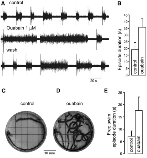
Na+/K+exchange pumps seemed the most likely contenders to mediate the usAHP because they (1) set the resting membrane potential, and (2) are affected by transmembrane Na+ and K+ gradients that determine the driving force for pump activity. Indeed the pump blocker ouabain (0.5–10mM; n = 10) caused a rapid and complete (but irreversible) block of the usAHP both after swimming (Figure 3A1) and after depolarizing pulses (Figure 3A3). In a complementary approach, the driving force for pump activity was reduced by removing extracellular K+ions. Zero-K+saline led to a rapid, total, and reversible block (Figures 3B1 and 3B3; n = 6), con-firming that the usAHP is not a K+conductance and supporting the conclusion that it involves a Na+spike-dependent increase in Na+/K+pump activity. Both ouabain and zero-K+ manipula-tions also increased swimming episode duramanipula-tions (Figures 3A1, 3A2, 3B1, and 3B2), suggesting that altering pump activity influences locomotor behavior.
Na+/K+Pumps Regulate Swim Bout Duration
Fictive swim episode duration correlates linearly with inter-swim interval (Figure 1B), but is this relationship causally related to an activity-dependent increase in Na+/K+ pump function? The fact that blocking the usAHP has the opposite effect on episode duration (an increase) to that of reducing the swim interval (a decrease) is consistent with the proposal that the usAHP dictates future network performance. To inves-tigate this possibility further, we first obtained relatively constant episode durations by using constant stimulus inter-vals (Figure 4A, upper trace). Shortly after applying ouabain (0.5–1mM), much longer swimming episodes, up to several minutes, initially appeared (Figure 4A, middle trace;Figure 4B; n = 9), disrupting the relationship between interswim interval and episode duration. After prolonged exposure to ouabain, an inhibitory effect on swimming occurred and episodes even-tually shortened to below control levels (data not shown), pre-sumably due to a gradual loss of ionic homeostasis following longer term inactivation of Na+
[image:4.603.57.546.78.376.2]/K+ pumps. Ouabain effects were sometimes partially reversible (Figure 4A, lower trace), but more usually they were irreversible even after 1 hr of wash. To explore the role of the usAHP at the behavioral level, we applied ouabain to freely swimming Xenopus tadpoles while their movements were videoed. The results paralleled the electrophysiological data: ouabain initially significantly increased the duration of swimming bouts (Figures 4C–4E;
Figure 3. The usAHP Is Mediated by Na+ /K+
Pumps (A1 and B1) Ouabain (0.5mM) or zero-K+
saline blocked the usAHP following swimming. Left panels, control; middle panels, ouabain or zero-K+
saline; right panels, wash.
(A2 and B2) Ouabain (p < 0.05) or zero-K+
saline (p < 0.01) increased swimming episode duration: control/wash, average of three episodes from each exper-iment; treatment, one episode during short application. Data are represented as mean6SEM.
(A3 and B3) Ouabain or zero-K+
n = 12; p < 0.05), but with prolonged exposure swimming even-tually reduced to only a few cycles or nonlocomotory twitches. Thus, enhanced Na+/K+pump activity and the resulting usAHP have a direct effect on locomotor bout duration.
Discussion
Locomotor CPG networks must adapt their output in light of changing environmental conditions, developmental states, or organismal demands. Locomotor activity is influenced by sensory information and descending signals from the brain, but CPGs also display intrinsic flexibility that modifies their output in an activity-dependent, self-regulatory manner. In this report, we provide the first direct demonstration that a potentiation of Na+/K+ pump activity plays an important role in determining the duration of vertebrate locomotor behavior. The increase in pump activity, presumably triggered by Na+influx, hyperpolarizes the membrane potential of spinal neurons in a manner that integrates spike frequency over time. We propose that the hyperpolarization of CPG neurons that accumulates during an episode will affect their firing pro-perties [1] and impair their recruitment during locomotion [10], hence reducing network excitability. In effect, this mech-anism endows the spinal network with a short-term memory of previous network performance: intense activity will lead to shorter subsequent bouts, whereas weaker activity affords
more prolonged locomotion, analogous to sprinting versus long-distance running.
The pump mechanism overlays other more conventional processes that unite to control locomotor bout duration including GABAergic [11], purinergic [12], peptidergic [16], and nitrergic [17, 18] signaling. Modulation of Na+-dependent channels by network activity also alters cell properties and CPG function [13–15], but evidence that membrane pumps play a role is limited. In zebrafish larvae, 5-hydroxytryptamine decreases the intervals between episodes, without affecting other episode parameters, by modulating the bumetanide-sensitive inward chloride cotransporter [2]. In a disinhibited spinal network of neonatal rats, blocking Na+/K+pump activity disrupts rhythmic bursting of lumbar MNs [3, 4]. InDrosophila larvae, MNs controlling crawling show a slow hyperpolariza-tion following depolarizing current injechyperpolariza-tion due to upregula-tion of Na+/K+ exchange pumps [1], similar to the usAHP. Genetic inhibition of the Na+/K+ ATPase reduces crawling suggesting the pumps directly control locomotion, as we demonstrate more directly here in tadpoles at the network and behavioral levels. Activity-dependent upregulation of pump function has also been demonstrated in sensory neurons, where it alters neuronal excitability [19, 20].
What advantage does the network accrue from a pump-based control mechanism? The usAHP does not involve a conductance change, so although the membrane potential hyperpolarizes, there is no shunting of the membrane, and neuronal excitability is relatively unimpaired. The usAHP was only detectable in a proportion of neurons (42%), suggesting that some circuit elements are only indirectly affected by the usAHP mechanism. For example, dINs, the electrically coupled glutamatergic interneurons that are thought to generate and maintain swimming once initiated [9, 21, 22], appear to lack a usAHP. This might ensure that the network always retains some rhythm generating capability to enable escape and enhance survival. However, it also raises the question of why episode durations are affected at all if the dINs have no usAHP. A likely explanation is that the dINs are conditional oscillators that receive feedback from CPG neurons, including midcycle inhibition from cINs. Any reduc-tion in this feedback due to the usAHP will impair dIN rebound firing properties and dampen dIN oscillations. Why don’t all neurons display a usAHP? One possibility is that the change in intracellular Na+
concentration during activity could be limited in large neurons and amplified in small ones. However, the presence of a usAHP did not correlate with input resistance (Figure S1A) and therefore cell size. A second possibility is neuron-specific differences in Na+/K+pump subunit expres-sion, which determines pump sensitivity to both intracellular Na+and ouabain [23]. Interestingly, a point mutation of the
[image:5.603.57.297.84.341.2]a3 subunit in humans leads to rapid-onset dystonia-parkin-sonism [24]. Thirdly, an endogenous ouabain-like molecule could block the usAHP in some neurons but not others [25]. Fourthly, Na+/K+ pump function is known to be regulated by a range of intracellular second-messenger pathways, including protein kinases and phosphatases [26]. Because these pathways can be accessed by familiar modulators of spinal CPGs, such as serotonin, noradrenaline, and nitric oxide, there could be differential intracellular regulation of pump function in different spinal neurons and neuron classes. The trigger for the usAHP, action potential firing in network neurons, is ubiquitous, making it likely that the same phenom-enon occurs elsewhere. It may serve different functions in different networks; increasing Na+/K+pump activity protects Figure 4. Role of usAHP
(A) Interswim interval was set to 15 s to generate relatively constant, short episodes in control (top trace). Ouabain resulted in longer swimming episodes (middle trace), and its effect was partially reversible (bottom trace).
(B) Histogram of ouabain effect on episode duration. Data are represented as mean6SEM.
(C) Real swimming in a stage 37/38Xenopustadpole with multiple consec-utive video frames overlapped to show swim path in response to touch. (D) Longer swimming episode in same animal after ouabain.
hippocampal neurons from ischemic damage [27], whereas excitotoxic calcium influx through glutamate receptors follow-ing intense activity, such as occurs durfollow-ing brain injury or ischemia, can inhibit pump function [28]. Activity-dependent potentiation of pump function could therefore serve as a useful homeostatic mechanism to maintain network activity within predamaging levels. In tadpole spinal neurons, the usAHP enables the locomotor network to set the excitability of the system in relation to previous activity and thereby provides a novel form of short-term memory that controls future loco-motor behavior.
Experimental Procedures
All experiments complied with UK Home Office regulations and were approved by the University of St Andrews Animal Welfare Ethics Committee. Experiments were performed on newly hatchedXenopus laevistadpoles (stage 37/38–42) [8] obtained by hormone-assisted mating of adults (chorionic gonadotropin injection; 1,000 U/ml, Sigma). All experimental procedures were described previously [10, 29]. For further details, see Supplemental Experimental Procedures.
Supplemental Information
Supplemental Information includes one figure and Supplemental Experi-mental Procedures and can be found with this article online atdoi:10. 1016/j.cub.2012.01.058.
Acknowledgments
We thank Stephen Currie, Bill Heitler, Gareth Miles, Mel Robertson, and Nick Scott for helpful discussion and comments on a draft of the manuscript. This work was supported by a project grant to K.T.S. from the BBSRC (UK), to whom we are grateful.
Received: November 23, 2011 Revised: January 9, 2012 Accepted: January 26, 2012 Published online: March 8, 2012
References
1. Pulver, S.R., and Griffith, L.C. (2010). Spike integration and cellular memory in a rhythmic network from Na+
/K+
pump current dynamics. Nat. Neurosci.13, 53–59.
2. Brustein, E., and Drapeau, P. (2005). Serotoninergic modulation of chlo-ride homeostasis during maturation of the locomotor network in zebra-fish. J. Neurosci.25, 10607–10616.
3. Rozzo, A., Ballerini, L., Abbate, G., and Nistri, A. (2002). Experimental and modeling studies of novel bursts induced by blocking Na+ pump and synaptic inhibition in the rat spinal cord. J. Neurophysiol.
88, 676–691.
4. Ballerini, L., Bracci, E., and Nistri, A. (1997). Pharmacological block of the electrogenic sodium pump disrupts rhythmic bursting induced by strychnine and bicuculline in the neonatal rat spinal cord. J. Neurophysiol.77, 17–23.
5. Rossignol, S., Dubuc, R.J., and Gossard, J.P. (2006). Dynamic sensori-motor interactions in locomotion. Physiol. Rev.86, 89–154.
6. Katz, P.S. (1999). Beyond Neurotransmission: Neuromodulation and Its Importance for Information Processing (New York: Oxford University Press).
7. Harris-Warrick, R.M. (2010). General principles of rhythmogenesis in central pattern generator networks. Prog. Brain Res.187, 213–222. 8. Nieuwkoop, P.D., and Faber, J. (1956). Normal Tables ofXenopus laevis
(Daudin) (Amsterdam: North Holland).
9. Roberts, A., Li, W.-C., and Soffe, S.R. (2010). How neurons generate behavior in a hatchling amphibian tadpole: an outline. Front Behav Neurosci4, 16.
10. Zhang, H.-Y., Issberner, J., and Sillar, K.T. (2011). Development of a spinal locomotor rheostat. Proc. Natl. Acad. Sci. USA108, 11674– 11679.
11. Reith, C.A., and Sillar, K.T. (1999). Development and role of GABA(A) receptor-mediated synaptic potentials during swimming in postembry-onicXenopus laevistadpoles. J. Neurophysiol.82, 3175–3187. 12. Dale, N., and Gilday, D. (1996). Regulation of rhythmic movements by
purinergic neurotransmitters in frog embryos. Nature383, 259–263. 13. Dale, N. (1993). A large, sustained Na+
- and voltage-dependent K+ current in spinal neurons of the frog embryo. J. Physiol.462, 349–372. 14. Walle´n, P., Robertson, B., Cangiano, L., Lo¨w, P., Bhattacharjee, A.,
Kaczmarek, L.K., and Grillner, S. (2007). Sodium-dependent potassium channels of a Slack-like subtype contribute to the slow afterhyperpola-rization in lamprey spinal neurons. J. Physiol.585, 75–90.
15. Hess, D., Nanou, E., and El Manira, A. (2007). Characterization of Na+
-activated K+
currents in larval lamprey spinal cord neurons. J. Neurophysiol.97, 3484–3493.
16. Pe´rez, C.T., Hill, R.H., and Grillner, S. (2007). Endogenous tachykinin release contributes to the locomotor activity in lamprey. J. Neurophysiol.97, 3331–3339.
17. McLean, D.L., and Sillar, K.T. (2004). Metamodulation of a spinal loco-motor network by nitric oxide. J. Neurosci.24, 9561–9571.
18. Kyriakatos, A., Molinari, M., Mahmood, R., Grillner, S., Sillar, K.T., and El Manira, A. (2009). Nitric oxide potentiation of locomotor activity in the spinal cord of the lamprey. J. Neurosci.29, 13283–13291.
19. Parker, D., Hill, R., and Grillner, S. (1996). Electrogenic pump and a Ca2+
- dependent K+
conductance contribute to a posttetanic hyperpo-larization in lamprey sensory neurons. J. Neurophysiol.76, 540–553. 20. Scuri, R., Lombardo, P., Cataldo, E., Ristori, C., and Brunelli, M. (2007).
Inhibition of Na+ /K+
ATPase potentiates synaptic transmission in tactile sensory neurons of the leech. Eur. J. Neurosci.25, 159–167.
21. Li, W.-C., Soffe, S.R., Wolf, E., and Roberts, A. (2006). Persistent responses to brief stimuli: feedback excitation among brainstem neurons. J. Neurosci.26, 4026–4035.
22. Li, W.-C., Roberts, A., and Soffe, S.R. (2010). Specific brainstem neurons switch each other into pacemaker mode to drive movement by activating NMDA receptors. J. Neurosci.30, 16609–16620. 23. Blanco, G., and Mercer, R.W. (1998). Isozymes of the Na-K-ATPase:
heterogeneity in structure, diversity in function. Am. J. Physiol.275, F633–F650.
24. Calderon, D.P., Fremont, R., Kraenzlin, F., and Khodakhah, K. (2011). The neural substrates of rapid-onset Dystonia-Parkinsonism. Nat. Neurosci.14, 357–365.
25. Schoner, W., and Scheiner-Bobis, G. (2007). Endogenous and exoge-nous cardiac glycosides and their mechanisms of action. Am. J. Cardiovasc. Drugs7, 173–189.
26. Therien, A.G., and Blostein, R. (2000). Mechanisms of sodium pump regulation. Am. J. Physiol. Cell Physiol.279, C541–C566.
27. Tian, D., Dmitrieva, R.I., Doris, P.A., Crary, J.F., Sondhi, R., Sacktor, T.C., and Bergold, P.J. (2008). Protein kinase M zeta regulation of Na/K ATPase: a persistent neuroprotective mechanism of ischemic pre-conditioning in hippocampal slice cultures. Brain Res.1213, 127–139. 28. Fukuda, A., and Prince, D.A. (1992). Excessive intracellular Ca2+
inhibits glutamate-induced Na+
-K+
pump activation in rat hippocampal neurons. J. Neurophysiol.68, 28–35.