1
The endogenous opioid system in schizophrenia and treatment resistant schizophrenia:
increased plasma endomorphin 2, and κ and μ opioid receptors are associated with
interleukin-6.
Shatha Rouf Moustafa,a Khalid F. Al-Rawi,b Arafat Hussein Al-Dujaili,c Thitiporn Supasitthumrong, d Hussein Kadhem Al-Hakeim, e Michael Maes. f,g,h
a Clinical Analysis Department, College of Pharmacy, Hawler Medical University,
Havalan City, Erbil, Iraq.
b College of Science, University of Anbar.
c Senior Clinical Psychiatrist at the Faculty of Medicine, University of Kufa, Iraq.
d Department of Psychiatry, Faculty of Medicine, Chulalongkorn University, Bangkok,
Thailand.
e Department of Chemistry, College of Science, University of Kufa, Iraq. f Department of Psychiatry, Medical University of Plovdiv, Plovdiv, Bulgaria; g School of Medicine, IMPACT Strategic Research Centre, Deakin University
Corresponding authors:
Thitiporn Supasitthumrong. M.D. and Prof. Dr. Michael Maes, M.D., Ph.D.
Department of Psychiatry
Faculty of Medicine
Chulalongkorn University
Bangkok
2
Thailand.
email: [email protected].
https//:scholar.google.co.th/citations?user=1wzMZ7UAAAAJ&hl=th&oi=ao
Shatha Rouf Moustafa: [email protected]
Khalid F. Al-Rawi: [email protected]
Arafat Hussein Al-Dujaili: [email protected].
Thitiporn Supasitthumrong:[email protected]
Hussein Kadhem Al-Hakeim: [email protected].
3
Abstract
Background: Activation of the immune-inflammatory response system (IRS) and the
compensatory immune-regulatory system (CIRS) play a key role in SCZ and treatment
resistant SCZ. There are only few data on immune and endogenous opioid system (EOS)
interactions in SCZ and treatment resistant SCZ.
Aim of the study: We examined serum β-endorphin, endomorphin-2 (EM2), mu-opioid
(MOR) and kappa-opioid (KOR) receptors, and interleukin (IL)-6 and IL-10 in 60 non
responders to treatment (NRTT), 55 partial RTT (PRTT) and 43 normal controls.
Results: Serum EM2, KOR, MOR, IL-6 and IL-10 were significantly increased in SCZ as
compared with controls. β-endorphin, EM2, MOR and IL-6 were significantly higher in
NRTT than in PRTT. There were significant correlations between IL-6, on the one hand,
and β-endorphin, EM2, KOR, and MOR, on the other, while IL-10 was significantly
correlated with MOR only. A large part of the variance in negative symptoms, psychosis,
hostility, excitation, mannerism, psychomotor retardation and formal thought disorders
was explained by the combined effects of EM2 and MOR with or without IL-6 while
increased KOR was significantly associated with all symptom dimensions. Increased
MOR, KOR, EM2 and IL-6 were also associated with neurocognitive impairments
including in episodic, semantic and working memory and executive functions.
Conclusion: The EOS contributes to SCZ symptomatology, neurocognitive impairments
and a non-response to treatment. In SCZ, EOS peptides/receptors may exert CIRS
functions, whereas increased KOR levels may contribute to the pathophysiology of SCZ
4
Keywords: inflammation, schizophrenia, treatment resistance, neurocognition,
5 Introduction
The first comprehensive neuro-immune theory of schizophrenia (SCZ) was
published by Smith and Maes in 1995 and suggested that activated monocytes and
T-lymphocytes are key phenomena in the pathophysiology of SCZ (Smith and Maes 1995).
Now, it is widely accepted that SCZ is accompanied by activation of the
immune-inflammatory response system (IRS) with activated M1 macrophages as indicated by
increased levels of interleukin (IL)-6, IL-1β and tumor necrosis factor alpha (TNFα) levels,
T-helper (Th)-1 cells and Th-17 cells (Al-Hakeim et al., 2019a; Almulla et al., 2019; Maes
et al., 2019c; Noto et al., 2015; 2019; Roomruangwong et al., 2019; Rubesa et al., 2018).
SCZ is also accompanied by activation of the compensatory immune-regulatory system
(CIRS) with increased activity of Th-2 immunocytes as indicated by increased levels of
IL-4 and IL-13, and T-regulatory (Treg) cells with increased production of IL-10, and
elevated levels of immune-regulatory compounds including acute phase proteins (e.g.
haptoglobin) and soluble interleukin receptors such as sIL-2R, sIL-1RA, sTNFR1 and
sTNFR2 (Noto et al., 2019; Roomruangwong et al., 2019). Importantly, IRS and CIRS
products including IL-1β, TNF-α, IL-6, IFN-γ, IL-4, IL-13, eotaxin (CCL11) and
neurotoxic tryptophan catabolites (TRYCATs) may all cause neuroprogression and, as a
consequence, deficits in memory and executive functions and, therefore, SCZ symptoms
(Al-Hakeim et al., 2019a; Kanchanatawan et al., 2018; Maes et al., 2019a; 2019b; 2019c;
2019d; Sirivichayakul et al., 2019a; 2019b).
Treatment resistant schizophrenia (TRS) is also accompanied by signs of IRS and
CIRS including M1 (especially IL-6 trans-signaling and TNFα) and Treg (IL-10) cell
6
(sIL-1RA), and sTNFR2 (Al-Hakeim et al., 2019a; Maes et al., 2000; Noto et al., 2015;
Roomruangwong et al., 2019).
Endogenous opioids (EOs) are expressed in the peripheral and central nervous
system where they modulate pain, reward, aversion, reinforcement, social bonding, and
neurotransmitter signaling including that of glutamate and dopamine (Charles et al., 2020;
Corder et al., 2018). During IRS activation, immunocytes produce and release EOs (Plein
and Rittner 2018) and express µ (MOR) and κ (KOR) opioid receptors (Li et al., 2009). In
humans and animal models, EOs negatively regulate immune responses (Hu et al., 1998;
Sacerdote, 2006) including downregulation of cytokine and chemokine levels and
associated receptors (Finley et al., 2008) and immune cell proliferation and activities
(McLaughlin et al., 2015).
Aberrations in EO system (EOS) activity are observed in SCZ including changes
in the levels of endorphins, and MOR and KOR expression (Schwarzer et al., 2009);
(Ashok et al., 2019). In post-mortem brain of SCZ patients, lowered MOR expression is
observed in the striatum (Ashok et al., 2019). Because MORs mediate hedonic and social
reward processing, lowered MOR expression may explain social impairments and other
negative symptoms of SCZ (Trezza et al., 2011). β-Endorphin concentrations are increased
in SCZ patients with negative symptoms and decreased in patients with positive symptoms
as compared with controls (Urban-Kowalczyk et al., 2016). Interestingly, clinical reports
indicate that some SCZ patients are less sensitive to pain (Szűcs et al., 2016), suggesting
increased EOS activity. Endomorphin-2, another EOS peptide, has high selectivity and
7
reward, neuroendocrine and neurocognitive functions (Fichna et al., 2007), suggesting that
this peptide may play a role in SCZ.
In major depression, significant associations were established between increased
plasma KOR/MOR levels, on the one hand, and elevated plasma IL-6 and IL-10, on the
other, indicating that immune - EOS interactions play a role in the pathophysiology of
depression (Al-Hakeim et al., 2019b). Furthermore, IL-6 regulates the KOR gene (Jenab
and Morris 2000) and MOR expression (Börner et al., 2004), while IL-10 increases
β-endorphin gene and protein expression (Wu et al., 2018). Since KOR/MOR/β-β-endorphin
have immune-regulatory effects and since these products are upregulated in major
depression, it was hypothesized that their increased levels may contribute to CIRS
functions in depression (Al-Hakeim et al., 2019b). However, in SCZ and TRS no data are
available on endomorphin 2 and possible associations between EOS compounds and
IRS/CIRS functions.
Hence, this study was performed to examine serum levels of β-endorphin and
endomorphin 2 as well as MOR and KOR in association with IL-6 and IL-10 in SCZ and
TRS.
Participants and Methods
Participants
In the current study, two groups of SCZ patients participated, namely 60 non
responders to treatment (NRTT) and 55 partial responders to treatment (PRTT) and 43
healthy volunteers. The participants were of both sexes and aged 18-65 years. NRTT and
8
Karbala, Iraq. All patients were diagnosed using DSM-IV-T criteria. NRTT was defined
as non-responders to two episodes of treatment (each for at least two months), with two
different antipsychotic drugs at adequate doses, without a decrease in symptom severity as
screened using the Clinical Global Impression Severity (CGI-S) scale (Guy, 2000). Healthy
volunteers were family members or friends of staff. Patients and controls were recruited
from same catchment area, namely Karbala city, Iraq. SCZ patients who showed axis-1
DSM-IV-TR diagnoses other than SCZ were excluded from the study including autism
spectrum disorders, major depression, psycho-organic disorders, bipolar disorder, and
schizoaffective disorder. Healthy controls were excluded if they had a current or lifetime
diagnosis of axis I diagnosis or had a positive family history of SCZ. Moreover, patients
and controls were excluded when they (a) suffered from medical disorders including
chronic obstructive pulmonary disease, diabetes, rheumatoid arthritis, inflammatory bowel
disease and psoriasis; and (b) suffered from neuro-inflammatory or neuro-degenerative
disorders including Parkinson’s disease, stroke, Alzheimer’s disease and multiple sclerosis.
Any subjects who had ever been taken immune-modulatory medications such as
glucocorticoids were excluded to participate. We also excluded subjects who took
therapeutic doses of antioxidant supplements 3 months prior to the blood aspiration. All
participants showed serum C-reactive protein (CRP) levels < 6 mg/L indicating absence of
overt inflammation.
All patients and controls, along with the guardians of patients (parents or the
first-degree family members), provided written informed consent prior to participation in the
study. The study was carried out according to international and Iraq ethics and privacy
9
Karbala (418/2019) and Karbala Health Department (1331/2019), which is in agreement
with the International Guidelines for Human Research protection as requested by the
Declaration of Helsinki, The Belmont Report, Council for International Organizations of
Medical Sciences guideline and International Conference on Harmonization in Good
Clinical Practice.
Measurements
Clinical evaluations
A senior psychiatrist specialized in SCZ diagnosed the patients according to
DSM-IV-TR criteria using the Mini-International Neuropsychiatric Interview (MINI), in a
validated Arabic translation (Iraqi dialect). The same psychiatrist employed a
semi-structured interview to assess socio-demographic and clinical data in all participants and
the same day he also assessed the Clinical Global Impression (CGI) Improvement (CGI-I)
and Severity (CGI-S) Scales (Guy, 2000). The CGI-I was used to identify NRTT when
they did not show any improvement on the CGI-I or showed poorer scores after treatment
(minimally worse, much worse, very much worse). The diagnosis “responders to
treatment” was made in case the CGI-I scores improved minimally, much or very much.
However, since no patients showed remission after treatment this group was named PRTT.
Furthermore, we assessed the Scale for the Assessments of Negative Symptoms (SANS)
to measure severity of negative symptoms (Andreasen, 1989). We also computed z
unit-weighted composite scores to reflect severity of psychosis, hostility, excitation,
mannerism, FTD (formal thought disorders) and PMR (psycho-motor retardation)
10
Toward this end, we assessed the Brief Psychiatric Rating Scale (BPRS) (Overall and
Gorham 1962), the Hamilton Depression Rating Scale (Hamilton, 1960) and the Positive
and Negative Syndrome Scale (PANNS) (Kay et al., 1987). The same psychiatrist also
completed the Brief Assessment of Cognition in SCZ (BACS) (Keefe et al., 2004) to assess
episodic memory (using the List learning test); working memory (using the Digit
Sequencing Task); verbal fluency (using the Controlled Word Association and Category
instances tests); attention (using the symbol coding test); and executive functions (using
the Tower of London). Tobacco use disorder (TUD) was examined according to
DSM-IV-TR criteria. Body mass index (BMI) was calculated from the formula: body weight (kg) /
squared length (m2).
Assays
Five milliliters of venous blood samples were collected after an overnight fast
between 8.00 and 9.00 a.m. After clotting, blood was centrifuged at 3000 rpm for 10 min,
and serum was separated and transferred into two Eppendorf tubes which were stored at
-80 °C until analysis. Serum levels of IL-10 (Elabscience®, Inc. CA, USA), MOR, KOR, and endomorphin 2 (Mybiosource®, Inc. CA, USA), and IL-6 and β-endorphin (Melsin Medical Co, Jilin, China) were assayed used commercial ELISA kits. All measured
concentrations of β-endorphins (sensitivity=0.1pg/mL), MOR (sensitivity=7.18 pg/mL),
KOR (sensitivity=1.0 ng/mL), endomorphin 2 (sensitivity=0.33 pg/mL), and IL-6
(sensitivity=0.1 pg/mL) were greater than their assay sensitivities. There was only one
IL-10 measurement (namely 4.05 pg/mL in a normal volunteer) that was lower than the
left-11
censoring of the data and employed the measured IL-10 concentration in our statistical
analyses (Al-Hakeim et al., 2019a). All intra-assay coefficients of variation were < 10.0%.
Serum CRP was measured using a kit supplied by Spinreact®, Spain, using a test based on the latex agglutination principle.
Statistical analysis
We used analysis of variance to check the differences in scale variables between
categories and Chi square test (χ2-test) to assess the associations between categorical
variables. Associations among biomarkers, cognitive and clinical scores were computed
employing Pearson’s product-moment and Spearman’s rank-order correlation coefficients.
Associations between diagnosis and biomarkers were examined using multivariate general
linear model (GLM) analysis while controlling for confounding variables including age,
sex, TUD, BMI, and education. Consequently, we performed tests for between-subject
effects to delineate the associations between diagnosis and each of the biomarkers. The
effect size was assessed using partial eta-squared values (η2). We also computed the model-generated estimated marginal mean (SE) values and used protected pairwise comparisons
among treatment means to assess differences between the diagnostic groups. Binary
logistic regression analysis was employed to delineate the best predictors of SCZ versus
controls and NRTT versus PRTT using the serum biomarkers levels as explanatory
variables. Nagelkerke values, which were used as pseudo-R2 values, were computed along with Odd’s ratios with 95% confidence intervals. We used multiple regression analysis to
assess the significant biomarkers, which predict the neurocognitive test results and
12
used an automatic stepwise method with the inclusion of variables with a p-to-entry of 0.05
and p-to-remove of 0.06 while checking the R2 change. All regression analyses were checked for collinearity using tolerance and variance inflation factor (VIF) values. Tests
were 2-tailed and a p-value of 0.05 was used for statistical significance. All statistical
analyses were performed using IBM SPSS windows version 25, 2017.
Results.
Socio-demographic data
Table 1 shows the sociodemographic data of the PRTT and NRTT and healthy
controls. We included 142 SCZ patients who were treated with antipsychotic drugs during
two trials with antipsychotic medications. During the first trial, patients were treated for 2
months and after this trial divided into those with a partial response (n=51) and those
without a clinical response (n=84) (we lost seven patients through this first trial). The
partial responders continued to take the similar medication for another two months while
we missed again seven patients in the follow up producing a final PRTT study group of
n=55. The non-responders to the initial antipsychotic drug were switched to another
antipsychotic drug for another 2 months and during this follow-up period we missed again
thirteen patients. Two months later, eleven patients had a partial response to treatment and
were categorized as PRTT, whereas sixty patients did not show any improvement on the
CGI-I and, therefore, were categorized as NRTT. Consequently, fifty-five PRTT and sixty
NRTT participated in the current study.
There were no significant differences in age, BMI, sex ratio, and TUD between
13
single as compared with the normal control group. SCZ patients were significantly more
unemployed as compared with controls while years of education were slightly lower in
NRTT. There were no differences in age at onset between both patients’ subgroups. All six
cognitive test scores were significantly different among the 3 study groups and the scores
decreased from controls to PRTT to NRTT. The total SANS score was significantly
different between the three study groups.
In Table 1, both the CGI-I and CGI-S scores were significantly higher in NRTT
than in PRTT. The CGI-I scores in PRTT were 2 (much improved) - 3 (minimally
improved) and in NRTT 4 (no change) - 5 (minimally worse). Patients in the NRTT group
were more often treated with clozapine, risperidone, and quetiapine than PRTT who were
more often treated with haloperidol and olanzapine.
The following symptom domains were significantly different between the three
study groups and increased from controls to PRTT to NRTT: excitement (F=320.71,
df=2/152, p<0.001), FTD (F=414.15, df=2/152, p<0.001), hostility (F=498.12, df=2/152,
p<0.001), mannerism (F=204.41, df=2/152, p<0.001), PMR (F=297.46, df=2/152,
p<0.001) and psychosis (F=772.55, df=2/152, p<0.001
In the whole study group, there were significant correlations between IL-6 and
β-endorphin (ρ=0.173, p=0.03), endomorphin 2 (ρ=0.253, p=0.001), KOR (ρ=0.491,
p<0.001), MOR (ρ=0.333, p<0.001) and a significant association between IL-10 and MOR
(ρ=0.385, p<0.001). MOR is correlated with KOR (ρ=0.449, p<0.001) while endomorphin
2 was correlated with β-endorphin (ρ=0.254, p=0.001) and KOR (ρ=0.385, p<0.001).
14
Table 2 displays the outcomes of a multivariate GLM analysis comparing the
differences in the biomarkers between the three study groups while adjusting for age, BMI,
and sex. There were significant differences in biomarkers between the study groups with
an effect size of 0.213, while the covariates had no significant effects. Tests for
between-subject effects and Table 3, which shows the estimated marginal mean (SE) values,
indicate that all six biomarkers (except β-endorphin) were significantly higher in SCZ
patients as compared with controls. Furthermore, β-endorphin, MOR, KOR and IL-6 were
significantly higher in NRTT than in PRTT and controls. Endomorphin 2 was significantly
different between the three groups and increased from controls to PRTT to NRTT. IL-10
was higher in NRTT than in controls while PRTT patients occupied an intermediate
position. We also performed a multivariate GLM analysis examining the associations
between the 6 biomarkers measured here and the diagnosis of SCZ versus normal controls
while controlling for age, sex and BMI. This analysis showed that endomorphin 2, KOR,
MOR, IL-6 and IL-10, but not β-endorphin (p=0.865) were significantly higher is SCZ than
in controls (with or without p correction for false discovery rate).
Table 4 shows the results of two binary logistic regression analyses examining the
best predictors of SCZ (versus controls) and NRTT (versus PRTT) using an automatic
stepwise method with biomarkers as explanatory variables while allowing for the effects
of age, sex and education. The first regression analysis showed that SCZ was best predicted
by increased levels of endomorphin 2, KOR, and IL-10 (χ2=39.338, df=3, p<0.001,
Nagelkerke=0.319) with an accuracy of 72.2%, sensitivity of 74.8% and specificity of
15
IL-6 best predicted NRTT versus PRTT (χ2=34.47, df=3, p<0.001, Nagelkerke=0.346)
with an accuracy of 72.2%, sensitivity of 80.0% and a specificity of 76.5%.
Effects of background variables
As shown above, age, sex, and BMI had no significant effects on serum biomarker
levels. TUD also had no significant effect on the measured biomarker levels (F=0.19,
df=6/146, p=0.979, partial η2 =0.008). We also examined the effects of antipsychotic drug
administration using multivariate GLM analysis and tests for between-subjects. We found
no significant effects of use of clozapine (F=0.63, df=6/146, p=0.704, partial η2 =0.025),
haloperidol (F=0.88, df=6/146, p=0.513, Partial η2 =0.035), risperidone (F=0.92, df=6/146, p=0.484, partial η2 =0.036), and quetiapine (F=0.93, df=6/146, p=0.476, partial η2 =0.037).
However, use of olanzapine had a significant effect on the measured biomarkers (F=2.89,
df=6/146, p=0.011, partial η2 =0.106), although after p-correction for multiple testing this
effect was no longer significant (p=0.195).
Prediction of symptom domains by biomarkers
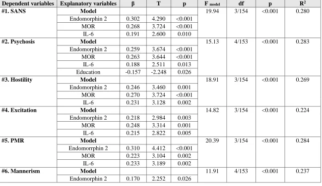
Table 5 shows different stepwise multiple regression analyses with the symptom
domains as dependent variables and the six biomarkers as explanatory variables while
allowing for the effects of education, age and sex. Regression #1 shows that 28.0% of the
variance in the total SANS score was explained by the regression on endomorphin 2, MOR,
and IL-6. Regressions #2, #3, #4 and #5 show that the same variables explained a
considerable part of the variance in psychosis (28.3%, but additionally with education),
16
of the variance in mannerism was explained by endomorphin 2, MOR, KOR, and IL-10.
Regression #7 shows that 27.8% of the variance in FTD was explained by the combined
effects of endomorphin 2, MOR, IL-6, IL-10, and education. Partial correlation coefficients
(adjusted for age, sex, and education) showed that all symptom profiles were significantly
associated with plasma KOR kevels (all r>0.314, p<0.001, n=153), but not with
β-endorphin.
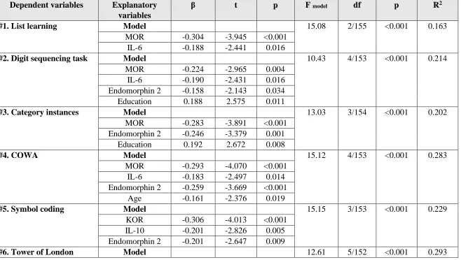
Prediction of cognitive impairments by biomarkers
Table 6 shows the outcome of 6 multiple regression analyses with the cognitive
test results as dependent variables and biomarkers as explanatory variables while allowing
for the effects of age, sex and education. We found that (regression #1) 16.3% of the
variance in List Learning scores was explained by the regression on IL-6 and MOR (all
inversely associated). Up to 21.4% of the variance in Digit Sequencing Task scores
(regression #2) was explained by the combined effects of MOR, IL-6, endomorphin 2
(inversely) and education (positively). Part of the variance (20.2%) in Category Instances
test scores (regression #3) was explained by MOR, endomorphin 2 (negatively) and
education (positively). We found that 28.3% of the variance in the COWA test scores
(regression #4) was explained by the cumulative effects of MOR, IL-6, endomorphin 2,
and age (negatively) while 22.9% of the variance in symbol coding scores (#5) was
negatively associated with KOR, IL-10, and endomorphin 2. Up to 29.3% of the variance
in Tower of London test scores (regression #6) was explained by the combined effects of
MOR, IL-6, endomorphin 2, sex (all inversely) and education (positively). Partial
17
tests results were significantly associated with plasma KOR (all r>0.206, p<0.01, n=152),
but not with β-endorphin.
Discussion
The first major finding of this study is that serum levels of KOR, MOR,
endomorphin 2, IL-6 and IL-10 are significantly increased in SCZ as compared with
controls. This is a first report on increased endomorphin 2 levels in SCZ. Volk et al. found
that increased MOR mRNA and protein levels in SCZ are largely independent of illness
severity, suggesting that increased MOR expression is part of the disease process rather
than a consequence of illness chronicity (Volk et al., 2011). In Han Chinese, a MOR
polymorphism may confer risk for SCZ (Ding et al., 2013) while an A118G polymorphism
of the MOR gene was associated with SCZ (Šerý et al., 2010). One study reported lowered
MOR availability in the brain of SCZ patients who died as a result of suicide, which would
be consistent with increased levels of EOS peptides occupying those receptors (Scarr et al.,
2012). Our negative findings on β-endorphin levels in SCZ are not in agreement with those
of a previous report (Ding et al., 2013). Animal models of SCZ are accompanied by
moderate alterations in EOS peptides (Ashok et al., 2019; Schwarzer et al., 2009; Volk et
al., 2011; Szűcs et al., 2016;). Our findings on increased IL-6 and IL-10 levels in SCZ are
in agreement with previous reports that SCZ is accompanied by enhanced IRS and CIRS
functions (Roomruangwong et al., 2019).
This is also a first report that a non-response to treatment is characterized by
increased MOR, endomorphin 2 and β-endorphin levels, indicating that the EOS
18
increased in NRTT as compared with PRTT is in accordance with previous reports that
IL-6 and macrophage M1 activation are associated with TRS (Maes et al., 2000;
Roomruangwong et al., 2019). In the current study we found that serum IL-10 was
significantly higher in NRTT than in controls, whereas PRTT occupied an intermediate
position. Previous reports showed that TRS is accompanied by increased IL-10 levels
(Maes et al., 2002).
The second major finding of this study is that IL-6 levels are strongly associated
with all four EOS biomarkers whilst IL-10 is associated with MOR concentrations only.
Similar findings were reported in major depression showing significant associations
between an immune activation index (based on IL-6 and IL-10) and KOR, MOR and
β-endorphin levels (Al-Hakeim et al., 2019b; Almulla et al., 2019). Many inflammatory
diseases are associated with up-regulation of opioid receptors (Jiménez et al., 2006) and,
in those conditions, inflammation is associated with increased MOR sensitivity in the
periphery and in the brainstem (Hipólito et al., 2015). Moreover, the combination of IL-6
with endomorphin 2 and MOR yielded a highly significant discrimination of NRTT from
PRTT indicating that upregulation of the immune - EOS axis is associated with the
pathophysiology of a non-response to treatment.
During activation of the IRS, immune cells produce a) opioid peptides, (Przewlocki
et al., 1992; Panerai and Sacerdote, 1997) with increased EO concentrations in blood and
inflammatory sites (Menzebach et al., 2003); and b) KOR and MOR (Bidlack 2000), which
are subsequently released in the circulation through shedding. Some T-cell subsets release
cytokines and EOS peptides/receptors that can promote, suppress, or resolve pain (Basso
19
immunocytes in inflamed subcutaneous tissues, whereas this peptide is almost absent in
non-inflamed tissue (Mousa et al., 2002). In addition, MOR is expressed in macrophages
and neutrophils, indicating that endomorphins produced during inflammation can stimulate
MORs on the surface of these cells (Ninković and Roy 2013). As such, the positive
intercorrelations detected in our study between increased IL-6 levels and EOS biomarkers,
including endomorphin 2, suggest that the enhanced IRS/CIRS responses in SCZ patients
and especially in NRTT are accompanied by increased production of EOS biomarkers. This
may be important to brain functions as some EOS peptides including endomorphin 2 may
cross the blood-brain barrier (BBB) following intraperitoneal administration (Chesnokova
et al., 2013). Ting et al. (1997) reported that increased MOR/KOR binding on the BBB
precedes BBB disruption (Ting et al., 1997). Moreover, recent research shows that SCZ
and especially deficit SCZ is accompanied by a breakdown of the tight junction and
vascular barriers of the BBB (Maes et al., 2019b).
There is now evidence that the EOS exerts immune-regulatory activities through
multiple feedback loops (Liang et al., 2016; Al-Hakeim et al., 2019b). For example, EOS
peptides/receptors modulate adaptive immune functions including attenuating Th functions
and neutrophil chemotaxis (Grimm et al., 1998; Martin et al., 2010), increasing T cell
apoptosis (Martin et al., 2010) and levels of immune-regulatory cytokines including IL-10
(Li et al., 2009). Increased MOR, KOR and β-endorphin levels display negative
immune-regulatory properties as reviewed in (Al-Hakeim et al., 2019b). Therefore, the latter authors
concluded that in, major depression, increased dynorphin/KOR and β-endorphin/MOR
signaling may contribute to CIRS functions (Al-Hakeim et al., 2019b). In addition,
ultra-20
low concentrations (Azuma and Ohura, 2003). Yang et al. (2012) reported that activated
dendritic cells induce their expression and secretion of endomorphins and that the latter, in
turn, may suppress T lymphocyte proliferation through stimulation of MOR (Yang et al.,
2012). Endomorphin 2 not only regulates the production of pro-inflammatory cytokines,
but also inhibits macrophage chemotaxis and the production of reactive oxygen species
(ROS) by macrophages and neutrophils (Azuma et al., 2002; Azuma and Ohura, 2003).
Importantly, in other inflammatory disorders, endomorphins exert anti-inflammatory
actions (Jessop 2006; Straub et al., 2008) for example by inhibiting IL-8, but not IL-6
(Straub et al., 2008). Moreover, endomorphin 2 is advocated as an agent to treat chronic
inflammatory disease (Jessop 2006). As such, all 4 EOS biomarkers measured here are
produced by activated immunocytes and may, consequently, exert CIRS activities thereby
regulating the immune response in SCZ patients and NRTT.
The third major finding of this study is that MOR, KOR and endomorphin 2 were
associated with PHEMN (psychosis, hostility, excitation, mannerism, negative) symptoms,
psychomotor retardation and formal thought disorders as well as neurocognitive deficits in
memory, attention and executive functions. The detrimental effects of IL-6 and, especially,
IL-6 trans-signaling on brain functions including neurocognitive functions are well
established (Maes et al., 2014; Maes et al., 2019c; Roomruangwong et al., 2019). Although
the EOS may exert CIRS (see above discussion) and neuroprotective effects (Yang et al.,
2012), this system may have detrimental effects as well. For example, endomorphins may
activate immune pathways, which may result in more detrimental effects, including
increased IL-1β signaling, macrophage adhesion, expression of adhesion molecules on
21
Moreover, KOR agonists may exhibit psychotomimic properties while opioid
antagonists may ameliorate SCZ symptoms (Clark and Abi-Dargham, 2019). KOR
activation and administration of KOR agonists including salvinorin A may induce
hallucinations, anxiety, depression, negative-like symptoms (lack of motivation and social
withdrawal), psychomotor retardation, dysphoria and neurocognitive impairments
including in attention, working memory and task performance, which are quite similar to
the effects of acute ketamine administration (Land et al., 2008; Nemeth et al., 2010;
Shekhar, 2019). These effects were explained by attenuated glutamate and dopamine
release in the prefrontal cortex, which play a key role in neuropsychological functioning
(Yoshikawa et al., 2009; Escobar et al., 2017). Therefore, increased KOR expression as
observed in our study may play a role in the pathophysiology of SCZ.
Moreover, endomorphin 2 may stimulate postsynaptic MORs causing postsynaptic
hyperpolarization of excitatory interneurons (Heinke et al., 2011; Chen et al., 2015).
Endomorphin 2 may additionally induce excitation, a bell-shaped dose-response curve for
locomotor enhancement and aversive effects, and place aversion (Gelmana et al., 2010).
As such, increased endomorphin 2 levels could, combined with increased KOR, contribute
to the pathophysiology of a non-response to treatment.
Conclusions
Serum levels of EOS biomarkers including endomorphin-2, MOR, KOR, IL-6 and
IL-10 are increased in SCZ patients as compared with controls, while increased
22
findings indicate that changes in the EOS and immune - EOS interactions play a role in the
pathophysiology of SCZ and a non-response to treatment.
Acknowledgment
We acknowledge the staff of the Psychiatry Unit at Al-Imam Al-Hussain Medical
City in Karbala city for their help in the collection of samples and the high-skilled staff
members of Asia Clinical Laboratory, Najaf city, for their help in the assays.
Funding
No specific funding
Conflict of interest
The authors have no conflict of interest with any commercial or other association
in connection with the submitted article.
Author’s contributions
All the contributing authors have participated in the preparation of the manuscript.
References
Al-Hakeim, H.K., Almulla, A.F., Maes M., 2019a. The Neuro-Immune Fingerprint of
Major Neuro-Cognitive Psychosis or Deficit Schizophrenia: A Supervised Machine
Learning Study. Neurotox Res, in press.
23
Al-Hakeim, H.K., Al-Fadhel, S.Z., Al-Dujaili, A.H., Maes M., 2019b. In major depression,
increased kappa and mu opioid receptor levels are associated with immune activation. Acta
Neuropsychiatr.: 1-38.
Almulla, A.F., Al-Hakeim, H.K., Carvalho, A., Maes M., 2019. Chronic fatigue and
fibromyxalgia symptoms are key components of deficit schizophrenia and are strongly
associated with activated immune-inflammatory pathways. Preprints 2019, 2019070262
Andreasen, N.C., 1989. The Scale for the Assessment of Negative Symptoms (SANS):
conceptual and theoretical foundations. Brit J Psychiatry Suppl 155(S7): 49-52.
Ashok, A.H., Myers, J., Marques, T.R., Rabiner, E.A., Howes, O.D., 2019. Reduced mu
opioid receptor availability in schizophrenia revealed with [11 C]-carfentanil positron
emission tomographic Imaging. Nat Commun 10(1): 1-9.
Azuma, Y., Ohura, K., Wang, P.L., Shinohara M.J., 2002. Endomorphins delay
constitutive apoptosis and alter the innate host defense functions of neutrophils. Immunol
Lett. 81(1): 31-40.
Azuma, Y., Ohura, K., 2003. Immunomodulation by Endomorphins 1 and 2 in Neutrophils,
Macrophages and Microglia. Curr. Med. Chem.: Anti-Inflammatory Anti-Allergy Agents
24
Basso, L., Garnier, L. Bessac, A. Boué, J. Blanpied, C. Cenac, N., et al., 2018.
T-lymphocyte-derived enkephalins reduce T h 1/T h 17 colitis and associated pain in mice. J
Gastroenterol 53(2): 215-226.
Bidlack, J.M., 2000. Detection and function of opioid receptors on cells from the immune
system. Clin Diagn Lab Immunol 7(5): 719-723.
Börner, C., Kraus, J., Schröder, H., Ammer, H., Höllt, V. J., 2004. Transcriptional
regulation of the human μ-opioid receptor gene by interleukin-6. Mol Pharmacol 66(6):
1719-1726.
Charles, S.J., Farias, M., Dunbar, R.I., 2020. The aetiology of social deficits within mental
health disorders: The role of the immune system and endogenous opioids. Brain, Behavior,
& Immunity-Health 1: 100003.
Chen, Y.B., Huang, F.S., Fen, B., Yin, J.B., Wang, W., Li, Y.Q., 2015. Inhibitory effects
of endomorphin-2 on excitatory synaptic transmission and the neuronal excitability of
sacral parasympathetic preganglionic neurons in young rats. Front Cell Neurosci 9: 206.
Chesnokova, E., Dubynin, V., Sarucheva, N., Kalikhevich, V., Ardemasova, Z.,
Kamensky, A. J., 2013. Opioid peptides endomorphin-2 and soymorphin-5-amide are able
to cross blood-brain barrier after intraperitoneal administration. J Neurochem 125(suppl.
25
Clark, S.D., Abi-Dargham, A.J., 2019. Dynorphin and the Kappa Opioid Receptor’s Role
in the Symptomatology of Schizophrenia: A Review of the Evidence. Biological Psychiatry
86: 502-511.
Corder, G., Castro, D. C., Bruchas, M.R., Scherrer, G., 2018. Endogenous and exogenous
opioids in pain. Annu Rev Neurosci 41: 453-473.
Ding, S., Chen, B., Zheng, Y., Lu, Q., Liu, L., Zhuge, Q.C. 2013. Association study of
OPRM1 polymorphisms with Schizophrenia in Han Chinese population. BMC Psychiatry
13(1): 107.
Escobar, A.P., González, M.P., Meza, R. C., Noches, V., Henny, P., Gysling, K., et al.,
2017. Mechanisms of Kappa Opioid Receptor Potentiation of Dopamine D2 Receptor
Function in Quinpirole-Induced Locomotor Sensitization in Rats. Int J
Neuropsychopharmacol 20(8): 660-669.
Fichna, J., Janecka, A., Costentin, J., Do Rego, J.C., 2007. The endomorphin system and
its evolving neurophysiological role. Pharmacol Rev 59(1): 88-123.
Finley, M.J., Chen, X., Bardi, G., Davey, P., Geller, E. B., Zhang, L., et al., 2008.
Bi-directional heterologous desensitization between the major HIV-1 co-receptor CXCR4 and
26
Gelman, P.L., Herrera, N.E.G., Ortega, M.E.M., Villanueva, E.B.C., Santillán, T., Juárez,
A.S., et al., 2010. Endomorphin peptides: pharmacological and functional implications of
these opioid peptides in the brain of mammals. Part two. Salud Ment 33(3): 257-272.
Grimm, M., Ben‐Baruch, A., Taub, D., Howard, O., Wang, J., Oppenheim, J.J., 1998.
Opiate inhibition of chemokine‐induced chemotaxis. Ann NY Acad Sci 840(1): 9-20.
Guy, W., 2000. Clinical Global Impressions (CGI) Scale, Modified. In Rush, John A.; Task
Force for the Handbook of Psychiatric Measures (eds.). Handbook of Psychiatric Measures
(1st ed.). Washington, DC: American Psychiatric Association.
Hamilton, M.J., 1960. A rating scale for depression. J Neurol Neurosurg Psychiatry 23(1):
56.
Heinke, B., E. Gingl, J. Sandkühler, 2011. Multiple targets of μ-opioid receptor-mediated
presynaptic inhibition at primary afferent Aδ- and C-fibers. J Neurosci. 31(4): 1313-1322.
Hipólito, L., Wilson-Poe, A., Campos-Jurado, Y., Zhong, E., Gonzalez-Romero, J., Virag,
L., et al., 2015. Inflammatory pain promotes increased opioid self-administration: role of
27
Hu, S., Peterson, P.K., Chao, C.C., 1998. κ-Opioid modulation of human microglial cell
superoxide anion generation. Biochem Pharmacol 56(3): 285-288.
Jenab, S., Morris, P.L. 2000. Interleukin-6 regulation of kappa opioid receptor gene
expression in primary sertoli cells. Endocrine. 13(1): 11-15.
Jessop, D.S., 2006. Endomorphins as agents for the treatment of chronic inflammatory
disease. BioDrugs. 20(3): 161-166.
Kanchanatawan, B., Hemrungrojn, S., Thika, S., Sirivichayakul, S., Ruxrungtham, K.,
Carvalho, A.F., et al., 2018. Changes in tryptophan catabolite (TRYCAT) pathway
patterning are associated with mild impairments in declarative memory in schizophrenia
and deficits in semantic and episodic memory coupled with increased false-memory
creation in deficit schizophrenia. Mol Neurobiol 55(6): 5184-5201.
Kay, S.R., Fiszbein A., Opler L.A., 1987. The positive and negative syndrome scale
(PANSS) for schizophrenia. Schizophr Bull 13(2): 261-276.
Keefe, R.S., Goldberg, T.E., Harvey, P.D., Gold, J.M., Poe, M.P., Coughenour L.J., 2004.
The Brief Assessment of Cognition in Schizophrenia: reliability, sensitivity, and
28
Land, B.B., Bruchas, M.R., Lemos, J.C., Xu, M., Melief, E.J., Chavkin, C.J., 2008. The
dysphoric component of stress is encoded by activation of the dynorphin κ-opioid system.
J Neurosci 28(2): 407-414.
Laumet, G., Ma, J., Robison, A.J., Susmita, K., Heijnen, C., Kavelaars, A.J., 2019. T cells
as an emerging target for chronic pain therapy. Front Mol Neurosci. 12: 216.
Li, Z.H., Chu, N., Shan, L.D., Gong, S., Yin, Q.Z., Jiang, X.H. 2009. Inducible expression
of functional mu opioid receptors in murine dendritic cells. J Neuroimmune Pharmacol
4(3): 359-367.
Liang, X., Liu, R., Chen, C., Ji, F., Li, T. J., 2016. Opioid system modulates the immune
function: a review. Transl Perioper Pain Med 1(1): 5.
Maes, M., Bocchio Chiavetto, L., Bignotti, S., Battista Tura, G., Pioli, R., Boin, F., et al.,
2000. Effects of atypical antipsychotics on the inflammatory response system in
schizophrenic patients resistant to treatment with typical neuroleptics. Eur
Neuropsychopharmacol. 10(2): 119-124.
Maes, M., Bocchio Chiavetto, L., Bignotti, S., Battista Tura, G., Pioli, R., Boin, F., et al.,
2002. Increased serum interleukin-8 and interleukin-10 in schizophrenic patients resistant
to treatment with neuroleptics and the stimulatory effects of clozapine on serum leukemia
29
Maes, M., Anderson, G., Kubera, M., Berk, M.J., 2014. Targeting classical IL-6 signalling
or IL-6 trans-signalling in depression? Expert Opin Ther Targets. 18(5): 495-512.
Maes, M., Kanchanatawan, B., Sirivichayakul, S., Carvalho, A.F., 2019a. In schizophrenia,
increased plasma IgM/IgA responses to gut commensal bacteria are associated with
negative symptoms, neurocognitive impairments, and the deficit phenotype. Neurotox Res
35(3): 684-698.
Maes, M., Sirivichayakul, S., Kanchanatawan, B., Vodjani, A.J., 2019b. Breakdown of the
paracellular tight and adherens junctions in the gut and blood brain barrier and damage to
the vascular barrier in patients with deficit schizophrenia. Neurotox Res. 36(2): 306-322.
Maes, M., Sirivichayakul, S., Matsumoto, A.K., Maes, A., Michelin, A.P., de Oliveira
Semeão, L., et al., 2019c. Increased Levels of Plasma Tumor Necrosis Factor-α Mediate
Schizophrenia Symptom Dimensions and Neurocognitive Impairments and Are Inversely
Associated with Natural IgM and Paraoxonase 1 Activity. Preprints 2019, 2019110135.
Maes, M., Vojdani, A., Geffard, M., Moreira, E.G., Barbosa, D.S., Michelin, A.P., Semeão,
L.O., Sirivichayakul, S., Kanchanatawan, B.J., 2019d. Schizophrenia phenomenology
comprises a bifactorial general severity and a single-group factor, which are differently
associated with neurotoxic immune and immune-regulatory pathways. Biomol Concepts
30
Martin, J.L., Koodie, L., Krishnan, A.G., Charboneau, R., Barke, R.A., Roy, S.J., 2010.
Chronic morphine administration delays wound healing by inhibiting immune cell
recruitment to the wound site. Am J Pathol 176(2): 786-799.
McLaughlin, P.J., McHugh, D.P., Magister, M.J., Zagon, I.S. 2015. Endogenous opioid
inhibition of proliferation of T and B cell subpopulations in response to immunization for
experimental autoimmune encephalomyelitis. BMC Immunol. 16(1): 24.
Menzebach, A., Hirsch, J., Hempelmann, G., Welters, I.J., 2003. Effects of endogenous
and synthetic opioid peptides on neutrophil function in vitro. Br J Anaesth 91(4): 546-550.
Mousa, S.A., Machelska, H., Schäfer, M., Stein, C.J., 2002. Immunohistochemical
localization of endomorphin-1 and endomorphin-2 in immune cells and spinal cord in a
model of inflammatory pain. J Neuroimmunol 126(1-2): 5-15.
Nemeth, C.L., Paine, T.A., Rittiner, J.E., Béguin, C., Carroll, F.I., Roth, B.L., Cohen, B.M.,
Carlezon, W.A., 2010. Role of kappa-opioid receptors in the effects of salvinorin A and
ketamine on attention in rats. Psychopharmacology (Berl). 210(2): 263-274.
Ninković, J., Roy, S.J., 2013. Role of the mu-opioid receptor in opioid modulation of
31
Noto, C., Maes, M., Ota, V.K., Teixeira, A.L., Bressan, R.A., Gadelha, A., Brietzke, E.,
2015. High predictive value of immune-inflammatory biomarkers for schizophrenia
diagnosis and association with treatment resistance. World J Biol Psychiatry 16(6):
422-429.
Noto, M.N., Maes, M. Nunes, S.O.V., Ota, V.K., Rossaneis, A. C., Verri Jr, W.A.,
Cordeiro, Q., Belangero, S. I., Gadelha, A., Bressan, R.A., 2019. Activation of the
immune-inflammatory response system and the compensatory immune-regulatory system in
antipsychotic naive first episode psychosis. Eur Neuropsychopharmacol 29(3): 416-431.
Overall, J.E., Gorham, D.R., 1962. The brief psychiatric rating scale. Psychol Rep 10(3):
799-812.
Panerai, A.E., Sacerdote, P.J., 1997. β-endorphin in the immune system: a role at last?
Immunol Today. 18(7): 317-319.
Plein, L.M., Rittner, H.L., 2018. Opioids and the immune system–friend or foe. Br J
Pharmacol 175(14): 2717-2725.
Przewlocki, R., Hassan, A., Lason, W., Epplen, C., Herz, A., Stein, C.J., 1992. Gene
expression and localization of opioid peptides in immune cells of inflamed tissue:
32
Roomruangwong, C., Noto, C., Kanchanatawan, B., Anderson, G., Kubera, M., Carvalho,
A.F., Maes, M., 2019. The role of aberrations in the immune-inflammatory response
system (IRS) and the compensatory immune-regulatory reflex system (CIRS) in different
phenotypes of schizophrenia: the IRS-CIRS theory of schizophrenia. Mol Neurobiol 1-20.
Rubesa, G., Gudelj, L., Makovac, D., 2018. Immunological characteristics of
schizophrenia. Psychiatria Danubina 30(4): S180-S187.
Sacerdote, P.J., 2006. Opioids and the immune system. Palliat. Med 20(8_suppl): 9-15.
Scarr, E., Money, T.T., Pavey, G., Neo, J., Dean, B.J, 2012. Mu opioid receptor availability
in people with psychiatric disorders who died by suicide: a case control study. BMC
Psychiatry 12(1): 126.
Schwarzer, C.J., 2009. 30 years of dynorphins-new insights on their functions in
neuropsychiatric diseases. Pharmacol Ther 123(3): 353-370.
Šerý, O., Přikryl, R., Častulík, L., Šťastný, F.J. 2010. A118G polymorphism of OPRM1
gene is associated with schizophrenia. J Mol Neurosci 41(1): 219-222.
Shekhar, A.J., 2019. Role of Kappa Opioid Receptors in Symptoms of Schizophrenia:
33
Sirivichayakul, S., Kanchanatawan, B., Thika, S., Carvalho, A.F., Maes, M., 2019a.
Eotaxin, an endogenous cognitive deteriorating chemokine (ECDC), is a major contributor
to cognitive decline in Normal people and to executive, memory, and sustained attention
deficits, formal thought disorders, and psychopathology in schizophrenia patients.
Neurotox Res 35(1): 122-138.
Sirivichayakul, S., Kanchanatawan, B., Thika, S., Carvalho, A.F., Maes, M.J., Targets,
N.D. 2019b. A New Schizophrenia Model: Immune Activation is Associated with the
Induction of Different Neurotoxic Products which Together Determine Memory
Impairments and Schizophrenia Symptom Dimensions. CNS Neurol Disord Drug Targets
18(2): 124-140.
Smith, R., Maes, M., 1995. The macrophage-T-lymphocyte theory of schizophrenia:
additional evidence. Med Hypotheses 45(2): 135-141.
Straub, R. H., Wolff, C., Fassold, A., Hofbauer, R., Chover‐Gonzalez, A., Richards, L.J.
Jessop, D.S. 2008. Antiinflammatory role of endomorphins in osteoarthritis, rheumatoid
arthritis, and adjuvant‐induced polyarthritis. Arthritis Rheum. 58(2): 456-466.
Szűcs, E., Büki, A., Kékesi, G., Horváth, G., Benyhe, S.J. 2016. Mu-Opioid (MOP)
receptor mediated G-protein signaling is impaired in specific brain regions in a rat model
34
Ting, P., Cushenberry, P., Friedman, T., Loh, Y., 1997. Enhanced Brain Opioid Receptor
Activity Precedes Blood-Brain Barrier Disruption. Brain Edema X, Springer: 250-253.
Trezza, V., Damsteegt, R., Achterberg, E.M., Vanderschuren, L.J. 2011. Nucleus
accumbens μ-opioid receptors mediate social reward. J Neurosci 31(17): 6362-6370.
Urban-Kowalczyk, M., Śmigielski, J., Strzelecki, D.J., 2016. Comparison of
beta-endorphin and CGRP levels before and after treatment for severe schizophrenia.
Neuropsychiatr Dis Treat 12: 863.
Volk, D.W., Radchenkova, P.V., Walker, E.M., Sengupta, E.J. Lewis D.A., 2011. Cortical
opioid markers in schizophrenia and across postnatal development. Cereb Cortex 22(5):
1215-1223.
Wu, H.Y., Mao, X.F., Tang, X.-Q., Ali, U., Apryani, E., Liu, H, Li, X.-Y., Wang Y.X.,
2018. Spinal interleukin-10 produces antinociception in neuropathy through microglial
β-endorphin expression, separated from antineuroinflammation. Brain Behav Immun 73:
504-519.
Yang, X., H. Xia, Y. Chen, X. Liu, C. Zhou, Q. Gao, Li Z. J., 2012. Inducible expression
35
Yoshikawa, S., Hareyama, N., Ikeda, K., Kurokawa, T., Nakajima, M., Nakao, K.,
Mochizuki, H., Ichinose, H., 2009. Effects of TRK-820, a selective kappa opioid receptor
36
Table 1: Demographic and clinical data of healthy controls (HC) and partial (PRTT) and non (NRTT) responders to treatment.
Variables HC A (n=43)
PRTT B (n=55)
NRTT C (n=60)
F/ψ/χ2 df p
Age (years) 33.2 (11.1) 36.5 (9.5) 36.2 (12.3) 1.29 2/155 0.280
Sex (Female/Male) 19/24 15/40 22/38 3.08 2 0.214
Married (No/Yes) 12/31 C 35/30 32/28 A 6.69 2 0.035
BMI (kg/m2) 27.9 (4.1) 29.6 (4.3) 28.4 (4.9) 1.90 2/155 0.153
TUD (No/Yes) 30/13 44/11 40/20 2.71 2 0.258
Employment (No/Yes) 17/26 B,C 36/19 A 43/17 A 11.63 2 0.003
Education (years) 11.1 (3.6) C 10.8 (4.5) C 8.9 (4.7) A,B 4.21 2/155 0.017
Age at onset (years) - 27.5 (7.5) 29.3 (10.2) 1.14 1/113 0.287
List learning * 54.9 (1.7) 48.2 (1.5) 21.4 (1.4) 142.21 1/151 <0.001
Digit sequencing task * 18.1 (0.5) 6.8 (0.4) 2.7 (0.4) 301.03 1/151 <0.001
Category instances * 50.5 (1.6) 41.4 (1.4) 29.7 (1.3) 52.09 1/151 <0.001
COWA * 49.1 (1.1) 20.3 (0.9) 6.5 (0.9) 447.92 1/151 <0.001
Symbol coding * 76.4 (1.1) 8.1 (0.9) 3.3 (0.9) 1564.46 1/151 <0.001
Tower of London * 16.4 (0.5) 8.6 (0.5) 2.5 (0.5) 198.70 1/151 <0.001
SANS total score * 4.4 (0.3) 52.5 (12.2) 91.95 (16.9) 591.70 2/155 <0.001
CGI-I - 2.73 (0.45) 4.20 (0.40) 342.92 1/113 <0.001
CGI-S - 4.38 (0.49) 5.95 (0.70) 190.63 1/113 <0.001
Clozapine (No/Yes) - 55/0 46/14 Ψ=0.356 - <0.001
Quietiapin (No/Yes) - 55/0 54/6 Ψ=0.225 - 0.016
Haloperidol (No/Yes) - 43/12 60/0 Ψ=0.357 - <0.001
Olanzapine (No/Yes) - 2/53 25/35 Ψ=0.448 - <0.001
37
Results are shown as mean (SD), except the neuropsychological test scores which are shown as estimated marginal mean (SE) values after considering the effects of age, sex and education.
A,B,C: pairwise comparisons between group means
*The test scores are significant different between the three study groups.
38
Table 2: Results of multivariate GLM analysis showing the associations between biomarkers and diagnosis while adjusting for background variables
Type Dependent variables Explanatory variables
F df p Partial η2
Multivariate β-Endorphin,
Endomorphin 2, KOR, MOR, IL-6, IL-10
Diagnosis 6.68 12/296 <0.001 0.213
Sex 1.23 6/147 0.296 0.048
Age 1.14 6/147 0.341 0.045
BMI 0.68 6/147 0.667 0.027
Tests for between-subject
effects
β-Endorphin Diagnosis 4.25 2/152 0.016 0.053
Endomorphin 2 Diagnosis 13.44 2/152 <0.001 0.150
KOR Diagnosis 13.38 2/152 <0.001 0.150
MOR Diagnosis 14.71 2/152 <0.001 0.162
IL-6 Diagnosis 15.22 2/152 <0.001 0.167
IL-10 Diagnosis 3.56 2/152 0.031 0.045
39
Table 3. Model-generated estimated marginal means values (SE) of the biomarkers in partial responders to treatment (PRTT), non-responders to treatment (NRTT) and healthy controls (HC)
Biomarkers HC A PRTT B NRTT C
β-Endorphin (pg/mL) 20.37(2.52) 16.57(2.32)C 24.62(2.14)B
Endomorphin 2 (pg/mL) 256.84(39.69) B,C 315.77(36.61) A,C 478.08(33.71) A,B
KOR (ng/mL) 4.24(1.07) B,C 7.70(0.98) A 7.32(0.91) A
MOR (pg/mL) 3.03(0.36) C 3.59(0.34) C 4.85(0.31) A,B
IL-6 (pg/mL) 4.82(0.86) C 5.73(0.80) C 7.79(0.73) A,B
IL-10 (pg/mL) 10.83(0.87) C 12.59(0.80) 14.12(0.74) A
A,B,C: pairwise comparisons between group means
40
Table 4: Results of two different binary logistic regression analyses with schizophrenia (versus healthy controls) and non-responders to treatment (NRTT) versus partial non-responders to treatment (PRTT) as dependent variables and the biomarkers as explanatory variables.
Dichotomies Explanatory
variables
B SE Wald df p OR 95% CI
Schizophernia/ Controls Endomorphin 2 0.496 0.237 4.386 1 0.036 1.642 1.032-2.61
KOR 0.979 0.280 12.231 1 <0.001 2.663 1.538-4.61
IL-10 0.591 0.225 6.902 1 0.009 1.806 1.162-2.81
NRTT / PRTT Endomorphin 2 0.711 0.261 7.434 1 0.006 2.037 1.22-3.40
MOR 0.673 0.260 6.705 1 0.010 1.960 1.18-3.26
IL-6 0.757 0.258 8.600 1 0.003 2.132 1.29-3.54
OR: Odds ratio, 95% CI: 95% confidence intervals.
41
Table 5: Results of multiple regression analysis with schizophrenia symptom domains as dependent variables.
Dependent variables Explanatory variables β T p F model df p R2
#1. SANS Model 19.94 3/154 <0.001 0.280
Endomorphin 2 0.302 4.290 <0.001
MOR 0.268 3.724 <0.001
IL-6 0.191 2.600 0.010
#2. Psychosis Model 15.13 4/153 <0.001 0.283
Endomorphin 2 0.259 3.674 <0.001
MOR 0.263 3.644 <0.001
IL-6 0.188 2.511 0.013
Education -0.157 -2.248 0.026
#3. Hostility Model 18.91 3/154 <0.001 0.269
Endomorphin 2 0.246 3.460 0.001
MOR 0.270 3.724 <0.001
IL-6 0.231 3.128 0.002
#4. Excitation Model 14.82 3/154 <0.001 0.224
Endomorphin 2 0.218 2.984 0.003
MOR 0.248 3.314 0.001
IL-6 0.215 2.822 0.005
#5. PMR Model 20.39 3/154 <0.001 0.284
Endomorrphin 2 0.310 4.412 <0.001
MOR 0.223 3.104 0.002
IL-6 0.233 3.189 0.002
#6. Mannerism Model 11.91 4/153 <0.001 0.237
42
MOR 0.183 2.310 0.022
KOR 0.211 2.613 0.010
IL-10 0.199 2.710 0.008
#7. FTD Model 11.71 5/152 <0.001 0.278
Endomorphin 2 0.203 2.850 0.005
MOR 0.246 3.285 0.001
IL-6 0.168 2.223 0.028
Education -0.159 -2.254 0.026
IL-10 0.147 2.047 0.042
IL: interleukin; KOR: κ-opioid receptor; MOR: µopioid receptor.
43
Table 6: Results of multiple regression analysis with neurocognitive test scores as dependent variables.
Dependent variables Explanatory variables
β t p F model df p R2
#1. List learning Model 15.08 2/155 <0.001 0.163
MOR -0.304 -3.945 <0.001
IL-6 -0.188 -2.441 0.016
#2. Digit sequencing task Model 10.43 4/153 <0.001 0.214
MOR -0.224 -2.965 0.004
IL-6 -0.190 -2.431 0.016
Endomorphin 2 -0.158 -2.143 0.034
Education 0.188 2.575 0.011
#3. Category instances Model 13.03 3/154 <0.001 0.202
MOR -0.283 -3.891 <0.001
Endomorphin 2 -0.246 -3.379 0.001
Education 0.192 2.672 0.008
#4. COWA Model 15.12 4/153 <0.001 0.283
MOR -0.293 -4.070 <0.001
IL-6 -0.183 -2.497 0.014
Endomorphin 2 -0.259 -3.669 <0.001
Age -0.161 -2.376 0.019
#5. Symbol coding Model 15.15 3/153 <0.001 0.229
KOR -0.306 -4.013 <0.001
IL-10 -0.201 -2.826 0.005
Endomorphin 2 -0.201 -2.647 0.009
44
MOR -0.273 -3.783 <0.001
IL-6 -0.150 -2.007 0.046
Endomorphin 2 -0.146 -2.070 0.040
Sex -0.183 -2.656 0.009
Education 0.290 4.178 <0.001
COWA: Controlled Oral Word Association Test; IL: interleukin; KOR: κ-opioid receptor; MOR: µopioid receptor.