INTRODUCTION
Medical defense against biological warfare or terrorism is an area of study unfamiliar to most military and civilian health care providers during peacetime. In the aftermath of Operations Desert Shield/Desert Storm, it became obvious that the threat of biological attacks against our soldiers was real. Increased incidents and threats of domestic terrorism (e.g., New York City World Trade Center bombing, Tokyo subway sarin release, Oklahoma City federal building bombing, Atlanta Centennial Park bombing) as well as numerous anthrax hoaxes around the country have brought the issue home to civilians as well. Other issues, including the disclosure of a sophisticated offensive biological warfare program in the Former Soviet Union (FSU), have reinforced the need for increased training and education of health care professionals on how to prevent and treat biological warfare casualties.
Numerous measures to improve preparedness for and response to biological warfare or terrorism are ongoing at local, state, and federal levels. Training efforts have increased both in the military and civilian sectors. The Medical Management of Chemical and Biological Casualties Course taught at both USAMRIID and USAMRICD trains over 560 military medical professionals each year on both biological and chemical medical defense. The highly successful 3-day USAMRIID satellite course on the Medical Management of Biological Casualties has reached over 40,000 medical personnel over the last three years.
Through this handbook and the training courses noted above, medical professionals will learn that effective medical countermeasures are available against many of the bacteria, viruses, and toxins which might be used as biological weapons against our military forces or civilian communities. The importance of this education cannot be overemphasized and it is hoped that our physicians, nurses, and allied medical professionals will develop a solid understanding of the biological threats we face and the medical armamentarium useful in defending against these threats.
The global biological warfare threat is serious, and the potential for devastating casualties is high for certain biological agents. There are at least 10 countries around the world currently that have offensive biological weapons programs. However, with appropriate use of medical countermeasures either already developed or under development, many casualties can be prevented or minimized.
HISTORY OF BIOLOGICAL WARFARE
AND CURRENT THREAT
The use of biological weapons in warfare has been recorded throughout history. Two of the earliest reported uses occurred in the 6th century BC, with the Assyrians poisoning enemy wells with rye ergot, and Solon’s use of the purgative herb hellebore during the siege of Krissa. In 1346, plague broke out in the Tartar army during its siege of Kaffa (at present day Feodosia in Crimea). The attackers hurled the corpses of plague victims over the city walls; the plague epidemic that followed forced the defenders to surrender, and some infected people who left Kaffa may have started the Black Death pandemic which spread throughout Europe. Russian troops may have used the same tactic against Sweden in 1710.
On several occasions, smallpox was used as a biological weapon. Pizarro is said to have presented South American natives with variola-contaminated clothing in the 15th century, and the English did the same when Sir Jeffery Amherst provided Indians loyal to the French with smallpox-laden blankets during the French and Indian War of 1754 to 1767. Native Americans defending Fort Carillon sustained epidemic casualties which directly contributed to the loss of the fort to the English.
In this century, there is evidence that during World War I, German agents inoculated horses and cattle with glanders in the U.S. before the animals were shipped to France. In 1937, Japan started an ambitious biological warfare program, located 40 miles south of Harbin, Manchuria, in a laboratory complex code-named “Unit 731”. Studies directed by Japanese General Ishii continued there until 1945, when the complex was burned. A post World War II investigation revealed that the Japanese researched numerous organisms and used prisoners of war as research subjects. Slightly less than 1,000 human autopsies apparently were carried out at Unit 731, mostly on victims exposed to aerosolized anthrax. Many more prisoners and Chinese nationals may have died in this facility - some have estimated up to 3,000 human deaths. Following reported overflights by Japanese planes suspected of dropping plague-infected fleas, a plague epidemic ensued in China and Manchuria. By 1945, the Japanese program had stockpiled 400 kilograms of anthrax to be used in a specially designed fragmentation bomb.
order. Between May 1971 and May 1972, all stockpiles of biological agents and munitions from the now defunct U.S. program were destroyed in the presence of monitors representing the United States Department of Agriculture, the Department of Health, Education, and Welfare, and the states of Arkansas, Colorado, and Maryland. Included among the destroyed agents were Bacillus anthracis, botulinum toxin, Francisella tularensis, Coxiella burnetii, Venezuelan equine encephalitis virus, Brucella suis, and Staphylococcal enterotoxin B. The United States began a medical defensive program in 1953 that continues today at USAMRIID.
In 1972, the United States, UK, and USSR signed the Convention on the Prohibition of the Development, Production and Stockpiling of Bacteriological (Biological) and Toxin Weapons and on Their Destruction, commonly called the Biological Weapons Convention. Over 140 countries have since added their ratification. This treaty prohibits the stockpiling of biological agents for offensive military purposes, and also forbids research into such offensive employment of biological agents. However, despite this historic agreement among nations, biological warfare research continued to flourish in many countries hostile to the United States. Moreover, there have been several cases of suspected or actual use of biological weapons. Among the most notorious of these were the “yellow rain” incidents in Southeast Asia, the use of ricin as an assassination weapon in London in 1978, and the accidental release of anthrax spores at Sverdlovsk in 1979.
Testimony from the late 1970’s indicated that Laos and Kampuchea were attacked by planes and helicopters delivering aerosols of several colors. After being exposed, people and animals became disoriented and ill, and a small percentage of those stricken died. Some of these clouds were thought to be comprised of trichothecene toxins (in particular, T2 mycotoxin). These attacks are grouped under the label “yellow rain”. There has been a great deal of controversy about whether these clouds were truly biological warfare agents. Some have argued that the clouds were nothing more than feces produced by swarms of bees.
In 1978, a Bulgarian exile named Georgi Markov was attacked in London with a device disguised as an umbrella. The device injected a tiny pellet filled with ricin toxin into the subcutaneous tissue of his leg while he was waiting for a bus. He died several days later. On autopsy, the tiny pellet was found and determined to contain the toxin. It was later revealed that the Bulgarian secret service carried out the assassination, and the technology to commit the crime was supplied by the former Soviet Union.
In April, 1979, an incident occurred in Sverdlovsk (now Yekaterinburg) in the former Soviet Union which appeared to be an accidental aerosol release of
difficulty breathing, and a large number died. The Soviet Ministry of Health blamed the deaths on the consumption of contaminated meat, and for years controversy raged in the press over the actual cause of the outbreak. All evidence available to the United States government indicated a massive release of aerosolized B. anthracis spores. In the summer of 1992, U.S. intelligence officials were proven correct when the new Russian President, Boris Yeltsin, acknowledged that the Sverdlovsk incident was in fact related to military developments at the microbiology facility. In 1994, Meselson and colleagues published an in-depth analysis of the Sverdlovsk incident (Science 266:1202-1208). They documented that all of the cases from 1979 occurred within a narrow zone extending 4 kilometers downwind in a southerly direction from Compound 19. There were 66 fatalities of the 77 patients identified.
In August, 1991, the United Nations carried out its first inspection of Iraq’s biological warfare capabilities in the aftermath of the Gulf War. On August 2, 1991, representatives of the Iraqi government announced to leaders of United Nations Special Commission Team 7 that they had conducted research into the offensive use of Bacillus anthracis, botulinum toxins, and Clostridium perfringens
(presumably one of its toxins). This open admission of biological weapons research verified many of the concerns of the U.S. intelligence community. Iraq had extensive and redundant research facilities at Salman Pak and other sites, many of which were destroyed during the war.
In 1995, further information on Iraq’s offensive program was made available to United Nations inspectors. Iraq conducted research and development work on anthrax, botulinum toxins, Clostridium perfringens, aflatoxins, wheat cover smut, and ricin. Field trials were conducted with Bacillus subtilis (a simulant for anthrax), botulinum toxin, and aflatoxin. Biological agents were tested in various delivery systems, including rockets, aerial bombs, and spray tanks. In December 1990, the Iraqis filled 100 R400 bombs with botulinum toxin, 50 with anthrax, and 16 with aflatoxin. In addition, 13 Al Hussein (SCUD) warheads were filled with botulinum toxin, 10 with anthrax, and 2 with aflatoxin. These weapons were deployed in January 1991 to four locations. In all, Iraq produced 19,000 liters of concentrated botulinum toxin (nearly 10,000 liters filled into munitions), 8,500 liters of concentrated anthrax (6,500 liters filled into munitions) and 2,200 liters of aflatoxin (1,580 liters filled into munitions).
agents. There is also growing concern that the smallpox virus, now stored in only two laboratories at the CDC in Atlanta and the Institute for Viral Precautions in Moscow, may be in other countries around the globe.
There is intense concern in the West about the possibility of proliferation or enhancement of offensive programs in countries hostile to the western democracies, due to the potential hiring of expatriate Russian scientists. It was reported in January 1998 that Iraq had sent about a dozen scientists involved in BW research to Libya to help that country develop a biological warfare complex disguised as a medical facility in the Tripoli area. In a report issued in November 1997, Secretary of Defense William Cohen singled out Libya, Iraq, Iran, and Syria as countries “aggressively seeking” nuclear, biological, and chemical weapons.
Finally, there is an increasing amount of concern over the possibility of the terrorist use of biological agents to threaten either military or civilian populations. There have been cases of extremist groups trying to obtain microorganisms that could be used as biological weapons. The 1995 sarin nerve agent attack in the Tokyo subway system raised awareness that terrorist organizations could potentially acquire or develop WMD's for use against civilian populations. Subsequent investigations revealed the organization had attempted to release botulinum toxins and anthrax on several occasions. The Department of Defense has been leading a federal effort to train the first responders in 120 American cities to be prepared to act in case of a domestic terrorist incident involving WMD. The program will be handed over to the Department of Justice on October 1, 2000. In the past two years, first responders, public health and medical personnel, and law enforcement agencies have dealt with the exponential increase in biological weapons hoaxes around the country.
DISTINGUISHING BETWEEN NATURAL AND
INTENTIONAL DISEASE OUTBREAKS
With a covert biological agent attack, the most likely first indicator of an event would be an increased number of patients presenting with clinical features caused by the disseminated disease agent. Therefore, health care providers must use epidemiology to detect and respond rapidly to a biological agent attack.
A sound epidemiologic investigation of a disease outbreak, whether natural or human-engineered, will assist medical personnel in identifying the pathogen, as well as instituting the appropriate medical interventions. Documenting the affected population, possible routes of exposure, signs and symptoms of disease, along with rapid laboratory identification of the causative agents, will greatly increase the ability to institute an appropriate medical and public health response. Good epidemiologic information can guide the appropriate follow-up of those potentially exposed, as well as assist in risk communication and responses to the media.
Many diseases caused by weaponized biological agents present with nonspecific clinical features that could be difficult to diagnose and recognize as a biological attack. The disease pattern that develops is an important factor in differentiating between a natural and a terrorist or warfare attack. Epidemiologic clues that can potentially indicate an intentional attack are listed in Table 1. While a helpful guide, it is important to remember that naturally occurring epidemics can have one or more of these characteristics and a biological attack may have none.
Once a biological attack or any outbreak of disease is suspected, the epidemiologic investigation should begin. The conduct of the investigation will not differ significantly whether or not the outbreak is intentional. The first step is to confirm that a disease outbreak has occurred. A case definition should be constructed to determine the number of cases and the attack rate. The case definition allows investigators who are separated geographically to use the same criteria when evaluating the outbreak. The use of objective criteria in the development of a case definition is very important in determining an accurate case number, as additional cases may be found and some cases may be excluded, especially as the potential exists for hysteria to be confused with actual disease. The estimated rate of illness should be compared with rates during previous years to determine if the rate constitutes a deviation from the norm.
peak may be in a matter of days or even hours. Later phases of the curve may also help determine if the disease appears to spread from person to person, which can be extremely important for determining effective disease control measures.
Well before any event, public health authorities must implement surveillance systems so they can recognize patterns of nonspecific syndromes that could indicate the early manifestations of a biological warfare attack. The system must be timely, sensitive, specific, and practical. To recognize any unusual changes in disease occurrence, surveillance of background disease activity should be ongoing, and any variation should be followed up promptly with a directed examination of the facts regarding the change.
It is important to remember that recognition of and preparation for a biological attack is similar to that for any disease outbreak, but the surveillance, response, and other demands on resources would likely be of an unparalleled intensity. A strong public health infrastructure with epidemiologic investigation capability, practical training programs, and preparedness plans are essential to prevent and control disease outbreaks, whether they are naturally occurring or otherwise.
Table 1. Epidemiologic Clues of a Biologic Warfare or Terrorist Attack
• The presence of a large epidemic with a similar disease or syndrome, especially in a discrete population
• Many cases of unexplained diseases or deaths
• More severe disease than is usually expected for a specific pathogen or failure to respond to standard therapy
• Unusual routes of exposure for a pathogen, such as the inhalational route for diseases that normally occur through other exposures
• A disease that is unusual for a given geographic area or transmission season • Disease normally transmitted by a vector that is not present in the local area • Multiple simultaneous or serial epidemics of different diseases in the same population
• A single case of disease by an uncommon agent (smallpox, some viral hemorrhagic fevers)
• A disease that is unusual for an age group
• Unusual strains or variants of organisms or antimicrobial resistance patterns different from those circulating
• Similar genetic type among agents isolated from distinct sources at different times or locations
• Higher attack rates in those exposed in certain areas, such as inside a building if released indoors, or lower rates in those inside a sealed building if released outside
• Disease outbreaks of the same illness occurring in noncontiguous areas • A disease outbreak with zoonotic impact
TEN STEPS IN THE MANAGEMENT OF
BIOLOGICAL CASUALTIES ON THE
BATTLEFIELD
Military personnel on the modern battlefield face a wide range of conventional and unconventional threats. Compared to conventional, chemical, and nuclear weapon threats, biological weapons are, perhaps, somewhat unique in their ability to cause confusion, disruption and panic. It is useful for medical care providers to understand the factors (Table 1) that account for this ability and for the difficulties they would be expected to face in dealing with biological casualties.
Potential for massive numbers of casualties
Ability to produce lengthy illnesses requiring prolonged and extensive care Ability of certain agents to spread via contagion
Paucity of adequate detection systems
Diminished role for self-aid & buddy aid, thereby increasing sense of helplessness
Presence of an incubation period, enabling victims to disperse widely Ability to produce non-specific symptoms, complicating diagnosis
Ability to mimic endemic infectious diseases, further complicating diagnosis
Table 1. Characteristics of Biological Weapons and Warfare
In light of these somewhat unique properties of biological weapons, medical personnel will require a firm understanding of certain key elements of biological defense in order to manage effectively the consequences of a biological attack amidst the confusion expected on the modern battlefield. Understanding the behavior, pathogenesis, modes of transmission, diagnostic modalities, and available treatment options for each of the potential agents thus becomes imperative. Acquiring such an understanding is relatively straightforward once the identity of the agent is known; many references (FM 8-9, FM 8-33, FM 8-284), including this handbook, exist to assist medical personnel in agent-based therapy. Proper and thorough evaluation and management of a potential biological attack, before a causative agent is identified, however, is likely to be complex and problematic. For this reason, we recommend a ten-step process to guide medical personnel in such evaluation and management.
prove fatal if therapy or prophylaxis is delayed until classic symptoms develop. Unfortunately, symptoms in the early, or prodromal, phase of illness are non-specific, making diagnosis difficult. Moreover, many potential BW diseases, such as Brucellosis, Q-fever, and Venezuelan Equine Encephalitis (VEE), may never present as more than non-specific febrile illnesses. Without a high index of suspicion, it is unlikely that the battlefield provider, especially at lower echelons, removed from sophisticated laboratory and preventive medicine resources, will promptly arrive at a proper diagnosis and institute appropriate therapy.
II. Protect Thyself. Before medical personnel approach a potential biological casualty, they must first take steps to protect themselves. These steps may involve a combination of physical, chemical, and immunologic forms of protection. On the battlefield, physical protection typically consists of a protective mask. Designed primarily with chemical vapor hazards in mind, the M-40 series mask certainly provides adequate protection against all inhalational BW threats. In fact, a HEPA-filter (or even a simple surgical) mask will afford adequate protection against BW (although not against chemical) threats. Chemical protection refers, in general, to the pre- and/or post-exposure administration of antibiotics; such strategies are discussed on an agent-specific basis elsewhere in this book. Immunologic protection principally involves active immunization and, in the present climate, applies mainly to protection against anthrax. Again, specific immunization strategies are discussed throughout this book.
III. Assess the Patient. This initial assessment is somewhat analogous to the primary survey of ATLS management. As such, airway adequacy should be assessed and breathing and circulation problems addressed before attention is given to specific management. The initial assessment is conducted before decontamination is accomplished and should thus be brief. Historical information of potential interest to the clinician might include information about illnesses in other unit members, the presence of unusual munitions, food and water procurement sources, vector exposure, immunization history, travel history, occupational duties, and MOPP status. Physical exam at this point should concentrate on the pulmonary and neuromuscular systems, as well as unusual dermatologic and vascular findings.
especially on human skin, however, is rarely warranted following a biological attack. More information on decontamination is included elsewhere in this text.
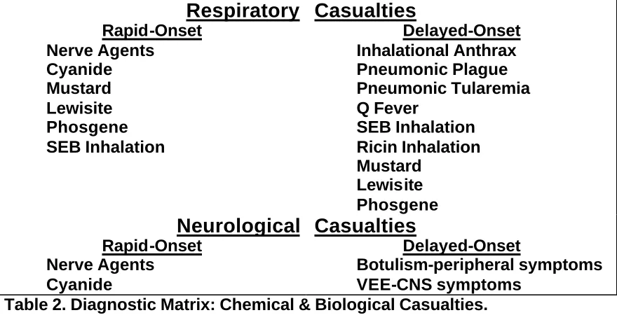
V. Establish a Diagnosis. With decontamination (where warranted) accomplished, a more thorough attempt to establish a diagnosis can be carried out. This attempt, somewhat analogous, to the secondary survey used in the ATLS approach, should involve a combination of clinical, epidemiologic, and laboratory examinations. The amount of expertise and support available to the clinician will vary at each echelon of care. At higher echelons, a full range of laboratory capabilities should enable definitive diagnosis. At lower echelons, every attempt should be made to obtain diagnostic specimens from representative patients and forward these through laboratory channels. Nasal swabs (important for culture and PCR, even if the clinician is unsure which
[image:10.612.87.527.308.533.2]organisms to assay for), blood cultures, serum, sputum cultures, blood and urine for toxin analysis, throat swabs, and environmental samples should be considered.
Respiratory
Rapid-Onset Nerve Agents Cyanide Mustard Lewisite Phosgene SEB InhalationCasualties
Delayed-Onset Inhalational Anthrax Pneumonic Plague Pneumonic Tularemia Q Fever SEB Inhalation Ricin Inhalation Mustard Lewisite PhosgeneNeurological
Rapid-Onset Nerve Agents CyanideCasualties
Delayed-Onset Botulism-peripheral symptoms VEE-CNS symptomsTable 2. Diagnostic Matrix: Chemical & Biological Casualties.
diagnosis, however, is complicated by the fact that many BW diseases (VEE, Q-Fever, Brucellosis) may present simply as undifferentiated febrile illnesses. Moreover, other diseases (Anthrax, Plague, Tularemia, Smallpox) have undifferentiated febrile prodromes.
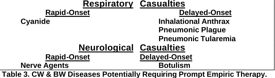
VI. Render Prompt Treatment. Unfortunately, it is precisely in the prodromal phase of many diseases that therapy is most likely to be effective. For this reason, empiric therapy of pneumonia or undifferentiated febrile illness on the battlefield might be indicated under certain circumstances. Table 3 is constructed by eliminating from consideration those diseases for which definitive therapy is not warranted, not available, or not critical. Empiric treatment of respiratory casualties (patients with undifferentiated febrile illnesses who might have prodromal anthrax, plague, or tularemia would be managed in a similar manner) might then be entertained. Doxycycline, for example, is effective against most strains of B. anthracis, Y. pestis, and F. tularensis, as well as against C. burnetii, and the Brucellae. Other tetracyclines and fluoroquinolones might also be considered. Keep in mind that such therapy is, in no way, a substitute for a careful and thorough diagnostic evaluation, when battlefield conditions permit such an evaluation.
Respiratory
Rapid-Onset Cyanide
Casualties
Delayed-Onset Inhalational Anthrax Pneumonic Plague Pneumonic Tularemia
Neurological
Rapid-Onset Nerve Agents
Casualties
[image:11.612.87.528.351.476.2]Delayed-Onset Botulism
Table 3. CW & BW Diseases Potentially Requiring Prompt Empiric Therapy.
VII. Practice Good Infection Control. Standard precautions provide adequate protection against most infectious diseases, including those potentially employed in BW. Anthrax, Tularemia, Brucellosis, Glanders, Q-Fever, VEE, and the Toxin-Mediated diseases are not generally contagious, and victims can be safely managed using standard precautions. Such precautions should be familiar to all clinicians. Under certain circumstances, however, one of three forms of transmission-based precautions would be warranted. Smallpox victims should, wherever possible, be managed using airborne precautions. Pneumonic Plague warrants the use of droplet precautions, and certain VHFs require contact precautions.
and Preventive Medicine personnel should be contacted to assist in the delineation of contaminated areas and the search for further victims.
IX. Assist in the Epidemiologic Investigation. All health care providers require a basic understanding of epidemiologic principles. Even under austere conditions, a rudimentary epidemiologic investigation may assist in diagnosis and in the discovery of additional BW victims. Clinicians should, at the very least, query patients about potential exposures, ill unit members, food/water sources, unusual munitions or spray devices, vector exposures, and develop a line listing of potential cases. Such early discovery might, in turn, permit post-exposure prophylaxis, thereby avoiding excess morbidity and mortality. Preventive medicine officers, field sanitation personnel, epidemiology technicians, environmental science officers, and veterinary officers are all available to assist the clinician in conducting an epidemiologic investigation.
BACTERIAL AGENTS
Bacteria are unicellular organisms. They vary in shape and size from spherical cells - cocci - with a diameter of 0.5-1.0 µm (micrometer), to long rod-shaped organisms - bacilli - which may be from 1-5 µm in size. Chains of bacilli may exceed 50 µm in length. The shape of the bacterial cell is determined by the rigid cell wall. The interior of the cell contains the nuclear material (DNA), cytoplasm, and cell membrane, that are necessary for the life of the bacterium. Many bacteria also have glycoproteins on their outer surfaces which aid in bacterial attachment to cell surface receptors. Under special circumstances some types of bacteria can transform into spores. The spore of the bacterial cell is more resistant to cold, heat, drying, chemicals and radiation than the vegetative bacterium itself. Spores are a dormant form of the bacterium and, like the seeds of plants, they can germinate when conditions are favorable.
The term rickettsia generally applies to very small, gram-negative coccobacillary organisms of the genera Rickettsia and Coxiella. Rickettsiae are unique from classical bacteria in their inability to grow (with rare exceptions) in the absence of a living host cell, but many are susceptible to treatment with antibiotics.
Bacteria generally cause disease in human beings and animals by one of two mechanisms: by invading host tissues, and by producing poisons (toxins). Many pathogenic bacteria utilize both mechanisms. The diseases they produce often respond to specific therapy with antibiotics. It is important to distinguish between the disease-causing organism and the name of the disease it causes (in parentheses below). This manual covers several of the bacteria or rickettsiae considered to be potential BW threat agents: Bacillus anthracis (Anthrax),
Brucella spp. (Brucellosis), Burkholderia mallei (Glanders), Burholderia pseudomallei (melioidosis), Yersinia pestis (Plague), Francisella tularensis
ANTHRAX
SUMMARY
Signs and Symptoms: Incubation period is generally 1-6 days, although longer periods have been noted. Fever, malaise, fatigue, cough and mild chest discomfort progresses to severe respiratory distress with dyspnea, diaphoresis, stridor, cyanosis, and shock. Death typically occurs within 24-36 hours after onset of severe symptoms.
Diagnosis: Physical findings are non-specific. A widened mediastinum may be seen on CXR in later stages of illness. The organism is detectable by Gram stain of the blood and by blood culture late in the course of illness.
Treatment: Although effectiveness may be limited after symptoms are present, high dose antibiotic treatment with penicillin, ciprofloxacin, or doxycycline should be undertaken. Supportive therapy may be necessary.
Prophylaxis: Oral ciprofloxacin or doxycycline for known or imminent exposure. An FDA-licensed vaccine is available. Vaccine schedule is 0.5 ml SC at 0, 2, 4 weeks, then 6, 12, and 18 months (primary series), followed by annual boosters.
OVERVIEW
Bacillus anthracis, the causative agent of Anthrax, is a gram-positive, sporulating rod. The spores are the usual infective form. Anthrax is primarily a zoonotic disease of herbivores, with cattle, sheep, goats, and horses being the usual domesticated animal hosts, but other animals may be infected. Humans generally contract the disease when handling contaminated hair, wool, hides, flesh, blood and excreta of infected animals and from manufactured products such as bone meal. Infection is introduced through scratches or abrasions of the skin, wounds, inhalation of spores, eating insufficiently cooked infected meat, or by biting flies. The primary concern for intentional infection by this organism is through inhalation after aerosol dissemination of spores. All human populations are susceptible. The spores are very stable and may remain viable for many years in soil and water. They resist sunlight for varying periods.
HISTORY AND SIGNIFICANCE
Anthrax spores were weaponized by the United States in the 1950's and 1960's before the old U.S. offensive program was terminated. Other countries have weaponized this agent or are suspected of doing so. Anthrax bacteria are easy to cultivate and spore production is readily induced. Moreover, the spores are highly resistant to sunlight, heat and disinfectants - properties which could be advantageous when choosing a bacterial weapon. Iraq admitted to a United Nations inspection team in August of 1991 that it had performed research on the offensive use of B. anthracis prior to the Persian Gulf War, and in 1995 Iraq admitted to weaponizing anthrax. A recent defector from the former Soviet Union's biological weapons program revealed that the Soviets had produced anthrax in ton quantities for use as a weapon. This agent could be produced in either a wet or dried form, stabilized for weaponization by an adversary and delivered as an aerosol cloud either from a line source such as an aircraft flying upwind of friendly positions, or as a point source from a spray device. Coverage of a large ground area could also be theoretically facilitated by multiple spray bomblets disseminated from a missile warhead at a predetermined height above the ground.
CLINICAL FEATURES
inhalational anthrax, known as Woolsorters’ disease, is also a rare infection contracted by inhalation of the spores. It occurs mainly among workers in an industrial setting handling infected hides, wool, and furs. In man, the mortality of untreated cutaneous anthrax ranges up to 25 per cent; in inhalational and intestinal cases, the case fatality rate is almost 100 percent.
DIAGNOSIS
After an incubation period of 1-6 days,* presumably dependent upon the dose and strain of inhaled organisms, the onset of inhalation anthrax is gradual and nonspecific. Fever, malaise, and fatigue may be present, sometimes in association with a nonproductive cough and mild chest discomfort. These initial symptoms are often followed by a short period of improvement (hours to 2-3 days), followed by the abrupt development of severe respiratory distress with dyspnea, diaphoresis, stridor, and cyanosis. Septicemia, shock and death usually follow within 24-36 hours after the onset of respiratory distress. Physical findings are typically non-specific, especially in the early phase of the disease. The chest X-ray may reveal a widened mediastinum ± pleural effusions late in the disease in about 55% of the cases, but typically is without infiltrates. Pneumonia generally does not occur; therefore, organisms are not typically seen in the sputum. Bacillus anthracis will be detectable by Gram stain of the blood and by blood culture with routine media, but often not until late in the course of the illness. Approximately 50% of cases are accompanied by hemorrhagic meningitis, and therefore organisms may also be identified in cerebrospinal fluid. Only vegetative encapsulated bacilli are present during infection. Spores are not found within the body unless it is open to ambient air. Studies of inhalation anthrax in non-human primates (rhesus monkey) showed that bacilli and toxin appear in the blood late on day 2 or early on day 3 post-exposure. Toxin production parallels the appearance of bacilli in the blood and tests are available to rapidly detect the toxin. Concurrently with the appearance of anthrax, the WBC count becomes elevated and remains so until death.
*During an outbreak of inhalational anthrax in the Soviet Union in 1979, persons are reported to have become ill up to 6 weeks after an aerosol release occurred.
MEDICAL MANAGEMENT
laboratory manipulation of organisms. All naturally occurring strains tested to date have been sensitive to erythromycin, chloramphenicol, gentamicin, and ciprofloxacin. In the absence of antibiotic sensitivity data, empiric intravenous antibiotic treatment should be instituted at the earliest signs of disease. Military policy (FM 8-284) currently recommends ciprofloxacin (400 mg IV q 12 hrs) or doxycycline (200 mg IV load, followed by 100 mg IV q 12 hrs) as initial therapy, with penicillin (4 million U IV q 4 hours) as an alternative once sensitivity data is available. Published recommendations from a public health consensus panel recommends ciprofloxacin as initial therapy. Therapy may then be tailored once antibiotic sensitivity is available to penicillin G or doxycycline. Recommended treatment duration is 60 days, and should be changed to oral therapy as clinical condition improves. Supportive therapy for shock, fluid volume deficit, and adequacy of airway may all be needed.
Standard Precautions are recommended for patient care. There is no evidence of direct person-to-person spread of disease from inhalational anthrax. After an invasive procedure or autopsy, the instruments and area used should be thoroughly disinfected with a sporicidal agent. Iodine can be used, but must be used at disinfectant strengths, as antiseptic-strength iodophors are not usually sporicidal. Chlorine, in the form of sodium or calcium hypochlorite, can also be used, but with the caution that the activity of hypochlorites is greatly reduced in the presence of organic material.
PROPHYLAXIS
Vaccine: A licensed vaccine (Anthrax Vaccine Adsorbed) is derived from sterile culture fluid supernatant taken from an attenuated strain. Therefore, the vaccine does not contain live or dead organisms. The vaccination series consists of six 0.5 ml doses SC at 0, 2, and 4 weeks, then 6, 12 and 18 months, followed by yearly boosters. A human efficacy trial in mill workers demonstrated protection against cutaneous anthrax. There is insufficient data to know its efficacy against inhalational anthrax in humans, although studies in rhesus monkeys indicate that good protection can be afforded after only two doses (15 days apart) for up to 2 years. However, it should be emphasized that the vaccine series should be completed according to the licensed 6 dose primary series. As with all vaccines, the degree of protection depends upon the magnitude of the challenge dose; vaccine-induced protection could presumably be overwhelmed by extremely high spore challenge. Current military policy is to restart the primary vaccine series only if greater than two years elapses between the first and second doses. For all other missed doses, administer the missed dose ASAP and reset the timeline for the series based on the most recent dose.
moderate. Up to 30 percent of recipients may experience mild discomfort at the inoculation site for up to 72 hours (e.g., tenderness, erythema, edema, pruritus), fewer experience moderate reactions, while less than 1 percent may experience more severe local reactions, potentially limiting use of the arm for 1-2 days. Modest systemic reactions (e.g., myalgia, malaise, low-grade fever) are uncommon, and severe systemic reactions such as anaphylaxis, which precludes additional vaccination, are rare. The vaccine should be stored between 2-6o C (refrigerator temperature, not frozen).
BRUCELLOSIS
SUMMARY
Signs and Symptoms: Illness, when manifest, typically presents with fever, headache, myalgias, arthralgias, back pain, sweats, chills, and generalized malaise. Other manifestations include depression, mental status changes, and osteoarticular findings (ie. Sacroiliitis, vertebral osteomyelitis). Fatalities are uncommon.
Diagnosis: Diagnosis requires a high index of suspicion, since many infections present as non-specific febrile illnesses or are asymptomatic. Laboratory diagnosis can be made by blood culture with prolonged incubation. Bone marrow cultures produce a higher yield. Confirmation requires phage-typing, oxidative metabolism, or genotyping procedures. ELISA, followed by Western blot are available.
Treatment: Antibiotic therapy with doxycycline + rifampin or doxycycline in combination with other medications for six weeks is usually sufficient in most cases. More prolonged regimens may be required for patients with complications of meningoencephalitis, endocarditis, or osteomyelitis.
Prophylaxis: There is no human vaccine available against brucellosis, although animal vaccines exist. Chemoprophylaxis is not recommended after possible exposure to endemic disease. Treatment should be considered for high-risk exposure to the veterinary vaccine, inadvertent laboratory exposure, or confirmed biological warfare exposure.
OVERVIEW
Brucellosis is one of the world’s most important veterinary diseases, and is caused by infection with one of six species of Brucellae, a group of gram-negative cocco-baccillary facultative intracellular pathogens. In animals, brucellosis primarily involves the reproductive tract, causing septic abortion and orchitis, which, in turn, can result in sterility. Consequently, brucellosis is a disease of great potential economic impact in the animal husbandry industry. Four species (B. abortus, B. melitensis, B. suis, and, rarely, B. canis) are pathogenic in humans. Infections in abattoir and laboratory workers suggest that the Brucellae are highly infectious via the aerosol route. It is estimated that inhalation of only 10 to 100 bacteria is sufficient to cause disease in man. Brucellosis has a low mortality rate (5% of untreated cases), with rare deaths caused by endocarditis or meningitis. Also, given that the disease has a relatively long and variable incubation period (5-60 days), and that many naturally occurring infections are asymptomatic, its usefulness as a weapon may be diminished. Large aerosol doses, however, may shorten the incubation period and increase the clinical attack rate, and the disease is relatively prolonged, incapacitating, and disabling in its natural form.
HISTORY AND SIGNIFICANCE
Marston described the manifestations of disease caused by B. melitensis
(“Mediterranean gastric remittent fever”) among British soldiers on Malta during the Crimean War. Work by the Mediterranean Fever Commission identified goats as the source, and restrictions on the ingestion of unpasteurized goat milk products soon decreased the number of brucellosis cases among military personnel.
In 1954, Brucella suis became the first agent weaponized by the United States at Pine Bluff Arsenal when its offensive BW program was active. Brucella weapons, along with the remainder of the U.S. biological arsenal, were destroyed in 1969, when the offensive program was disbanded.
CLINICAL FEATURES
Brucellosis, also known as “undulant fever”, typically presents as a nonspecific febrile illness resembling influenza. Fever, headache, myalgias, arthralgias, back pain, sweats, chills, generalized weakness, and malaise are common complaints. Cough and pleuritic chest pain occurs in up to twenty percent of cases, but acute pneumonitis is unusual, and pulmonary symptoms may not correlate with radiographic findings. The chest x-ray is often normal, but may show lung abscesses, single or miliary nodules, bronchopneumonia, enlarged hilar lymph nodes, and pleural effusions. Gastrointestinal symptoms (anorexia, nausea, vomiting, diarrhea and constipation) occur in up to 70 percent of adult cases, but less frequently in children. Ileitis, colitis, and granulomatous or mononuclear infiltrative hepatitis may occur, with hepato- and spleno-megaly present in 45-63 percent of cases.
Lumbar pain and tenderness can occur in up to 60% of brucellosis cases and are sometimes due to various osteoarticular infections of the axial skeleton. Vertebral osteomyelitis, intervertebral disc space infection, paravertebral abscess, and sacroiliac infection occur in a minority of cases, but may be a cause of chronic symptoms. Consequently, persistent fever following therapy or the prolonged presence of significant musculoskeletal complaints should prompt CT or MR imaging. 99mTechnetium and 67Gallium scans are also reasonably sensitive means for detecting sacroiliitis and other axial skeletal infections. Joint involvement in brucellosis may vary from pain to joint immobility and effusion. While the sacroiliac joints are most commonly involved, peripheral joints (notably, hips, knees, and ankles) may also be affected. Meningitis complicates a small minority of brucellosis cases, and encephalitis, peripheral neuropathy, radiculoneuropathy and meningovascular syndromes have also been observed in rare instances. Behavioral disturbances and psychoses appear to occur out of proportion to the height of fever, or to the amount of overt CNS disease. This raises questions about an ill-defined neurotoxic component of brucellosis.
DIAGNOSIS
the use of selective isolation media and the implementation of Biosafety Level-3 (BSL-3) safety precautions. A serum agglutination test (SAT) is available to detect both IgM and IgG antibodies; a titer of 1:160 or greater is indicative of active disease. ELISA and PCR methods are becoming more widely utilized.
MEDICAL MANAGEMENT
Standard precautions are adequate in managing brucellosis patients, as the disease is not generally transmissible from person-to-person. As noted previously, BSL-3 practices should be used when handling suspected brucella cultures in the laboratory because of the danger of inhalation in this setting.
Oral antibiotic therapy alone is sufficient in most cases of brucellosis. Exceptions involve uncommon cases of localized disease, where surgical intervention may be required (e.g., valve replacement for endocarditis). A combination of Doxycycline 200 mg/d PO + Rifampin 600 mg/d PO is generally recommended. Both drugs should be administered for six weeks. Doxycycline 200 mg/d PO for six weeks in combination with two weeks of Streptomycin (1 g/d IM) is an acceptable alternative. Regimens involving Doxycycline + Gentamicin, TMP/SMX + Gentamicin, and Ofloxacin + Rifampin have also been studied and shown effective. Long-term triple-drug therapy with rifampin, a tetracycline, and an aminoglycoside is recommended by some experts for patients with meningoencephalitis or endocarditis.
PROPHYLAXIS
The risk of endemic brucellosis can be diminished by the avoidance of unpasteurized goat-milk and dairy products, especially while travelling in areas where veterinary brucellosis remains common. Live animal vaccines are used widely, and have eliminated brucellosis from most domestic animal herds in the United States, although no licensed human brucellosis vaccine is available.
GLANDERS AND MELIOIDOSIS
SUMMARY
Signs and Symptoms: Incubation period ranges from 10-14 days after inhalation. Onset of symptoms may be abrupt or gradual. Inhalational exposure produces fever (common in excess of 102 F.), rigors, sweats, myalgias, headache, pleuritic chest pain, cervical adenopathy, hepatosplenomegaly, and generalized papular / pustular eruptions. Acute pulmonary disease can progress and result in bacteremia and acute septicemic disease. Both diseases are almost always fatal without treatment.
Diagnosis: Methylene blue or Wright stain of exudates may reveal scant small bacilli with a safety-pin bipolar appearance. Standard cultures can be used to identify both B. mallei and B. pseudomallei. CXR may show miliary lesions, small multiple lung abscesses, or infiltrates involving upper lungs, with consolidation and cavitation. Leukocyte counts may be normal or elevated. Serologic tests can help confirm diagnosis, but low titers or negative serology does not exclude the diagnosis.
Treatment: Therapy will vary with the type and severity of the clinical presentation. Patients with localized disease, may be managed with oral antibiotics for a duration of 60-150 days. More severe illness may require parenteral therapy and more prolonged treatment.
Prophylaxis: Currently, no pre-exposure or post-exposure prophylaxis is available.
OVERVIEW
The causative agents of Glanders and Melioidosis are Burkholderia mallei and Burkholderia pseudomallei, respectively. Both are gram-negative bacilli with a “safety-pin” appearance on microscopic examination. Both pathogens affect domestic and wild animals, which, like humans, acquire the diseases from inhalation or contaminated injuries.
B. mallei is primarily noted for producing disease in horses, mules, and donkeys. In the past man has seldom been infected, despite frequent and often close contact with infected animals. This may be the result of exposure to low concentrations of organisms from infected sites in ill animals and because strains virulent for equids are often less virulent for man. There are four basic forms of disease in horses and man. The acute forms are more common in mules and donkeys, with death typically occurring 3 to 4 weeks after illness onset. The chronic form of the disease is more common in horses and causes generalized lymphadenopathy, multiple skin nodules that ulcerate and drain, and induration, enlargement, and nodularity of regional lymphatics on the extremities and in other areas. The lymphatic thickening and induration has been called farcy. Human cases have occurred primarily in veterinarians, horse and donkey caretakers, and abattoir workers.
B. pseudomallei is widely distributed in many tropical and subtropical regions. The disease is endemic in Southeast Asia and northern Australia. In northeastern Thailand, B. pseudomallei, is one of the most common causative agents of community-acquired septicemia. Melioidosis presents in humans in several distinct forms, ranging from a subclinical illness to an overwhelming septicemia, with a 90% mortality rate and death within 24-48 hours after onset. Also, melioidosis can reactivate years after primary infection and result in chronic and life-threatening disease.
These organisms spread to man by invading the nasal, oral, and conjunctival mucous membranes, by inhalation into the lungs, and by invading abraded or lacerated skin. Aerosols from cultures have been observed to be highly infectious to laboratory workers. Biosafety level 3 containment practices are required when working with these organisms in the laboratory. Since aerosol spread is efficient, and there is no available vaccine or reliable therapy, B. mallei
and B. pseudomallei have both been viewed as potential BW agents.
HISTORY AND SIGNIFICANCE
the Middle East and South America. During World War I, glanders was believed to have been spread deliberately by agents of the Central Powers to infect large numbers of Russian horses and mules on the Eastern Front. This had an effect on troop and supply convoys as well as on artillery movement, which were dependent on horses and mules. Human cases in Russia increased with the infections during and after WWI. The Japanese deliberately infected horses, civilians, and prisoners of war with B. mallei at the Pinfang (China) Institute during World War II. The United States studied this agent as a possible BW weapon in 1943-44 but did not weaponize it. The former Soviet Union is believed to have been interested in B. mallei as a potential BW agent after World War II. The low transmission rates of B. mallei to man from infected horses is exemplified by the fact that in China, during World War II, thirty percent of tested horses were positive for glanders, but human cases were rare. In Mongolia, 5-25% of tested animals were reactive to B. mallei, but no human cases were seen. B. mallei exists in nature only in infected susceptible hosts and is not found in water, soil, or plants.
In contrast, melioidosis is widely distributed in the soil and water in the tropics, and remains endemic in certain parts of the world, even to this day. It is one of the few genuinely tropical diseases that are well established in Southeast Asia and northern Australia. As a result of its long incubation period, it could be unknowingly imported.
B. pseudomallei was also studied by the United States as a potential BW agent, but was never weaponized. It has been reported that the former Soviet Union was experimenting with B. pseudomallei as a BW agent.
CLINICAL FEATURES
Both glanders and melioidosis may occur in an acute localized form, as an acute pulmonary infection, or as an acute fulminant, rapidly fatal, sepsis. Combinations of these syndromes may occur in human cases. Also, melioidosis may remain asymptomatic after initial acquisition, and remain quiescent for decades. However, these patients may present with active melioidosis years later, often associated with an immune-compromising state.
Aerosol infection produced by a BW weapon containing either B. mallei
The pulmonary form may follow inhalation or arise by hematogenous spread. Systemic symptoms as described for the septicemic form occur. Chest radiographs may show miliary nodules (0.5-1.0 cm) and/or a bilateral bronchopneumonia, segmental, or lobar pneumonia, consolidation, and cavitating lung lesions
Acute infection of the oral, nasal and/ or conjunctival mucosa can cause mucopurulent, blood streaked discharge from the nose, associated with septal and turbinate nodules and ulcerations. If systemic invasion occurs from mucosal or cutaneous lesions then a papular and / or pustular rash may occur that can be mistaken for smallpox (another possible BW agent). Evidence of dissemination of these infections includes the presence of skin pustules, abscesses of internal organs, such as liver and spleen, and multiple pulmonary lesions. This form carries a high mortality, and most patients develop rapidly progressive septic shock.
The chronic form is unlikely to be present within 14 days after a BW aerosol attack. It is characterized by cutaneous and intramuscular abscesses on the legs and arms. These lesions are associated with enlargement and induration of the regional lymph channels and nodes. The chronic form may be asymptomatic, especially with melioidosis. There have been cases associated with the development of osteomyelitis, brain abscess, and meningitis.
DIAGNOSIS
Gram stain of lesion exudates reveals small gram negative, bipolar bacteria. These stain irregularly with methylene blue or Wright’s Stain. The organisms can be cultured and identified with standard bacteriological media. The addition of 1-5% glucose, 5 % glycerol, or meat infusion nutrient agar may accelerate growth. Primary isolation requires 48 hours at 37.5º C. For B. mallei,
agglutination tests are not positive for 7-10 days, and a high background titer in normal sera (1:320 to 1:640) makes interpretation difficult. Complement fixation tests are more specific and are considered positive if the titer is equal to, or exceeds 1:20. For B. pseudomallei, a four fold increase in titer supports the diagnosis of melioidosis. A single titer above 1:160 with a compatible clinical picture suggests active infection. Occurrence in the absence of animal contact and / or in an epidemic, is presumptive evidence of a BW attack. Mortality will be high despite antibiotic use. In the hamster 1 to 10 organisms administered by aerosol is lethal
MEDICAL MANAGEMENT
have been suggested for localized disease: Amoxicillin / clavulanate 60 mg/kg/day in three divided doses; Tetracycline 40 mg/kg/day in three divided doses; or Trimethoprim / sulfa (TMP 4 mg/kg/day-sulfa 20 mg/kg/day) in two divided doses. The duration of treatment should be for 60 - 150 days.
If the patient has localized disease with signs of mild toxicity, then a combination of two of the oral regimens is recommended for a duration of 30 days, followed by monotherapy with either amoxicillin / clavulanate or TMP / sulfa for 60 - 150 days. If extrapulmonary suppurative disease is present, then therapy should continue for 6-12 months. Surgical drainage of abscesses may be required.
For severe disease, parental therapy with Ceftazidime 120 mg/kg/day in three divided doses combined with TMP/sulfa (TMP 8 mg/kg/day – sulfa 40 mg/kg/day) in four divided doses for 2 weeks, followed by oral therapy for 6 months.
Other antibiotics that have been effective in experimental infection in hamsters include doxycycline, rifampin, and ciprofloxacin. The limited number of infections in humans has precluded therapeutic evaluation of most of the antibiotic agents; therefore, most antibiotic sensitivities are based on animal and
in vitro studies. Various isolates have markedly different antibiotic sensitivities; therefore, each isolate should be tested for its own resistance pattern.
PROPHYLAXIS
Vaccine: There is no vaccine available for human use.
PLAGUE
SUMMARY
Signs and Symptoms: Pneumonic plague begins after an incubation period of 1-6 days, with high fever, chills, headache, malaise, followed by cough (often with hemoptysis), progressing rapidly to dyspnea, stridor, cyanosis, and death. Gastrointestinal symptoms are often present. Death results from respiratory failure, circulatory collapse, and a bleeding diathesis. Bubonic plague, featuring high fever, malaise, and painful lymph nodes (buboes) may progress spontaneously to the septicemic form (septic shock, thrombosis, DIC) or to the pneumonic form.
Diagnosis: Suspect plague if large numbers of previously healthy individuals develop fulminant Gram negative pneumonia, especially if hemoptysis is present. Presumptive diagnosis can be made by Gram, Wright, Giemsa or Wayson stain of blood, sputum, CSF, or lymph node aspirates. Definitive diagnosis requires culture of the organism from those sites. Immunodiagnosis is also helpful.
Treatment: Early administration of antibiotics is critical, as pneumonic plague is invariably fatal if antibiotic therapy is delayed more than 1 day after the onset of symptoms. Choose one of the following: streptomycin, gentamicin, ciprofloxacin, or doxycyclinefor 10-14 days. Chloramphenicol is the drug of choice for plague meningitis.
Prophylaxis: For asymptomatic persons exposed to a plague aerosol or to a patient with suspected pneumonic plague, give doxycycline 100 mg orally twice daily for seven days or the duration of risk of exposure plus one week. Alternative antibiotics include ciprofloxacin, tetracycline, or chloramphenicol. No vaccine is currently available for plague prophylaxis. The previously available licensed, killed vaccine was effective against bubonic plague, but not against aerosol exposure.
Isolation and Decontamination: Use Standard Precautions for bubonic plague, and Respiratory Droplet Precautions for suspected pneumonic plague. Y. pestis
OVERVIEW
Yersinia pestis is a rod-shaped, non-motile, non-sporulating, gram-negative bacterium of the family Enterobacteraceae. It causes plague, a zoonotic disease of rodents (e.g., rats, mice, ground squirrels). Fleas that live on the rodents can transmit the bacteria to humans, who then suffer from the bubonic form of plague. The bubonic form may progress to the septicemic and/or pneumonic forms. Pneumonic plague would be the predominant form after a purposeful aerosol dissemination. All human populations are susceptible. Recovery from the disease is followed by temporary immunity. The organism remains viable in water, moist soil, and grains for several weeks. At near freezing temperatures, it will remain alive from months to years but is killed by 15 minutes of exposure to 55°C. It also remains viable for some time in dry sputum, flea feces, and buried bodies but is killed within several hours of exposure to sunlight.
HISTORY AND SIGNIFICANCE
The United States worked with Y. pestis as a potential biowarfare agent in the 1950's and 1960's before the old offensive biowarfare program was terminated, and other countries are suspected of weaponizing this organism. The former Soviet Union had more than 10 institutes and thousands of scientists who worked with plague. During World War II, Unit 731, of the Japanese Army, reportedly released plague-infected fleas from aircraft over Chinese cities. This method was cumbersome and unpredictable. The U.S. and Soviet Union developed the more reliable and effective method of aerosolizing the organism. The interest in the terrorist potential of plague was brought to light in 1995 when Larry Wayne Harris was arrested in Ohio for the illicit procurement of a Y. pestis
culture through the mail. The contagious nature of pneumonic plague makes it particularly dangerous as a biological weapon.
CLINICAL FEATURES
are positive for the organism in patients with bubonic plague. However, only about a quarter of bubonic plague patients progress to clinical septicemia.
In those that do progress to secondary septicemia, as well as those presenting septicemic but without lymphadenopathy (primary septicemia), the symptoms are similar to other Gram-negative septicemias: high fever, chills, malaise, hypotension, nausea, vomiting, and diarrhea. However, plague septicemia can also produce thromboses in the acral vessels, with necrosis and gangrene, and DIC. Black necrotic appendages and more proximal purpuric lesions caused by endotoxemia are often present. Organisms can spread to the central nervous system, lungs, and elsewhere. Plague meningitis occurs in about 6% of septicemic and pneumonic cases.
Pneumonic plague is an infection of the lungs due to either inhalation of the organisms (primary pneumonic plague), or spread to the lungs from septicemia (secondary pneumonic plague). After an incubation period varying from 1 to 6 days for primary pneumonic plague (usually 2-4 days, and presumably dose-dependent), onset is acute and often fulminant. The first signs of illness include high fever, chills, headache, malaise, and myalgias, followed within 24 hours by a cough with bloody sputum. Although bloody sputum is characteristic, it can sometimes be watery or, less commonly, purulent. Gastrointestinal symptoms, including nausea, vomiting, diarrhea, and abdominal pain, may be present. Rarely, a cervical bubo might result from an inhalational exposure. The chest X-ray findings are variable, but most commonly reveal bilateral infiltrates, which may be patchy or consolidated. The pneumonia progresses rapidly, resulting in dyspnea, stridor, and cyanosis. The disease terminates with respiratory failure, and circulatory collapse.
Nonspecific laboratory findings include a leukocytosis, with a total WBC count up to 20,000 cells with increased bands, and greater than 80 percent polymorphonuclear cells. One also often finds increased fibrin split products in the blood indicative of a low-grade DIC. The BUN, creatinine, ALT, AST, and bilirubin may also be elevated, consistent with multiorgan failure.
DIAGNOSIS
Diagnosis is based primarily on clinical suspicion. The sudden appearance of large numbers of previously healthy patients with severe, rapidly progressive pneumonia with hemoptysis strongly suggests plague. A presumptive diagnosis can be made microscopically by identification of the coccobacillus in Gram, Wright, Giemsa, or Wayson's stained smears from lymph node needle aspirate, sputum, blood, or cerebrospinal fluid samples. When available, immunofluorescent staining is very useful. Definitive diagnosis relies on culturing the organism from blood, sputum, CSF, or bubo aspirates. The organism grows slowly at normal incubation temperatures, and may be misidentified by automated systems because of delayed biochemical reactions. It may be cultured on blood agar, MacConkey agar or infusion broth. Most naturally occurring strains of Y. pestis produce an F1-antigen in vivo, which can be detected in serum samples by immunoassay. A four-fold rise in antibody titer in patient serum is retrospectively diagnostic. PCR (using specific primers), although not sufficiently developed and evaluated for routine use, is a very sensitive and specific technique, currently able to identify as few as 10 organisms per ml. Most clinical assays can be performed in Biosafety Level 2 (BSL-2) labs, whereas procedures producing aerosols or yielding significant quantities of organisms require BSL-3 containment.
MEDICAL MANAGEMENT
Use Standard Precautions for bubonic plague patients. Suspected pneumonic plague cases require strict isolation with Droplet Precautions for at least 48 hours of antibiotic therapy, or until sputum cultures are negative in confirmed cases. If competent vectors (fleas) and reservoirs (rodents) are present, measures must be taken to prevent local disease cycles. These might include, but are not limited to, use of flea insecticides, rodent control measures (after or during flea control), and flea barriers for patient care areas.
Usual supportive therapy includes IV crystalloids and hemodynamic monitoring. Although low-grade DIC may occur, clinically significant hemorrhage is uncommon, as is the need to treat with heparin. Endotoxic shock is common, but pressor agents are rarely needed. Finally, buboes rarely require any form of local care, but instead recede with systemic antibiotic therapy. In fact, incision and drainage poses a risk to others in contact with the patient; aspiration is recommended for diagnostic purposes and may provide symptomatic relief.
PROPHYLAXIS
Vaccine: No vaccine is currently available for prophylaxis of plague. A licensed, killed whole cell vaccine was available in the U.S. from 1946 until November 1998. It offered protection against bubonic plague, but was not effective against aerosolized Y. pestis. Presently, an F1-V antigen (fusion protein) vaccine is in development at USAMRIID. It protected mice for a year against an inhalational challenge, and is now being tested in primates.
Q FEVER
SUMMARY
Signs and Symptoms: Fever, cough, and pleuritic chest pain may occur as early as ten days after exposure. Patients are not generally critically ill, and the illness lasts from 2 days to 2 weeks.
Diagnosis: Q fever is not a clinically distinct illness and may resemble a viral illness or other types of atypical pneumonia. The diagnosis is confirmed serologically.
Treatment: Q fever is generally a self-limited illness even without treatment, but tetracycline or doxycycline should be given orally for 5 to 7 days to prevent complications of the disease. Q fever endocarditis (rare) is much more difficult to treat.
Prophylaxis: Chemoprophylaxis begun too early during the incubation period may delay but not prevent the onset of symptoms. Therefore, tetracycline or doxycycline should be started 8-12 days post exposure and continued for 5 days. This regimen has been shown to prevent clinical disease. An inactivated whole cell IND vaccine is effective in eliciting protection against exposure, but severe local reactions to this vaccine may be seen in those who already possess immunity. Therefore, an intradermal skin test is recommended to detect pre-sensitized or immune individuals.
OVERVIEW
The endemic form of Q fever is a zoonotic disease caused by the rickettsia, Coxiella burnetii. Its natural reservoirs are sheep, cattle, goats, dogs, cats and birds. The organism grows to especially high concentrations in placental tissues. The infected animals do not develop the disease, but do shed large numbers of the organisms in placental tissues and body fluids including milk, urine, and feces. Exposure to infected animals at parturition is an important risk factor for endemic disease. Humans acquire the disease by inhalation of aerosols contaminated with the organisms. Farmers and abattoir workers are at greatest risk occupationally. A biological warfare attack with Q fever would cause a disease similar to that occurring naturally. Q fever is also a significant hazard in laboratory personnel who are working with the organism.
HISTORY AND SIGNIFICANCE
Q fever was first described in Australia and called “Query fever” because the causative agent was initially unknown. Coxiella burnetii, discovered in 1937, is a rickettsial organism that is resistant to heat and desiccation and highly infectious by the aerosol route. A single inhaled organism may produce clinical illness. For all of these reasons, Q fever could be used by an adversary as an incapacitating biological warfare agent.
CLINICAL FEATURES
Following the usual incubation period of 2-14 days, Q fever generally occurs as a self-limiting febrile illness lasting 2 days to 2 weeks. The incubation period varies according to the numbers of organisms inhaled, with longer periods between exposure and illness with lower numbers of inhaled organisms (up to forty days in some cases). The disease generally presents as an acute non-differentiated febrile illness, with headaches, fatigue, and myalgias as prominent symptoms. Physical examination of the chest is usually normal. Pneumonia, manifested by an abnormal chest x-ray, occurs in half of all patients, but only around half of these, or 28 percent of patients, will have a cough (usually non-productive) or rales. Pleuritic chest pain occurs in about one-fourth of patients with Q fever pneumonia. Chest radiograph abnormalities, when present, are patchy infiltrates that may resemble viral or mycoplasma pneumonia. Rounded opacities and adenopathy have also been described.
DIAGNOSIS
Differential Diagnosis: Since Q fever usually presents as an undifferentiated febrile illness, or a primary atypical pneumonia, it may be difficult to distinguish from viral illnesses and must be differentiated from pneumonia caused by Mycoplasma pneumoniae, Legionella pneumophila, Chlamydia psittaci, and Chlamydia pneumoniae (TWAR). More rapidly progressive forms of Q fever pneumonia may look like bacterial pneumonias such as tularemia or plague. Significant numbers of soldiers (from the same geographic area) presenting over a one to two week period with a nonspecific febrile illness, and associated pulmonary symptoms in about a quarter of cases, should trigger the treating physicians to consider the possibility of an attack with aerosolized Q fever. The diagnosis will often rest on the clinical and epidemiologic picture in the setting of a possible biowarfare attack.
Laboratory Diagnosis: A leukocytosis may be present in one third of patients. Most patients with Q fever have a mild elevation of hepatic transaminase levels. Identification of organisms by examination of the sputum is not helpful. Isolation of the organism is impractical, as the organism is difficult to culture and a significant hazard to laboratory workers. Serological tests for Q fever include identification of antibody to C. burnetii by indirect fluorescent antibody (IFA), enzyme-linked immunosorbent assay (ELISA), and complement fixation. Specific IgM antibodies may be detectable as early as the second week after onset of illness. ELISA testing is available at USAMRIID. A single serum specimen can be used to reliably diagnose acute Q fever with this test as early as 1 1/2 - 2 weeks into the illness. The most commonly available serologic test is the complement fixation test (CF) which is relatively insensitive and may not be useful if sera have intrinsic anti-complement activity.
MEDICAL MANAGEMENT
PROPHYLAXIS
Vaccine: A formalin-inactivated whole cell IND vaccine is available for immunization of at-risk personnel on an investigational basis, although a Q fever vaccine is licensed in Australia. Vaccination with a single dose of this killed suspension of C. burnetii provides complete protection against naturally occurring Q fever, and greater than 95 percent protection against aerosol exposure. Protection lasts for at least 5 years. Administration of this vaccine in immune individuals may cause severe local induration, sterile abscess formation, and even necrosis at the inoculation site. This observation led to the development of an intradermal skin test using 0.02 mg of specific formalin-killed whole-cell vaccine to detect presensitized or immune individuals.
TULAREMIA
SUMMARY
Signs and Symptoms: Ulceroglandular tularemia presents with a local ulcer and regional lymphadenopathy, fever, chills, headache and malaise. Typhoidal tularemia presents with fever, headache, malaise, substernal discomfort, prostration, weight loss and a non-productive cough.
Diagnosis: Clinical diagnosis. Physical findings are usually non-specific. Chest x-ray may reveal a pneumonic process, mediastinal lymphadenopathy or pleural effusion. Routine culture is possible but difficult. The diagnosis can be established retrospectively by serology.
Treatment: Administration of antibiotics (streptomycin or gentamicin) with early treatment is very effective.
Prophylaxis: A live, attenuated vaccine is available as an investigational new drug. It is administered once by scarification. A two week course of tetracycline is effective as prophylaxis when given after exposure.
OVERVIEW
Francisella tularensis, the causative agent of tularemia, is a small, aerobic non-motile, gram-negative cocco-bacillus. Tularemia (also known as rabbit fever and deer fly fever) is a zoonotic disease that humans typically acquire after skin or mucous membrane contact with tissues or body fluids of infected animals, or from bites of infected ticks, deerflies, or mosquitoes. Less commonly, inhalation of contaminated dusts or ingestion of contaminated foods or water may produce clinical disease. Respiratory exposure by aerosol would typically cause typhoidal or pneumonic tula