Thesis by R. McKell Carter
In Partial Fulfillment of the Requirements for the Degree of
Doctor of Philosophy
California Institute of Technology Pasadena, California
For my family
new or old
Acknowledgements
It seems unfair that I receive a degree for the work that many have done. To all of you, I can only hope I have helped you, or can help, in return. Research grinds to a halt without funding to keep it moving. I would like to thank the following for their sponsorship: Sandia National Labs, the David and Lucile Packard Foundation, the Gordon and Betty Moore Foundation, the Keck Foundation, the National Institutes of Health, the National Institutes of Mental Health, the National Science Foundation (and ERC), the Wellcome Trust and the William T. Gimbel Discovery Fund in Neuroscience at Caltech
All of my collaborators were both great scientists and enjoyable colleagues. I would like to thank all of you. Nao and Connie spent time on the early conditioning work, transforming its direction. Conferences would not have been the same without you. Ben Seymour was the king of pain and scotch eggs at the FIL. The study of trace and delay conditioning was made a great deal more entertaining with him and John
O’Doherty in the control room. I also thank the rest of the FIL for their endless help and discussion. For those who have the opportunity, the FIL is a great place to live and work. Brian Cleary for his time spent thinking about functional DTI, a project that I hope will be continued. Thanks to Tony Bruguier for a great deal of time spent scratching our heads over noise in an MRI environment. Beena, Kats, and Romi all deserve thanks for
introducing me to psychophysics and for a great project not included in this thesis (Khurana et al., 2006). I would like to thank the members of the Conscious Mouse Club for thought provoking discussion at meetings well after most have gone home.
helped make both skin conductance recording during fMRI and the rapid acquisition of DTI images possible. Nothing would be accomplished at the CBIC without Mary Munoz.
There are a number of professors who had to suffer my harassment and
sometimes serve on my committees. They have all provided valuable input and I am glad to have spent time interacting with them, thank you. Ralph Adolphs, John Allman, David Anderson, Michael Fanselow and Shinsuke Shimojo all had the thankless task of serving on my committees. Ray Dolan was my host at the FIL for two projects. I rotated in Gilles Laurent’s lab and still consider his lab to be one of the best at Caltech. Henry Lester was part of the Conscious Mouse Club and was a tireless advocate of clarity in presentation. John O’Doherty deserves special thanks for introducing me to SPM and MRI as well as The Queen’s Larder. I would still be lost in colored blobs had it not been for his capable direction. You have been a great friend, and I wish you even greater success here at Caltech.
I would like to thank my advisor Christof Koch. The time spent analyzing, climbing, debating, driving, running, talking and writing have all been valuable to me. You have served as a model for both academics and attitude. I thank you for your guidance and friendship. I also thank you for providing opportunities that have greatly enriched my stay at Caltech.
Abstract
The ability to adapt to a changing environment is central to an organism’s success. The process of associating two stimuli (as in associative conditioning) requires very little in the way of neural machinery. In fact, organisms with only a few hundred neurons show conditioning that is specific to an associated cue. This type of learning is commonly referred to as implicit learning. The learning can be performed in the absence of the subject’s ability to describe it. One example of learning that is thought to be implicit is delay conditioning. Delay conditioning consists of a single cue (a tone, for example) that starts before, and then
overlaps with, an outcome (like a pain stimulus).
In addition to associating sensory cues, humans routinely link abstract concepts with an outcome. This more complex learning is often described as explicit since subjects are able to describe the link between the stimulus and outcome. An example of conditioning that requires this type of knowledge is
trace conditioning. Trace conditioning includes a separation of a few seconds between the cue and
outcome. Explicit learning is often proposed to involve a separate system, but the degree of separation between implicit associations and explicit learning is still debated.
We describe aversive conditioning experiments in human subjects used to study the degree of interaction that takes place between explicit and implicit systems. We do this in three ways. First, if a higher order task (in this case a working memory task) is performed during conditioning, it reduces not only explicit learning but also implicit learning. Second, we describe the area of the brain involved in explicit learning during conditioning and confirm that it is active during both trace and delay conditioning. Third, using functional magnetic resonance imaging (fMRI), we describe hemodynamic activity changes in perceptual areas of the brain that occur during delay conditioning and persist after the learned association has faded.
Table of Contents
1 INTRODUCTION ... 1
1.1 Explicit and Implicit Aspects of Conditioning...2
1.2 Our Approach ...5
2 WORKING MEMORY AND FEAR CONDITIONING... 8
2.1 Introduction...8
2.2 Materials and Methods ...10
2.2.1 Equipment ...10
2.2.2 Subjects ...11
2.2.3 Procedure...11
2.2.4 Conditioning Stimuli (Figure 2-1A)...12
2.2.5 Experimental Phases (Figure 2-1B)...13
2.2.6 Distracting Tasks (Figure 2-1C)...14
2.2.7 Analysis of SCR...15
2.2.8 Trial Effects...16
2.3 Results ...17
2.3.1 Differential Conditioning ...17
2.3.2 Awareness of CS/US Contingency...18
2.3.3 Single Cue Conditioning ...18
2.4 Discussion...20
2.5 Figures and Legends ...25
3 THE NEURAL CORRELATES OF IMPLICIT AND EXPLICIT PROCESSES IN CONDITIONING ... 31
3.1 Introduction...32
3.2 Methods...34
3.2.1 Participants...34
3.2.2 Experimental Procedure ...35
3.2.3 Online Subject Reports of Contingency Awareness...35
3.2.4 Post-Experimental Questionnaire...36
3.2.5 Unconditioned Stimuli ...36
3.2.6 Online Measure of SCR ...37
3.2.7 fMRI...37
3.2.8 Learning: Contingency awareness and Conditioning ...39
3.3 Results ...40
3.3.1 Conditioned skin conductance responses ...40
3.3.2 Contingency awareness ...40
3.5 Tables ...45
3.6 Figures...47
3.7 Supplementary Methods...50
4 PERSISTENT CHANGES IN VISUAL CORTEX DUE TO AVERSIVE CONDITIONING ... 51
4.1 Introduction...51
4.2 Methods...54
4.2.1 Participants...54
4.2.2 Experimental Procedure ...54
4.2.3 Stimulus Presentation...55
4.2.4 Skin Conductance Conditioning...56
4.2.5 fMRI...57
4.3 Results ...58
4.3.1 Skin Conductance Conditioning...58
4.3.2 Brain Activity during Acquisition...59
4.3.3 Extinction ...61
4.4 Discussion...62
4.5 Tables ...67
4.6 Figures...69
5 DISCUSSION ... 77
5.1 Explicit Influences on Implicit Processes ...77
5.2 Implicit Influences on Explicit Processes ...79
5.3 Continuum or Separate Systems...81
5.4 Work on the Conscious Mouse...82
5.5 Conclusion ...83
6 APPENDIX – SCR RECORDING DURING FMRI ACQUISITION ... 85
6.1 Abstract...85
6.2 Introduction...86
6.3 Material and methods...88
6.3.1 Equipment ...88
6.3.2 Body simulation ...88
6.3.3 Leads and electrodes ...89
6.3.4 Heat insulation ...90
6.3.6 Head coils...91
6.3.7 Resonance testing...92
6.3.8 Measurement of induced voltages...92
6.4 Results ...93
6.4.1 Resonances...93
6.4.2 Recorded waveform ...93
6.4.3 Recorded voltages ...94
6.4.4 GSR recording quality...95
6.5 Discussion...96
6.6 Tables ...97
Table list
TABLE 3-1...45
TABLE 3-2...46
TABLE 4-1...67
TABLE 4-2...68
Figure List
FIGURE 2-1 ...25
FIGURE 2-2 ...26
FIGURE 2-3 ...27
FIGURE 2-4 ...28
FIGURE 2-5 ...29
FIGURE 2-6 ...30
FIGURE 3-1 ...47
FIGURE 3-2 ...48
FIGURE 3-3 ...49
FIGURE 4-1 ...69
FIGURE 4-2 ...70
FIGURE 4-3 ...71
FIGURE 4-4 ...72
FIGURE 4-5 ...73
FIGURE 4-6 ...74
FIGURE 4-7 ...75
FIGURE 4-8 ...76
FIGURE 6-1 ...99
FIGURE 6-2 ...100
FIGURE 6-3 ...101
FIGURE 6-4 ...102
FIGURE 6-5 ...103
FIGURE 6-6 ...104
Abbreviations
ACC Anterior Cingulate Cortex
BOLD Blood Oxygenation Level Dependent CS conditioned stimulus
CS+ the conditioned stimulus sometimes followed by a US CS- the conditioned stimulus never followed by a US DLPFC Dorsal Lateral Prefrontal Cortex
FFA Fusiform Face Area
fMRI functional Magnetic Resonance Imaging FWE Family Wise Error
FWHM Full Width at Half Maximum. GP Globus Pallidus
GSR Galvanic Skin Response Ins. Insula
IPL Inferior Parietal Lobule IPS Inferior Parietal Sulcus MFG Middle Frontal Gyrus MRI Magnetic Resonance Imaging Operc. Operculum
PFC Prefrontal Cortex ROI Region of Interest
S Siemen (as in micro Siemen or nano Siemen) SC Superior Colliculus
1 Introduction
Every extension of knowledge arises from making the conscious the unconscious – Nietzsche
Organisms need to learn. Learning provides the basis for adaptation to a diverse and changing environment. Those organisms that are better at learning are more successful both in the acquisition of resources and the avoidance of potentially detrimental
situations. When learning and memory are discussed in an everyday context, it is usually to retain information for a test or to avoid forgetting a person’s name. This type of memory usually involves facts or concepts and is described as explicit. There are cases where individuals have shown an extraordinary associative capacity to the point where sensory associations from their explicit memories overpower current experience.
One such individual is the famous Solomon Shereshevskii, known in the literature simply as ‘S’. The extent of his success (and difficulties) was described by the
neuropsychologist Alexander Luria (Luria, 1968). From the Wikipedia entry for
abilities, he became a very successful mnemonist, able to retain a great deal more than he could without the techniques. Most surprising was that ‘S’ scored absolutely average on intelligence tests. While his abilities sound incredibly useful to anyone who has ever stood stammering while trying to recall a name or reference, this ability had its cost. The extent of his associations eventually left him unable to interact normally; for instance he once explained that he was unable to eat strawberry ice cream because the tone of the ice cream’s vendor left the taste of coal in his mouth. He spent the later portion of his life in an asylum. ‘S’ is an example of nearly perfect explicit association; each stimulus had such a rich sensory representation that he was able to form episodic memories that were very robust.
A type of learning that is better studied and more common is that of conditioned association, often referred to as implicit since it does not require the subject to be aware of the association. Conditioning is studied in a wide variety of organisms from mollusks to fruit flies, rodents, monkeys and humans (Baer and Fuhrer, 1982; Mackintosh, 1983; Gallistel, 1990; Thompson and Krupa, 1994; Connolly et al., 1996; Eichenbaum, 1997; Pearce et al., 1997; Tully, 1998; Squire and Kandel, 1999; Kocorowski and Helmstetter, 2001). This gives scientists a large number of tools to study how this association takes place. The most notable model used to study association is that of Pavlovian
conditioning.
1.1 Explicit and Implicit Aspects of Conditioning
reaction, such as salivation, to only the meaningful stimulus. Over time, the subject begins to respond to the previously neutral stimulus in the same way as the meaningful one. The subject has formed an association; he or she now begins salivating to the presence of the bell alone without food. In Pavlovian conditioning terms, the initially neutral stimulus (the bell) is referred to as the Conditioned Stimulus or CS. The initially meaningful stimulus (the food) is referred to as the Unconditioned Stimulus or US.
This thesis examines the interaction between implicit and explicit learning. The simple association that takes place in most organisms is often described as implicit,
occurring without any relationship to conscious knowledge (Manns et al., 2002). Learning a person’s name or associating two abstract concepts is described as explicit
since it occurs, essentially by definition, with conscious knowledge.
It is easy to understand how conditioning could be considered an implicit process; even the 302 neurons in the roundworm Caenorhabditis elegans show learning in a large
number of association paradigms (Rankin et al., 1990; Wen et al., 1997; Law et al., 2004). With such a small number of neurons involved, it becomes more difficult to imagine that explicit knowledge is involved in the process.
Although the differentiation between “explicit” and “implicit” has intuitive weight, what makes a particular learned relationship explicit? The study of a patient referred to as HM served as a means to split the two learning systems. HM’s case was first described by Scoville and Milner in 1957 (Scoville and Milner, 1957). HM was involved in an accident at a young age that eventually resulted in epilepsy. His condition was bad
memories. He retained older memories, but was unable to recall what he had eaten for breakfast or what he had done yesterday. He could carry on a conversation as long as there weren’t too many changes of topic, and as long as it was less than a few minutes. While HM could not form any new explicit memories, his implicit learning remained intact. Over several days he learned to trace objects in a mirror without ever being able to report that he had tried the task before. He could learn new skills, motor associations, without any explicit knowledge. This finding has been generally interpreted to mean that explicit and implicit learning depend on two separate memory systems (Squire and Kandel, 1999).
A set of experiments by Larry Squire and colleagues described a further dissociation between explicit and implicit learning using two different types of eye-blink
conditioning. In eye-blink conditioning, a neutral stimulus, such as a tone (the CS), is paired with a puff of air to the eye, the US. As the experiment progresses, the subject learns to blink (non-consciously) at the appropriate time to protect their eye.
Delay conditioning is an example of Pavlovian conditioning that has been described as occurring independently of awareness (Manns et al., 2002). For the association to be described as delay conditioning, the CS must precede the US, either overlapping with it or directly before it. It is called delay conditioning because the US presented is delayed
with respect to the CS.
the neutral (CS) and meaningful (US) stimulus requires that a memory trace of the CS be
kept after the CS terminates in order to associate it with the US. In spite of its relative complexity, there is evidence that this type of learning occurs in the fruit fly Drosophila
melanogaster (Tully and Quinn, 1985).
There are many examples of explicit influences in implicit processes. Studies from as early as 1937 describe conditioning physiological responses using only verbal instruction (Cook and Harris, 1937); that is, the subject begins to respond to a previously neutral stimulus because of false instructions that say the stimulus may now be paired with a shock.
1.2 Our Approach
We sought to further examine the interactions between explicit and implicit processes in a classical conditioning paradigm. We chose to use a fear conditioning paradigm rather than an eye-blink conditioning paradigm because fear conditioning is well studied in a wide variety of organisms. Fear conditioning’s widespread use provided us not only with better information about the paradigm being studied, but also made model systems
lesion studies in rodents applicable to behavioral results with human subjects. Details of the collaboration are contained in the discussion. This thesis is organized in three main parts.
Chapter 2 describes the effects of performing a working memory task during delay and trace conditioning. We reasoned that if trace conditioning depended on high level mental resources, such as working memory, then having subjects perform a working memory task during conditioning would eliminate trace conditioning, leaving delay unaffected. Instead, we discovered that the working memory task affected not only trace conditioning, but delay as well. These effects could be partially overcome by
simplification of the protocol; for example, reducing the number of stimuli or providing the subject with information before the experiment. This study provides strong evidence of the influence that explicit processes have on implicit ones.
Chapter 3 describes areas of the brain that are important for the acquisition of both explicit and implicit information. Subjects were aversively conditioned using both trace and delay protocols during fMRI acquisition. We also recorded skin conductance
responses for use as a correlate of implicit learning, and shock expectancy responses as a correlate of explicit learning. Our analysis identified portions of the brain where
The identification of visual cortex as an area correlating with implicit learning led us to specifically examine the visual areas representing the CS.
Chapter 4 describes an experiment assessing blood oxygenation changes in order to assess differences in the fusiform face area that occur as a result of delay conditioning to images of faces. These changes are consistent with an increase in the representation of the paired stimulus. They are also persistent beyond extinction of the conditioned
2 Working Memory and Fear Conditioning
This work was published under the same title in 2003 (Carter et al., 2003).Constanze Hofstötter conducted the single cue trace uninformed 0-back experiment and all of the informed experiments. She was also involved in the analysis and write up. Her contributions in the writing process made the manuscript far better than it would have been otherwise. Naotsugu Tsuchiya conducted unpublished control experiments and was also involved in the analysis and write up. Christof Koch initiated the project and secured funding. His input in the early stages of the project (while we entered a field we had no experience in) was always very useful. He also advised on analysis procedures and made substantial contributions in the write up and review processes. Experiments and analysis were conducted at the California Institute of Technology.
Here, we investigate the extent to which human classical fear conditioning depends on working memory.
2.1 Introduction
Pavlovian conditioning is widely used to study associative learning in species ranging from mollusks to flies, rodents, monkeys and humans (Baer and Fuhrer, 1982;
Mackintosh, 1983; Gallistel, 1990; Thompson and Krupa, 1994; Connolly et al., 1996; Eichenbaum, 1997; Pearce et al., 1997; Tully, 1998; Squire and Kandel, 1999;
which certain forms of classical conditioning depend on higher-level cognitive processes including selective attention, working memory and awareness (Hilgard et al., 1937; Dawson and Furedy, 1976; Clark and Squire, 1998; Ohman and Soares, 1998; Carrillo et al., 2000; Knuttinen et al., 2001; Lovibond and Shanks, 2002). Eye-blink conditioning is an associative learning paradigm where the role of explicit knowledge / awareness is being investigated. The paradigm involves the association of an eye-blink (a somatic motor response) with previously meaningless stimuli (CS).
other is not (CS-).
We chose fear conditioning to replicate and extend these findings with human subjects on the basis of a conditioning protocol easily extendible to mice, animals for which well established molecular tools used for manipulating genetically identifiable cell populations are available. Fear conditioning differs from eye-blink conditioning in its underlying neuronal implementation, due in part to the fact that the association involves an autonomic, rather than a somatic, motor response. Fear conditioning is easy to establish in humans and rodents, is acquired in a fraction of the trials needed for eye-blink conditioning and is tolerant to long trace periods, making it amenable to fMRI investigations (Buchel et al., 1998b; LaBar et al., 1998; Buchel et al., 1999; Knight et al., 1999). Finally, the neural circuits underlying fear conditioning, particularly the lateral nucleus of the amygdala, hippocampus and prefrontal cortex, are being vigorously
explored (Fendt and Fanselow, 1999; Medina et al., 2002). We use transient elevations in skin conductance (skin conductance response or SCR) as our measure of autonomic arousal when testing responses to auditory stimuli that have been previously paired with a shock. At the same time, we distract our subjects with tasks of variable working memory load. There were parallel efforts to reproduce selected aspects of this work in mice (Han et al., 2003).
2.2 Materials and Methods
2.2.1 Equipment
Silver Chloride electrodes filled with Med Associates paste TD-246 were used for shock presentation and recording skin conductance. CS presentations were mixed into stereo headphones. Distracting tasks were written in Matlab (Mathworks) utilizing the Psychophysics Toolbox (Brainard, 1997). Analysis was carried out using programs written in Matlab as well as SPSS 10.
2.2.2 Subjects
Subjects were recruited from Caltech and were paid 20 dollars for their participation, based on informed consent. Their age ranged from 18-31 with a mean of 21 years. The following differential conditioning groups consisted of six subjects each: (i) delay no
task, (ii) delay 1-back, (iii) delay 2-back (iv) trace no task, (v) trace 1-back, (vi) trace
2-back. The following single cue conditioning groups consisted of four subjects each: (i)
delay no task, (ii) delay 2-back, (iii) uninformed trace no task, (iv) uninformed trace
0-back, (v) uninformed trace 2-back, (vi) informed trace no task, (vii) informed trace
0-back, (viii) informed trace 2-back.
2.2.3 Procedure
Skin conductance electrodes were attached to the palmar surface of the first and second fingers of the non-dominant hand. Shocking electrodes were attached to the palmar surface of the third and fourth fingers of the dominant hand. Each individual’s shock level was determined using a subjective rating protocol that sought a level that was “uncomfortable but not painful”. This shock level was used throughout the experiment.
three sessions of approximately five minutes each to ensure the subject had reached plateau performance. Prior to conditioning, subjects were read instructions asking them to focus on either their visual task or the wall in front of them. Naïve subjects had no
previous specific knowledge of the experiment except that it was a “...learning and memory experiment that involves electric shocks.” Subjects in the informed groups were read instructions that explicitly stated that an “electric shock shortly follows most
presentations of a tone” and that “the tone generally predicts the occurrence of the electric shock.” They were asked to confirm verbally that they “understand that the tone usually predicts the occurrence of the electric shock.” Subjects were given a post-experimental questionnaire to assess their knowledge of the CS/US relationship (Clark and Squire, 1998) and were debriefed. The questionnaire for differential conditioning included 17 questions to assess the subject’s explicit knowledge of stimulus relationships. Subjects were not allowed to correct previous answers. The awareness index is a number between 0 and 17, corresponding to the number of correct responses. The higher the index, the more detailed the subject’s ability to recall the presence or absence of a contingency relationship between stimuli.
The informed consent procedure was reviewed and approved by the Caltech committee for the protection of human subjects.
2.2.4 Conditioning Stimuli (Figure 2-1A)
were always 1 second in length. The 2 kHz tone was always used as the CS+ during single cue conditioning. During delay conditioning, reinforced CS+ presentations coterminated with the US. Reinforced CS+ presentations during trace conditioning were followed by a shock 4 seconds after the CS+ onset, leaving a 3 second trace period.
2.2.5 Experimental Phases (Figure 2-1B)
The learning procedure consisted of three phases: habituation, acquisition and extinction. In the first phase, habituation, subjects received two presentations of the CS+ and two of the CS-, in that order, to familiarize them with both stimuli. During acquisition, subjects received 24 CS+ and 24 CS- presentations, a total of 48 trials. Twenty of the 24 CS+ presentations were reinforced with a US, while four were not reinforced to allow for conditioning assessment. These four stimuli were positioned by randomly removing the US following one of the six CS+ presentations in each of the four blocks of 12 trials (six CS+, six CS-) during acquisition (excluding the first two CS+/US pairings in the
experiment). During the extinction phase, subjects received twelve nonreinforced CS+ and twelve CS- presentations. CS+/CS- presentations occurred in random order with the limiting factors being a) that no more than two presentations of a specific CS occurred in a row and b) six of each occurred in each block of twelve trials. Intertrial intervals were uniformly distributed from 15-25 seconds.
method, our procedure has the disadvantage of not controlling for unassociated stimulus SCR; however, it also has several advantages. It allows a comparison within subjects, a more effective means of detecting conditioning. This method also avoids the pitfalls of using a US only protocol where the US/CS- relationship is randomized or explicitly unpaired. The former may be associated with elevated CS- responses due to a generally elevated anxiety level. The latter tests the subject’s ability to learn the anticorrelated relationship between the CS and US to enable suppression of the aforementioned general anxiety. It should be noted that our results show that working memory tasks interfere with our single cue trace conditioning protocol, adding validity to the idea that using the phantom CS- allows for accurate and sensitive detection of conditioning.
2.2.6 Distracting Tasks (Figure 2-1C)
To confirm that the conditioning protocols were effective, one group of subjects was excluded from performing a task (i.e. for each procedure they simply stared at the wall). The degree to which conditioning depends on working memory was assessed by asking a group of subjects to perform an n-back memory task during a conditioning procedure. Subjects had to press a key every time a given number appeared (0-back), when the present number matched the one before it (1-back), or whenever it was identical to the one before the previous (2-back). Only single cue trace subjects were asked to perform the 0-back task. The 0-back task involves the same input and the same motor output, including frequency of response, as the 1 and 2-back tasks, but is only minimally dependent on working memory.
adjusted for each subject to achieve a performance of approximately 85%. The mean rate of 2-back presentation was 1 Hz for differential subjects (88% correct), 1.33 Hz for uninformed single cue subjects (84% correct), and 1.2 Hz for informed single cue
subjects (85% correct). All 1-back and 0-back tasks were performed at a presentation rate of 1.33 Hz. The mean performance for subjects focusing on the 1-back task was 93.5%. The mean task performance for single cue subjects in the 0-back group was 98% for uninformed subjects and 99% for informed subjects.
2.2.7 Analysis of SCR
A skin conductance response was measured as the maximal amplitude difference of more than 10 nS that occurred in a 1 to 4 second window after the delay CS onset, or in a 1 to 7 second window following the trace CS onset. Valid responses were range corrected by the largest amplitude response for each subject (Lykken, 1972). When there was no response, a zero-amplitude response was included in the analysis.
Habituation analysis was performed for differential conditioning using a paired t-test and a normalized ANOVA. No significant SCR differences were observed between the CSs, with one exception. Only the differential delay group performing no task showed an SCR difference (p<0.05) using the normalized ANOVA. However, no
difference was observed using the paired t-test. The discrepancy between these statistical tests, the robustness of the conditioning for this group and the biased presentation order of the CSs lead us to regard this difference as inconsequential.
Reported ‘p’ values for conditioning were ranked F-statistics for bootstrapped ANOVAs (105 re-samples per test). Four other tests were performed for confirmation: ranked F-statistics for a permuted ANOVA (105 re-samples); a square root corrected ANOVA; a permutation test (Efron and Tibshirani, 1998) (105 re-samples); and a paired t-test (averaging each trial across subjects). These confirmation statistics yielded similar results, with the exceptions noted below. Differential awareness correlations used a least squares fit. Analysis of main factors and interactions were performed using the GLM univariate ANOVA in SPSS (v10, Macintosh). These tests utilized the mean CS+, CS- difference for each subject.
2.2.8 Trial Effects
Trial effects were analyzed overall for acquisition and extinction phases of the
experiment to assess the possible presence of consistent trends, such as a gamblers fallacy effect. Whether or not conditioning has occurred is assessed by comparing the results of the habituation analysis to the results of the acquisition/extinction phases of the
experiment. In general, no CS+ / CS- (or phantom CS-) difference is present during habituation. There is a significant difference (p<0.05) between CS+ and CS- (or phantom CS-) responses during acquisition and extinction when conditioning has occurred.
2.3 Results
2.3.1 Differential Conditioning
No task Differential conditioning relationships were first established for trace and
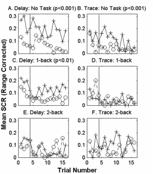
delay paradigms, using six subjects per group who were not asked to perform any task during conditioning. The delay group (Figure 2-2A) shows larger SCRs to the CS+ test trials than to adjacent CS- presentations (p<0.001). The same is true of SCRs during trace conditioning (Figure 2-2B, p<0.001 paired t-test p<0.01). No significant trial effects are present in either group. Thus, trace and delay differential protocols are suffcient to produce conditioning when performed alone, without distraction.
Concurrent distracting task The n-back working memory task served as a
distraction from the concurrently performed conditioning protocol. When six subjects performed the 1-back working memory task during differential delay conditioning (Figure 2-2C), there is a statistically significant difference between responses to CS+ and CS- during conditioning (p<0.01). However, when a 1-back working memory task was performed by six subjects during differential trace conditioning, there is no significant difference between SCRs to CS+ and SCRs to CS- (Figure 2-2D). When subjects carried out the 2-back task, there is no significant difference between responses to CS+ and CS- for either delay (n=6) or trace (n=6) conditioning (Figure 2-2E, F). No significant trial effects are present.
Differential main effects A univariate ANOVA using the mean CS+/CS- differences
2.3.2 Awareness of CS/US Contingency
Correlations between awareness and CS+/CS- amplitude differences There is a
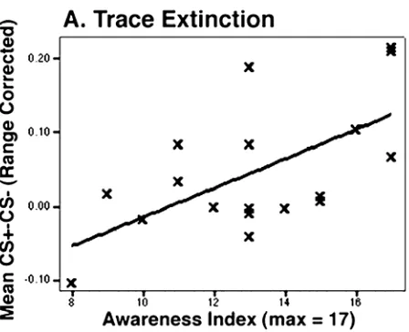
positive correlation between the awareness index and strength of conditioning (mean [CS+ - CS-]) during extinction for the 18 subjects carrying out the differential trace learning procedure (Figure 2-3). The correlation has an adjusted r2 value of 0.334 (Pearson coeff. = 0.611, p < 0.01). No significant correlations between contingency awareness and CS+/CS- difference are present for trace acquisition, or for either acquisition or extinction during delay conditioning.
Differential conditioning task interference The twelve subjects who were not
performing a task during differential conditioning (six delay, six trace) have an average awareness index of 15.2 (maximum 17). Twenty-four subjects who were performing a task during differential conditioning (trace and delay, 1-back and 2-back, six subjects in each combination of conditions) have an average index of 13.4. A univariate ANOVA utilizing the awareness questionnaire score to test factors that influence awareness show significant main effects for both task (p<0.05) and delay/trace (delay mean = 14.8, trace mean = 13.2, p<0.05) with no significant interaction. In summary, both the addition of a task and the addition of a short trace interval reduce the subject’s ability to report the CS/US contingency relationship in a post-experimental questionnaire.
2.3.3 Single Cue Conditioning
No task Single cue conditioning relationships were established in a group of four
significant differences between CS+ test trials and adjacent phantom CS- presentations (p<0.001). No significant trial effects are present.
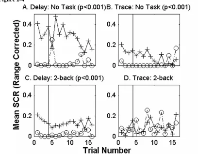
Concurrent distracting task A group of four single cue delay subjects and a group of
four single cue trace subjects were asked to focus on the 2-back working memory task during conditioning (Figure 4C and D respectively). The subjects that carried out the 2-back task during single cue delay conditioning show greater SCRs to CS+ test trials than to phantom CS- trials (p<0.001). The 4 subjects performing the same 2-back task during a trace conditioning protocol show no significant conditioning for the experiment. No significant trial effects are present. While the 2-back task interferes with single cue trace and differential delay conditioning (Figure 2-2E), there is still a significant CS difference in single cue delay conditioning during the 2-back task.
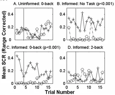
Uninformed 0-back task A group of four subjects had to signal whenever a
particular number appeared on the screen (0-back) during the single cue trace conditioning procedure (Figure 2-5A). There is no statistically significant difference between responses to the CS+ and the phantom CS- for this group. No significant trial effects are present. Although the 0-back task is a simple signal-detection task, there is no significant CS difference during single cue trace conditioning.
Informed subjects For the group of four informed subjects not distracted by any
present in any group. Prior explicit knowledge of the stimulus contingency facilitates, but does not guarantee, single cue trace conditioning.
2.4 Discussion
It is generally held in both eye-blink and fear conditioning that acquired trace and delay CS/US associations are distinct forms of learning. While the key difference between the two is the interposition of a temporal gap between the end of the CS and the start of the US, they involve different neural circuits and obey different regularities. For instance, acquisition of trace but not delay conditioning is critically dependent on hippocampus and certain prefrontal structures (Kim and Fanselow, 1992; Phillips and LeDoux, 1992; Maren et al., 1997; Weible et al., 2000; McLaughlin et al., 2002; Quinn et al., 2002) . In addition, Clark and Squire (Clark and Squire, 1998) showed that differential trace eye blink conditioning depends on CS/US contingency awareness, while this is not the case for delay conditioning (see also (Manns et al., 2000a; Clark et al., 2001; Manns et al., 2002)). This claim has been challenged. For example, Carrillo, Gabrieli and Disterhoft (Carrillo et al., 2000) demonstrated that not only single cue delay, but also single cue trace conditioning, was unaffected by division of attention. They used a dual-task paradigm to study the ability of subjects to acquire eye blink conditioning while their attention is concurrently engaged by watching a silent movie or verbal shadowing. Differential delay conditioning is, however, affected by the division of attention.
et al., 2001), and above results).
In this paper, we present experiments on fear conditioning. Fear conditioning differs from eye-blink conditioning in that it is dependent on the amygdala for both delay and trace conditioning, while eye-blink conditioning shows a similar pattern of
dependence on the cerebellum (Medina et al., 2002). Our experimental paradigm involves association between tones or noises as CSs and electric shocks as USs. As a measure of autonomic conditioning, we utilize increases in skin conductance in a comparatively young population (college students). We choose fear conditioning since it can easily be adapted to rodents, allowing the use of molecular and genetic tools to study the
underlying neuronal substrates of conditioning.
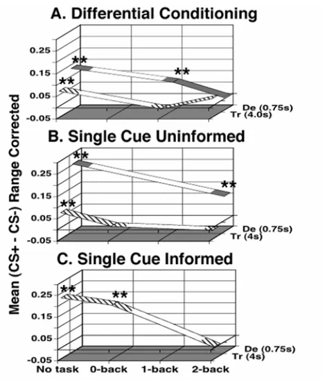
The general pattern of our findings is that the extent of associative autonomic conditioning depends on the cognitive load involved. The larger the demand on the system, the less conditioning occurs. We use the mean CS+, CS- difference for each group as a measure of strength of conditioning. This measure of conditioning is plotted in Figure 2-6 for each of our experiments. Figure 2-6 A, B, and C represent the transition from uninformed differential (A) to uninformed single cue (B, removing the second anticorrelated CS) and then the addition of explicit knowledge of the CS+/US
from the simplest protocol supports the hypothesis that as conditioning complexity increases, the amplitude/probability of conditioning decreases. This is reflected in a univariate ANOVA where the main effects single/differential, delay/trace, task level, and informed/uninformed effects are all significant. The only significant interaction is
between single/differential and delay/trace. The lack of a significant delay/trace task effect could be due to a floor effect, because the conditioning amplitude has reached zero for trace conditioning protocols in the first level, where a concurrent task has been added. We are not making any claims about the uniqueness of this representation. Others are possible and might prove advantageous.
It should be noted that Figure 2-6 is compatible with the existence of secondary tasks that do not interfere with trace conditioning in naïve subjects. A similar plot might also prove beneficial in summarizing the eye blink conditioning literature.
reliably induce trace conditioning under a 0-back task. We conjecture that this focused their attention onto the CS/US relationship and boosted learning.
The evaluation of the post-experimental questionnaire showed a correlation (r2=0.395) between differential trace subjects’ awareness scores and conditioning during the extinction phase. We found no significant correlation in the acquisition phase, nor did we find a correlation for either phase of delay conditioning. The correlation found
establishes a link between explicit knowledge of the CS/US relationship and the expression of trace fear conditioning during extinction. It is different from the explicit knowledge/conditioning correlations reported in (Clark and Squire, 1998), because our correlation occurs in fear conditioning and is true for the extinction phase as opposed to acquisition. A challenge for the future will be to develop on-line measures of CS/US contingency awareness (LaBar and Disterhoft, 1998; Lovibond and Shanks, 2002).
One might expect that subjects who are aware of the stimulus contingency would show a gambler’s fallacy effect where the differential response amplitude during
extinction phase increases for a number of extinction trials. Such a pattern was reported during eye blink conditioning (Clark et al., 2001). We failed to find any significant trend in response slope. In fact, it is likely that if higher awareness scores cause stronger conditioning, this may lead to more than one response strategy (for example, higher initial responses with rapid extinction or gambler’s fallacy). Our results also show a reduction in awareness in those groups who were performing a task compared to the no task controls.
complex types of conditioning. When that explicit knowledge cannot be acquired, conditioning cannot be established. This is supported by the fact that task performance reduces both the awareness index and the efficacy of differential conditioning. In
addition, explicit prior knowledge of the CS/US relationship compensates for some of the interference in single cue trace conditioning caused by concurrent task performance. Two, it is possible that concurrent task performance suppresses amygdala activity and subsequently suppresses the establishment of a conditioned fear response. Medial prefrontal cortex stimulation in rodents shows suppression of the basolateral complex of the amygdala (Rosenkranz and Grace, 2001). Furthermore, the n-back task shows an increased fMRI BOLD signal in human prefrontal areas that could be linked to suppression of normal brain activity under adverse conditions (Pochon et al., 2002). Either of these observations could explain fear conditioning interference by concurrent task performance.
2.5 Figures and Legends
Figure 2-1Figure 2-1 A) Delay conditioning consisted of a 0.25 second long electric shock that overlapped and co-terminated with the 1 second long CS+ (tone or noise). In trace conditioning, the CS+ was followed 3 second later by the US. B) The conditioning
Figure 2-2
Figure 2-2 Mean range corrected SCRs to CS presentations for each trial. Thirty-six subjects (6 per group) participated in either the differential delay (A, C or E) or trace (B, D, or F) learning procedure without any task or while being distracted by a 1-back or a 2-back task. Mean range corrected SCRs to CS+ are shown in solid lines with cross
Figure 2-3
Figure 2-4
Figure 2-4 Mean range corrected SCRs to CS presentations for each trial. Sixteen subjects (4 per group) participated in either single cue delay (A or C) or trace (B or D) conditioning without any distraction or while carrying out a 2-back task. Mean range corrected SCRs to CS+ are shown in solid lines with cross markers. Mean range corrected SCRs to marked phantom CS- time points are indicated by dashed lines with circles. Significant conditioning exists for delay conditioning with no concurrent task and while performing the 2-back task. Significant trace conditioning is present only while no task was performed. The vertical line marks the last test trial presented during
Figure 2-5
Figure 2-6
Figure 2-6 Summary of our data plotted in a 3-D space capturing the contingencies of our protocol. The vertical axis marks the group average for each subject’s average range corrected and normalized CS+, CS- difference. The horizontal axis marks the task
difficulty. The axis into the plane of the paper marks the group as trace or delay using the difference in CS/US onset (SOA) in seconds. In addition, the line for trace is hatched while the line for the delay group is solid. "**" indicates significant conditioning at p<0.01. Areas of the lines that are not filled in are meant to assist the stability of the figure, not to imply any prediction about the magnitude of conditioning in that area. A) Mean group differences for differential subjects. B) Mean group differences for
3 The Neural Correlates of Implicit and Explicit
Processes in Conditioning
Portions of this work were published as “Contingency Awareness in Human Aversive Conditioning Involves the Middle Frontal Gyrus” in 2006 (Carter et al., 2006). The authors were: Ronald McKell Carter, John P. O’Doherty, Ben Seymour, Christof Koch and Raymond J. Dolan. John O’Doherty served as a very knowledgeable teacher; he was involved in the experimental design, fMRI sequence choice and all data collection. He introduced me to SPM and provided base code to make the analysis easier, then continued to follow up with ideas to improve our analysis. He also helped arrange my visit to UCL and was a part of extensive manuscript review. Ben Seymour aided in the experimental design, was a great help in getting the electrical stimulation equipment, stimulation code and IRB protocols in place, helped in preparing subjects and data
collection, and completed extensive manuscript reviews. He was also the artist behind the abstract images we used. Christof Koch and Raymond J. Dolan not only found funding to make the project possible and initiated the collaboration, but were also involved in the design of the experimental procedure and statistical analysis as well as extensive manuscript review. Experiments and some of the analysis were conducted at the Functional Imaging Laboratories at University College London.
3.1 Introduction
Learning about aversive stimuli in the environment is necessary for an organism’s success. One of the simplest and best studied mechanisms by which this is realized is classical conditioning, whereby a predictive association is learned between a neutral stimulus (the conditioned stimulus or CS) and a biologically meaningful signal (the unconditioned stimulus or US) (Pavlov, 1906; Cook and Harris, 1937; Wilensky et al., 1999; Buchel and Dolan, 2000; Maren, 2001; Clark et al., 2002; LeDoux, 2003; Maren and Quirk, 2004). Typically, after repeated pairings of CS and US, the CS comes to elicit a response that is appropriate to the anticipated US. In aversive conditioning, this
conditioned response (CR) will often be a change in heart rate or skin conductance, and is taken as an implicit measure of successful conditioning in experimental studies.
However, it is also possible to become consciously aware of the predictive contingency between CS and US, a phenomenon referred to as contingency awareness. An individual can acquire both implicit associations and contingency awareness or either may be acquired independently (Bechara et al., 1995), indicating some degree of dissociation between the two systems. Currently, a major question in conditioning (and
The acquisition of contingency awareness and its interaction with conditioning differs across conditioning protocols (Clark and Squire, 1998; Ohman and Soares, 1998; Knuttinen et al., 2001; Han et al., 2003). For instance, in trace conditioning, in which there is temporal separation between CS and US, contingency awareness has been shown to positively correlate with the amplitude of conditioned responses (Clark and Squire, 1998). Those subjects in a trace conditioning experiment who do not display contingency knowledge fail to be trace conditioned. By contrast, in delay conditioning, in which there is no separation between the CS and US, no correlation between contingency awareness and successful conditioning has been observed; either can be acquired in the absence of the other. However, the simplicity of the delay protocol often results in immediate acquisition of explicit knowledge, making separation of explicit from implicit processes difficult using a delay paradigm alone.
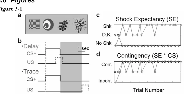
In this study, we used functional magnetic resonance imaging (fMRI) to identify brain regions that were specifically related to the explicit acquisition of contingency awareness during both delay and trace conditioning, independent of individual protocols. We simultaneously conditioned human subjects to predict an aversive electrical stimulus (US) from arbitrary visual cues (CS) with concurrent delay and trace protocols (see Figure 3-1a/b). The use of simultaneous conditioning allowed us to identify brain
activity in dorsolateral prefrontal cortex and hippocampus would correlate with these measures of explicit knowledge based on evidence that these structures are involved in working memory (Leung et al., 2002), memory formation (Fanselow, 2000) and revaluation (Corlett et al., 2004), as well as from lesion studies of trace conditioning deficits (Compton et al., 1997; Clark and Squire, 1998; Kronforst-Collins and Disterhoft, 1998; McEchron et al., 1998). In addition, we identified those regions that correlated with an implicit measure of learning, differential skin conductance responses. Consistent with previous work, we found that the amygdala correlated with implicit learning as measured by skin conductance changes. Surprisingly, changes in skin conductance were also a good predictor of activity in visual cortex and the hippocampus.
3.2 Methods
3.2.1 Participants
3.2.2 Experimental Procedure
We performed concurrent trace and delay Pavlovian conditioning. The CSs were abstract colored images (see Figure 3-1a) presented for 2 seconds and the US was a 1second electrical stimulus (see below). The study comprised 160 individual trials involving four separate CSs (each presented 40 times). One of the images acted as the trace conditioning cue (trace CS), which was followed on 50% of occasions by the US after a 3 second trace interval. Another image acted as the delay conditioning cue (delay CS), followed on 50% of occasions by the US, with a 0.5 second overlap between the end of the CS and the start of the US. The remaining two images acted as neutral cues (CS-), never followed by the US. Images were counter-balanced across conditions between subjects. Presentations of the CSs were arranged randomly, such that two of each CS type appeared in a block of eight. The delay and trace CS were each reinforced once in every block of eight trials. Trials were triggered on the nearest slice using a pseudo-randomized inter-trial onset asynchrony of 8, 9.25, 10.5, or 12 seconds. Presentation of stimuli and timing were controlled using Cogent 2000 (Wellcome Department of Imaging Neuroscience, Institute of Neurology, London, UK).
3.2.3 Online Subject Reports of Contingency Awareness
experiment were participants explicitly informed about any relationship between the images (CSs) and shock (US). Failures to respond or responses where latencies exceeded 1.5 seconds were scored as “don’t know”.
3.2.4 Post-Experimental Questionnaire
Following scanning, subjects were given a post-experimental questionnaire similar to that used by Clark and Squire (Clark and Squire, 1998). The questionnaire assessed their knowledge of the CS/US contingency relationships for both delay and trace protocols (see supplementary material). Subjects rated each statement on a 7 point scale ranging from “not true” through “don’t know” to “true”, capturing their degree of confidence. A response that was both accurate and very confident received a score of +3 and a response that was inaccurate and very confident received a score of -3, with all other responses falling on a scale between these limits. Scores for each subject were then totaled, giving a potential range of -48 to +48 for each protocol. More positive scores reflect greater contingency awareness.
3.2.5 Unconditioned Stimuli
chosen for each subject before the experiment, starting at a low level and using an ascending rating method where the current amplitude was raised until the subject gave a rating of 9 on a scale of 1-10, where 1 indicated the subject could barely feel the shock and 10 indicated the shock was too uncomfortable to be used in the experiment.
3.2.6 Online Measure of SCR
Skin conductance data was collected at a minimum of 100Hz, and was aligned to the first slice pulse where scanning had started. Data collected at a rate higher than 100Hz was first down-sampled to that frequency. Before analysis, all skin conductance data was median filtered to reduce noise. Skin conductance responses (SCRs) were defined as the maximum amplitude response initiated no earlier than 1second with a peak no later than 5seconds after the CS onset. SCR amplitudes were range corrected by the maximum response for that subject (Lykken, 1972). A two-tailed, single sample t-test across subjects (n=14) showed a significant difference between the mean nonreinforced CS+ response and the mean CS- response for both delay (P<0.01) and trace (P<0.01) conditioning.
3.2.7 fMRI
Forty-four slice whole brain tilted axial BOLD images were acquired in a 3 Tesla
Functional image analysis was performed using SPM2 (Wellcome Department of Imaging Neuroscience, Institute of Neurology, London, UK). Prior to analysis, all
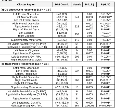
functional images were realigned, slice time acquisition corrected, normalized to the MNI EPI template and smoothed using a Gaussian kernel (8mm FWHM including contrast image smoothing). Individual subject models were then constructed and random effects analysis conducted as noted below. Each trial was modeled in two segments: first, an initial event-related response to each CS presentation, and second, a 3second period following image termination. This was done for both trace and delay conditioning to ensure that correlations between the stimulus event and trace period models were treated similarly. Trace period regressors were orthogonalized with respect to their event-related CS onset equivalents to minimize any contamination of the trace period response by the CS onset response. Only results for the CS onset event responses are reported here.
The acquisitions of both conditioning and contingency awareness were modeled as a parametric modulation (Buchel et al., 1998a) of responses to a CS presentation (indexed by skin conductance and US expectancy respectively). In testing for brain activity that correlated with learning, we examined BOLD responses specific to the CS by performing a conjunction analysis (Friston et al., 1999). This technique identifies only those regions that show significant activation across the included conditions. Statistically, a conjunction analysis is identical to performing an F test with the constraint that the individual effects are positive. For all reported results, we identified regions whose activity reflected learning for both delay and trace conditioning during all CS+ trials
absence of a shock and those due to peculiarities of the specific conditioning protocol. A region is reported as active if it violates the null hypothesis that on average members of the conjunction showed no effect (global null). An identified region’s consistent
activation for all members of the conjunction is confirmed by looking at the least significant P value for any member of that conjunction. We include plots of correlations in Figure 3-3 to demonstrate the consistency of a given effect across conditions included in the conjunction. This procedure lessens response ambiguity due to either the presence or absence of a US or any differences in protocol. Using a conjunction across these conditions allows us to infer a network that relates to the overall learned differences between CS+ (delay and trace) and CS-(neutral) representations. In areas where there was a prior hypothesis, results were family wise error (FWE) corrected for multiple
comparisons using small volume correction (20mm diameter sphere centered at the peak of activation). FWE correction for multiple comparisons for the whole brain is applied for brain regions where there was no prior hypothesis.
3.2.8 Learning: Contingency awareness and Conditioning
Explicit learning accuracy was defined by the interaction between CS type (CS+ or CS-)
and the reported US expectancy for each trial. Brain activity that correlates with US expectancy alone corresponds to those areas relevant for explicit fear. A shock
expectancy by CS type (+ or -) interaction tests for brain activity that correlates with the accuracy of shock expectancy on each trial. The magnitude of explicit learning defined
Implicit learning accuracy was defined by the interaction between CS type (+ or -) and
the normalized amplitude of the skin conductance response for each trial. The magnitude
of implicit learning was defined as the average difference between CS+ and CS- skin
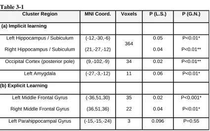
conductance responses and was used as a subject covariate in a second level random effects analysis (Table 3-1a).
3.3 Results
3.3.1 Conditioned skin conductance responses
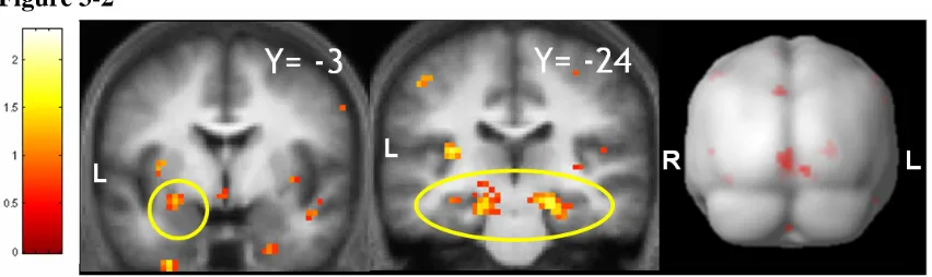
We recorded skin conductance responses associated with cues to provide an implicit measure of conditioning. Activity in the left amygdala (-27,-3,-12) correlated with the trial-by-trial time course of conditioning, indexed by the level of discriminatory skin conductance responses (P<0.01 corrected, see methods and Table 3-1a). This result confirms previous findings (Buchel et al., 1998b; Buchel et al., 1999; Knight et al., 2004) and in addition demonstrates that amygdala activity correlates with the specific time course of learning. It also indicates that activity in the amygdala correlates with the relative success of conditioning in different subjects.
3.3.2 Contingency awareness
described as the accuracy of contingency awareness (Figure 3-1d). The magnitude of a
subject’s contingency awareness was defined by a post-experimental questionnaire score that assessed the individual’s overall contingency knowledge via a series of true/false statements about the CS/US relationship (see supplementary methods – section 3.7 ). Brain regions that correlated with contingency awareness had greater activity during trials where a subject accurately expected a shock. We accounted for inter-subject differences by testing for regions that showed greater activity in those subjects who scored higher on the post-experimental questionnaire. This revealed responses correlated with contingency awareness in bilateral middle frontal gyri (MFG) (Figure 3-3, Table 3-1b, left -36,51,30; right 36,51,36), significant after correction for multiple comparisons. We also noted correlated activity in the para-hippocampal gyrus (-15,-15,-24, P=0.055 corrected for multiple comparisons, see methods).
3.4 Discussion
Our data indicate a clear role for the middle frontal gyrus in contingency
awareness during conditioning, correlated specifically with the acquisition of awareness on a trial-by-trial basis. To our knowledge, this is the first time such a trial by trial link has been demonstrated during conditioning. The role of the middle frontal gyrus in contingency awareness is contrasted with involvement of the amygdala, which we show reflects the acquisition of implicit knowledge, as indexed by autonomic activity,
In delay eye-blink conditioning, the magnitude of conditioning is independent of explicit knowledge (Manns et al., 2002). However, explicit learning is likely to be expressed in delay protocols, even if it is not correlated with the degree of conditioning. Since the degree of implicit knowledge is not correlated with the degree of explicit knowledge in delay conditioning, the substrates mediating both forms of learning can be separated. We confirmed that those same neural substrates were active during trace conditioning by testing for areas whose activation was consistent across both delay and trace conditioning. The fact that the middle frontal gyrus is active in both trace and delay conditioning, even though trace conditioning is correlated with contingency knowledge and delay is not (Clark and Squire, 1998), has implications for the mechanism by which contingency knowledge facilitates conditioning. The middle frontal gyrus is unlikely to directly facilitate conditioned associations, since doing so would require a second
inhibitory mechanism active during delay conditioning. It is therefore more likely that the middle frontal gyrus facilitates conditioning by means of another brain area, such as the hippocampal complex (see below). While it is unlikely that prefrontal areas directly facilitate conditioning, it is important to keep in mind that there is evidence that areas of prefrontal cortex inhibit activity in the amygdala (Rosenkranz and Grace, 2001; Quirk et al., 2003).
An area homologous to the middle frontal gyrus, the medial prefrontal cortex in the rabbit, is necessary for trace eye-blink conditioning (Kronforst-Collins and Disterhoft, 1998). This region is also strongly implicated in tasks requiring maintenance and
(Goldman-Rakic, 1987; Petrides, 2000; Castner et al., 2004). In the previous chapter, we showed that working memory distraction during fear conditioning reduces explicit knowledge of the CS/US contingency (Carter et al., 2003). This reduction of explicit knowledge is consistent with the middle frontal gyri’s involvement in both working memory and contingency awareness.
Brain areas central to the expression of explicit knowledge, as required in
reporting contingencies, may play a role in abstract, symbolic manipulation. In line with this notion, neurons in the middle frontal gyrus of behaving macaque monkeys respond to specific rules (Wallis et al., 2001) or limit responses to a given stimulus to only those times when a specific practiced task is being performed (Asaad et al., 2000). Thus, our finding that activity in this region correlates with contingency awareness is consistent with a putative role in the representation of abstract concepts.
We also found activity in the left para-hippocampal gyrus correlated with contingency awareness during conditioning. These results point to a role for the
hippocampal complex in mediating the integration of explicit knowledge of contingencies (Eichenbaum et al., 1996; Clark and Squire, 1998). It is interesting that while we found significant contingency related activation in the para-hippocampal gyrus, we did not find such effects in the hippocampus proper. By contrast, we observed a significant
related to explicit knowledge (Chun and Phelps, 1999; Schendan et al., 2003; Degonda et al., 2005). Future studies will need to address the mechanism of integration.
3.5 Tables
Table 3-1
Cluster Region MNI Coord. Voxels P (L.S.) P (G.N.)
(a) Implicit learning
Left Hippocampus / Subiculum
Right Hippocampus / Subiculum
(-12,-30,-6) (21,-27,-12) 364 0.05 0.04 P<0.01* P<0.01**
Occipital Cortex (posterior pole) (9,-102,-9) 34 0.02 P<0.01**
Left Amygdala (-27,-3,-12) 11 0.06 P<0.01*
(b) Explicit Learning
Left Middle Frontal Gyrus
Right Middle Frontal Gyrus
(-36,51,30) (36,51,36) 35 22 0.02 0.04 P<0.001* P<0.01*
Left Parahippocampal Gyrus (-15,-15,-24) 3 0.096 P=0.55
* FWE corrected for small volume (20mm diameter sphere) ** FWE corrected for whole brain
Table 3-1 Brain regions whose BOLD responses correlate with implicit and explicit measures of learning are shown in (a) and (b). This table specifies the anatomical labels for responsive clusters with the location
Table 3-2
Cluster Region MNI Coord. Voxels P (L.S.) P (G.N.)
(a) CS onset event responses (CS+ > CS-)
Left Frontal Operculum Left Anterior Insula Left Inf. Frontal Gyrus
(-48,12,3) (-33,21,6) (-57,9,12) 241 0.03 0.003 0.02 P<0.05** P<0.0001** P<0.05** Right Frontal Operculum
Right Anterior Insula Right Inf. Frontal Gyrus
(48,21,6) (42,21,-3) (51,18,18) 323 0.002 0.002 0.002 P<0.001** P<0.05** P<0.01* Left Caudate Right Caudate (-12,9,3)
(9,6,3) 152
0.01 0.02
P<0.01** P<0.01**
Supplementary Motor Area (3,21,60) 145 0.01 P<0.05**
Left Middle Frontal Gyrus (DLPFC) Right Middle Frontal Gyrus (DLPFC)
(-33,45,18) (33,48,15) 50 49 0.03 0.06 P<0.001* P<0.01* Left Anterior Cingulate
Right Anterior Cingulate
(-9,42,30) (6,30,24) 9 8 0.08 0.007 P<0.01* P<0.01* Left Supramarg. Gyr. / IPL
Right Supramarginal Gyrus
(-60,-48,24) (66,-36,33) 209 37 0.04 0.06 P<0.05** P<0.01*
(b) Trace Period Responses (CS+ > CS-)
Left Frontal Operculum Left Frontal Insula Left Inf. Frontal Gyr
(-42,15,6) (-36,21,27) (-60,18,3) 337 0.01 0.001 0.008 P<0.01* P<0.05** P<0.01* Right Frontal Operculum
Right Frontal Insula Right Inf. Frontal Gyr
(51,18,6) (36,15,-9) (51,18,18) 266 0.001 0.003 0.02 P<0.05** P<0.001* P<0.05*
Supplementary Motor Area (-12,-12,69) 15 0.005 P<0.01*
Left Middle Frontal Gyrus (DLPFC) Right Middle Frontal Gyrus (DLPFC)
(-45,54,6) (33,60,21) 6 34 0.01 0.003 P<0.01* P<0.001*
Right Anterior Cingulate (12,24,36) 34 0.006 P<0.01*
Left Supramarg. Gyr. /IPL Right Supramarg. Gyr. / IPL
(-48,-48,33) (54,-42,42) 90 564 0.005 0.00005 P<0.01* P<0.0001**
* FWE corrected for small volume (20mm diameter sphere) ** FWE corrected for whole brain
Table 3-2- Brain regions whose mean BOLD responses are greater for the CS+ than CS- during (a) the
transient stimulus onset (across delay and trace conditioning and reinforced and nonreinforced trials), and (b) the 3 second long trace period (see Figure 3-1) for trace conditioning trials (reinforced and
3.6 Figures
Figure 3-1Figure 3-1 Experimental design – 14 subjects reported shock expectancy during concurrent delay and trace aversive conditioning while functional brain images were acquired. Each CS presentation was 2 seconds long. Reinforced CS+ trials were followed by an electric shock lasting 1 second. For each subject, the four images in (a, presented in
color) acted as either the delay CS+ (image presentation overlaps with the shock, see b),
trace CS+ (image presentation ends 3 s before the onset of the shock, see b), or as one of
two CS- baselines. The point used for modeling event related analysis for each trial is marked with a dotted line in b. The memory trace period, 3 seconds marked by a shaded
box (also in b), was analyzed separately and is not discussed here. As soon as each image
appeared, subjects had to judge the likelihood of being shocked (shock expectancy; c)
using one of three keys indicating “shock likely,” “don’t know,” or “no shock likely” (* = CS+; o = CS-). d. The accuracy of contingency awareness is the interaction between
Figure 3-2
Figure 3-3