doi: 10.1093/glycob/cwy061 Advance Access Publication Date: 27 July 2018 Original Article
Glycan Recognition
Biophysical analysis of sialic acid recognition
by the complement regulator Factor H
Christoph Q Schmidt
2,1, Agnes L Hipgrave Ederveen
3, Markus J Harder
2,
Manfred Wuhrer
3, Thilo Stehle
4, and Bärbel S Blaum
4,12
Institute of Pharmacology of Natural Products and Clinical Pharmacology, Ulm University, Ulm, Germany,
3Center for Proteomics and Metabolomics, Leiden University Medical Center, Albinusdreef 2, 2333 ZA, Leiden,
The Netherlands, and
4Interfaculty Institute of Biochemistry, University of Tübingen, 72076 Tübingen, Germany
1To whom correspondence should be addressed: Tel:+49-7071-29-75-359; Fax:+49-7071-29-55-65;
e-mail: [email protected] (B.S.B.); Tel:+49-731-500-65615; Fax:+49-731-500-65602; e-mail: [email protected] (C.Q.S.)
Received 9 November 2017; Revised 28 June 2018; Editorial decision 28 June 2018; Accepted 1 July 2018
Abstract
Complement factor H (FH), an elongated and substantially glycosylated 20-domain protein, is a
soluble regulator of the complement alternative pathway (AP). It contains several glycan binding
sites which mediate recognition of
α
2-3-linked sialic acid (FH domain 20) and glycosaminoglycans
(domains 6
–
8 and 19
–
20). FH also binds the complement C3-activation product C3b, a powerful
opsonin and focal point for the formation of C3-convertases of the AP feedback loop. In freely
cir-culating FH the C3b binding site in domains 19
–
20 is occluded, a phenomenon that is not fully
understood and could be mediated by an intramolecular interaction between FH
’
s intrinsic
sialy-lated glycosylation and its own sialic acid binding site. In order to assess this possibility, we
char-acterized FH
’
s sialylation with respect to glycosidic linkage type and searched for further potential,
not yet characterized sialic acid binding sites in FH and its seven-domain spanning splice variant
and fellow complement regulator FH like-1 (FHL-1). We also probed FH binding to the sialic acid
variant Neu5Gc which is not expressed in humans but on heterologous erythrocytes that restrict
the human AP and in FH transgenic mice. We
fi
nd that FH contains mostly
α
2-6-linked sialic acid,
making an intramolecular interaction with its
α
2-3-sialic acid speci
fi
c binding site and an
asso-ciated self-lock mechanism unlikely, substantiate that there is only a single sialic acid binding site
in FH and none in FHL-1, and demonstrate direct binding of FH to the nonhuman sialic acid
Neu5Gc, supporting the use of FH transgenic mouse models for studies of complement-related
diseases.
Key words:carbohydrate, complement, glycans, mass spectrometry, saturation transfer difference NMR
Introduction
Complement factor H (FH), a serum protein composed of 20 ellips-oid complement control protein repeats (CCPs) that are connected head-to-tail, is a negative regulator of the complement alternative pathway (AP), a branch of innate immunity that requires active down-regulation. FH contains eightN-glycosylation sites, each car-rying single- to multiantennae sialoglycans, adding almost 18 kDa to
its mass (Fenaille et al. 2007). Its regulatory action targets the com-plement component C3-activation product C3b, the central protein of a positive feedback loop in which all complement pathways con-verge. FH accelerates the decay of the AP C3 convertase (C3bBb) and serves as a co-factor to the serine protease factor I (FI) that degrades C3b (Xue et al. 2017). FH-mediated down-regulation of C3b activity is targeted to host tissue to which FH localizes via one
© The Author(s) 2018. Published by Oxford University Press. 765
or both of its two host glycan recognition sites, located in CCPs 6–8 and CCPs 19–20. These domains engage host-specific glycans, namely glycosaminoglycans (GAGs, CCPs 6–8 and CCPs 19–20) or sialic acid (Sia)-containing glycans (CCP 20). Both GAG sites prefer the heparan sulfate (HS)/heparin type over other GAG types but dif-fer in their relative affinities for differentially (de-)sulfated forms of heparin (Clark et al. 2013). FH also binds sialylated glycans (Fearon 1978;Pangburn and Müller-Eberhard 1978;Michalek et al. 1988a), likely simultaneously with surface-bound C3b (Wu et al. 2009; Kajander et al. 2011;Morgan et al. 2011;Blaum et al. 2015). In full length FH the two binding sites for C3b (located in CCPs 1–4 and 19–20) are not equally accessible (Schmidt et al. 2013;Herbert et al. 2015). Instead, free FH adopts one or several conformation(s) in which the C-terminal C3b binding site is obscured (DiScipio 1992; Oppermann et al. 2006; Schmidt et al. 2010; Makou et al. 2012; Herbert et al. 2015;Baud et al. 2016). The structural basis for the FH conformational heterogeneity or for a possible regulatory mech-anism that could adjust the equilibrium between conformations with accessible and obscured C-terminal C3b binding sites is unknown. One possibility could be a self-locking mechanism in which the FH sialylated glycan chains interact with the FH sialic acid binding site in domain 20.
In our past structural analysis of Sia recognition by FH (Blaum et al. 2015), we focused on the previously established Sia binding site in FH’s most C-terminal domain CCP 20 and the most common Sia variant in humans,α-N-acetylneuraminic acid (αNeu5Ac). We found that FH bindsα2-3- but notα2-6- orα2-8-linked Neu5Ac, in agreement with functional investigations by other groups (Michalek et al. 1988b;Ram et al. 1998;Gulati et al. 2005;Hyvärinen et al. 2016). We now characterized the glycosidic linkage types present in the eight FH glycan chains. We further probed for the existence of additional, not yet characterized Neu5Ac binding sites in FH and its N-terminal splice variant FH-like 1 (FHL-1) (Schwaeble et al. 1987; Ripoche et al. 1988) to exclude that potential Sia-recognition by
FHL-1, which is the main complement regulator on Bruch’s mem-brane, is implicated in the pathophysiology of age-related macular degeneration (AMD) (Clark et al. 2014). We also wanted to cross-validate the common approach of investigating (human) FH functions in FH transgenic mice models as well as on heterologous erythrocytes, which traditionally serve in complement activity assays (e.g., sheep erythrocytes). To this end we probed direct binding of FH toα-N-glycolylneuraminic acid (αNeu5Gc), a common mamma-lian Sia variant that is not expressed in humans but in the FH trans-genic mice model and on the surface of heterologous erythrocytes.
Results
The glycan recognition site in FH CCPs 6
–
8 does not
contribute to Sia-mediated erythrocyte protection
from complement attack by FH
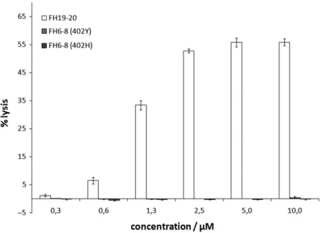
Sheep erythrocytes (RBCs) are non-activators of the human AP and are stable in normal human serum (NHS) supplemented with Mg-EGTA (which preserves activity of the AP but not the classical or mannose-binding lectin complement pathways). Hemolysis can be induced by neuraminidase treatment or by addition of recombinant FH CCPs 19–20 (FH19–20), which necessarily requires the presence of RBC sialylation in order to compete FH and induce lysis (Schmidt et al. 2013;Hyvärinen et al. 2016). The current model for these observations is direct competition of FH19–20 with serum FH for simultaneous binding of plasma-membrane Sia and small amounts of cell-bound C3b (Ferreira et al. 2006).
We employed this assay to assess the possibility that FH CCPs 6–8, which contain one of the two FH GAG binding sites, also bind to plasma membrane sialoglycans. To this end, we prepared washed sheep RBCs in NHS supplemented with Mg-EDTA and added recombinant FH6–8 (both the Y402 and H402 variant that is linked to AMD) or recombinant FH19–20 (for comparison). Even at a con-centration of 10μM FH6–8 virtually no hemolysis was observed,
whereas 2.5μM of FH19-20 induced well over 50% of hemolysis (where 100% equals complete osmotic hemolysis with distilled water) (Figure1). This observation suggests that recombinant FH6–8, unlike FH19–20, does not compete with serum FH for Sia binding on sheep RBCs and hence that FH CCPs 6–8 do not contribute to Sia recognition by FH.
Neither FH6
–
8 nor FHL-1 bind directly to sialylated
glycans
Because of the so-called AP“tick-over”, which constantly produces a small amount of C3-convertase and thus deposits C3b in a rather unselective manner, the hemolytic assay may assess FH’s capacity to simultaneously bind to Sia and cell-bound C3b rather than to Sia alone (Ferreira et al. 2006). While FH19–20 also contains a C3b binding site directly adjacent to the Sia binding site in CCP 20 (Blaum et al. 2015, 2016), it is not entirely clear if FH6–8 also engages C3b. A potential binding site in FH CCPs 6–8 for C3b was reported but the binding affinity was too low for an accurate deter-mination (Schmidt et al. 2008). Thus, to complement the hemolytic assay with FH6–8 by an experiment that is entirely independent of FH’s interaction with C3 activation fragments, we conducted satur-ation transfer difference (STD) NMR experiments with two
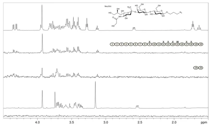
recombinant FH6–8 variants (Y402 and H402) and sialylated gly-can representatives of the three glycosidic linkage types that are most commonly observed in human sialoglycans (i.e., withα2-3-, α2-6-, orα2-8-linked Neu5Ac caps, respectively). We also included FHL-1 and a FH construct containing CCP 13 (for which a potential carbohydrate recognition site has been proposed but could not be confirmed (Pangburn et al. 1991;Ormsby et al. 2006;Schmidt et al. 2008)) in the STD NMR experiments. Unlike FH and FH19–20, none of the other shortened FH constructs (FH6–8Y, FH6–8H and FH8–15) nor FHL–1 bound to any of the three soluble sialy-lated glycans (3′sialyllactose (3′SL), 6′sialyllactose (6′SL), and the GD3 glycan) (Figure2A). Together with the hemolytic assay these experiments confirm the Sia binding site in CCP 20 as the only Sia binding site in FH and suggest that FHL-1 does not bind to Sia in vitro.
FH binds the nonhuman Sia variant Neu5Gc
Using STD NMR experiments, we analyzed whether serum-derived FH recognizes Neu5Gc, which is not expressed in humans but in other mammals, including sheep and mice (Chou et al. 1998;Hedlund et al. 2007). First, we used the monosaccharide 2-O-methyl-α -N-glycolyl-neuraminic acid (Neu5GcαOMe, 2-O-methylated in order to prevent
Fig. 2.FHL-1 and recombinant fragments of FH devoid of CCPs 19–20 do not bind to sialylated glycans. Representatives ofα2-3,α2-6, andα2-8-linked Neu5Ac (3’SL, 6’SL, and the GD3 glycan, respectively, chemical structures shown with Neu5Ac glycosidic linkage types in red) were measured at 2 mM each and STD NMR experiments were recorded at 283 K in PBS containing D2O instead of H2O with different proteins at around 10μM present. Spectra from top to bottom:
proton 1D reference spectrum of the sugar mix (top), STD NMR difference spectra with FH (second from top), with FH8-15 (third from top), with FH6–8 402Y (fourth from top), with FHL-1 (fifth from top), and, for comparison, without protein (bottom). Proteins used are shown schematically with naturally occurring gly-cosylation sites highlighted as“Y”. All recombinantly produced proteins (i.e., all apart from FH) were deglycosylated with Endo Hfduring the purification
mutarotation and formation of the non-physiologicalβ-anomer) but did not observe any interaction with FH (Figure3). However, with a larger Neu5Gcα-containing glycan, namely the trisaccharide Neu5Gcα2-3Galβ1-4Glcβ-propyl-N3(glycolyl-3′SL-ProN3), good
mag-netization transfer from FH and from FH19–20 was detected (Figure3). This experiment, together with indirect evidence from bio-chemical studies (Gulati et al. 2005), demonstrates that nonhuman Neu5Gc is recognized by human FH just as well as Neu5Ac. Our observation that a single Sia ring (i.e., Neu5GcαOMe) is not capable of FH binding is in agreement with our previous crystallographic and NMR spectroscopic analysis, which showed that further pyranoses attached to the specifically recognized Sia ring (for example in sialy-lated trisaccharides such as 3′SL) entertain so-called CH–πinteractions with the W1183 side chain in FH and also receive substantial magnet-ization transfer in STD NMR experiments (Blaum et al. 2015,2016). For glycolyl-3′SL-N3 such transfer of magnetization to the non-Sia
rings Gal and Glc (but, notably, not to the non-physiological propargyl linker) is also observed (Figure 3), suggesting that Neu5Gc-capped glycans are bound by FH in much the same manner as Neu5Ac-capped glycans.
FH is primarily
α
2-6-sialylated
The FH sequence contains nine theoreticalN-glycosylation sites, eight of which are in fact glycosylated (Fenaille et al. 2007). Glycosylation is restricted to domains 9 to 18. The most common non-reducing end oligosaccharide sequences of human complexN-glycans are Neu5Acα2-3Galβ1-4GlcNAc and Neu5Acα2-6Galβ1-4GlcNAc.
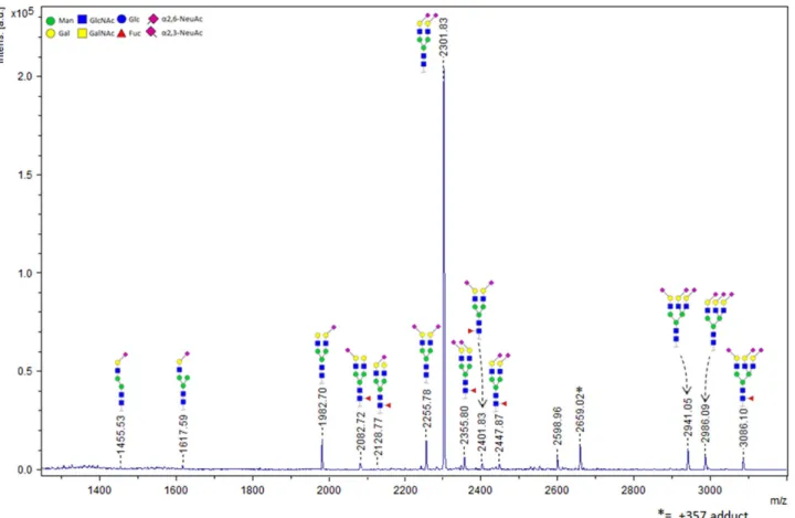
We have previously shown that FH specifically binds to trisaccharides which terminate in the Neu5Acα2-3Gal disaccharide (Blaum et al. 2015). Thus, FH’s own glycosylation may resemble the sialylated host markers on which it relies for self-recognition purposes. In order to clarify in how far FH itself isα2-3- orα2-6-sialylated, we undertook mass spectrometry-based characterization of the indi-vidual glycosidic linkage types. We employed a chemical modifi-cation strategy with whichα2-6-linked Neu5Ac is ethyl esterified while the α2-3-linked variant is lactonized, introducing a mass difference between both Sia types and achieving charge neutral-ization and sialic acid stabilneutral-ization at the same time (Reiding et al. 2014). The glycans were enzymatically released, derivatized, and subsequently analyzed using matrix-assisted laser desorption/ ionization time-of-flight tandem mass spectrometry (MALDI-TOF-MS). We identified 15 different glycan compositions in FH from human serum, with the A2S2 type (two-antennary disialylated) being the most abundant type by far (79.5%±0.1%). In total, 74.8%±0.3% of the FH glycans are diantennary with both Sia caps in theα2-6 link-age variant. Only 4.7%±0.2% of the diantennary glycans have a singleα2-3-linked Sia cap, and no diantennary glycans with two α2-3-linked Sia caps are observed (Figure 4 and Table I). The total percentage ofα2-6 andα2-3 linked Sia, with respect to the total level of sialylation and taking mixed species into account, was found to be 91.5% and 8.5%, respectively. Consistent with the absence ofO-glycosylation, treatment with a deglycosylation mix containing PNGase F andO-glycosidase did not reduce the apparent molecular weight of FH (as observed by SDS-PAGE) fur-ther than treatment with PNGase F only (data not shown).
Fig. 3.FH binds the nonhuman Neu5Gc variant of sialic acid. From top: Proton 1D reference spectrum of Neu5Gcα2-3Galβ1-4Glcβ-propyl-N3(glycolyl-3’
SL-ProN3, chemical structure shown; Neu5Gc differs only by one additional OH-group in the acetyl side chain from the Neu5Ac Sia-variant in 3’SL); STD NMR
dif-ference spectrum of 2 mM glycolyl-3’SL-ProN3with 8μM of FH; STD NMR difference spectrum of 2 mM glycolyl-3’SL-ProN3with 50μM of FH19–20; Proton 1D
Discussion
Carbohydrate recognition by FH is linked via genetic disposition to tissue-specific complement-mediated diseases, in particular AMD (on Bruch’s membrane) and atypical hemolytic uremic syndrome (aHUS, in
the kidney glomerulus) (Carroll and Sim 2011;Kavanagh et al. 2013). Due to the low affinities with which oligosaccharides bind to FH in vitro and the chemical complexity of mammalian glycosylation a comprehensive understanding of the role that glycans play in AP
Fig. 4.FH itself is primarilyα2-6-sialylated. MALDI-TOF-MS spectrum and structural assignment of glycans released from FH. Mostly disialylated diantennary (A2S2 and A2S2F) but also trisialylated triantennary (A3S3 and A3S3F) and traces of monosialylated diantennary glycans (A2S1 and A2S1F) are observed. The chemical composition, including the Sia glycosidic linkage type(s) of each species, is depicted schematically. Linkage types were assigned based on mass differ-ences that were introduced via chemical derivatization prior to MALDI-TOF-MS analysis.
Table I.Relative abundances of ethyl esterified glycans compared to HPCE-LIF analyzed APTS derived glycansa
Ethyl esterifiedN-glycansa Mean (±SD) Mean (±SD) Relative proportiona APTS derivedN-glycansb
H5N4E1 3.8 (0.28) 3.8 (0.28) 9.2 A2S1
H5N4F1L1 1.0 (0.06) 1.3 (0.09) 2.0 A2S1F
H5N4F1E1 0.3 (0.03)
H5N4E1L1 4.7 (0.16) 79.5 (0.10) 67.4 A2S2
H5N4E2 74.8 (0.26)
H5N4F1L2 2.3 (0.06) 4.7 (0.03) 8.9 A2S2F
H5N4F1E1L1 1.4 (0.02)
H5N4F1E2 1.0 (0.01)
H6N5E2L1 4.2 (0.15) 7.0 (0.18) 3.9 A3S3
H6N5E3 2.8 (0.04)
H6N5F1E2L1 2.0 (0.07) 2.0 (0.07) 4.0 A3S3F
H3N3E1 0.3 (0.03) 1.6 (0.14) 4.6 Other
H4N3E1 0.4 (0.06)
H6N5E1 0.8 (0.13)
H6N5E1 0.1 (0.02)
regulation is still lacking. It is, however, certain that both types of FH self-markers (i.e., HS-proteoglycans and sialylated glycans) are not functionally redundant with respect to their interaction with FH. Instead, it seems that both glycan types act as tissue-specific self-mar-kers to FH, i.e. proteoglycans appear to be the dominant self-marself-mar-kers in Bruch’s membrane and sialylated glycans in the glomerulus (possibly with additional contribution by proteoglycans whose GAG chains are structurally distinct from those on Bruch’s membrane) (Clark et al. 2013;Blaum et al. 2015;Langford-Smith et al. 2015). Ourfinding that FHL-1, the main AP regulator on Bruch’s membrane, and FH CCPs 6–8, previously shown to bind stronger to Bruch’s membrane than FH CCPs 19–20, do not bind to sialylated oligosaccharides further sup-ports the hypothesis that GAGs act as the major or sole self-markers in Bruch’s membrane (Clark et al. 2013). It is, however, possible that recruitment of FH by Sia plays a role in complement protection in other parts of the eye where full length FH may be present, for example in the retinal interphotoreceptor matrix for which a prevalence ofα 2-3-linked Neu5Ac was previously suggested on the basis of lectin bind-ing experiments (Bishop et al. 1993).
Like many serum proteins, FH itself is heavily glycosylated, and it is possible that the intrinsic glycosylation plays a direct or indirect role in FH’s activity – including the reported conformational heterogeneity of FH, which impacts C3b binding. A comparison of N-glycosylation consensus sequence sites across different mamma-lian FH sequences, which could give hints as to which glycan chains are functionally important, shows that theN-glycosylation sites in FH are not subject to strict conservation but nevertheless tend to cluster in certain regions, namely those that are not directly impli-cated in ligand binding. Most glycosylation sites are loimpli-cated between CCPs 12 and 15, in CCP 17 and 18, and in the linker region between CCPs 8 and 9 (Supplementary data, Figure S1). Thus far, none of these regions have been convincingly implicated in direct interactions with FH’s principal ligands. A possible interpretation of this observation is that the glycan chains in FH serve predominantly indirect purposes, such as solubility or resistance to proteolytic deg-radation. In order to experimentally evaluate if the FH glycan chains may directly mediate a back-bending mechanism in which the FH Sia binding site in CCP 20 would bind to its own glycan chains and modulate accessibility of the C-terminal C3b binding site, we char-acterized the FH glycan chains with respect to the Neu5Ac glyco-sidic linkage types and demonstrate that the vast majority of glycan chains in pooled serum-derived FH is decorated withα 2-6-linked Neu5Ac. Thisfinding is in line with a reported excess ofα 2-6-over α2-3-linked Neu5Ac in pooled human plasma proteins (Gagneux et al. 2003;Reiding et al. 2014). Because FH itself has a clear preference for binding α2-3-linked Sia (Blaum et al. 2015; Hyvärinen et al. 2016) thisfinding makes a scenario in which the FH Sia binding site interacts with its own glycan chains (or those of other FH molecules) unlikely. Since sialylation levels and Sia-linkage types change with age and disease, such a regulatory mechanism would also be highly unlikely from a theoretical point of view as FH conformational equilibria would otherwise shift with age (Garcia et al. 2005;Pousset 1997). Of note, desialylation and further deglycosylation of plasma-purified FH with bacterial neuraminidase and fungal endo-N-acetyl-glucosaminidase did not decrease the protein’s ability to decay cell-bound AP C3-convertase nor its ability to distinguish between activators and non-activators of the AP (Jouvin et al. 1984). The same is true for full-length FH produced inPichia pastorisand subsequently deglycosylated with Endo Hf(Schmidt et al. 2011). Together, these observations
sug-gest that the glycan chains are not critical for FH’s structural
architecture–although they could still influence the protein’s con-formational dynamics.
The sialic acid family consists of more than 50 differentα-keto sugars. Most of these differ from the prevalent Sia type in humans, the nine-carbon pyranoseαNeu5Ac, through additional acetyl and/ or hydroxyl groups (Varki et al. 2017). Absolute and relative expression levels of individual Sia variants are species- and tissues specific, with the Neu5Ac and Neu5Gc being the overall most com-mon mammalian variants. Humans lost the ability to hydroxylate Neu5Ac to yield Neu5Gc but can incorporate Neu5Gc directly from dietary sources (Tangvoranuntakul et al. 2003; Ng et al. 2014; Springer et al. 2014). Sheep RBCs, on the other hand, contain Neu5Gc (Klimas et al. 1982). The observation that sheep RBCs are non-activators of the human AP, therefore, hints to a certain promis-cuity of human FH with respect to this nonhuman Sia variant. Notably, wild type mice and thus human FH transgenic mouse mod-els that are used to study complement diseases express Neu5Gc in multiple organs including the kidney, making the question if human FH binds Neu5Gc highly relevant for the interpretation of data obtained with these animal models (Hedlund et al. 2007; Pickering et al. 2007). From a structural point of view, the Sia binding site in FH CCP 20 should be able to accommodate an additional hydroxyl group positioned at the Neu5Ac acetyl func-tion, i.e., Neu5Gc (Blaum et al. 2015), and our STD NMR experi-ment with a Neu5Gc-containing trisaccharide confirms this assumption. Potential physiological benefits of FH’s incapacity to discriminate between Neu5Ac and nonhuman Neu5Gc, however, remain enigmatic. Presentation of Sia and related nonulosonate sugars on microbial lipooligosaccharide (LOS) is an AP evasion strategy (Ram et al. 1998). For Neisseria gonorrhoeae(Ng) the specificity of glycosylation-mediated serum resistance is well documented and it was shown that both Neu5Ac- and Neu5Gc-modification of Ng LOS convey human AP-resistance (Gulati et al. 2015). However, in a physiological settingNg scavenges CMP-activated sugars for LOS-decoration from its host, and humans provide only traces of CMP-Neu5Gc (possibly from diet-ary intake). Therefore, no evolutiondiet-ary disadvantages appear associated with the lack of discrimination between the two Sia types by FH. Nevertheless, thefinding that FH directly binds to Neu5Gc-containing glycans has medical implications as human FH transgenic mice (which produce Neu5Gc) are used as animal models for infection with serum-resistant Ng strains and complement-specific diseases such as aHUS (Pickering et al. 2007; Gulati et al. 2015). Since bothNg serum resistance and aHUS predisposition are linked to FH-Sia recognition (Blaum et al. 2015; Gulati et al. 2015; Hyvärinen et al. 2016) such animal models would be highly questionable if FH was not promiscuous with respect to Neu5Ac hydroxylation.
Sia glycerol chain (Blaum et al. 2015). It was proposed that the S1191L/V1197A double mutation could alter sialic acid specificity (de Jorge et al. 2018). We do not think this likely (with the excep-tion of Neu5Gc) since the mutaexcep-tions are both well away from the Sia glycosidic linkage and the preceding pyranoses in the crystal structure of the complex. It is, however, possible that the conform-ational dynamics of the hypervariable loop (FH residues 1182–1189) are affected by the mutations. Since the double mutant reduces C3b binding by FH19–20 (Ferreira et al. 2009), and since C3b binding may promote the open, Sia-binding conformation of this loop (Blaum 2017) altered structural dynamics in this region of mutated FH and FHR-1 could also be the reason for impaired Sia binding by FHR-1.
Materials and methods
Proteins, recombinant production and puri
fi
cation
In terms of full length FH, a commercial source (CompTech, USA) of FH purified from human plasma was used. FHL-1 (402Y) and FH fragments FH6–8, FH19–20 and FH8–15 were produced as secreted proteins inPichia pastorisas described previously (Schmidt et al. 2008; Clark et al. 2014). In brief, P. pastoriscells (strain KM71H, Invitrogen) that had been stably transformed with the expression cassette (pPICZαB, Invitrogen) containing the relevant coding DNA were grown in a fermenter and protein expression was induced by supplementation with methanol (according to standard procedure described by the supplier’s manual and as published in detail inSchmidt et al. (2011)). The recombinant proteins were puri-fied by consecutive cation- and/or anion-exchange chromatography steps. The construct FHL-1 (corresponding to FH1–7), FH6–8 and FH19–20 are not glycosylated inP. pastoris, in contrast to FH8-15. However, the N-linked glycans of FH8–15 were removed during the purification process with the endoglycosidase Endo Hf (New
England Biolabs). Optionally a size-exclusion chromatography was employed to further increase purity if necessary.
AP hemolytic assay
This assay is based on the findings that sialic acid moieties on sheep erythrocytes are crucial to restrict the alternative comple-ment pathway (Fearon 1978; Pangburn and Müller-Eberhard 1978; Michalek et al. 1988a). 1 mL of defibrinated sheep blood (TCS Biosciences) was diluted with 10 mL precooled buffer (20 mM HEPES, 145 mM NaCl, 0.1% (w/v) gelatin (from pork skin, Fluka) and 10 mM EDTA, pH 7.3 at 25°C). The cell suspen-sion was mixed gently and spun for 10 min at 500 ×g, at 4°C. The supernatant and a layer of leukocytes were aspirated off and the procedure was repeated two more times with buffer and sub-sequently three times with an identical buffer without EDTA but with 5 mM MgCl2 and 5 mM EGTA (Mg-EGTA). 2.5μL of the
concentrated cell suspension was then mixed with 20μL of nor-mal human serum and 1μL of 100 mM Mg-EGTA. Recombinant FH fragments (FH19–20, FH6–8 402Y or FH6–8 402 H) were added from 30μM stocks prepared in HEPES/NaCl buffer with-out further supplements. Final recombinant protein concentra-tions were 0.3μM, 0.6μM, 1.3μM, 2.5μM, 5.0μM and 10μM. Individual reaction volumes were adjusted to 40μL with HEPES/ NaCl buffer without supplements. Individual reactions were incu-bated for 20 min at 37°C, stopped by addition of 150μL cold buf-fer without gelatin but with 10 mM EDTA, and spun at 1500×gfor 5 min at 4°C. A control experiment was conducted without
recombinant FH fragments and heat-inactivated human serum (56°C for 30 min). Hemolysis was determined as absorbance of the supernatant at 415 nm (A415) and compared to the A415 value for the same amount of cells lysed with distilled water. Four independ-ent experimindepend-ents were conducted and A415 values averaged for each data point.
NMR spectroscopy
NMR spectra were recorded at 283 K using 3 mm tubes (200μL sample volume) and a Bruker AVIII-600 spectrometer equipped with a room temperature probe head and processed with TOPSPIN 3.0 (Bruker). Samples for STD NMR spectra with Neu5Ac-containing glycans contained 2 mM of each of the three glycans (3′ SL, 6′SL and the GD3 glycan) and varying concentrations (10μM±
2μM) of either full-length FH (Complement Technology, Inc.), recombinant FHL-1μM, recombinant FH6–8 402Y, or recombinant FH8–15. Proteins were buffer-exchanged prior to NMR experiments in 10 kDa MWCO centrifugal concentrators to 20 mM potassium phosphate pH 7.4, 150 mM NaCl in D2O. Individual glycans were
added to the protein samples from 40 mM stock solutions prepared in D2O. A sample with 2 mM of each of the three glycans but
with-out protein was used to verify that no direct excitation of the gly-cans took place during the on-resonance irradiation step of the STD pulse program, and also to record a proton 1D reference spectrum of the sugar mix.
Off- and on-resonance irradiation frequencies were set to −30 ppm and 7.3 ppm, respectively. The irradiation power of the selective pulses was 57 Hz, the saturation time was 2 s, and the total relaxation delay was 3 s. A 50 ms continuous-wave spin-lock pulse with a strength of 3.2 kHz was employed in order to suppress residual protein signals. A total number of 512 scans were recorded. A total of 12 k points were collected, and spectra were multiplied with a Gaussian window function prior to Fourier transformation. The sample with glycolyl-3′SL-ProN3 contained 8μM FH and
2 mM glycolyl-3′SL-ProN3and was recorded at 288 K for technical
reasons with otherwise identical settings.
Enzymatic deglycosylation
FH at 1μg/μL in PBS (Complement Technologies, Texas) was dena-tured for 10 min at 95°C with the NEB protein denaturation buffer supplied with the NEB deglycosylation mix, and subsequently cen-trifuged for 5 min at 16,900×g. One microgram of denatured FH was then mixed 1:0.25 with 4-fold reducing SDS-PAGE buffer (29% (v/v) glycerol, 290 mM Tris pH 6.8, 1.5% (w/v) SDS, 12 mM EDTA, 3 M 2-mercaptoethanol) and left to incubate at room tem-perature for 10 min while anotherμg was treated with PNGase F (NEB, glycerol-free, fromElizabethkingia miricola) and 1μg was treated with the NEB deglycosylation mix (containing PNGase F fromElizabethkingia miricola, O-glycosidase fromEnterococcus faecalis, α2-3,6,8,9 neuraminidase A from Arthrobacter ureafa-ciens,β1-4 galactosidase S from Streptococcus pneumoniae, and β-N-acetylhexosaminidaseffromStreptomyces plicatus). The
N
-glyan release and linkage-speci
fi
c sialic acid
derivatisation
Five microgram of the protein was denatured with 10μL 2% sodium dodecyl sulfate (SDS; Merck, Darmstadt, Germany) by incu-bation for 10 min at 60°C. Subsequently the release step was per-formed by adding 10μL of a mixture containing 2% Nonidet P-40 (NP-40) and 1 U recombinant peptide-N-glycosidase F (PNGase F; Roche Diagnostics, Mannheim, Germany) in 2.5×PBS, followed by overnight incubation at 37°C. The released glycans were derivatized by ethyl esterification as described by elsewhere (Reiding et al. 2014). Briefly, the derivatization reagent mixture contains 0.25 M 1-ethyl-3-(3-dimethylaminopropyl) carbodiimide ((EDC); Fluorochem, Hadfield, UK) and 0.25 M 1-hydroxybenzotriazole ((HOBt); Sigma-Aldrich, Steinheim, Germany), in ethanol (Merck). Two microliter of releasedN-glycans were added to 20μL of derivatization reagent and incubated 1 h at 37°C. Allowing to cool down to room tem-perature, the derivatized glycans were purified by hydrophilic inter-action chromatography (HILIC) using pipette tipsfilled with cotton as stationary phase (Selman et al. 2011). Briefly, 20μL of aceto-nitrile ((ACN); Biosolve, Valkenswaard, The Netherlands) was added to the reaction mixture. The cotton inserted tips were washed with three-times 20μL of MQ water and three-times 20μL of 85% ACN followed by sample loading by pipetting 20 times up and down. Finally, cotton tips were washed by pipetting three-times 20μL 85% ACN containing 1% trifluoroacetic acid ((TFA); Merck) and three-times 20μL 85% ACN prior to the elution of the deriva-tized glycan sample using 10μL MQ.
MALDI-TOF-MS-(/MS) analysis of released
N
-glycans
Two microliter of purified derivatized glycans were spotted on a MTP AnchorChip 800/384 TF MALDI target plate (Bruker Daltonics) together with 1μL of matrix (5 mg/mL super-DHB, 1 mM NaOH in 50% ACN) and left to dry at room temperature. Mass spectrometry was performed in reflectron positive mode on an UltrafleXtreme mass spectrometer with a Smartbeam-II laser (Bruker Daltonics) controlled byflexControl 3.4 (Build 135). Within a window ofm/z 1000–5000, spectra were recorded with 10,000 laser shots at a frequency of 1000 Hz, employing a full sample ran-dom walking pattern of 100 shots per raster spot. Tandem mass spectrometry (MALDI-TOF/TOF-MS/MS) was performed on the most abundant peaks via laser-induced dissociation to confirm gly-can composition (data not shown).
Data analysis of released
N
-glycans
For relative quantification and quality control of the spectra, the raw spectra were exported fromflexAnalysis 3.4 (Build 76; Bruker Daltonics) as.txt file (x,y), and further processed by MassyTools (version 0.1.8.0) (Jansen et al. 2015). Internal calibration based on a predefined list of analytes was applied, and 15 visually detected gly-can compositions were extracted by integrating the areas of 95% of the theoretical isotopic envelope and subtracting from these the background found within a window of 20m/z. The areas were nor-malized to the sum of all areas, and derived traits were calculated based on the compositional features. Analyses were performed in duplicate.
Supplementary data
Supplementary data is available atGLYCOBIOLOGYonline.
Funding
This work was supported by the German Research Council (DFG, BL 1294/ 2-1, to B.S.B.; SCHM 3018/2-2, to C.Q.S.).
Con
fl
ict of interest statement
None declared.
Abbreviations
ACN, acetonitrile; AMD, age-related macular degeneration; APTS, (3-Aminopropyl)triethoxysilane); CCP, complement control protein; GAG, glycosaminoglycans; LOS, lipooligosaccharide NHS, normal human serum; STD, saturation transfer difference; TFA, trifluoroacetic acid.
References
Baud A, Gonnet F, Salard I, Le Mignon M, Giuliani A, Mercère P, Sclavi B, Daniel R. 2016. Probing the solution structure of factor H using hydroxyl radical protein footprinting and cross-linking. Biochem J. 473: 1805–1819.
Bishop PN, Boulton M, McLeod D, Stoddart RW. 1993. Glycan localization within the human interphotoreceptor matrix and photoreceptor inner and outer segments.Glycobiology. 3:403–412.
Blaum BS. 2017. The lectin self of complement factor H.Curr Opin Struct Biol. 44:111–118.
Blaum BS, Frank M, Walker RC, Neu U, Stehle T. 2016. Complement Factor H and Simian Virus 40 bind the GM1 ganglioside in distinct conforma-tions.Glycobiology. 26:532–539.
Blaum BS, Hannan JP, Herbert AP, Kavanagh D, Uhrín D, Stehle T. 2015. Structural basis for sialic acid-mediated self-recognition by complement factor H.Nat Chem Biol. 11:77–82.
Chou HH, Takematsu H, Diaz S, Iber J, Nickerson E, Wright KL, Muchmore EA, Nelson DL, Warren ST, Varki A. 1998. A mutation in human CMP-sialic acid hydroxylase occurred after the Homo-Pan divergence.Proc Natl Acad Sci USA. 95:11751–11756.
Clark SJ, Ridge LA, Herbert AP, Hakobyan S, Mulloy B, Lennon R, Würzner R, Morgan BP, Uhrín D, Bishop PN, Day AJ. 2013. Tissue-specific host recognition by complement factor H is mediated by differential activities of its glycosaminoglycan-binding regions.J Immunol. 190:2049–2057. Clark SJ, Schmidt CQ, White AM, Hakobyan S, Morgan BP, Bishop PN.
2014. Identification of Factor H–like Protein 1 as the Predominant Complement Regulator in Bruch’s Membrane: Implications for Age-Related Macular Degeneration.J Immunol. 193:4962–4970.
Carroll MV, Sim RB. 2011. Complement in health and disease.Adv Drug Deliv Rev. 63:965–975.
de Jorge EG, Yebenes H, Serna M, Tortajada A, Llorca O, de Córdoba SR. 2018. How novel structures inform understanding of complement func-tion.Semin Immunopathol. 40:3–14.
DiScipio RG. 1992. Ultrastructures and interactions of complement factors H and I.J Immunol. 149:2592–2599.
Fearon DT. 1978. Regulation by membrane sialic acid of beta1H-dependent decay-dissociation of amplification C3 convertase of the alternative com-plement pathway.Proc Natl Acad Sci USA. 75:1971–1975.
Fenaille F, Le Mignon M, Groseil C, Ramon C, Riandé S, Siret L, Bihoreau N. 2007. Site-specific N-glycan characterization of human complement factor H.Glycobiology. 17:932–944.
Ferreira VP, Herbert AP, Cortés C, McKee KA, Blaum BS, Esswein ST, Uhrín D, Barlow PN, Pangburn MK, Kavanagh D. 2009. The binding of factor H to a complex of physiological polyanions and C3b on cells is impaired in atypical hemolytic uremic syndrome.J Immunol. 182:7009–7018. Ferreira VP, Herbert AP, Hocking HG, Barlow PN, Pangburn MK. 2006.
Gagneux P, Cheriyan M, Hurtado-Ziola N, van der Linden ECMB, Anderson D, McClure H, Varki A, Varki NM. 2003. Human-specific regulation of
α2–6-linked sialic acids.J Biol Chem. 278:48245–48250.
Garcia GG, Berger SB, Akha AAAS, Miller RA. 2005. Age-associated changes in glycosylation of CD43 and CD45 on mouse CD4 T cells. Eur J Immunol. 35:622–631.
Gulati S, Cox A, Lewis LA, Michael FS, Li J, Boden R, Ram S, Rice PA. 2005. Enhanced Factor H binding to sialylated gonococci is restricted to the sialylated lacto-N-neotetraose lipooligosaccharide species: Implications for serum resistance and evidence for a bifunctional lipooli-gosaccharide sialyltransferase in gonococci. Infect Immun. 73: 7390–7397.
Gulati S, Schoenhofen IC, Whitfield DM, Cox AD, Li J, St. Michael F, Vinogradov EV, Stupak J, Zheng B, Ohnishi M et al. 2015. Utilizing CMP-sialic acid analogs to unravel Neisseria gonorrhoeae lipooligosaccharide-mediated complement resistance and design novel therapeutics.PLoS Pathog. 11:e1005290.
Hedlund M, Tangvoranuntakul P, Takematsu H, Long JM, Housley GD, Kozutsumi Y, Suzuki A, Wynshaw-Boris A, Ryan AF, Gallo RL et al. 2007. N-glycolylneuraminic acid deficiency in mice: Implications for human biology and evolution.Mol Cell Biol. 27:4340–4346.
Herbert AP, Makou E, Chen ZA, Kerr H, Richards A, Rappsilber J, Barlow PN. 2015. Complement evasion mediated by enhancement of captured Factor H: Implications for protection of self-surfaces from complement.
J Immunol. 195:4986–4998.
Hyvärinen S, Meri S, Jokiranta TS. 2016. Disturbed sialic acid recognition on endothelial cells and platelets in complement attack causes atypical hemo-lytic uremic syndrome.Blood. 127:2701–2710.
Jansen BC, Reiding KR, Bondt A, Hipgrave Ederveen AL, Palmblad M, Falck D, Wuhrer M. 2015. MassyTools: A high-throughput targeted data pro-cessing tool for relative quantitation and quality control developed for glycomic and glycoproteomic MALDI-MS. J Proteome Res. 14: 5088–5098.
Jouvin MH, Kazatchkine MD, Cahour A, Bernard N. 1984. Lysine residues, but not carbohydrates, are required for the regulatory function of H on the amplification C3 convertase of complement. J Immunol. 133: 3250–3254.
Kajander T, Lehtinen MJ, Hyvärinen S, Bhattacharjee A, Leung E, Isenman DE, Meri S, Goldman A, Jokiranta TS. 2011. Dual interaction of factor H with C3d and glycosaminoglycans in host-nonhost discrimination by complement.Proc Natl Acad Sci USA. 108:2897–2902.
Kavanagh D, Goodship TH, Richards A. 2013. Atypical hemolytic uremic syndrome.Semin Nephrol. 33:508–530.
Klimas NG, Caldwell KE, Whitney PL, Fletcher MA. 1982. Comparison of receptor properties of erythrocyte membrane glycoproteins.Dev Comp Immunol. 6:765–774.
Langford-Smith A, Day AJ, Bishop PN, Clark SJ. 2015. Complementing the Sugar Code: Role of GAGs and sialic acid in complement regulation.
Front Immunol. 6:25.
Makou E, Mertens HDT, Maciejewski M, Soares DC, Matis I, Schmidt CQ, Herbert AP, Svergun DI, Barlow PN. 2012. Solution structure of CCP modules 10–12 illuminates functional architecture of the complement regulator, factor H.J Mol Biol. 424:295–312.
Michalek MT, Bremer EG, Mold C. 1988b. Effect of gangliosides on activa-tion of the alternative pathway of human complement.J Immunol. 140: 1581–1587.
Michalek MT, Mold C, Bremer EG. 1988a. Inhibition of the alternative path-way of human complement by structural analogues of sialic acid.
J Immunol. 140:1588–1594.
Morgan HP, Schmidt CQ, Guariento M, Blaum BS, Gillespie D, Herbert AP, Kavanagh D, Mertens HDT, Svergun DI, Johansson CM et al. 2011. Structural basis for engagement by complement factor H of C3b on a self surface.Nat Struct Mol Biol. 18:463–470.
Ng PSK, Böhm R, Hartley-Tassell LE, Steen JA, Wang H, Lukowski SW, Hawthorne PL, Trezise AEO, Coloe PJ, Grimmond SM et al. 2014. Ferrets exclusively synthesize Neu5Ac and express naturally humanized influenza A virus receptors.Nat Commun. 5:5750.
Oppermann M, Manuelian T, Józsi M, Brandt E, Jokiranta TS, Heinen S, Meri S, Skerka C, Götze O, Zipfel PF. 2006. The C-terminus of comple-ment regulator Factor H mediates target recognition: Evidence for a com-pact conformation of the native protein. Clin Exp Immunol. 144: 342–352.
Ormsby RJ, Jokiranta TS, Duthy TG, Griggs KM, Sadlon TA, Giannakis E, Gordon DL. 2006. Localization of the third heparin-binding site in the human complement regulator factor H1. Mol Immunol. 43: 1624–1632.
Pangburn MK, Atkinson MA, Meri S. 1991. Localization of the heparin-binding site on complement factor H.J Biol Chem. 266:16847–16853. Pangburn MK, Müller-Eberhard HJ. 1978. Complement C3 convertase: Cell
surface restriction of beta1H control and generation of restriction on neuraminidase-treated cells.Proc Natl Acad Sci USA. 75:2416–2420. Pickering MC, de Jorge EG, Martinez-Barricarte R, Recalde S, Garcia-Layana A,
Rose KL, Moss J, Walport MJ, Cook HT, de Córdoba SR et al. 2007. Spontaneous hemolytic uremic syndrome triggered by complement factor H lacking surface recognition domains.J Exp Med. 204:1249–1256. Pousset D. 1997. Increased α2,6 sialylation of N-glycans in a transgenic
mouse model of hepatocellular carcinoma.Cancer Res. 57:4249–4256. Ram S, Sharma AK, Simpson SD, Gulati S, McQuillen DP, Pangburn MK, Rice
PA. 1998. A novel sialic acid binding site on Factor H mediates serum resist-ance of sialylated Neisseria gonorrhoeae.J Exp Med. 187:743–752. Reiding KR, Blank D, Kuijper DM, Deelder AM, Wuhrer M. 2014.
High-throughput profiling of protein N-glycosylation by MALDI-TOF-MS employing linkage-specific sialic acid esterification. Anal Chem. 86: 5784–5793.
Ripoche J, Day AJ, Harris TJ, Sim RB. 1988. The complete amino acid sequence of human complement factor H.Biochem J. 249:593–602. Schmidt CQ, Bai H, Lin Z, Risitano AM, Barlow PN, Ricklin D, Lambris JD.
2013. Rational engineering of a minimized immune inhibitor with unique triple-targeting properties.J Immunol. 190:5712–5721.
Schmidt CQ, Herbert AP, Kavanagh D, Gandy C, Fenton CJ, Blaum BS, Lyon M, Uhrín D, Barlow PN. 2008. A new map of glycosaminoglycan and C3b binding sites on factor H.J Immunol. 181:2610–2619. Schmidt CQ, Herbert AP, Mertens HDT, Guariento M, Soares DC, Uhrín D,
Rowe AJ, Svergun DI, Barlow PN. 2010. The central portion of factor H (modules 10–15) is compact and contains a structurally deviant CCP module.J Mol Biol. 395:105–122.
Schmidt CQ, Slingsby FC, Richards A, Barlow PN. 2011. Production of bio-logically active complement factor H in therapeutically useful quantities.
Protein Expr Purif. 76:254–263.
Schwaeble W, Zwirner J, Schulz TF, Linke RP, Dierich MP, Weiss EH. 1987. Human complement factor H: expression of an additional truncated gene product of 43 kDa in human liver.Eur J Immunol. 17:1485–1489. Selman MHJ, Hemayatkar M, Deelder AM, Wuhrer M. 2011. Cotton HILIC
SPE microtips for microscale purification and enrichment of glycans and glycopeptides.Anal Chem. 83:2492–2499.
Springer SA, Diaz SL, Gagneux P. 2014. Parallel evolution of a self-signal: humans and new world monkeys independently lost the cell surface sugar Neu5Gc.Immunogenetics. 66:671–674.
Tangvoranuntakul P, Gagneux P, Diaz S, Bardor M, Varki N, Varki A, Muchmore E. 2003. Human uptake and incorporation of an immuno-genic nonhuman dietary sialic acid. Proc Natl Acad Sci USA. 100: 12045–12050.
Varki A, Schnaar RL, Schauer R. 2017. Sialic acids and other nonulosonic acids. In: Varki A, Cummings RD, Esko JD, Stanley P, Hart GW, Aebi M, Darvill AG, Kinoshita T, Packer NH, Prestegard JH, Schnaar RL, Seeberger PH, editors.Essentials of Glycobiology, 3rd ed. Cold Spring Harbor (NY): Cold Spring Harbor Laboratory Press.
Wu J, Wu Y-Q, Ricklin D, Janssen BJC, Lambris JD, Gros P. 2009. Structure of complement fragment C3b-factor H and implications for host protec-tion by complement regulators.Nat Immunol. 10:728–733.