ACKNOWLEDGEMENTS
The completion of this thesis was made possible by the collective effort of several
individuals. First and foremost, I would like to express my sincere gratitude to my
advisor, Dr. Jonathan Berg, for not only his guidance throughout this project, but also for
his mentorship throughout the latter portion of my undergraduate career. I would also like
to thank Dr. Bryce Seifert, who played an invaluable role in the completion of this project
and subsequent manuscript. I am truly grateful for the kind words, encouragement, and
help you have provided throughout the time we have worked together.
Furthermore, I would also like to express my gratitude to the following
individuals for their contributions to this project and manuscript: Julianne O’Daniel,
Daniel Marchuk, Nirali Patel, Joel Parker, Alan Hoyle, Lisle Mose, Andrew Marron,
Michele Hayward, Christopher Bizon, Kirk Wilhelmsen, James Evans, H. Shelton Earp
III, Norman Sharpless, and Neil Hayes.
Finally, I offer my deepest thanks to Amy Maddox, Alain Laederach, and the rest
of the UNC Department of Biology for making this undergraduate research opportunity
ABSTRACT
In the past decade, tumor-germline next generation sequencing has become a
routine part of personalized oncology care. Via this method, germline mutations are
typically subtracted from those in the tumor to identify somatic mutations, thus negating
the possibility of discovering germline variants. Previously, it has been proposed that the
identification of germline variants could have significant clinical implications for patients
with hereditary cancers and their family members. In this exploratory research study, we
sought to investigate the prevalence of germline variants identified through clinical
tumor-germline sequencing among a cohort of patients across ten major cancer types.
Germline sequencing data from 439 individuals undergoing tumor-germline sequencing
through the LCCC1108/UNCseq™ (NCT01457196) study were analyzed for genetic
variants in 36 hereditary cancer susceptibility genes. Variants indicative of hereditary
cancer predisposition were identified in 19 (4.3%) patients. For about half (10/19), these
findings represent new molecular diagnostic information with potentially important
implications for the patient and their family. Genes with pathogenic variants included the
hereditary cancer genes: ATM, BRCA1, BRCA2, CDKN2A, and CHEK2. Furthermore, a
substantial proportion of patients (178, or 40.5%) had Variants of Uncertain Significance
(VUS), 24 of which had VUS in genes pertinent to the presenting cancer. Overall, with
approximately 4% of cases harboring pathogenic variants in known hereditary cancer
susceptibility genes, diagnostic germline findings such as these could be beneficial for
INTRODUCTION
Cancer is a leading cause of death worldwide, with an estimated 1.68 million new
cases in 20151. Recently, major technological advances in massively parallel sequencing
coupled with dramatic reductions in cost have positioned next-generation sequencing as
an integral tool used in cancer care. The application of this technology has enabled
clinicians and scientists to recognize the potential of personalized oncology, particularly
with respect to diagnosing tumors and determining effective courses of action for cancer
patients2.
Cancer is primarily a genetic disease caused by mutations in a wide variety of
genes, including proto-oncogenes and tumor suppressor genes. These genetic changes can
either be acquired post-conception (somatic mutation) or be present constitutively in all
cells of the body as a result of inheritance or early post-zygotic events (hereafter referred
to as constitutional or germline mutations)3. In patients with hereditary cancer
syndromes, the presence of germline mutations can complicate the use of next-generation
sequencing in identifying somatic mutations in a patient’s tumor. Typically, when
tumor-germline sequencing is performed on entire genomes, exomes, or selected genes,
germline variants are “subtracted out” from those found in the tumor in order to identify
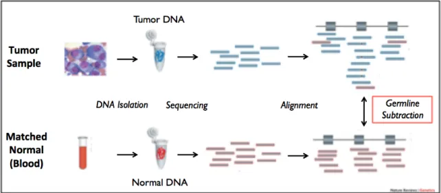
somatic mutations in the tumor (figure 1). Previous studies have shown that germline
variant subtraction enhances the specificity of detecting somatic mutations4. As a result, pathogenic germline mutations that predispose a patient to increased cancer susceptibility
may be overlooked when seeking out somatic variants via tumor-germline sequencing.
Thus, ignoring the germline variants post “subtraction” will likely miss these critical
syndromes. For instance, knowledge of these variants can guide the preventive care of the
patient and their family members. Moreover, in a fraction of cases, knowledge of these
variants can also influence treatment targets, as is the case with the use of PARP
inhibitors in patients with BRCA1 and BRCA2 mutation-associated cancers4.
Figure 1. Tumor-germline sequencing workflow. Germline variants derived from normal tissue are typically subtracted out from tumor sequences in order to identify somatic mutations, thus negating the possibility of discovering germline mutations.
While the majority of cancer cases arise sporadically via acquired somatic mutations,
inherited germline mutations are estimated to play a major role in roughly 5 to 10 percent
of all cancers5. The burden of germline mutations in cancer patients varies by cancer type, with some tumor types having lesser-known germline etiology (e.g. lung cancer,
kidney cancer, etc.) and others with well-known germline etiologies (e.g. breast cancer,
ovarian cancer). Despite our knowledge of the relative frequencies of hereditary cancers,
empirical data illustrating the burden of germline mutations identified through routine
Given the potential ramifications of missing critical germline variants in patients
with cancer, we conducted an exploratory study within patients undergoing
tumor-germline sequencing to explore the frequency of opportunistically identified pathogenic
germline variants within cancer predisposing genes.
METHODS
Summary
The experimental design for this study can be divided into three main steps: (1)
Tumor-germline sequencing, (2) Variant calling, and (3) Variant Classification. Germline
sequencing data from 439 individuals undergoing tumor-germline dyad sequencing were
analyzed for genetic variants in 36 hereditary cancer susceptibility genes. Patients across
10 major cancer types were included in the study. In order to realistically assess the
burden of germline variants among individuals who receive tumor-germline sequencing,
patients were included in the study irrespective of having prior clinical indicators of
hereditary cancer predisposition. The germline variants found in the patients were then
evaluated for pathogenicity using a variant classification framework previously published
by the American College of Medical Genetics.
Steps (1) and (2) were performed by other members of the Berg Lab, while step
(3) was conducted by Krunal Amin, Bryce Seifert, and Julianne O’Daniel.
(1) Tumor-germline sequencing
Participants were enrolled in the LCCC1108 study (UNC clinical sequencing
buccal as appropriate) were obtained from all patients through an institutional review
board (IRB)-approved protocol at the Lineberger Comprehensive Cancer Center and the
University of North Carolina, Chapel Hill (NCT01457196). The UNCseq™ study aims to
associate known molecular alterations with clinical outcomes in oncology and uses this
information to support treatment decisions through reporting of genetic profiling to
clinicians. The overarching study consent describes the collection and analysis of both
tumor and germline tissue including the explicit possibility for identification of an
underlying hereditary cancer predisposition. Participants consent to the reporting of all
results deemed clinically significant. Consent was obtained by UNCseq™ study staff for
the primary study at enrollment. Patients were referred into the UNCseq™ study team by
their clinic physician and enrolled according to their treated cancer (Table 1) and thus the
tumor tissue to be analyzed. All patients enrolled between 11/2011 and 06/2014 for the
cancer types listed in Table 1 were included in our data capture for exploratory germline
analysis.
(2) Variant Calling
Library preparation and gene capture methods have been described previously7. Briefly, DNA was extracted from blood using a Puregene DNA Purification kit (Gentra
Systems), DNeasy Blood and Tissue Kit (Qiagen), or a Maxwell MDx16TM (Promega,
Inc.). In each methodology, DNA was extracted according to the manufacturer’s
protocol. DNA was fragmented to approximately 180-225 base pairs 7 using a Covaris
E220 focused ultrasonicator instrument (Covaris, Inc.). Postfragmentation, the sample
employing AMPure beads (Beckman Coulter). Fragment size enrichment and subsequent
library preparation steps involving precise liquid handling steps were performed using the
Agilent basic Bravo A and/or the Bravo B robot(s) (Agilent Technologies). Gene capture
was performed using a SureSelectXT custom capture kit according to the manufacturer’s
protocol (Agilent Technologies). All exons of the 247 genes on the UNCseq™ panel
were sufficiently captured with average coverage depth of 750X (see Supplementary
Table 1-Capture V6 within Jeck et al. listing all 247 genes).
Library quality was assessed with a Bioanalyzer or Tapestation 2200 (Agilent
Technologies) using either D1K Screentapes or High Sensitivity D1K Screentapes
(Agilent Technologies). Completed libraries were normalized and pooled using Bravo
robots guided by vWorks automation control software (Agilent Technologies), and
sequenced at the UNC High Throughput Sequencing Facility (HTSF) using a
HiSeq2500TM (Illumina). Alignment and variant calling of the sequencing reads have
been described previously, with the addition of Isaac and FreeBayes for variant calling as
well as ABRA for read realignment7-10. In brief, germline sequencing reads were mapped
to the hg18 reference genome using the Burrows Wheeler Aligner and ABRA. ABRA is
a bioinformatics platform designed to improve indel detection and accuracy for
estimation of variant allele frequency11. The germline variants were then called using
Varscan12, FreeBayes haplotype-based variant detector9, and Isaac to improve calling near indels by local realignment13. Lastly, variants were 8 annotated using ANNOVAR14.
Generally, mean target coverage for all patients ranged from 100-2000X, with the
average being approximately 750X. Germline variants and variant annotations were
While the NGS methods used here may detect copy number variation (CNV), we
did not use it for this purpose. If we had, any CNV would have been verified through a
Clinical Laboratory Improvement Amendments (CLIA)-certified laboratory at UNC
Chapel Hill. Validation of the assay including assessment of sensitivity and specificity to
detect germline variants was not performed because this is an exploratory research study.
Any variants deemed clinically significant, and thus warranting return to the patient, are
confirmed on a new sample through an orthogonal method within the CLIA certified
Molecular Genetics Laboratory at the University of North Carolina at Chapel Hill.
(3) Variant classification
Variants were first filtered through a list of 36 known hereditary cancer genes
and then prioritized for analysis based on minor allele frequencies, protein effect, and
existence in databases of previously reported pathogenic variants (see Table 1 for
analyzed genes). Allele frequency data were obtained from The 1000 Genomes Browser
(http://browser.1000genomes.org/index.html), National Heart, Lung, and Blood Institute
Exome Variant Server ESP6500 Data Set (http://evs.gs.washington.edu/EVS/), and/or
The Exome Aggregation Consortium (ExAC, http://exac.broadinstitute.org/). In order to
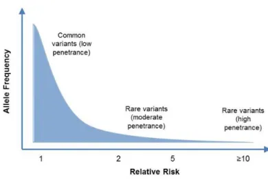
focus our analysis on rare, highly penetrant variants, variants with a maximum allele
Figure 2. Analysis was focused on rare, highly penetrant variants. Given the low incidence of most hereditary cancer syndromes in the population, common variants are unlikely to contribute to hereditary cancer predisposition.
Online resources for variant classification included The National Center for
Biotechnology Information ClinVar database (http://www.ncbi.nlm.nih.gov/clinvar/), the
Leiden Open-Source Variation Database (LOVD, http://www.lovd.nl/2.0/index_list.php),
and the Catalogue of Somatic Mutations in Cancer (COSMIC,
http://cancer.sanger.ac.uk/cosmic). COSMIC was used to determine if a variant existed in
tumors from similar tissues of origin. After a preliminary computational classification,
variant counts were generated using an in-house python script and validated manually.
Variants underwent tiered review by trained molecular analysts in conjunction with
discussion in a multidisciplinary group. Evidence curation and variant classification was
performed in a manner similar to the more recently published guidelines from the
American College of Medical Genetics and Genomics and the Association for Molecular
suggestive of a hereditary cancer predisposition, this phenotype information was not
available during the variant review process. Therefore, the molecular analysts utilized an
incidental or secondary variant analysis approach such that a high threshold for
pathogenicity must be met for variant result. The medical and family history presented in
Table 2 was obtained from medical record review after variant analysis. Following
stringent review, variants classified as Likely Pathogenic or Known Pathogenic were
identified as eligible for return to patients. Prior to results return, these variants will be
confirmed through analysis of a new sample via an orthogonal method (e.g. Sanger
sequencing) and verified by an American Board of Medical Genetics and Genomics
(ABMGG)-certified molecular pathologist. The confirmation step was ongoing at the
time of submission. Once confirmed, the hereditary cancer predisposing variants will be
returned to the patients through a board certified genetic counselor experienced in
hereditary cancer. When medical record review documented a clinically known
hereditary cancer predisposing variant, no additional steps for confirmation and results
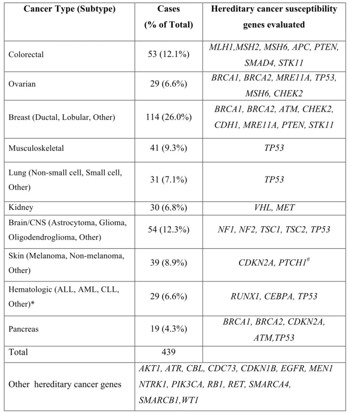
Cancer Type (Subtype) Cases (% of Total)
Hereditary cancer susceptibility genes evaluated
Colorectal 53 (12.1%) MLH1,MSH2, MSH6, APC, PTEN,
SMAD4, STK11
Ovarian 29 (6.6%) BRCA1, BRCA2, MRE11A, TP53,
MSH6, CHEK2
Breast (Ductal, Lobular, Other) 114 (26.0%) BRCA1, BRCA2, ATM, CHEK2,
CDH1, MRE11A, PTEN, STK11
Musculoskeletal 41 (9.3%) TP53
Lung (Non-small cell, Small cell,
Other) 31 (7.1%) TP53
Kidney 30 (6.8%) VHL, MET
Brain/CNS (Astrocytoma, Glioma,
Oligodendroglioma, Other) 54 (12.3%) NF1, NF2, TSC1, TSC2, TP53
Skin (Melanoma, Non-melanoma,
Other) 39 (8.9%) CDKN2A, PTCH1
#
Hematologic (ALL, AML, CLL,
Other)* 29 (6.6%) RUNX1, CEBPA, TP53
Pancreas 19 (4.3%) BRCA1, BRCA2, CDKN2A,
ATM,TP53
Total 439
Other hereditary cancer genes
AKT1, ATR, CBL, CDC73, CDKN1B, EGFR, MEN1 NTRK1, PIK3CA, RB1, RET, SMARCA4,
SMARCB1,WT1
*Hematologic cancer abbreviations: Acute Lymphoblastic Leukemia (ALL), Acute Myeloid Leukemia (AML), Chronic Lymphocytic Leukemia (CLL).
#PTCH1 variants were considered relevant only in skin cancer cases that were of the
non-melanoma type.
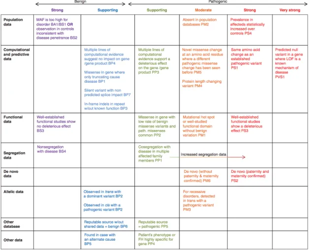
Figure 3. Evidence framework. This chart published by the American College of Medical Genetics organizes each of the criteria by the type of evidence as well as the strength of the criteria for a benign (left) or pathogenic (right) assertion.
RESULTS
To assess the frequency of pathogenic variants in a group of unselected cancer
patients undergoing tumor sequencing, we analyzed germline variants from 439 patients
ascertained through the UNCseq™ study. Although all 247 genes of the UNCseq™ panel
were sequenced, we specifically investigated germline variants in 36 genes that were
present on the somatic sequencing panel. Based on current knowledge about the
spectrum of cancers associated with these hereditary cancer syndromes, 24 of the 36
genes were considered concordant with the cancer types of the patients being analyzed
(Table 1). These cancers included colorectal, ovarian, breast, musculoskeletal, lung,
kidney, brain/CNS, melanoma, hematologic, and pancreatic cancers12, 15-20. Of all cases
examined, 19/439 (4.3%) had pathogenic germline variants in a hereditary cancer
predisposing gene. Of these, 12 were in genes concordant with the presenting cancer at
enrollment and 7 were in other hereditary cancer genes (Figure 4, Table 2). The majority
of these findings occurred in patients with colorectal, ovarian, breast, and pancreatic
cancers; very few such findings occurred in patients with musculoskeletal, lung, kidney,
brain, skin, or hematologic malignancies.
Overall, BRCA1 and BRCA2 harbored 11/19 (57.9%) of the pathogenic variants,
the majority of which were classified Likely Pathogenic because they were novel variants
expected to result in an early truncation or for which existing evidence suggested a
pathogenic role based on classification guidelines10 (Figure 5). As would be expected in
an unselected cancer patient population, a small percentage had previously undergone
clinical genetic assessment for hereditary cancer predisposition. Medical record review
following variant classification revealed that the BRCA1/2 variants identified in breast
and ovarian cancer patients in this study had all been previously identified through
routine clinical genetic testing, indicated based on medical and family history21. The
breast cancer patients that would be missed in individuals whose clinical testing was
restricted to BRCA1 and BRCA222-23.
The table in Appendix 1 shows a clinical summary of UNCseq™ patients with
germline pathogenic variants. Some patients had a personal history of cancer consistent
with the variant identified in the germline analysis, but had been enrolled for cancers that
were presumably unrelated (Table 2). For example, Patient 11 was previously diagnosed
with breast cancer at age 41 and was enrolled in UNCseq™ when diagnosed with
colorectal cancer at age 49. She was found to have a pathogenic canonical splice site
variant in BRCA1 (NM_007294.3:c.594-2A>C) that provides an explanation for her breast cancer (Appendix 1), but is not likely to have any relevance to her colorectal
cancer diagnosis. Similarly, Patient 17 was previously diagnosed with breast cancer at
age 52, but was enrolled in the UNCseq™ study for non-small cell lung cancer. She was
found to have a pathogenic nonsense variant in ATM (NM_000051.3:c.352C>T,
(p.Gln118Ter)) (Appendix 1) that provides a plausible explanation for her breast cancer
and could potentially suggest a role of ATM in lung cancer, although previous studies
have found limited evidence to support this role or suggested that environmental factors
may contribute much more significantly.
Although relatively few patients had clearly pathogenic variants, 178/439 (40.5%)
had a germline Variant of Uncertain Significance (VUS) (Figure 6). In 24 patients, a
VUS was found in a gene relevant to the presenting cancer type, while 143 patients had a
VUS in hereditary cancer genes unrelated to their cancer type. Not surprisingly, 11
Figure 5. Germline pathogenic variants identified in all UNCseqTM patients. The numbers of Known Pathogenic (KP) and Likely Pathogenic (LP) variants across all UNCseq™ patients analyzed are depicted as a bar graph, divided by gene.
Figure 6A Variants occurring in genes relevant to the patient’s cancer diagnosis.
DISCUSSION
Frequency of Pathogenic Germline Variants
This thesis explores the yield of clinically relevant findings from germline
analysis in patients undergoing tumor-germline dyad sequencing. The proportion of
patients found harboring a pathogenic germline mutation (4.3%) is consistent with the
range reported in previous hereditary cancer studies3, 24-25. It should be noted that this
frequency is only an estimate, and may vary based on which genes are included in the
capture panel as well as the cancer types considered. For example, a similar exploratory
study conducted by Schrader et al. 20164 reported a frequency of 12% of cancer patients sequenced via tumor-germline dyad sequencing harboring germline mutations. However,
Schrader and colleagues4 expanded the survey to include 187 genes associated with
Mendelian diseases while also including a wider array of tumor types than this study (e.g.
prostate cancer, thyroid cancer, liver cancer, etc.). If the data published by Schrader and
colleagues4 were limited to the same cancer types and hereditary cancer predisposition genes in our current analysis, a pathogenic variant would be found in 3.9% of cases,
which is consistent with our findings. This also demonstrates a limitation in our analysis
in that only hereditary cancer predisposition genes in patients from 10 major cancer types
were considered. Future studies should aim to ascertain the frequency of incidental
germline findings in other Mendelian disorders besides hereditary cancer syndromes, as
these incidental findings may also reveal information critical diagnostic information
about a patient.
Moreover, the distribution of pathogenic variants across cancer types (Figure 4)
the patient cohort, the majority of pathogenic variants were in patients with breast and
ovarian cancer, which are tumor types that both have well-documented genetic etiologies.
Perhaps more surprisingly, none of the patients diagnosed with colorectal cancer were
found to have a mutation in a gene associated with Lynch Syndrome, which accounts for
roughly 3 to 5% of all colorectal cancer cases. However, this is likely due to the relatively
small number of colorectal cancer cases evaluated (N = 53). In tumor types with
lesser-known genetic etiologies (i.e. musculoskeletal, lung, kidney, brain/CNS, skin,
hematologic cancers), no pathogenic germline variants were found as expected.
Furthermore, while the majority of pathogenic germline findings occurred in
genes that were concordant with the presenting cancer at enrollment, a significant portion
(7/19, or 36.8%) of the pathogenic variants were found in discordant genes – meaning
that either the mutation was found in a gene that was concordant with another cancer type
or in one of the “Other hereditary cancer genes” listed in Table 1. For example, a BRCA1
pathogenic variant was found in patient 11 with colorectal cancer, BRCA2 in patient 15 with AML, and so on (Appendix 1). The vast majority of the discordant findings occurred
in patients who had cancer types with lesser-known genetic etiologies, such as acute
myeloid leukemia, non-small cell lung cancer, and musculoskeletal cancer. These
incidental findings
Finally, among the 19 patients with positive findings in a hereditary cancer
predisposition gene, half of the pathogenic variants had previously been identified
through clinical evaluation. The other half, representing roughly ~2% of patients in the
entire cohort, were not associated with any prior clinical cancer genetic evaluation. For
that was not otherwise known to the patient and their family, enabling potential lifesaving
interventions26-27. Identifying pathogenic germline variants could also provide important prognostic information, guiding surgical procedures and or targeted therapeutic options
for the individual cancer patient, thereby providing immediate applications2, 28. However, we recognize that such unexpected germline susceptibility information might be
unwelcome to some patients depending on their personal situation or preference for
information. Therefore, further long-term follow-up is needed in order to assess what
portion of patients would ultimately welcome and benefit from such information.
Variants of Uncertain Significance
Often, variants of uncertain significance (VUS) are returned to patients after the
diagnostic evaluation of hereditary cancer risk29. However, when tumor-germline sequencing is performed for prognostic or therapeutic indications, the identification of
germline variation would be considered an incidental or secondary finding. In this
situation, as per the evidence-based guidelines published by the American College of
Medical Genetics and Genomics10, only Pathogenic or Likely Pathogenic findings should be reported to patients. This notion is supported by our data, in which we discovered at
least one VUS in almost half (40.5%) of all patients. By definition, the clinical relevance
of these variants remains to be determined. Based on the low prior probability of clinical
CONCLUSION
Though it is a major focus of precision medicine efforts, whether next generation
sequencing should be applied on a routine basis for tumor mutation profiling remains to
be determined30. Here, we demonstrate that utilizing an incidental/secondary variant analysis approach for germline sequence data in unselected patients undergoing
tumor-germline sequencing may provide a small but important benefit with regard to the
detection of clinically relevant, highly penetrant variants in hereditary cancer
predisposition genes. Most of these findings can be ascertained through cancer genetics
evaluation recommended on the basis of family history, age at presentation, ancestry or
tumor phenotype. However, some of these patients may not be referred to a cancer
genetic service31-34 and a minority will be missed due to lack of typical clinical and/or
family history indications35-36.
Potentially unsuspected pathogenic variants have now been reported in a small,
but not insignificant, proportion of cancer patients undergoing therapeutically indicated
tumor-germline testing4,6, and our data provide further support to this scenario. Disclosing the identification of a hereditary cancer predisposition would be highly
relevant to the clinical care of these cancer patients and have important implications for
their relatives’ medical guidance. Providers who obtain tumor sequencing will need to be
cognizant of the implications of tumor-germline analysis with respect to potential
incidental findings37, understand the differences between tumor sequencing and clinical genetic testing for hereditary cancer susceptibility, and be able to effectively
REFERENCES
1. American Cancer Society. Cancer Facts & Figures 2015. Atlanta: American Cancer Society; 2015.
2. Roychowdhury S, Iyer MK, Robinson DR, Lonigro RJ, Wu Y-M, Cao X, et al. Personalized oncology through integrative high-throughput sequencing: a pilot study. Sci Transl Med. 2011;3:111ra121.
3. Jones S, Anagnostou V, Lytle K, Parpart-Li S, Nesselbush M, Riley DR, et al. Personalized genomic analyses for cancer mutation discovery and interpretation. Sci Transl Med. 2015;7:283ra53–283ra53.
4. Schrader KA, Cheng DT, Joseph V, Prasad M, Walsh M, Zehir A, et al. Germline Variants in Targeted Tumor Sequencing Using Matched Normal DNA. JAMA Oncol. 2016;2:104–11.
5. Schlussel AT, Gagliano RA, Seto-Donlon S, Eggerding F, Donlon T, Berenberg J, et al. The evolution of colorectal cancer genetics—Part 1: from discovery to practice. J Gastrointest Oncol. 2014;5:326–35.
6. Meric-Bernstam F, Brusco L, Daniels M, Wathoo C, Bailey A, Strong L, et al. Incidental germline variants in 1000 advanced cancers on a prospective somatic genomic profiling protocol. Ann Oncol. 2016.
7. Jeck WR, Parker J, Carson CC, Shields JM, Sambade MJ, Peters EC, et al. Targeted next generation sequencing identifies clinically actionable mutations in patients with melanoma. Pigment Cell Melanoma Res. 2014;27:653–63.
8. Zhao X, Wang A, Walter V, Patel NM, Eberhard DA, Hayward MC, et al. Combined Targeted DNA Sequencing in Non-Small Cell Lung Cancer (NSCLC) Using UNCseq and NGScopy, and RNA Sequencing Using UNCqeR for the Detection of Genetic Aberrations in NSCLC. PLoS ONE. 2015;10:e0129280.
9. Garrison E, Marth G. Haplotype-based variant detection from short-read sequencing. arXiv [Internet]. 2012 [cited 2015 Jul 22];1207.3907v2. Available from: http://arxiv.org/pdf/1207.3907.pdf
10. Richards S, Aziz N, Bale S, Bick D, Das S, Gastier-Foster J, et al. Standards and guidelines for the interpretation of sequence variants: a joint consensus
recommendation of the American College of Medical Genetics and Genomics and the Association for Molecular Pathology. Genet Med. 2015;17:405–23.
12. Economopoulou P, Dimitriadis G, Psyrri A. Beyond BRCA: New hereditary breast cancer susceptibility genes. Cancer Treatment Reviews. 2015;41:1–8.
13. Raczy C, Petrovski R, Saunders CT, Chorny I, Kruglyak S, Margulies EH, et al. Isaac: ultra-fast whole-genome secondary analysis on Illumina sequencing platforms. Bioinformatics. 2013;29:2041–3.
14. Evans DG. Neurofibromatosis 2. In: Pagon RA, Adam MP, Ardinger HH, Wallace SE, Amemiya A, Bean LJ, et al., editors. GeneReviews(®) [Internet]. Seattle (WA): University of Washington, Seattle; 1993 [cited 2015 May 18]. Available from: http://www.ncbi.nlm.nih.gov/books/NBK1201/
15. Walsh T, Casadei S, Lee MK, Pennil CC, Nord AS, Thornton AM, et al. Mutations in 12 genes for inherited ovarian, fallopian tube, and peritoneal
carcinoma identified by massively parallel sequencing. Proc Natl Acad Sci USA. 2011;108:18032–7.
16. Friedman JM. Neurofibromatosis 1. In: Pagon RA, Adam MP, Ardinger HH, Wallace SE, Amemiya A, Bean LJ, et al., editors. GeneReviews(®) [Internet]. Seattle (WA): University of Washington, Seattle; 1993 [cited 2015 May 18]. Available from: http://www.ncbi.nlm.nih.gov/books/NBK1109/
17. Northrup H, Koenig MK, Au K-S. Tuberous Sclerosis Complex. In: Pagon RA, Adam MP, Ardinger HH, Wallace SE, Amemiya A, Bean LJ, et al., editors. GeneReviews(®) [Internet]. Seattle (WA): University of Washington, Seattle; 1993 [cited 2015 May 18]. Available from:
http://www.ncbi.nlm.nih.gov/books/NBK1220/
18. Song WJ, Sullivan MG, Legare RD, Hutchings S, Tan X, Kufrin D, et al.
Haploinsufficiency of CBFA2 causes familial thrombocytopenia with propensity to develop acute myelogenous leukaemia. Nat Genet. 1999;23:166–75.
19. Ghiorzo P. Genetic predisposition to pancreatic cancer. World J Gastroenterol. 2014;20:10778–89.
20. Matloff E. Cancer Principles and Practice of Oncology: Handbook of Clinical Cancer Genetics. Philadelphia, PA: Lippincott Williams & Wilkins; 2013.
21. Hampel H, Bennett RL, Buchanan A, Pearlman R, Wiesner GL. A practice guideline from the American College of Medical Genetics and Genomics and the National Society of Genetic Counselors: referral indications for cancer
22. LaDuca H, Stuenkel AJ, Dolinsky JS, Keiles S, Tandy S, Pesaran T, et al. Utilization of multigene panels in hereditary cancer predisposition testing: analysis of more than 2,000 patients. Genet Med. 2014;16:830–7.
23. Minion LE, Dolinsky JS, Chase DM, Dunlop CL, Chao EC, Monk BJ. Hereditary predisposition to ovarian cancer, looking beyond BRCA1/BRCA2. Gynecol Oncol. 2015;137:86–92.
24. Green RC, Berg JS, Grody WW, Kalia SS, Korf BR, Martin CL, et al. ACMG recommendations for reporting of incidental findings in clinical exome and genome sequencing. Genet Med. 2013;15:565–74.
25. Schrader KA, Cheng DT, Joseph V, et al. Germline variants in targeted tumor sequencing using matched normal DNA. JAMA Oncol. 2015;1–8.
26. Bombard Y, Robson M, Offit K. Revealing the incidentalome when targeting the tumor genome. JAMA. 2013;310:795–6.
27. Parsons DW, Roy A, Plon SE, Roychowdhury S, Chinnaiyan AM. Clinical tumor sequencing: an incidental casualty of the American College of Medical Genetics and Genomics recommendations for reporting of incidental findings. J Clin Oncol. 2014;32:2203–5.
28. Rahman N. Realizing the promise of cancer predisposition genes. Nature. 2014;505:302–8.
29. Plon SE, Eccles DM, Easton D, Foulkes WD, Genuardi M, Greenblatt MS, et al. Sequence variant classification and reporting: recommendations for improving the interpretation of cancer susceptibility genetic test results. Hum Mutat.
2008;29:1282–91.
30. Tripathy D, Harnden K, Blackwell K, Robson M. Next generation sequencing and tumor mutation profiling: are we ready for routine use in the oncology clinic? BMC Med. 2014;12:140.
31. Mai PL, Vadaparampil ST, Breen N, McNeel TS, Wideroff L, Graubard BI. Awareness of cancer susceptibility genetic testing: the 2000, 2005, and 2010 National Health Interview Surveys. Am J Prev Med. 2014;46:440–8.
32. van Riel E, van Dulmen S, Ausems MGEM. Who is being referred to cancer genetic counseling? Characteristics of counselees and their referral. J Community Genet. 2012;3:265–74.
34. McCarthy AM, Bristol M, Fredricks T, Wilkins L, Roelfsema I, Liao K, et al. Are physician recommendations for BRCA1/2 testing in patients with breast cancer appropriate? A population-based study. Cancer. 2013;119:3596–603.
35. Karageorgos I, Mizzi C, Giannopoulou E, Pavlidis C, Peters BA, Zagoriti Z, et al. Identification of cancer predisposition variants in apparently healthy individuals using a next-generation sequencing-based family genomics approach. Hum Genomics. 2015;9:12.
36. Amendola LM, Dorschner MO, Robertson PD, Salama JS, Hart R, Shirts BH, et al. Actionable exomic incidental findings in 6503 participants: challenges of variant classification. Genome Res. 2015;25:305–15.
APPENDIX 1:
Clinical summary of UNCSeq
TMpatients with germline pathogenic variants
Patient Cancer type at Enrollment
Sex Agea Gene cDNA changeb Protein
change
Variant type Classification Clinical Genetics Evaluation Personal and/or Family History Prior Clinical Testing Clinical Test Resultc Concordance with Cancer at
Enrollmentd
1 Ovarian F 48 BRCA1 NM_007294.3:
c.5266_5267insC
p.(Gln1756fs) Frameshifting
indel
KP Yes Ashkenazi
Jewish F:Lung, Bladder MA: Pancreas, 72
Yes (+) Yes
2 Ovarian F 41 BRCA1 NM_007294.3:
c.5193+1G>T
N/A Splice-site KP Yes No Cancer
History
Yes (+) Yes
3 Breast F 37 CDKN2A NM_000077.4:
c.35C>A
p.(Ser12Ter) Nonsense LP Yes Personal:
Melanoma, 21,31 Family: Adopted
Yes (-) No
4 Breast F 55 BRCA1 NM_007294.3:
c.2457_2457delC
p.(Asp821fs) Frameshifting
indel
KP Yes MMaR:Breast,
Ovarian, Pancreas M:Breast, 36
Yes (+) Yes
5 Breast F 29 BRCA1 NM_007294.3:
c.211A>G
p.(Arg71Gly) Missense KP Yes S:Breast, 28;
M:Breast,42 MA:Breast,33; MA:Breast,37
Yes (+) Yes
6 Breast F 63 ATM NM_000051.3:
c.1561_1562delAG p.(Glu522fs) Frameshifting indel LP Yes M:Breast,55; MA:Breast,30s&
40s 3MU:Blood MA:Cancer,60s
Yes (-) Yes
7 Breast F 29 BRCA2 NM_000059.3:
c.7538_7539insA
p.(Thr2515fs) Frameshifting
indel
LP Yes M:Breast,
39&49, and Brain/CNS, 58
Yes (+) Yes
8 Breast F 35 BRCA1 NM_007294.3:
c.131G>A
p.(Cys44Tyr) Missense KP Yes PA: Bilateral
Breast, 45
Yes (+) Yes
9 Breast F 37 BRCA2 NM_000059.3:
c.8575delC
p.(Gln2859fs) Frameshifting
indel
KP Unknown Unknown
(adopted)
a"Age"at"the"time"of"diagnosis"
b Transcripts are listed according to the HGVS nomenclature recommendations or the commonly accepted transcript.
c Clinical Test Result: (+) = Same Mutation Reported, (-) = Gene was not included in the clinical genetic test and these negative
results indicate new diagnostic results.
10 Breast M 53 BRCA2 NM_000059.3:
c.5718_5719delCT p.(Leu1908fs) Frameshifting indel LP Yes S:Breast,50; B:Colon,53 PA:Breast, 55; PC:Colon, 35 Known Familial Mutation
Yes (+) Yes
11 Colorectal F 49 BRCA1 NM_007294.3:
c.594-2A>C N/A Splice-site LP Yes Personal: Breast, 41
Family: PA: Breast, 29; PC: Breast, 50
Yes (+) No
12 AML M 54 BRCA1 NM_007294.3:
c.594-2A>C
N/A Splice-site LP Unknown Not Reported No N/A No
13 GI-other M 54 ATM NM_000051.3:
c.8545C>T
p.(Arg2849Ter) Nonsense LP No No Cancer
History
No N/A No
14 Breast F 59 CHEK2 NM_007194.3:
c.1100delC
p.(Thr367fs) Frameshifting indel
KP No
Non-Contributory M:Lymph node, 80
U: Liver
No N/A Yes
15 AML M 57 BRCA2 NM_000059.3:
c.5233_5233delA
p.(Met1745fs) Frameshifting indel
LP No F:Pancreas, 72 No N/A No
16 Pancreas M 61 ATM NM_000051.3:
c.170G>A
p.(Trp57Ter) Nonsense LP Yes B:Pancreas, 52 No N/A Yes
17 NSCLC F 66 ATM NM_000051.3:
c.352C>T
p.(Gln118Ter) Nonsense LP No Personal:
Breast, 52 Family: S:Breast, F: Bone (myeloma), B:Amyloidosis
No N/A No
18 Musculoskeletal F 57 CHEK2e NM_007194.3: c.1100delC
p.(Thr367fs) Frameshifting indel
KP No F:Kidney
PGM:Lung, B:CNS (2)
No N/A No
19 Ovarian F 52 CHEK2 NM_007194.3:
c.1486C>T
p.(Gln496Ter) Nonsense LP No Personal:
Melanoma, 57 Family: PU: Stomach
d Concordance with cancer at enrollment: Yes = Pathogenic variant is in a gene that is concordant with the presenting cancer at
enrollment. No = Pathogenic variant is in a gene that is discordant with the presenting cancer at enrollment.
e CHEK2 has been implicated as a susceptibility gene for a Li-Fraumeni-like cancer syndrome. However, the current evidence for this
association is disputed(50).
Abbreviations:
Cancer type: AML=Acute Myelogenous Leukemia; NSCLC= Non-small cell lung cancer.
Gender: F= female; M= male. Classification: KP= Known Pathogenic; LP= Likely Pathogenic; VUS= Variant of Uncertain Significance.
Family History: M=Mother; F=Father; S=Sister; B=Brother; MA= Maternal Aunt; MU= Maternal Uncle; MGM= Maternal Grandmother; MGF= Maternal Grandfather; MMaR=Multiple Maternal Relatives; PA= Paternal Aunt; PU= Paternal Uncle; PGF=Paternal Grandfather; PGM=Paternal Grandmother; PC= Paternal Cousin; U=Uncle.