By
Carson Elizabeth Rouse
Senior Honors Thesis Curriculum in Archaeology
University of North Carolina at Chapel Hill 2016
Approved:_________________________ Dr. Dale H. Hutchinson, Thesis Advisor Dr. C. Margaret Scarry, Reader
1. Piedmont North Carolina and Southern Virginia
Piedmont of North Carolina and Southern Virginia ………….……..………. 1
Geographic Background for Piedmont North Carolina and Southern Virginia...……..4
Maize Agriculture in the Piedmont………..………..4
Historic Account of the Piedmont ……….………....5
Archaeological Background……….6
Archaeological Sample ……….………...8
Town Creek Indian Mound (Mg2-3)………....8
Stockton (Vir231) ………..……….10
Fredricks (Or231) ………..………..11
Materials and Methods ………..………..12
2. Osteological Evidence for Changes in Health Metabolic Stress Indicators ………..……...15
Porotic Hyperostosis and Cribra Orbitalia………..….15
Scurvy………..………16
Rickets ………..………...18
Oral Health………..……….20
Dental Caries………..……..20
Periodontal Disease and Ante-Mortem Tooth-Loss………..………..21
Dental Enamel Hypoplasia ………..………...23
Infectious Disease ………..……….25
Sinusitis………..………..25
Periosteal Reaction………..……….25
Osteomyelitis………..……….26
Treponemal Infection………...………27
3. Results Osteological Evidence of Metabolic Disease in the Piedmont…………..…………..30
Cribra Orbitalia………..………..30
Porotic Hyperostosis………..………..31
Scurvy…………..………33
Rickets…..………36
Osteological Evidence of Changes to Oral Health Environments in the Piedmont….38 Dental Caries………..………..38
Periodontal Disease and Ante-Mortem Tooth Loss………..………...39
Alveolar Infection…………..………..41
Osteological Evidence of General Stress Indicators in the Piedmont…………..……44
Dental Enamel Hypoplasia………..………44
Osteological Evidence of Infectious Diseases in the Piedmont……….……….45
Sinusitis ……….……….45
Periosteal Reaction……….46
4. Effect of Changing Diet on the Native Populations of the Piedmont
Metabolic Diseases………53
Oral Health……….58
General Changes in Health………..…..60
List of Tables 1. Age Categories ………..……..13
2. Frequency of Metabolic Bone Disease………30
3. Frequency of Cribra Orbitalia………..31
4. Individuals with Cribra Orbitalia ………31
5. Frequency of Porotic Hyperostosis………..32
6. Individuals with Porotic Hyperostosis……….33
7. Frequency of Scurvy………35
8. Individuals with Scurvy………36
9. Frequency of Rickets………...37
10. Frequency of Dental Carious Lesions………..38
11. Frequency of Ante-Mortem Tooth Loss………..39
12. Individuals with Ante-Mortem Tooth Loss……….40
13. Frequency of Alveolar Infection………..41
14. Individuals at Town Creek (Mg2-3) with Alveolar Infections ………...42
15. Individuals at Stockton (Vir231) with Alveolar Infections………...42
16. Individuals at Fredricks (Or231) with Alveolar Infection………...43
17. Frequency of Dental Enamel Hypoplasias………44
18. Individuals with Dental Enamel Hypoplasias………44
20. Individuals with Sinusitis………...45
21. Frequency of Periosteal Reactions……….46
22. Individuals with Periosteal Reactions………....47
23. Frequency of Osteomyelitis………...48
24. Individuals with Osteomyelitis………..48
25. Comparative Studies………..54
List of Figures 1. Location of Archaeological Sites from Lambert 2000, 173………..………..7
2. Mg2-3 Individual 10 with scorbutic lesions on eye orbits………..………...34
3. Mg2-3 Individual 36 with scorbutic lesions on the parietal.…………..………...34
4. Mg2-3 Individual 4 with scorbutic lesions on eye orbits……..……….…34
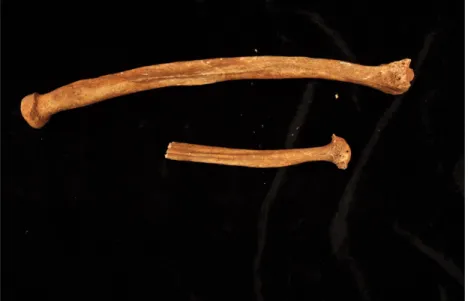
5. Vir231 Individual 4 with bowed radii ………..……….…37
6. Vir231 Individual 4, X-ray of bowed radii…………..…………...………...37
7. Vir231 Individual 13 with Ante-Mortem Tooth Loss………..………...41
8. Vir231 Individual 13 with Osteomyelitis of right femur………..……….49
9. Vir231 Individual 13 with osteomyelitis of tibiae with cloaca..………….………...49
10. Vir231 Individual 8 with osteomyelitis of fibula, tibia, and humerus…...………50
11. Vir231 Individual 15 with stellate lesions and nodes on mandible ………..……51
12. Vir231 Individual 8 with stellate lesions………..……….52
Appendices I. Individuals with Cavities………69
I would like to thank my advisor, Dr. Dale L. Hutchinson, for his constant support, insight, and guidance throughout this process; my committee members Dr. C. Margaret Scarry and Dr. R.P. Stephen Davis, Jr; my family for their constant love and patience through all of the highs and the lows; the Dunlevie Honors Undergraduate Research Award for their generous financial support which funded the radiographs; and Crystal Martin for her
ABSTRACT
Carson Elizabeth Rouse: Nutrition and Health in the Piedmont of North Carolina and Virginia: A Bioarchaeological Study
Nutrition and Health in the Piedmont of North Carolina and Virginia
Chapter One: Piedmont North Carolina and Southern Virginia
Piedmont of North Carolina and Southern Virginia
According to the North Carolina Commission of Indian Affairs, there were more than 80,000 Indian People living in North Carolina as of 2001 which is about 1.2% of the state’s overall population. The majority of the Indian population (39%) resides in Robeson County which is located in the Piedmont region of the state. Although much is known historically about current Indian inhabitants of the Piedmont, little is known about the past native inhabitants of this area (Price 2001, xii-xiii). According to James H. Merrell, the Piedmont natives were peoples who historically “lived and died in obscurity, an obscurity that, for the most part, has continued to this day” demonstrating the lack of information pertaining to the native peoples of Piedmont North Carolina and Southern Virginia (Merrell 1987, 20).
The bioarchaeology of metabolic diseases can reveal a great deal about the lives of past peoples (Brickley and Ives 2008, 1). Brickley and Ives, in their 2008 book, The
Bioarchaeology of Metabolic Bone Disease, define metabolic bone disease as bone modeling
and remodeling that is disrupted due to disease. Studying these changes in bone growth and regrowth can reveal concepts such as the lifestyle, culture, diet, socio-economic status, and environments of past peoples (Brickley and Ives 2008, 1).
In addition to metabolic bone disease, I also examined oral health changes and general stress indicators because they could indicate underlying changes in health due to diet during specific time periods. Changes in the oral health environment can indicate changes in diet and nutrition in past peoples. General stress indicators are skeletal and dental lesions for which the exact etiology may be unknown. They differ from metabolic disease indicators because their origin may or may not be related back to nutrition and diet but instead can be linked back to sources such as genetics. They include periosteal reactions, osteomyelitis, and enamel hypoplasias. Although the exact cause of these lesions in the human body varies, there is abundance of evidence that they increase in frequency with urbanization and with the transition to agriculture (Roberts and Manchester 2005, 173).
A prime example of this increase in frequency can be seen in Clark Spencer Larsen’s study of prehistoric populations from the Georgia Coast (Larsen 1984, 367). He focused on disease-nutritional stress by looking for periosteal reactions and dental caries, and
primarily relied on hunting, gathering, and fishing for their main sources of food. After A.D. 1150, they began to cultivate crops, primarily maize. By comparing these groups for
evidence of disease-nutritional stress and mechanical stress, Larsen concluded that there was a decline in health with the switch from preagricultural to agricultural groups evidenced by the increase in periosteal reactions (Larsen 1984, 387-388). Based on these three categories, I hypothesis that the overall health of the individuals increased during the historic period due to contact with the Europeans affecting their foodways and lifestyles.
In 2000, Patricia M. Lambert published a study of 649 individuals from thirteen sites throughout the Piedmont of North Carolina and Virginia as part of a NAGPRA inventory project, including Town Creek Indian Mound, Stockton, and Fredricks (Lambert 2000, 169). She observed dental caries, cribra orbitalia, scurvy, iron-deficiency anemia, periosteal reactions, treponematosis, tuberculosis, and enamel hypoplasias (Lambert 2000, 174).
She noted that there was an increase in dental caries during the prehistoric period, but a decrease in dental caries during the protohistoric and historic periods. She also noted that the frequency of infectious diseases such as treponemotosis and tuberculosis decreased after contact with Europeans (Lambert 2000, 192). In contrast to these results, the number of enamel hypoplasias increased during the Contact Period (Lambert 2000, 185). Based on these varied results, Lambert concluded that, “variation within this region further suggest that variables such as resource distribution, quality of arable land, microclimatic variability, and unique cultural practices influenced heath and thus the quality of life in various regions of North Carolina and Virginia” (Lambert 2000, 192).
are frequently associated with diet and nutrition. I further place those data within the context of cultural change, and distribution brought about by the colonial process.
Geographic Background for Piedmont North Carolina-Southern Virginia
Piedmont North Carolina is located between the coastal plain and the Blue Ridge Mountains. Piedmont Virginia is located between the Potomac River and the Blue Ridge Mountains. The Piedmont is a “highly dissected plateau that contains some 20,000 square miles” (Ward 1983, 53-54). Its elevation spans from 400 to 2,000 feet above sea level. The landscape of this area consists of rolling hills and ridges that run from the northeast to the southwest. Although the topography is fairly consistent throughout the Piedmont, there are a few abnormalities such as Kings Mountain in Cleveland and Gaston counties, North
Carolina. A plethora of resources were available to the native inhabitants of this area
including an abundant lithic supply, abundant plant and animal resources, many waterways to aid in travel and trade, and fertile soil for growing crops (Ward 1983, 54-56).
Maize Agriculture in the Piedmont
A study published in 1996 by Trimble demonstrated that stable isotope carbon levels varied across North Carolina through time. For example, the Donnaha site (A.D.1040-1480) had carbon values of about 15.8% while the Koehler site (A.D. 1300-1400) in Virginia showed levels of carbon at about 19.1% (Trimble 1996 cited in Lambert 2000, 169). Based on Trimble’s results, it is apparent that there was an increasing reliance on maize during the prehistoric period in the Piedmont. However, “there is some archaeological evidence for a return to a more mixed economy perhaps as a result of fur trade activities, in the
protohistoric/ historic period” [Ward and Davis 1993, 1999 cited in Lambert 2000, 170). Historic Account of the Piedmont
Although there is only some early historic information pertaining to the indigenous peoples of the Piedmont, one of the most famous historic accounts of the Piedmont is contained in John Lawson’s 1709 book: “A New Voyage to Carolina.” Little is known of Lawson’s personal background until he journeyed to the New World in 1700. Lawson states that a gentleman had informed him that “Carolina was the best Country I could go to; and, that there then lay a Ship in the Thames, in which I might have my passage. I laid hold on this Opportunity, and was not long on Board” (Lefler 1967, 7).
miles; however, recent scholarly work indicates that he only travelled about 550 miles during this trip (Lefler 1967, xv).
As he traveled, Lawson kept a detailed journal including diary entries, notes about the flora and fauna, and translations of the native language. In the chapter, “An Account of the Indian of North Carolina,” Lawson described the lives of the native peoples he encountered on his journey by explaining their customs, burial practices and foodways. He lists the wide variety of foods that they ate including “Bear and Bever; Panther; Pole-cat; Wild-cat; Possum; Raccoon; Hares; and Squirrels” (Lefler 1967,182). In addition, he emphasized the use of shell-fish and gourds as well as their reliance on flora such as maize, potatoes, acorns (Lefler 1967,182). According to Lawson, there were “no Indians having greater plenty of Provisions than these. The Savages do, indeed, still possess the Flower of Carolina,” (Lefler 1967, 61).
Based on Lawson’s observational notes, it appears that resources were abundant and were readily available for the native populations to achieve an adequate diet. This
maintenance of an adequate diet would hinder the occurrence of metabolic bone diseases in the populations because they stem from inadequate nutrition. Although Lawson’s history paints a crude picture of the lives of the native inhabitants of North Carolina during the turn of the 18th century, the osteological information reported in this paper provides additional information regarding the lives and diets of the past native populations of North Carolina.
Archaeological Background
According to some scholars, North Carolina archaeology truly commenced when archaeologists began to excavate the Piedmont in the early 1930s. The North Carolina
1934. The Piedmont was divided in those early years into zones to be surveyed by individuals including Douglas Rights, Guy B. Johnson, and Joffre Coe. The main goal of these initial surveying expeditions was to learn more information and find historic towns associated with the Siouan groups such as the Keyauwee site located in Randolph County, North Carolina.
Following World War II, the focus of archaeological work in the Piedmont shifted to attempting to find earlier sites (Ward 1983, 57-59). And finally, in the 1970s another major archaeological shift occurred in North Carolina. The “passage of national conservation legislation emphasizing cultural resource management (CRM)” began to be the main focus of archaeology (Ward 1983, 59). Researchers began to focus on survey and settlement studies with the main excavations being test operations (Ward 1983, 59-60). The three sites
described below (chosen for this study) serve witness to the archaeological efforts that were an outgrowth of that early research.
Archaeological Sample
Town Creek Indian Mound (Mg2-3)
Town Creek Indian Mound is an archaeological site located on the Little River in Southern Montgomery County, North Carolina. This site is associated with the Pee Dee culture and contained a ceremonial mound, a plaza and village center (Cunningham 2010, 1). Excavations at this site began in 1937 under the supervision of Joffre Coe, an undergraduate student at the University of North Carolina at Chapel Hill. The excavations were associated with the Work Progress Administration, the State Museum, and University of North Carolina at Chapel Hill (Coe 1995, 31). The site was excavated intermittently and a reconstruction of the site was completed in 1964. Coe remained in charge of research at the site until his retirement in 1982 (Ward and Davis 1999, 13).
not work at Town Creek because the levels of fluoride in the environment were too low to gain accurate fluor-apatite (Cunningham 2010,12-13).
There are 563 identified burials at Town Creek Indian Mound, but only 239 of them have been excavated (Cunningham 2010, 10). Of those excavated, seven came from the Yadkin group, fourteen came from the proto-historic Siouan group and 218 of them came from the Pee Dee Culture (Cunningham 2010, 10). The burials were often single inhumations found in the floors of buildings surrounding the plaza, but some were buried within the mound and others were buried in open areas of the site (Cunningham 2010, 11). In addition, the types of burials varied greatly at this site. Most of the individuals were “loosely flexed,” however, some of them were fully extended. In addition, infants and subadults were often wrapped and buried in burial urns (Ward and Davis 1999, 124).
Stockton (Vir231)
Stockton is an archaeological site located in Henry County, Virginia and dates to the Dan River phase (A.D. 1000-1450) (Davis et al. 1997, 1). This site was originally discovered by R.D. Harris Jr. in 1967 after uncovering artifacts such as potsherds and shells while plowing the land for a field (Davis et al. 1997, 1). Excavation of the site began in March 1969 and was conducted mainly by Richard P. Gravely Jr. and R.D. Harris. Work on the site was conducted intermittently until June 1970 (Davis et al. 1997,1).
The site is circular in shape and contained sixty-six archaeological features (approximately 250 postholes were also discovered but were not labeled as features).
Twenty-three of these features were burials. Most of these were simple pit burials, with a few shaft-and-chamber burials. Each of the pit burials was filled with soil, ash, and refuse. The majority of the burials were located on the southwestern edge of the site. Fourteen were oriented facing the east, which was a common trend in Dan River drainage sites (Davis et al. 1997, 7-17).
Fredricks (Or231)
Fredricks is an archaeological site located in Orange County, North Carolina near the Eno River. This site is believed to be an Occaneechi village established after these natives had to migrate from their settlement on the Roanoke River (Dickens, Ward, and Davis 1987, 1). Originally the Occaneechi people had control over multiple trade endeavors (such as deerskin trade) from Georgia to Virginia because of their strategic settlement location on the Roanoke River. In 1676 Bacon’s Rebellion, a civil war broke out in the Virginia Colony between Governor William Berkeley and Nathaniel Bacon over how to handle conflicts with the neighboring Native American tribes (Rice 2014, 728). Bacon and his followers
eventually began to attack groups of Indians, which led to a war with the Susquehannocks, a neighboring tribe (Rice 2014, 730). Soon the Susquehannocks took refuge near the
Occaneechi settlement on the Roanoke River. The Occaneechi sent word to Virginia about the location of the Susquehannocks and assisted in the attacks on two of their forts. Although the attacks on the Susquehannocks were well coordinated between the Occaneechi and Virginia colonists, a dispute broke out between the two groups leading to the death of one hundred Occaneechi (Rice 2014, 737-738). The tribe was unable to maintain and protect their island settlement, so they migrated to present day Hillsborough, North Carolina (Dickens, Ward, and Davis 1987, 2). This new site was occupied between A.D. 1680 and 1710 and is believed to be the last major village of the Occaneechi tribe (Dickens, Ward, and Davis 1987, 1).
Indian groups of the North Carolina-Southern Virginia Piedmont during the historic period” (Dickens, Ward, and Davis 1987, 1).
During the first field season at Fredricks in 1983, researchers discovered a plethora of European artifacts such as scissors, knives, and glass beads, and aboriginal artifacts such as shell gorgets, shell beads, and ceramic vessels. Rectangular pits with post holes and four burial pits which contained three sets of human remains were also found at the site. Each of the pits with skeletal remains also had grave goods within them (Dickens, Ward, and Davis 1987, 14). However, these graves did not follow the pattern of burials seen in surrounding sites potentially indicating a “depopulation from European diseases” (Driscoll, Davis, and Ward 2001, 130). A second cemetery was found in 1989 and a third was discovered in 1995
and 1996 (Driscoll, Davis, and Ward 2001, 130). All three cemeteries were similar in age and
sex composition (Driscoll, Davis, and Ward 2001, 136).
In addition to the burial and structural data collected during these field excavations, multiple features also included food remains, which serve to inform us of the diet of these past people. Large amounts of corn and corn kernels were found indicating that agriculture was a large staple in the diets of these people. Multiple types of seeds were found including maypops, sumac and hickory nutshells, acorns, peach pits, walnut shells. There was also evidence of animals including turtle, raccoon, deer, bear, fox and horse (Merrell 1987, 63-71).
Materials and Methods
studied macroscopically to estimate age and sex and for indications of oral health and dietary deficiency. Sixteen individuals were studied from Town Creek (Mg2-3), twenty-two were examined from Stockton (Vir231), and eleven were examined from Fredricks (Or231) for a total of forty-nine individuals examined for this thesis.
Age estimation was performed using methods detailed in Standards: For Data Collection From Human Skeletal Remains (Buikstra and Ubelaker, 1994). Dental
development and eruption was used to estimate age for subadults. If dentition was
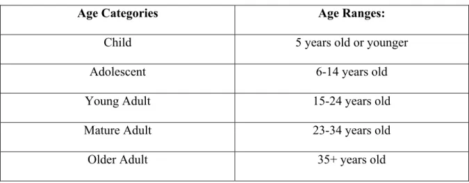
unavailable, then the subadult age was estimated using epiphyseal union. For adults, age was determined using the auricular surface of the innominate. If the auricular surface estimate was unavailable (due to missing elements or poor preservation), age estimation was performed by analysis of the pubic symphysis. When possible, these age estimations were compared to those obtained by studying the sternal rib ends (Buikstra and Ubelaker 1994, 21-44). All age estimations were grouped into one of five categories (table 1) (Boudreaux 2005, 270).
Table 1: Age Categories
Age Categories Age Ranges:
Child 5 years old or younger
Adolescent 6-14 years old
Young Adult 15-24 years old
Mature Adult 23-34 years old
Sex estimation was only conducted for the adult samples (twenty years or older) because “skeletal material [for sex estimation] is most accurate after the individual reaches maturity” (White and Folkens 2005, 385). The Phenice method developed in 1996 was used to estimate the sex of the individuals by analyzing the ventral arc, subpubic concavity, and the medial aspect of the ischiopubic ramus (White and Folken 2005, 395-397). If the Phenice method could not be used on the sample (due to missing elements or poor preservation), then visual estimations of the crania and innominates using methods described in Standards book (Buikstra and Ubelaker 1994, 18-21).
Chapter Two: Osteological Evidence for Changes in Health
Metabolic stress indicators
The forty-nine individuals from the three sites were examined macroscopically for potential metabolic stress indicators. These indicators include porotic hyperostosis and cribra orbitalia, and pathological lesions indicative of scurvy and rickets. Each of these indicators can be attributed to poor nutrition and can indicate major changes in the health of these native individuals due to dietary deficiency.
Porotic Hyperostosis and Cribra Orbitalia
Porotic hyperostosis and cribra orbitalia are defined as lesions found on the cranial vault and eye orbits that, in their most extreme expression, have a hair-on-end appearance. When found on the cranial vault, the lesions are referred to as porotic hyperostosis. When found on the orbital roofs, the lesions are referred to as cribra orbitalia. Some researchers believe that the lesions on the cranial vault are indicators of a more severe case of poor nutrition (Roberts and Manchester 2005, 230-231). The cause of these lesions is iron deficiency anemia.
There are two forms of iron that are found in foods. The first type is non-haem and is often found in cereals such as maize. Non-haem iron is only absorbed through the intestines and they often do not absorb much of it. The other form of iron found in diets is haem iron. This form of iron is consumed when an individual eats red meats and vegetables, and it is more easily absorbed (Roberts and Manchester 2005, 226). Researchers often believe that populations switching from hunter-gather to agriculture could have caused increases in iron-deficiency anemia because of the decrease in haem and the increase in non-haem in these past populations’ diets (Roberts and Manchester 2005, 226).
Because of this lack of iron, the diploe in the cranial vault often thickens in size due to the bone marrow attempting to compensate for the lack of red blood cells caused by the low levels of iron in the blood stream. This thickening creates lesions with a hair-on-end appearance on the parietals and the occipital bone. Some types of anemia that could cause porotic hyperostosis and cribra orbitalia are hereditary, such as thalassemia and sickle cell anemia (Goodman, Martin, Armelagos, and Clark 1984, 29). The most common form of anemia, however, is acquired iron-deficiency anemia -- as of 2005, it was found in over 500 million people worldwide (Roberts and Manchester 2005, 226). Skeletal changes due to this anemia often only occur during an individual’s childhood but can remain present into their adult life (Roberts and Manchester 2005, 229).
Scurvy
Although it is most common in infants, vitamin C deficiency does not manifest until the infant is about four months old because the infant has stores of vitamin C from the mother (Ortner 1985, 270).
A prolonged vitamin C deficiency causes increased bleeding beneath the surface of the skin and the membrane around the bones. This deficiency also affects the binding agents in the blood vessels, causing increased bleeding in the soft tissue which can lead to new bone formations in the affected areas. In addition to these new bone formations (often in the long bones), small lesions on the skull have also been attributed to vitamin C deficiencies (Roberts and Manchester 2005, 235-236).
Subadults suffering from a vitamin C deficiency tend to have bilateral lesions on the greater wings of the sphenoid, maxilla and orbital plate. Researchers believe that these lesions are associated with the temporalis muscle. This muscle is located next to the sphenoid. Between the muscle and the sphenoid bone are two temporal arteries. These arteries can be affected by the lack of vitamin C and hemorrhaging would likely occur causing an inflammatory action between the muscle, the sphenoid and the posterior portion of the maxilla and orbital plate. This inflammatory action can lead to the lesions that researchers see on subadults (Ortner and Ericson 1997, 212-220).
Although many mammals have the ability to synthesize vitamin C, anthropoid primates lost this ancestral trait due to a mutation of the L-gulono--lactone oxidase (GLO) gene, thus causing humans to have to obtain vitamin C through food and drink (Drouin, Godin and Page 2011, 371-373). To prevent the development of these skeletal
(Brickley and Ives 2008, 47-48). A prolonged lack of vitamin C often can have lasting effects on the skeleton. Vitamin C is found in fresh fruits and uncooked vegetables, and small
amounts can be found in milk, meat, and fish (Brickley and Ives 2008, 41).
In addition, a study conducted by Geber et al. focused on scurvy in individuals who died during The Great Famine (1845-1852) in Ireland (Geber and Murphy 2012, 512). The potato was a major source of food for the poor in Ireland during the mid-1800s and contains very high amounts of Vitamin C. For example, “freshly dug potatoes hold approximately 30 mg of Vitamin C per 100 g of edible matter” (Geber and Murphy 2012, 514). When a blight destroyed the potato crop, a “widespread occurrence of infectious and metabolic diseases” and “mass starvation” occurred throughout the population (Geber and Murphy 2012, 512). Of the 970 individuals they examined, 16% of them had evidence of scurvy (Geber and Murphy 2012, 512), demonstrating the relationship between scurvy and changes in diet.
Rickets
Vitamin D deficiency can also have a lasting effect on an individual’s skeleton but not related to direct mortality (Ortner 1985, 273). Most individuals obtain the necessary vitamin D through exposure to sunlight, which is a precursor to vitamin D production, and a supplemental diet of items such as eggs, fortified milk, liver, and oily fish (Brickley and Ives 2008, 75). Although the duration of sun exposure is debated, most researchers believe that five to fifteen minutes of exposure to sunlight should provide individuals with enough
vitamin D to be healthy. The amount of vitamin D needed in an individual’s diet varies based on location, sex, and skin pigmentation (Brickley and Ives 2008, 85).
vitamin D (Brickley and Ives 2008, 9). Vitamin D is necessary in the mineralization of osteoid during bone formation and is also important in maintaining homeostatic calcium levels (Brickley and Ives 2008, 75). Vitamin D deficiencies do not manifest until the child is at least four months old because the nutrients were passed from the mother and child through the placenta. Children are most often affected between six months and two years (Ortner 1985, 274). Often these indicators peak when the subadult is three to eighteen months old (Brickley and Ives 2008, 91).
In children, severe vitamin D deficiencies can cause malformation of the weight-bearing elements of the skeleton, such as changes to the pelvis and bowing of the long bones (Brickley and Ives 2008, 92). The ribs can also be affected, causing the ends of the ribs to flair. In addition, some of the long bones might have flaring bone growth (Brickley and Ives 2008, 92). Vitamin D deficiency can also cause porosity in the cranial bones and growth plates (Brickley and Ives 2008, 91). These skeletal manifestations are often categorized as Rickets in subadult skeletons.
Oral Health
The forty-nine individuals were also macroscopically examined for indicators of degenerative oral health. The indicators of oral health include carious lesions, periodontal disease and ante-mortem tooth loss, and alveolar infections. Each of these indicators can be attributed to poor nutrition and can indicate major changes in the health of these native skeletal series due to changes in diet.
Dental Caries
The presence of dental carious lesions (or cavities) can indicate major changes in the oral environments of individuals potentially caused by changes in diet. Dental caries is a process defined as the, “destruction of enamel, dentine and cement resulting from acid production by bacteria in dental plaque, ultimately leading to the formation of a cavity in the crown or root surface” (Hillson 1996, 269). The lesions (cavities or dental carious lesions) are the most reported dental disease in archaeology and range from small opaque spots to large cavities that extend into the pulp cavity of the tooth, and can be found on both the crowns and the roots of the teeth (Roberts and Manchester 2005, 65).
There are multiple potential causes for dental carious lesions. Mary Powell divided these causes into four categories: environmental factors, pathogenic agents, exogenous facts and endogenous factors (Roberts and Manchester 2005, 65). In addition, dental problems can also be a factor for the development of dental carious lesions because they can weaken the teeth and severe wear can increase the likelihood that bacteria could get inside the pulp cavity causing the decay (Roberts and Manchester 2005, 66). However, the most commonly referred to cause of caries is the presence of large amounts of sugar in the diet of the
Lactobacillus acidophilus and Streptococcus mutans (Roberts and Manchester 2005, 65).
This fermentation process causes microbial action of the teeth that can lead to the
demineralization of the tooth. This demineralization allows the tooth to be susceptible to the development of dental carious lesions (Roberts and Manchester 2005, 65).
Certain aspects of an individual’s life may also make them predisposed to developing dental carious lesions. Miura et al. (1997) conducted a cross-national study of dental caries and determined that the number of dental carious lesions increased significantly if the individual lived in an urban area (Miura et al. 1997 in Roberts and Manchester 2005, 65). In addition, low levels of fluoride in the surrounding environment may also make the individual more susceptible to cavities. Also, multiple studies have been conducted that have concluded that the likelihood of an individual developing cavities increases with age as well as their gender and status within the environment (Roberts and Manchester 2005, 67).
Finally, there appears to be a correlation between cavities and agricultural societies. Clark Larsen conducted a study in 1984 of the Georgia coast to compare pre-agricultural (1000 BC-A.D. 1150) and agricultural (A.D. 1150-1550) communities. In his study, he noted that there appeared to be a 10% increase in dental caries in all tooth types across genders in the shift from pre-agricultural to agricultural societies. Larsen believed that the main cause of this increase stemmed from the increase in sucrose in their diet caused by maize becoming a main staple in their diets (Roberts and Manchester 2005, 67).
Periodontal Disease and Ante-Mortem Tooth Loss
ligament, cement, gingivae, and mucosa (Hillson 1996, 260). Having large traces of calculus between the gums and the teeth often are a predisposing factor for the development of the disease. Initially this disease begins with gingivitis, or the inflammation of the gums. Then, if it spreads past the three initial stages (initial lesion, early lesion and established lesion), it then enters into the fourth stage: advanced lesion. When the last stage is reached, the disease is then classified as periodontis instead of gingivitis. Once in the periodontal stage, all of the periosteal tissue around the infected tooth are affected (Hillson 1996, 262-263). It affects the bones and can lead to the loss of the periodontal ligament that holds teeth in their place. Eventually the gap between the tooth and bone grow so much that the tooth is lost (Hillson 1996, 262-263).
Many researchers believe that it is hard to identify periodontal disease in the archaeological record because there is no standardized method of identifying this disease. Some archaeologists even argue that periodontal disease is over-diagnosed within the archaeological record. However, the presence of healing around the tooth socket or bone growth in the empty tooth socket are two major indicators of the disease in the archaeological record (Roberts and Manchester 2005, 73).
Alveolar Infection
smooth-walled lesion forms around the roots which are indicative of an alveolar infection. However, researchers indicate that bone damaged post mortem may mimic these abscesses and that researchers should also look for evidence of healing around the bone as an indicator of alveolar infection (Roberts and Manchester 2005, 70-71).
General Stress Indicators
Furthermore, the forty-nine individuals were macroscopically examined for general stress indicators. The one stress indicator examined in this paper is enamel hypoplasias The exact cause of this general stress indicator is unknown, but studies have been conducted that show an increase in hypoplasias with urbanization and the shift to agricultural societies. In addition, the presence of general stress indicators may be positively correlated with both metabolic bone diseases and changes in oral health. Other stressors, such as infections and inflammatory responses, also can be related back to the diet of the skeletal series.
Dental Enamel Hypoplasia
Enamel is the hard mineralized substance that encapsulates the crown of human teeth. It is formed by the soft tissue epithelium which is, “a sheet of closely and regularly packed cells called ameloblasts” in two stages (Hillson 1996, 148). During matrix secretion crystallites and minerals combine to grow and during maturation the ameoloblasts break down the organic matter in the matrix to make the teeth mineralize (Hillson 1996, 148-149).
There are multiple different causes of dental enamel hypoplasias that can be grouped into, “hereditary anomalies, localized trauma, and systemic metabolic stress such as
nutritional deficiency or a childhood illness” (Goodman and Rose 1991 in Roberts and
starvation or a major disease when the enamel of their teeth was still developing (during their childhood), slight lines, grooves or pits may have formed on their teeth, which are
categorized as enamel defects (Larsen and Hutchinson 1992, 151-169). Enamel defects are thus interpreted as non-specific indicators of stress in an individual’s life (Roberts and Manchester 2005, 75).
There are three suspected causes of the formation of hypoplasias during childhood: genetic abnormalities, trauma, or metabolic stress. Although there are three possible causes for the development of dental enamel hypoplasias, cases of hypoplasias caused by genetic abnormalities and trauma are rare in the archeological realm because those individuals have a significantly lower chance of survival which would not give the defect enough time to
manifest on the enamel surface. Because of that low chance of survival, the majority of dental enamel hypoplasia (especially linear enamel hypoplasia) cases in archaeology are thought to be caused by metabolic stress (Goodman and Rose 1990, 59-110).
When there is a period of stress or trauma, such as a fever, lack of nutrients, or hormonal changes, for an individual, they cause disruptions with the ameloblasts production of the enamel matrix, which manifests as dental enamel hypoplasias in the individual
(Hillson 1986, 130). The severity of these hypoplasias corresponds with the length of the stress or trauma that the individual went through (Goodman and Rose 1990, 59-110). For example, if an individual had a severe illness, the hypoplasia might manifest in a wave-like pattern which corresponds to the severe periods of the illness.
society to one focused on agriculture increased stress and created an influx of enamel hypoplasias in native populations in Georgia (Roberts and Manchester 2005, 75-76). These periods of stress often cause dental enamel hypoplasia and can often reveal stress related to changes in diet in past populations.
Infectious Disease
Sinusitis
Sinusitis is defined as an infection of the throat, ear, nasal sinuses, or chest. Although this infection was probably very common in past populations, it is difficult to diagnose in the archaeological record because the “nasal sinuses are air-filled cavities within the bone of the face and are therefore difficult to inspect (Roberts and Manchester 2005, 174). However, if the air-filled cavities are exposed in the archaeological record, then sinusitis can be identified by irregular pitting or new bone formation within the sinuses (Roberts and Manchester 2005, 174).
One study conducted by Roberts et al. at St Helen-on-the-Walls in York, England found that 72% of the individuals in their sample suffered from maxillary sinusitis. This high percentage may arise from a correlation between sinus infections and dental caries. When a tooth is severely infected, the infection can spread into the sinuses thus allowing sinusitis to occur. In addition, sinusitis has been shown to occur more often in urban sites than in rural sites (Roberts and Manchester 2005, 174-175).
Periosteal Reaction
periosteal reactions are often interpreted as non-specific stress indicators, they are often associated with infectious disease process.
There can be multiple causes for periosteal reactions within the body. Some reactions can occur through trauma while others can occur through an infection in the body. Although there are multiple potential causes for periosteal reactions, the tibia is the most commonly affected bone in the human body. Many researchers believe that this bone is mainly affected because it is so close to the skin surface, allowing it to be susceptible to trauma. Others argue that, because the bone is so close to the skin surface, the temperature of the bone is cooler thus creating a “physiologically inactive surface, leading to bacterial colonization” and “blood tends to stagnate in the lower legs, allowing bacteria to accumulate” (Roberts and Manchester 2005, 172-173).
Osteomyelitis
Osteomyelitis is a nonspecific disease indicator that occurs in the body and affects the marrow of bones (Ortner 1985, 112), but like periostitis it is often associated with specific disease processes. It is often characterized by bone destruction, pus formation and
simultaneous bone repair (Roberts and Manchester 2005, 168-169). Bacteria from outside sources, usually spreading to the bone through the bloodstream from an infection of the throat nose, is the major cause of osteomyelitis; however, the bacteria can also enter the bloodstream through a penetrating bone injury, skin ulcers, trauma, or surgery.
Staphylococcus aureaus is the bacteria responsible for 90% of present day cases of
body begins to produce new bone growth around the sequestrum (Ortner 1985, 112). This new bone is made by osteoblasts in the periosteum and lacks microstructure, causing the new bone to be labeled as ‘woven bone.’ In extreme cases, the original bone underneath the newly formed bone can die, thus forcing the new and less efficient bone to support the the body (Roberts and Manchester 2005, 168-17). In conjunction with the bone necrosis and regrowth, within the bone, pus abscess fill the cavities of the bone. These abscesses can escape through the involucrum and soft tissue which cause distinctive cloaca to form on the bone (Ortner 1985, 113).
Evidence of osteomyelitis is mainly found on the femura and tibiae about 80% of the time and about 10% is found on the humerus. Other long bones, tubular bones, cancellous bones, and the skull have also been reported but are more rarely affected (Ortner 1985, 111). Treponemal Infection
There are four different types of treponemal infection: venereal syphilis, nonvenereal (endemic) syphilis, yaws and pinta. These infections are caused by bacterial spirochetes: Treponema carterum, Treponema pallidum pertenue, Treponema pallidum pallidum,and
Treponema pallidum endemicum, respectively (Larsen 1997, 94). An individual contracts
later stage then causes the destruction and reconstruction of bones. This process is a form of osteomyelitis (Roberts and Manchester 2005, 208).
In yaws, the most commonly affected element is the tibia. New bone formation creates a woven bone pattern on the bone. This new bone growth causes the tibia to have a ‘saber-shin’ appearance. In addition, irregular depressions may form on the cranial vault, maxilla, and nasal bones (Larsen 1997, 94-95). This bone involvement occurs in about 5-15% of all cases of yaws (Roberts and Manchester 2005, 208).
Similar to yaws, the bones of the nasal region and upper jaw can also be affected in nonvenereal syphilis. In addition, the tibiae may also be affected creating the ‘saber-shin’ appearance. The amount of bone involvement increases in nonvenereal syphilis to between 15-20%. The major difference between nonvenereal syphilis and yaws depends on the environment. Yaws tends to manifest in humid, tropical environments while nonvenereal syphilis manifests in more arid regions (Roberts and Manchester 2005, 209). In addition, nonvenereal syphilis tends to manifest stellate lesions on the cranial vault and some consider these lesions, “the best single criterion for endemic treponematosis” in specific regions (Larsen 1997, 97).
and parietal bones. About 20% of bones are involved in venereal syphilis (Roberts and Manchester 2005, 208).
Chapter Three: Results
Osteological Evidence of Metabolic Disease in the Piedmont
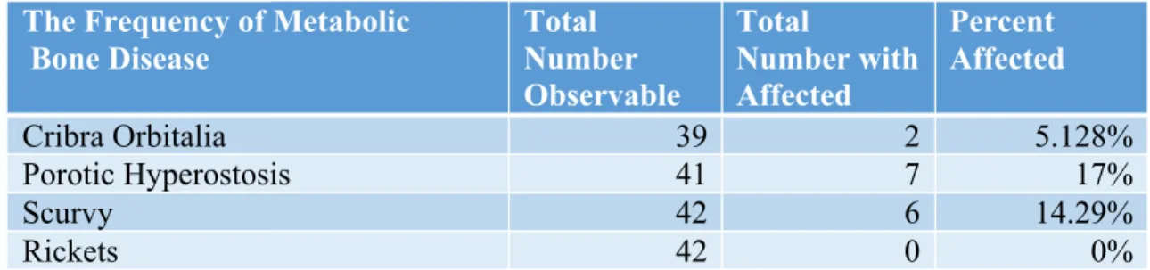
Of the forty-nine individuals examined for this thesis, 30.6% of them had evidence of metabolic bone disease consistent with inadequate diets. Of this 30.6%, about 5% suffered from cribra orbitalia, 17% suffered from porotic hyperostosis, 23% suffered from vitamin C deficiency and none appeared to suffer from a vitamin D deficiency as seen in Table 2. This next section will take a closer look at the specific occurrences of metabolic bone disease and what elements were affected.
Table 2: Frequency of Metabolic Bone Disease The Frequency of Metabolic
Bone Disease
Total Number Observable
Total
Number with Affected
Percent Affected
Cribra Orbitalia 39 2 5.128%
Porotic Hyperostosis 41 7 17%
Scurvy 42 6 14.29%
Rickets 42 0 0%
Cribra Orbitalia
Table 3: Frequency of Cribra Orbitalia
Table 4: Individuals with Cribra Orbitalia Site Individuals Age
Category Sex Elements Affected Clarifying Notes
Vir231 11 Subadult (2
± 8 months) n/a Frontal bone Lesions in right eye orbit
Vir231 22 Subadult
(5± 16 months)
n/a Frontal bone, zygomas
Lesions in right eye
orbit
Porotic Hyperostosis
Although there was only slight evidence of anemia with the presence of cribra orbitalia in the three series examined, there was a larger proportion of individuals that exhibited porotic hyperostosis (Table 5). Seven individuals (17%) exhibited lesions on the cranial vault that were consistent with the diagnosis of porotic hyperostosis. These
individuals ranged in age and sex estimations as seen in Table 6. One subadult exhibited lesions consistent with porotic hyperostosis. Three young adults, one mature adult, and two older adults also had lesions consistent with porotic hyperostosis. There were four females and two males that exhibited evidence of porotic hyperostosis.
At Town Creek Indian Mound (Mg2-3), one individual (8.33%), individual 5, had heavily remodeled of porotic hyperostosis lesions on the left and right parietal bones along the sagittal suture. This individual was an adolescent about 12-16 years old. In contrast to Town Creek (Mg2-3), five individuals (26.32%) from Stockton (Vir231) had lesions
Individuals with Cribra Orbitalia Total Number Observed Total Number Affected Percent Affected
Town Creek (Mg2-3) 10 0 0%
Stockton (Vir231) 19 2 10.53
consistent with porotic hyperostosis on the cranial vault. These individuals all exhibited porotic hyperostosis on the cranial vault in various degrees. Individuals 1, 3 and 8 had porotic lesions extensively along the cranial vault while individual 5 only had slight lesions on the right parietal bone. Finally, individual 13 had evidence of porotic hyperostosis lesions along the sagittal suture on both the left and right parietals. In summary, 26.3% of the
individuals studied from Stockton (Vir231) that could be examined for porotic hyperostosis lesions exhibited evidence of iron deficiency. Of those affected, four were females and one was a male.
Finally, one individual (10%) from Fredricks (Or231) also had evidence of porotic hyperostosis on. Individual 3 was a young adult male about 20-24 years old who had extensive lesions along both parietal bones and along the frontal bone. In total, 10% of the individuals examined for Fredricks had evidence of iron deficiency.
Table 5: Frequency of Porotic Hyperostosis Individuals with Porotic
Hyperostosis
Total Number Observed
Total Number Affected
Percent Affected
Town Creek (Mg2-3) 12 1 8.33%
Stockton (Vir231) 19 5 26.32%
Table 6: Individuals with Porotic Hyperostosis Site Individual Age
Category
Sex Elements Affected
Mg2-3 5 Subadult n/a Left and right parietals, occipital extensive
Vir231 1 Young
adult
Female Frontal, right parietal, and occipital
Vir231 3 Older
adult
Female Frontal, very slight left and right parietals
Vir231 5 Young
adult
Female Left and right parietals
Vir231 8 Older
adult
Male Frontal, left and right parietals, and occipital
Vir231 13 Mature
adult Female Left and right parietals
Or231 3 Young
adult Male Frontal and left and right parietals
1
Scurvy
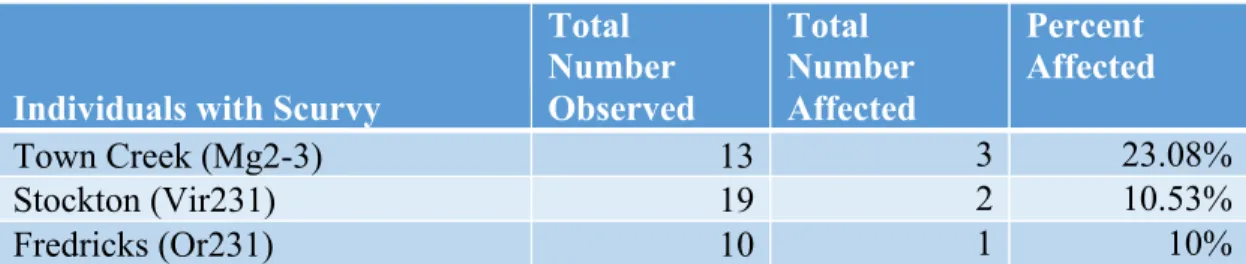
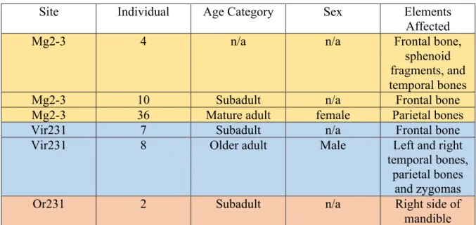
As seen in Table 7, six individuals had evidence of scurvy. Three of the individuals from Town Creek (Mg2-3), 4, 10 (Fig. 2), and 36, (23%) showed evidence of scorbutic lesions that ranged from localized lesions such as the ones from individual 36 (Fig. 3), found along the parietal bones, to widespread lesions such as the ones seen with individual 4 (Fig. 4), who had extensive lesions on the frontal bone, sphenoid fragments, and temporal bones (Table 8). That latter case could indicate a more severe case of vitamin C deficiency.
1 Colors refer to site in all tables: yellow is Town Creek (Mg2-3), blue is Stockton (Vir231), and
Fig. 2: Mg2-3 Individual 10, scorbutic lesions on eye orbits
Fig. 3: Mg2-3 Individual 36, scorbutic lesions on parietal
Table 7: Frequency of Scurvy
Individuals with Scurvy
Total Number Observed
Total Number Affected
Percent Affected
Town Creek (Mg2-3) 13 3 23.08%
Stockton (Vir231) 19 2 10.53%
Fredricks (Or231) 10 1 10%
At Stockton (Vir231), individuals 7 and 8 also exhibited lesions that were consistent with a vitamin C deficiency. These two individuals out of the nineteen observable for scurvy like lesions make up 10.53% of the individuals examined for this study. Individual 7, a subadult, had evidence of scurvy on the frontal bone while individual 8, an older adult male, had lesions suggestive of scurvy on left and right temporal bones, parietal, and zygomas as well as on the frontal bone. In addition, there appeared to be scorbutic nodes on the
mandible; however, there were no scurvy-like lesions on either side of the ascending ramus, which is a common location for lesions indicating a vitamin C deficiency.
Table 8: Individuals with Scurvy
Site Individual Age Category Sex Elements
Affected
Mg2-3 4 n/a n/a Frontal bone,
sphenoid fragments, and temporal bones
Mg2-3 10 Subadult n/a Frontal bone
Mg2-3 36 Mature adult female Parietal bones
Vir231 7 Subadult n/a Frontal bone
Vir231 8 Older adult Male Left and right
temporal bones, parietal bones
and zygomas
Or231 2 Subadult n/a Right side of
mandible
Rickets
Although all of the individuals in this study were examined for evidence of rickets, none of the individuals had any of the key indicators suggesting a vitamin D deficiency (Table 9). Such changes include pelvic deformation, potential bowing in the long bone, and the flaring of rib ends. One individual, individual 4 from Stockton (Vir231), did have bowing of the arm bones (Fig. 5 and 6). However, this bowing could be due to mechanical stress and thus was not included in the analysis of a vitamin D deficiency.
vitamin D deficiency may have passed away before changes to the skeleton could have occurred, thus making it impossible to see any deficiency in the skeletal remains.
Fig. 5 Vir 231 Individual 4 bowed radii
Fig. 6 Vir231 Individual 4, x-ray of bowed radii
Table 9: Frequency of Rickets
Individuals with Rickets
Total Number of Individuals with Rickets
Town Creek (Mg2-3) 0
Stockton (Vir231) 0
Osteological Evidence of Changes to Oral Health Environments in the Piedmont
Dental Carious Lesions
Thirty individuals (Appendix I) from the three sites had at least one cavity (Table 10). Six of these individuals were children and adolescents, nine were young adults, thirteen were mature adults, and two were older adults. Twelve of these individuals were female and seven were male. The rest of the individuals were either subadults for whom sex estimates were not attempted (seven individuals) or there was not enough of the skeleton preserved for an accurate sex estimation to be made for older individuals (four individuals).
Nine individuals (56%) from Town Creek (Mg2-3) had at least one cavity. There was one male, three females, and five sex indeterminate because they either a subadult or not enough of the skeletal materials were available to make an accurate sex estimation. At Stockton (Vir231) thirteen individuals (59%)- eight females, four males and one
indeterminate- had at least one cavity. Eight individuals (72%) from Fredricks (Or231) had at least one cavity. Of these eight individuals, four of them were too young to have accurate sex estimates, one was not complete enough to have an accurate sex estimation, two were male, and one was female. The number, the size, and the location of these cavities varied
throughout the individuals.
Table 10: Frequency of Dental Carious Lesions Individuals with Dental Carious
Lesions
Total Number Observed
Total Number Affected
Percent Affected
Town Creek (Mg2-3) 12 9 75%
Stockton (Vir231) 17 13 76.47%
Periodontal Disease and Ante-Mortem Tooth Loss
Of the forty-nine individuals examined, eighteen (43.9%) showed evidence of ante-mortem tooth loss as seen in Table 11. This ante-ante-mortem tooth loss was identified through the presence of additional bone growth around empty teeth sockets (Fig. 7). Ten of these individuals were female, five were male, and three were of indeterminate sex due either to age or lack of remains to accurately estimate sex. Four (28.5%) of these individuals were from Town Creek. From Stockton, thirteen individuals (65%) showed evidence of ante-mortem tooth loss and one individual (10%) from Fredrick, individual 5, showed evidence of ante-mortem tooth loss.
(Table 11).
Table 11 Frequency of Ante-Mortem Tooth Loss
Individuals with Ante-Mortem Tooth Loss
Total Number Observed
Total Number Affected
Percent Affected
Town Creek (Mg2-3) 14 4 28.57%
Stockton (Vir231) 20 13 65%
Table 12: Individuals with Ante-Mortem Tooth Loss
Site Individual Age
Categor y
Age Sex Location of Ante-Mortem Tooth Loss
Mg2-3 6 Child 4-5
years
n/a Left and right mandible (m1,m2)
Mg2-3 8 Mature
Adult
28+ Female Left and right mandible (m1,m2)
Mg2-3 27 Mature
Adult
mid 20s
Female Left and right mandible (m1,m2)
Mg2-3 41 Young
Adult
16-22 Female Left mandible (m1,m2, m3)
Vir231 1 Young
adult
20-24 Female Right mandible (pm4, m1,m2,m3)
Vir231 3 Older
adult
38.2 ±10
Female Left and right mandible (m1,m2,m3)
Vir231 5 Young
Adult
20 Female Right mandible (pm4, m1,m2,m3)
Vir231 6 Mature
Adult
About 30
n/a Left mandible (m1,m2)
Vir231 8 Older
Adult
Mid 30s
Male Left maxilla (m2,m3)
Vir231 13 Mature
Adult
24-32 Female Left mandible (m1,m2,m3) right mandible (pm4,
m1,m2,m3)
Vir231 14 Mature
Adult
24-32 Female Left mandible (m1,m2) right mandible (m1,m2)
Vir231 15 Mature
Adult
24-28 Male Left maxilla (m1,m2,m3), right maxilla (m1,m2,m3) Left mandible (m1) Right mandible (pm4,m1,m2,m3)
Vir231 18 Mature
Adult
About 25
Female Right mandible (pm3,pm4, m1,m2,m3)
Vir231 20 Mature
Adult
25-28 Male Left mandible (i1,i2,c,pm3,pm4,m1,m2m,
3)
Vir231 21 Mature
Adult
25-29 Male Right mandible (m1,m2)
Vir231 X Young
Adult
21-24 Female Left mandible (all teeth) right mandible (i1,i2,c,pm3)
Vir231 22 Subadul
t
Aroun d birth
n/a Right maxilla (il)
Or231 5 Young
Adult
Fig. 7 Vir231 Individual 13 Ante-Mortem Tooth Loss Alveolar Infection
Over 30% of individuals examined (Table 13) had evidence of an alveolar infection ranging from a slight infection of one tooth (such as individual 1b from Town Creek (Mg2-3) to more severe cases affecting multiple teeth on both the mandible and maxillae (such as individual 9 from Stockton (Vir231) that had the infection underneath eight teeth). All of the individuals that showed evidence of at least one alveolar infection were young adults or older. There were no cases of subadults having evidence of alveolar infections. The severity of the infections appeared to varied between the sites. At Town Creek (Mg2-3), four
individuals (33.3%) showed evidence of an alveolar infection (Table 14).
Table 13: Frequency of Alveolar Infection Individuals with Alveolar
Infection
Total Number Observed
Total Number Affected
Percent Affected
Town Creek (Mg2-3) 12 4 33.33%
Stockton (Vir231) 17 12 70.59%
Table 14: Individuals at Town Creek (Mg2-3) with Alveolar Infections
Table 15: Individuals at Stockton (Vir231) with Alveolar Infections Site Individual Age
Category Age Sex Location of Alveolar Infection
Mg2-3 1B Mature
adult 31+ Male Left mandible
Mg2-3 27 Mature
adult mid 20s Female Left and right mandible
Mg2-3 36 Mature
adult
25-29 Female Left and right maxillae
Mg2-3 41 Young
adult
16-22 Female Left and right mandible
Site Individual: Age
Category: Age: Sex: Location of Alveolar Infection:
Vir231 1 Young
adult
20-24 female Right mandible (c, pm3)
Vir231 3 Older
adult
38 ± 10 Female Left mandible(c)
Vir231 5 Young
adult
16-22 Female Left maxilla (pm4, m2) right maxilla (c, m1, m2)
Vir231 6 Mature
adult slightly 30 or younger
n/a Right maxilla (pm3, m1,m2,m3) left mandible (m2) right mandible
(i1, m1,m2)
Vir231 8 Older
adult
mid 30s male Right maxilla (m1,m2)
Vir231 9 Young
adult 20-24 female Left maxillae (c, pm3), Right maxilla (pm4, m2,m3) Left mandible (m1) Right mandible
(m1, m3)
Vir231 13 Mature
adult
24-32 female Right maxilla
(c,pm3,pm4,m1,m2,m3), left mandible (m1,m2,m3) right
mandible (i1,i2,c)
Vir231 14 Mature
Twelve individuals out of seventeen, or 70.5%, examined from Stockton (Vir231) showed evidence of alveolar infections on either the maxillae or mandible (Table 15).
Three individuals (30%) from Fredricks (Or231) suffered from alveolar infections. This means that 30% of the individuals examined from Fredricks (Or231) had evidence of at least one alveolar infection (Table 16).
Table 16: Individuals at Fredricks (Or231) with Alveolar Infections
Vir231 18 Mature
adult 25 Female Left maxilla (pm3), right Maxilla (pm3, pm4, m1, and m2),
Vir231 21 Mature
adult 25-29 Male Right maxilla (canine, pm3, pm4), right mandible (i2) Vir231 22 Subadult Around
birth
n/a Left and right mandible and maxillae
Site Individuals Age category
Age Sex Location of alveolar infection
Or231 3 Young
adult
20-24 Male Left maxilla (c, m1) right maxilla (i1,i2,c)
Or231 4 Mature
adult 35+ Female Left maxilla (m1,m2,m3) right mandible (m2, m3)
Or231 5 Young
adult
22-24 Male Left mandible
Osteological Evidence of General Stress Indicators in the Piedmont
Dental enamel hypoplasia
Four individuals (Table 18) (8.3%) showed lines, grooves, or pits consistent with dental enamel hypoplasias (Table 17). One individual from Town Creek (Mg2-3), individual 29, had hypoplasias on the right maxillary incisors. The three other individuals (30%) with linear enamel hypoplasias came from Fredricks (Or231) and were individuals 2, 6, and 11. Individual 2 had hypoplasias on the left and right first mandibular incisor and one on the left mandibular first molar. Individual 6 had hypoplasias on the left and right first and second maxillary incisor and canines of mandible on both sides. Finally, individual 11 had
hypoplasias on the first and second incisor and canines on both maxillae and right mandible. The left half of the mandible only had one hypoplasia on the left canine. These four
individuals make up about 10% of the total sample studied.
Table 17: Frequency of Dental Enamel Hypoplasias Individuals with Dental Enamel
Hypoplasias Total Number Observed Total Number Affected Percent Affected
Town Creek (Mg2-3) 12 1 8.33%
Stockton (Vir231) 17 0 0%
Fredricks (Or231) 10 3 30%
Table 18: Individuals with Dental Enamel Hypoplasias
Site Individual Age Category Sex Elements
Affected
Mg2-3 29 Mature adult n/a right maxillary
I1
Or231 2 Subadult n/a Left and right
mandibular I1, mandibular m1
Or231 6 Subadult n/a Left and right
mandibular I1
Or231 11 Subadult n/a right
Osteological Evidence of Infectious Diseases in the Piedmont General Lesions
Sinusitis
Of the thirty individuals examined for this study, ten (33.3%) showed evidence of maxillary sinus infections (Table 19). Only one individual (41) from Town Creek Indian Mound (Mg2-3) had evidence of a sinus infection indicating that only about 12.5% of the observable individuals from this site examined suffered from this infection. In contrast, eight individuals (Table 20) showed evidence of sinus infections in their maxillae, which makes up about 61.5% of the individuals from Stockton (Vir231). Finally, one individual (5), from the observable nine individuals (11.11%) from Fredricks (Or231) had evidence of a sinus infection indicating that about 9% of the individuals examined from Fredricks suffered from a sinus infection.
Table 19: Frequency of Sinusitis
Individuals with Sinusitis
Total Number Observed
Total Number Affected
Percent Affected
Town Creek (Mg2-3) 8 1 12.5%
Stockton (Vir231) 13 8 61.54%
Fredricks (Or231) 9 1 11.11%
Table 20: Individuals with Sinusitis
Site Individual Age Category Sex Elements
Affected
Mg2-3 41 Young Adult Female Maxilla
Vir231 3 Older Adult Female Maxilla
Vir231 6 Mature Adult n/a Maxilla
Vir231 8 Older Adult Male Maxilla
Vir231 9 Young Adult Female Maxilla
Vir231 10 Young Adult n/a Maxilla
Vir231 12 Child n/a Maxilla
Vir231 15 Mature Adult Male Maxilla
Vir231 22 Child n/a Maxilla
Periosteal Reaction
Of the forty-four individuals examined for this study that had long bones and/or patellae present, seventeen (38.6%) showed evidence of periosteal reactions (Table 21). Four individuals (30.7%) from Town Creek showed evidence of these reactions on at least one bone. Twelve individuals showed evidence of periosteal reactions on at least one bone from Stockton (Vir231), which is about 60% of the individuals studied from this site. Finally, one individual (4) from the eleven observed at Fredricks (Or231) showed evidence of periosteal reactions, which is about 9% of the skeletal series examined (Table 22).
Table 21: Frequency of Periosteal Reactions Individuals with Periosteal
Reactions
Total Number Observed
Total Number Affected
Percent Affected
Town Creek (Mg2-3) 13 4 30.77%
Stockton (Vir231) 20 12 60%
Table 22: Individuals with Periosteal Reactions
Site Individual with Periosteal Reactions
Age Category
Age Sex Number
of Elements
Elements
Mg2-3 1B Mature
Adult
31+ Male 2 Left and right tibiae
Mg2-3 6 Child 4-5 n/a 2 Left and right femur
shaft
Mg2-3 8 Mature
Adult
28+ Female 1 Right innominate Mg2-3 31 Adolescent 6±24
months n/a 4 femur, left tibia, Left radius, left right tibia
Vir231 4 Young
Adult 23+ Female Patellae
Vir231 6 Mature
Adult
mid to late 20s
n/a 1 Right patella
Vir231 8 Older
Adult
mid 30s Male 2 Patellae
Vir231 9 Young
adult
20-24 Female 2 Left tibia, left fibula
Vir231 11 Child 2+-8
months
n/a 8 Tibiae, humeri,
radii, ulnas
Vir231 14 Mature
Adult
30+ Female 1 Left patella
Vir231 15 Mature
Adult
24-28 Male 4 Ulnas, patellae
Vir231 17 Young
Adult
20-24 Female 1 Right patella
Vir231 18 Mature
Adult
25 Female 4 Tibiae, patellae
Vir231 20 Mature
Adult 25-28 Male 2 patellae
Vir231 21 Mature
Adult
25-29 Male 3 Patellae and right tibia
Vir231 X Young
Adult
21-24 Female 2 Tibiae
Or231 4 Mature
Adult
Osteomyelitis
Of the forty-three individuals examined for this study, only four individuals (20%), all from Stockton (Vir231), had evidence of new bone formation consistent with
osteomyelitis (Table 23). Individual 8 (Table 24) had new bone formation on the right femur and left tibia, left humerus and a fibula (unable to be sided due to lack of both proximal and distal ends of the bone). Individual 9 had new bone growth on the left femur and left tibia and the left fibula. There was also a cloaca on the left tibia of this individual. Individual 13 has new bone formation on the scapula, patellae, clavicles, tibiae, and femur (Fig. 8), as well as a cloaca on the tibia (Fig. 9). Individual 15 had new bone formation on a femur, tibia, humerus, and radius (Fig. 10).
Table 23: Frequency of Osteomyelitis
Table 24: Individuals with Osteomyelitis
Site Individual Age Category Sex Elements
Affected
Vir231 8 Older Adult Male Both tibiae,
left humerus and fibula
Vir231 9 Young Adult Female Left femur,
tibia and fibula
Vir231 13 Mature Adult Female Scapula,
patellae, clavicles, tibiae, femur
Vir231 15 Mature Adult Male Femur, tibia,
humerus Individuals with Osteomyelitis
Total Number Observed
Total Number Affected
Percent Affected
Town Creek (Mg2-3) 12 0 0%
Stockton (Vir231) 20 4 20%
Fig. 8: Vir231 Individual 13 osteomyelitis of a right femur
Fig. 10 Vir231 Individual 8 osteomyelitis of fibula, tibia and humerus
Specific Lesions
Based on the presence of stellate lesions on the cranial vaults, individuals 8 and 15 appear to have suffered from a treponemal infection, which was manifested in the
osteomyelitis in the long bones. Individual 8 also had sclerotic nodes on the mandible,
consistent with a treponemal infection. At least six stellate lesions were located on the cranial vault along the parietals and frontal bones (Fig. 12). Although the left tibia did not have the “saber-shin” appearance, there was evidence of systemic infection with bone reformation on the diaphysis of the bone. Individual 15 had at least seven of these stellate lesions across the parietals and frontal bone (Fig. 11). In addition, the new bone formation on the right tibia had the classic “saber-shin” appearance.
lesions, and the “saber-shin” of individual 15 and the history of the disease. The other two individuals, 9 and 13, lacked stellate lesions and thus were not placed in the treponenmal infection category. However, these two individuals could have been suffered from a treponemal infection and had not yet reached the tertiary stage of the disease and thus the manifestation of the stellate lesion would not have occurred yet. Additional diagnostic identification studies need to be conducted on these two individuals to conclude or rule out a treponemal infection.