Original Article
Shear Bond Strength of Nanocomposites to Dentin Substrate
Treated with Er:YAG Laser Followed by Two Different
Bonding Systems
M. Mirzaee 1, P. Fasih 2,E. Yasini 3, N. Chiniforush 4, MJ. Kharrazifard 5, Sh.Rafiezadeh 6, L. Rajbar Omrani 7.
1 Associate Professor, Dental Research Center, Dentistry Research Institute, Tehran University of Medical Sciences, Tehran, Iran
AND Department of Operative Dentistry, School of Dentistry, Tehran University of Medical Sciences, Tehran, Iran
2 Postgraduate Student, Department of Prosthetic, School of Dentistry, Shahid Beheshti University of Medical Sciences, Tehran,
Iran
3 Professor, Department of Operative Dentistry, School of Dentistry, Tehran University of Medical Sciences, Tehran, Iran 4 PhD Laser, Laser Research Center of Tehran University of Medical Sciences, Tehran, Iran
5 Research Member, Dental Research Center, Dentistry Research Institute, Tehran University of Medical Sciences, Tehran, Iran 6 Postgraduate Student, Department of Operative Dentistry, School of Dentistry, Tehran University of Medical Sciences, Tehran,
Iran
7 Assistant Professor, Department of Operative Dentistry, School of Dentistry, Tehran University of Medical Sciences, Tehran,
Iran
Corresponding author: Sh. Rafizadeh, Postgraduate Student, Department of Operative Dentistry, School of Dentistry, Tehran University of Medical Sciences, Tehran, Iran
Received: 1 Jan 2017 Accepted: 3 Apr 2017
Abstract
Background and Aim: Preparation of tooth structure by laser systems produces a surface with different characteristics. Therefore, selecting an appropriate bonding system is necessary to achieve the maximum bond strength. The aim of this study was to compare the bond strength of Adper Single Bond 2 adhesive and Single Bond Universal to dentin prepared by bur or laser.
Materials and Methods: In this experimental study, 48 third molars were collected. The enamel was abraded by wet grinding to achieve flat surfaces of dentin. Samples were divided into 4 groups. Group 1: bur preparation + Single Bond Universal adhesive, Group 2: bur preparation + Adper Single Bond 2, Group 3: Er:YAG laser preparation + Single Bond Universal adhesive and Group 4: Er:YAG laser + Adper Single Bond 2. Nanocomposites were bonded to the samples. The shear bond strength was measured. Failure mode was evaluated under a stereomicroscope. Data were analyzed using two-way ANOVA. The level of significance was considered as 0.05.
Results: The shear bond strength of the laser group was significantly lower than that of bur group (P<0.001). No significant difference was observed in shear bond strength regarding the type of adhesive used (P>0.05). The failure mode in all groups was mainly mixed.
Conclusion: Preparation of dentin by Er: YAG laser decreased the bond strength in comparison to diamond bur preparation. The type of adhesive did not affect the bond strength, regardless of the preparation method.
Key Words: Adper Single Bond 2, Lasers, Solid-State,Shear Strength
Cite this article as: Mirzaee M, Fasih P, Yasini E, Chiniforush N, Kharrazifard MJ, Rafiezadeh Sh, et al. Shear Bond Strength of Nanocomposites to Dentin Substrate Treated with Er:YAG Laser Followed by Two Different Bonding Systems. J Islam Dent Assoc Iran. 2017; 29(2):51-57. DOI: 10.30699/jidai.29.2.51
Introduction
Nowadays, tooth-colored restorations such as composite restorations are widely used due to their esthetic results, minimally invasive preparation, and bonding to tooth structure [1]. Ideal adhesion of resin composites to tooth structure causes marginal adaptation and decreases microleakage, marginal staining, pulpal damage and recurrent caries [2]. Bonding to dentin is more complex than enamel because dentin is a vital tissue containing dentinal tubules with water content [3].
In the recent years, different bonding systems with different formulations have been introduced by the manufacturers in order to achieve an ideal bond
and simplify the bonding procedure [4].
Multimode adhesives are among the newest generations of adhesives, which can be used on different substrates, as etch-and-rinse or self-etch adhesive [5].
Adper Single Bond 2 is the gold standard for total-etch two step bonding systems, and contains monomers, silica nano-fillers and polyalkenoic
acid copolymer [6]. Scotchbond Universal
adhesive, is a type of filled multimode adhesive that contains polyalkenoic acid copolymer. Additionally, methacrylate monomers (UDMA and GDMA) have been partially replaced with acidic 10-methacryloyloxydecyl dihydrogen phosphate monomer (10- MDP) that has the ability to chemically bond to tooth structure [7].
Using laser for cavity preparation is desirable for patients, due to less noise and vibration, and no need for anesthesia. Er:YAG Laser emits a wavelength of 2.94 micrometer that coincides with the major absorption band of water. This emitted energy is well absorbed by hydroxyapatite and has the capacity to remove dental hard tissues more effectively than other laser systems [8]. It has been reported that when Er:YAG laser is used in conjugation with water spray, the thermal damage is minimal [9]. Laser creates a surface topography on the tooth structure different from bur preparation, which affects the quality of bonding of adhesives. Studies have shown that laser irradiated dentin surfaces are acid resistance, and total-etch and self-etch adhesive systems do not bond effectively to laser prepared dental surfaces [8,10,11].
The aim of this study was to evaluate the effect of
a multipurpose bonding system with lower pH and acidic monomer on shear bond strength of nanocomposite to dentin. The null hypothesis was that the bond strength of composite to laser prepared dentin would not be different in use of Scotchbond Universal adhesive in total-etch mode and Adper Single Bond 2.
Materials and Methods
According to a study by Dunn et al, [12] and using functional design level 2 comparison in Minitab software and considering α=0.05, and β=0.2, the sample size in each group was found to be 12. A total of 48 extracted third molars were selected within three months (n=12). The teeth were caries-free, unrestored and without cracks or hypoplasia (ethical approval number 93-1-69-2298). Any remaining soft tissues were completely removed from the tooth surfaces with a dental scaler.
All teeth were then stored in 0.5% chloramine T for 7 days, rinsed with distilled water and stored in saline at 4°C before mounting. The teeth were mounted in self-cure acrylic resin (Acropars 200 Malik Medical Industries Co., Tehran, Iran) in metal molds (1.5x2.5x3.5 mm); the buccal surface was at the level of acrylic resin. Enamel was abraded with 150 grit silicon carbide paper in a polishing machine (Struers Dap-7, Denmark). The teeth were evaluated under a stereomicroscope (Nikon, SMZ10; Tokyo, Japan) to ensure that the enamel was completely removed. Dentin surfaces were then polished with 340, 400 and 600 grit silicon carbide paper using a polishing machine for 60 seconds.
The specimens were randomly divided into four groups (n=12):
Group 1: Fissure bur + Scotchbond Universal adhesive (3M, ESPE, St. Paul, MN, USA)
Group 2: Fissure Bur + Adper Single Bond 2 (3M, ESPE, St. Paul, MN, USA)
Group 3: Er:YAG laser+ Scotchbond Universal adhesive
Group 4: Er:YAG laser+ Adper Single Bond 2 On the prepared dentin surface, a cavity was prepared with 0.5-mm depth either by bur or by Er: YAG laser and the depth was measured and controlled by a periodontal probe [8].
In the bur groups, cavities were prepared by a
diamond fissure bur (D&Z, Diamant, Germany) under water and air coolant.
In the laser groups, cavities were prepared by Er:YAG laser (USD20, DEKA, Italy) with 2.94 nm wavelength, 3 W output power, and 10 Hz frequency for removal of dentin with a pulse duration of 230 μs. The handpiece was placed 4 mm above the surface [13]. An endodontic file was attached to the hand piece to standardize the distance.
All the samples were air dried by light air for 5 seconds from 5 cm distance and acid etched by 35% phosphoric acid (3M, ESPE, St. Paul, MN, USA) for 15 seconds and rinsed for 10 seconds with water spray. Excess water was removed by cotton pellet so that a moist dentin surface remained.
In groups 1 and 3, two layers of Scotchbond Universal adhesive was applied with a microbrush according to the manufacturer’s instructions. After 15 seconds, it was air-dried for 5 seconds from 5 cm distance and cured for 10 seconds with LED curing device (Guilin Woodpecker Medical Instrument Co, Guilin, Guangxi, China) with 1000mW/cm² light intensity. The output was
periodically checked by a radiometer
(Radiometer LED, Demetron, Kerr, Orange, CA, USA).
In groups 2 and 4, after etching, two layers of Adper Single Bond 2 (3M ESPE, ST. Paul, MN,
USA) were applied according to the
manufacturer’s instructions and after 15 seconds, it was air dried for 5 seconds from 5 cm distance, and light cured.
All the cavities were restored by composite resin (Filtek Z350 nanocomposite; 3M ESPE). A cylindrical plastic mold (3.5x4 mm) was placed on the treated dentin surface; composite was applied in two layers and each layer was light-cured for 40 seconds from the upper surface [14]. After complete curing, the mold was removed by a scalpel. The composite surfaces were polished by aluminum oxide discs (Sof-Lex Pop On XT; 3M ESPE). Each disc was used for 30 seconds.
The specimens were placed in distilled water and stored at 37°C for 24 hours. They were then thermocycled for 1500 cycles between 5°C and 55°C (TC300; Vafaei Industrial, Tehran, Iran). Dwell time in each bath was 15 seconds, and the
transfer time between the two baths was 10 seconds.
After thermocycling, the shear bond strength test was performed by a universal testing machine (Z050; Zwick/Roell, Ulm, Germany) with a knife-edge crosshead. The specimens were positioned so that the loading head was perpendicular to the bonding area with less than 0.25 mm distance from the tooth surface.
Tests were performed with a crosshead speed of 0.5 mm/minute until the composite cylinder was separated from the tooth surface. The shear bond strength was calculated as the ratio of fracture load to the bonding area, calculated in megapascals (MPa). The mode of failure was determined at x20 magnification using a stereomicroscope (SZX9; Olympus, Tokyo, Japan) and recorded as adhesive failure, when there were less than 25% composite left on the tooth surface, cohesive failure when there were more than 75% composite left, and mixed failure, when there were 25-75% composite on the tooth surface.
Then bond strength of each group was recorded and analyzed using two-way ANOVA to determine whether there was any significant difference among the groups. The level of significant was considered at 0.05.
Results
The mean and standard deviation of shear bond strength in each group are shown in Table 1 in
megapascals. Two-way ANOVA revealed
significant differences in shear bond strength values between bur and laser groups (P<0.001). While, no significant difference was observed in shear bond strength regarding the type of adhesive used (P=0.56).
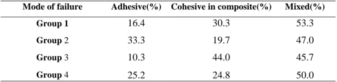
The failure mode of each group is shown in Table 2. Failure mode was mostly mixed in all groups.
Discussion
Availability of different bonding systems with different characteristics and compositions creates a great challenge to select a suitable bonding system for bonding of substrates with different surface topographies [15].
Bonding systems in this study were used with total-etch approach, as cutting the tooth by rotary instruments creates a smear layer with low surface
Table 1. Shear bond strength and standard deviation of experimental groups (MPa)
a, b: the same letters are not significant at the 0.05 level
Table 2. Failure mode distribution in each group
energy that inhibits resin infiltration into the dentin structure, and etching the surface removes the smear layer and increases the surface energy and intertubular porosities [8]. In laser prepared surfaces, a micromechanical pattern forms with more surface area and free from the smear layer [9,16-18]. Studies have shown that etching the surfaces prepared by laser before bonding, creates
a hybrid layer and resin tags, reduces
microleakage, and increases the bond strength [19,20].
The results of this study showed that the shear bond strength of composite to bur prepared dentin in both types of bonding systems was significantly higher than the Er:YAG prepared surfaces. This finding is in accordance with other studies [8,21-23].
The difference between bur and laser might be related to morphological changes of dentin surfaces prepared by laser. Er:YAG laser has
thermomechanical effect, and produces
microexplosions and thermochemical ablation in hard dental tissues [8]. This thermomechanical
effect of laser extends into deeper layers of dentin and threatens the integrity of the bonded restoration and might reduce the bond strength [12]. Caballous et al. [24] reported that ablation of dentin with laser melts and denatures the collagen fibers, and some of their cross links might be lost; subsequently, the fibrils will attach to each other. As a result, the resin would not infiltrate into the collagen fibrils, and the main bond strength is the result of infiltration of resin tags into dentinal tubules which has small contribution to bond strength [8,24].
In scanning electron microscopic evaluations of dentin surface prepared by laser, cracks were seen in denatured dentin under the hybrid layer that might have a negative effect on the quality of the bond and the bond strength. Additionally, thermographic analysis showed a denatured layer due to thermal effect and an amorphous and nonfibrous layer at the top of the lased dentin surface [12,25,26].
However, results of some studies are not consistent with ours [11,14,15,24]. This difference in findings
Std. deviation
Mean
Bond strength Groups
4.05 16.33a
1
4.44 17.50a
2
1.97 10.42b
3
2.74 8.08b
4
Mixed(%) Cohesive in composite(%)
Adhesive(%) Mode of failure
53.3 30.3
16.4
Group 1
47.0 19.7
33.3
Group 2
45.7 44.0
10.3
Group 3
50.0 24.8
25.2
Group 4
may be related to difference in the power, energy and frequency of Er:YAG laser, different bonding agents, experimental conditions, the method of bond strength evaluation, variations of dentin properties and expertise of the operator [8,27,28]. It is believed that acidic monomers in the Universal bonding agents dissolve the mineral crystals around the collagen fibrils and result in simultaneous monomer penetration. Also, 10-MDP creates a chemical bond with calcium in hydroxyapatite, and creates insoluble calcium MDP [12]. However, this bonding agent did not increase bond strength in laser prepared group. It seems that acid resistance of laser prepared dentin is due to reduction of calcium and phosphorous in the dentin surface, as well as entrapment of water in the porosities created in the surface that remains even after air drying the surface [29,30]. This probably decreases the efficacy of Single Bond Universal adhesive since 10-MDP is not able to effectively bond to dentin.
Another finding was that there was no significant difference in bond strength between the two bonding systems. This finding was not in accordance with that of Munoz et al, [31] since they reported that the bond strength of Adper Single Bond 2 was significantly higher. This difference might be related to difference in bond strength test, since they measured microtensile bond strength.
The failure mode was mainly mixed in all groups in our study; this finding is related to the adhesive interface, variations in the modulus of elasticity and stress absorption quality of the adhesive layer [32].
This was an in vitro study. In the oral environment, restorations are exposed to tensile, shear, torsional, thermal and occlusal stresses. These situations cannot be perfectly simulated in vitro. Thus, the results should be interpreted with caution. However, in vitro experiments are the best reliable methods to assess the quality of materials before their use in clinical situations.
Conclusion
Within the limitation of this study, it seems that multimode universal adhesive system was not able to increase the bond strength in bur or laser treated
dentin, and their bond strength was comparable by that of total-etch systems.
Acknowledgement
This study was supported by Dental School of Tehran University of Medical Sciences (No: 93-1-69-22989).
References
1. Betrisey E, Krejci I, Di Bella E, Ardu S. The influence of stratification on color and appearance of resin composites. Odontology. 2016 May;104 (2):176-83.
2. Breschi L, Mazzoni A, Ruggeri A, Cadenaro M, Di Lenarda R, Dorigo ED. Dental adhesion review: aging and stability of the bonded interface. Dent Mater. 2008 Jan;24(1):90-101.
3. Korkmaz Y, Gurgan S, Firat E, Nathanson D. Shear bond strength of three different nano-restorative materials to dentin. Oper Dent. 2010 Jan-Feb;35(1):50-7.
4. Bekes K, Gernhardt CR, Makowski M, Blunck U, Schaller HG. Micro tensile bond strength of
adhesive systems combined with flowable
composites. Int Poster J Dent Oral Med. 2005;7 (3): Poster 282.
5. Perdigão J, Loguercio AD. Universal or multi-mode adhesives: why and how? J Adhes Dent. 2014 Apr;16(2):193-4.
6.Stanislawczuk R, Amaral RC, Zander-Grande C,
Gagler D, Reis A, Loguercio AD. Chlorhexidine-containing acid conditioner preserves the longevity of resin-dentin bonds. Oper Dent. 2009 Jul-Aug; 34(4):481-90.
7. Muñoz MA, Luque I, Hass V, Reis A,
Loguercio AD, Bombarda NH. Immediate bonding properties of universal adhesives to dentine. J Dent. 2013 May;41(5):404-11.
8. Pahlavan A, Mehmanchi M, Omrani LR,
Chiniforush N. Effect of air abrasion and erbium-doped yttrium aluminum garnet (Er: YAG) laser preparation on shear bond strength of composite to dentin. J Lasers Med Sci. 2013 Summer;4(3):127-130.
9.Armengol V, Jean A, Rohanizadeh R, Hamel H.
Scanning electron microscopic analysis of diseased and healthy dental hard tissues after Er: YAG laser irradiation: in vitro study. J Endod. 1999 Aug;25
(8):543-6.
10. Gurgan S, Kiremitci A, Cakir FY, Yazici E, Gorucu J, Gutknecht N. Shear bond strength of composite bonded to erbium: yttrium-aluminum-garnet laser-prepared dentin. Lasers Med sci. 2009 Jan;24(1):117-22.
11.Ramos TM, Ramos-Oliveira TM, Moretto SG,
de Freitas PM, Esteves-Oliveira M, de Paula Eduardo C. Microtensile bond strength analysis of adhesive systems to Er:YAG and Er,Cr: YSGG laser-treated dentin. Lasers Med Sci. 2014 Mar;29 (2):565-73.
12. Dunn WJ, Davis JT, Bush AC. Shear bond strength and SEM evaluation of composite bonded to Er: YAG laser-prepared dentin and enamel. Dent Mater. 2005 Jul;21(7):616-24.
13. Arami S, Shahabi S, Tabatabaie M,
Chiniforush N, Morshedi E, Torabi S. Assessing microleakage of composite restorations in class V cavities prepared by Er: YAG laser irradiation or diamond bur. J Conserv Dent. 2014 May;17(3): 216-9.
14. Omrani LR, Sabouri P, Abbasi M, Ahmadi E, Ghavam M. Shear bond strength of two types of glass ionomer to bleached dentin: Effect of delayed bonding and antioxidant agent. Open Dent J. 2016 Dec 30;10:720-727.
15. Türkmen C, Sazak-Öveçoğlu H, Günday M,
Güngör G, Durkan M, Öksüz M. Shear bond strength of composite bonded with three adhesives
to Er, Cr: YSGG laser-prepared enamel.
Quintessence Int. 2010 Jun;41(6):e119-24.
16.Jafari A, Shahabi S, Chiniforush N, Shariat A. Comparison of the shear bond strength of resin modified glass ionomer to enamel in bur-prepared or lased teeth (Er: YAG). J Dent (Tehran). 2013 Mar;10(2):119-123.
17. Keller U, Hibst R, Geurtsen W, Schilke R, Heidemann D, Klaiber B, et al. Erbium:YAG laser application in caries therapy. Evaluation of patient perception and acceptance. J Dent. 1998 Nov;26 (8):649-56.
18.Visuri SR, Gilbert JL, Wright DD, Wigdor HA,
Walsh JT Jr. Shear strength of composite bonded to Er: YAG laser-prepared dentin. J Dent Res. 1996 Jan;75(1):599-605.
19. Shahabi S, Chiniforush N, Bahramian H, Monzavi A, Baghalian A, Kharazifard MJ. The effect of erbium family laser on tensile bond
strength of composite to dentin in comparison with conventional method. Lasers Med Sci. 2013 Jan;28 (1):139-42.
20. Kataumi M, Nakajima M, Yamada T, Tagami J. Tensile bond strength and SEM evaluation of Er: YAG laser irradiated dentin using dentin adhesive. Dent Mater J 1998;17(2):125-38.
21. Gurgan S, Kiremitci A, Cakir FY, Yazici E, Gorucu J, Gutknecht N. Shear bond strength of composite bonded to erbium: yttrium-aluminum-garnet laser-prepared dentin. Lasers Med Sci. 2009 Jan;24(1):117-22.
22.Koliniotou-Koumpia E, Kouros P, Zafiriadis L, Koumpia E, Dionysopoulos P, Karagiannis V. Bonding of adhesives to Er: YAG laser-treated dentin. Eur J Dent. 2012 Jan;6(1):16-23.
23. da Graça Lopes RM, Steiner-Oliveira C,
Aranha AC, Castellan CS, Bello-Silva MS, Júnior ND, et al. Microtensile bond strength to Er: YAG laser pretreated dentin. Braz Dent Sci. 2014 Jan;17 (1):54-61.
24. Ceballos L, Toledano M, Osorio R, García-Godoy F, Flaitz C, Hicks J. ER-YAG laser pretreatment effect on in vitro secondary caries formation around composite restorations. Am J Dent. 2001 Feb;14(1):46-9.
25.Schein MT, Bocangel JS, Nogueira GE, Schein
PA. SEM evaluation of the interaction pattern between dentin and resin after cavity preparation using Er: YAG laser. J Dent. 2003 Feb;31(2):127-35.
26. Bertrand MF, Hessleyer D, Muller-Bolla M, Nammour S, Rocca JP. Scanning electron microscopic evaluation of resin–dentin interface after Er: YAG laser preparation. Lasers Surg Med. 2004;35(1):51-7.
27. Sasaki LH, Lobo PD, Moriyama Y, Watanabe
IS, Villaverde AB, Tanaka CS, et al. Tensile bond strength and SEM analysis of enamel etched with Er:YAG laser and phosphoric acid: a comparative study in vitro. Braz Dent J. 2008;19(1):57-61. 28. Korkmaz Y, Gurgan S, Firat E, Nathanson D. Shear bond strength of three different nano-restorative materials to dentin. Oper Dent. 2010 Jan-Feb;35(1):50-7.
29.Shirani F, Birang R, Malekipur MR, Zeilabi A, Shahmoradi M, Kazemi S, et al. Adhesion to Er: YAG laser and bur prepared root and crown dentine. Aust Dent J. 2012 Jun;57(2):138-43.
30. Hibst R, Keller U. Experimental studies of the application of the Er: YAG laser on dental hard substances: I. Measurement of the ablation rate. Lasers Surg Med. 1989;9(4):338-44.
31. Muñoz MA, Luque I, Hass V, Reis A,
Loguercio AD, Bombarda NH. Immediate
bonding properties of universal adhesives to
dentine. J Dent. 2013 May;41(5):404-11.
32. Bouillaguet S, Gysi P, Wataha JC, Ciucchi B, Cattani M, Godin C, et al. Bond strength of composite to dentin using conventional, one-step, and self-etching adhesive systems. J Dent. 2001 Jan;29(1):55-61.