Cystic Fibrosis Transmembrane Regulator-independent
Release of ATP
ITS IMPLICATIONS FOR THE REGULATION OF P2Y2RECEPTORS IN AIRWAY EPITHELIA*
(Received for publication, January 13, 1998, and in revised form, March 25, 1998)
William C. Watt, Eduardo R. Lazarowski‡, and Richard C. Boucher
From the Cystic Fibrosis Research and Treatment Center, Department of Medicine, University of North Carolina, Chapel Hill, North Carolina 27599-7248
The cystic fibrosis (CF) transmembrane regulator (CFTR) is a cyclic AMP-dependent Cl2channel that is defective in CF cells. It has been hypothesized that CFTR exhibits an ATP release function that controls the airway surface ATP concentrations. In airway epithelial cells, CFTR-independent Ca21-activated Cl2 conduct-ance is regulated by the P2Y2receptor. Thus, ATP may function as an autocrine signaling factor promoting Cl2 secretion in normal but not CF epithelia if ATP release is defective. We have tested for CFTR-dependent ATP release using four independent detection systems. First, a luciferase assay detected no differences in ATP con-centrations in the medium from control versus cyclic AMP-stimulated primary normal human nasal epithelial (HNE) cells. A marked accumulation of extracellular ATP resulted from mechanical stimulation effected by a medium displacement. Second, high pressure liquid chromatography analysis of3H-labeled species released from [3H]adenine-loaded HNE cells revealed no differ-ences between basal and cyclic AMP-stimulated cells. Mechanical stimulation of HNE cells again resulted in enhanced accumulation of extracellular [3H]ATP and [3H]ADP. Third, when measuring ATP concentrations via nucleoside diphosphokinase-catalyzed phosphoryla-tion of [a-33P]dADP, equivalent formation of [33P]dATP was observed in the media of control and cyclic AMP-stimulated HNE cells and nasal epithelial cells from wild-type and CF mice. Mechanically stimulated [33P]dATP formation was similar in both cell types. Fourth, 1321N1 cells stably expressing the human P2Y2 receptor were used as a reporter system for detection of ATP via P2Y2receptor-promoted formation of [3 H]inosi-tol phosphates. Basal [3H]inositol phosphate accumula-tion was of the same magnitude in control and CFTR-transduced cells, and no change was observed following addition of forskolin and isoproterenol. In both cell types, mechanical stimulation resulted in hexokinase-attenuable [3H]inositol phosphate formation. In sum-mary, our data suggest that ATP release may be trig-gered by mechanical stimulation of cell surfaces. No evidence was found supporting a role for CFTR in the release of ATP.
The wide distribution of cell surface P2 receptors (1, 2) and the presence in most tissues of ectoenzymes that rapidly de-grade extracellular nucleotides (3) support the notion that reg-ulated process(es) for the cellular release of ATP may exist. Indeed, ATP has been found in an extracellular location in many tissues (4 –10), and ATP secretion from intracellular granules during platelet activation as well as nerve transmis-sion are well described events where physiological release of nucleotides occurs (4). However, in most tissues where signifi-cant ATP concentrations in the extracellular space have been detected, the mechanism(s) of ATP release have not been identified.
In the airways, the volume and composition of the liquid secretions may be regulated by extracellular nucleotides (11– 14). In cystic fibrosis (CF)1airway epithelia, the P2Y
2receptor
is linked to a Ca21-dependent chloride channel that provides an alternative Cl2 secretory pathway to the CF transmem-brane regulator (CFTR) Cl2channel (11). This alternative chlo-ride channel (Cla) has been identified as a potential target for
therapy of CF lung disease. The localization of P2Y2receptors
on the apical surface of airway epithelia suggests the possibil-ity that these receptors are regulated endogenously by the release of ATP onto the lumen.
It has been recently proposed that CFTR itself modulates the composition of airway surface liquids by acting as a channel for ATP, regulating Clapathways via activation of P2Y2receptors (15). In CF with defective CFTR, an implication of this hypoth-esis is that resting levels of ATP would be reduced, resulting in reduced Claas well as CFTR activation. The notion that CFTR
mediates the release of ATP evolved from studies showing that protein kinase A stimulated a single channel (CFTR) current in CFTR-expressing cells when 100 mM ATP was present in the
intracellular compartment (15, 16). In addition, CFTR was shown to regulate the activity of a second chloride channel in excised patches, an activity thought to reflect ATP release and activation of outwardly rectifying Cl2channels by P2Y2
recep-tors (15). However, other studies found either no evidence for CFTR-mediated ATP conductance (17–19) or that some but not all CFTR Cl2channels could be associated with an ATP per-meability (20). Measurements of ATP released from cells have also produced results that either support (15, 21) or do not support (22, 23) a role for CFTR in the regulation of ATP release. The current debate (24 –26) reflects the uncertainty that prevails on this issue (reviewed in Ref. 27). In this study, we tested the function of CFTR as a pathway for ATP release
* This work was supported by National Institutes of Health Grants HL34322 and HL42384 and by Cystic Fibrosis Foundation Grant R026. W. C. W. and E. R. L. contributed equally to this work. The costs of publication of this article were defrayed in part by the payment of page charges. This article must therefore be hereby marked “advertisement” in accordance with 18 U.S.C. Section 1734 solely to indicate this fact.
‡ To whom correspondence should be addressed: Dept. of Medicine, CB 7248, School of Medicine, University of North Carolina, Chapel Hill, NC 27599-7248. Tel.: 919-966-7046: Fax: 919-966-7524; E-mail: [email protected].
1The abbreviations used are: CF, cystic fibrosis; CFTR, cystic fibrosis
transmembrane regulator; Cla, alternative chloride channel; HNE,
hu-man nasal epithelial; DMEM-H, Dulbecco’s modified high glucose Ea-gle’s medium; HPLC, high pressure liquid chromatography; NDPK, nucleoside diphosphokinase.
© 1998 by The American Society for Biochemistry and Molecular Biology, Inc. Printed in U.S.A.
using both biochemical methods for directly measuring ATP release in intact cells and an assay utilizing the human P2Y2
receptor as a biological reporter for ATP release within the relevant cellular biophase (receptor domain).
MATERIALS AND METHODS
Cell Culture—Primary cultures of human nasal epithelial (HNE) cells and immortalized nasal epithelial cells from either normal or CF mice (28) were grown as polarized epithelia on 12.5-mm porous Transwell Col filters (Costar) as reported previously (29). Assays with HNE cells were carried out 7–10 days after seeding, a time coincident with the development of the maximal ion transport activity (29). T84 human colonic carcinoma cells, a cell line expressing high levels of endogenous CFTR (30), were grown as a polarized epithelium onto cross-linked collagen supports as described previously (30). Control and CFTR-transduced NIH-3T3 fibroblasts were grown on 12-well plastic plates as described (31). HP2U-1321N1 cells, a clonal cell line derived from human astrocytoma cells stably expressing the human P2Y2
re-ceptor (32), were grown on 12-well plastic plates (inositol phosphate assay) or on 6-well plates (36Cl2 efflux measurement and Western
blotting) in DMEM-H containing 5% fetal bovine serum and antibiotics as described previously (9).
Luciferin/Luciferase Assay—HNE cells were preincubated for 1 h in a Krebs-Ringer solution (2.4 mMK2HPO4, 0.4 mMKH2PO4, 115 mM
NaCl, 1.2 mMMgCl2, 1.2 mMCaCl2, 25 mMNaH2CO3, and 5 mMglucose)
at 37 °C and 5% CO2. The cells were exposed for the indicated times to
the designated drug. The mucosal medium (0.3 ml) was collected, cen-trifuged to remove potentially detached cells, and boiled for 1 min. A 100-ml sample aliquot was diluted with 200ml of H2O prior to
meas-urements. The luciferin/luciferase mixture (300mMluciferin, 5mg/ml
luciferase, 25 mMHEPES (pH 7.8), 6.25 mMMgCl2, 0.63 mMEDTA, 75 mMdithiothreitol, and 1 mg/ml bovine serum albumin) was added to
samples via an LB953 AutoLumat luminometer (Berthold GmbH, Wild-bad, Germany), and the sample luminescence was compared with an ATP standard curve performed for each individual experiment. To assess intracellular ATP content, cells were lysed with 5% trichloroace-tic acid followed by ethyl ether extraction and neutralization.
Release of [3H]Adenine-labeled Nucleotides—The cells were labeled
for 3 h with 10mCi/ml [3H]adenine as described previously (33). Labeled
cells were exposed to the indicated drug without washing away the label to avoid unnecessary agitation of the cells. The mucosal medium was collected, and3H-labeled species were resolved by high pressure liquid
chromatography (HPLC).
Nucleoside Diphosphokinase (NDPK)-catalyzed Formation of [a-33P]dATP—NDPK catalyzes the phosphorylation of nucleoside
diphosphates utilizing ATP as theg-phosphate donor molecule (34). We used [a-33P]dADP, obtained as described previously (29), as the
accep-tor molecule to quantitatively determine the formation of [a-33P]dATP
as a function of ATP concentration. HNE cells and nasal epithelial cells from either wild-type or CF mice were preincubated for 1 h in 0.3 ml of (mucosal) HEPES (pH 7.4)-buffered DMEM-H (HEPES/DMEM). Incu-bations were in the presence of 0.5 units/ml NDPK and 10 nM
[a-33P]dADP (0.2mCi) added to the mucosal bath.33P-Labeled species
were resolved by HPLC.
Quantification of Nucleotides by HPLC—Nucleotides were separated by HPLC (Shimadzu) via a strong anion-exchange column (Rainin In-strument Co. Inc.) with a mobile phase developed from 0.45 M
NH4COOH (pH 4.8) to 0.5MNa2H2PO4(pH 2.7) over a 30-min period
(9). Radioactivity was measured on line with a Radiomatic 500TR analyzer (Packard Instrument Co.). Species were identified and quan-tified as described previously (9).
Release of51Cr from HNE Cells—Confluent polarized HNE cells were
incubated for 3 h in HEPES/DMEM containing 10mCi of [51Cr]Na 2CrO3
added to the mucosal bath. The cells were washed (four times) and preincubated for 1 h in HEPES/DMEM. The mucosal solution (0.5 ml) was removed at the times indicated, and the radioactivity was quanti-fied with a Cobra Autogamma counter (Packard Instrument Co.).
Expression of CFTR in HP2U-1321N1 Astrocytoma Cells— Subcon-fluent HP2U-1321N1 cells grown on 12-well plastic plates were infected with adenoviral vector (53109particles/well) containing DNA
encod-ing either the CFTR gene (AdCFTR) or LacZ gene (AdLacZ) as de-scribed (31). Cells were assayed for CFTR expression 48 h after infec-tion. For Western blot analysis, CFTR was localized with an antiserum raised against a carboxyl-terminal peptide of CFTR (35). [36Cl]Chloride
efflux was quantified as described previously (31).
Measurement of [3H]Inositol Phosphates—HP2U-1321N1 cells were
labeled overnight in 0.5 ml of inositol-free DMEM-H containing 4
mCi/mlmyo-[3H]inositol. At the time of assay, 10 m
MLiCl was added to the cells for 15 min, followed by a further 15-min incubation in the presence of the indicated drugs. Incubations were terminated by the addition of 5% trichloroacetic acid, and the resulting [3H]inositol
phos-phates were separated and quantified by chromatography on Dowex columns as described previously (9).
Reagents—ATP and dATP were purchased from Pharmacia (Upp-sala, Sweden). Hexokinase, NDPK, forskolin, and dADP were from Boehringer Mannheim. Luciferin, luciferase, and isoproterenol were obtained from Sigma. [3H]Adenine (17 Ci/mmol) andmyo-[3H]inositol
(20 Ci/mmol) were from American Radiolabeled Chemicals (St. Louis, MO). [a-33P]dATP (3000 Ci/mmol), [51Cr]Na
2CrO3(300 –500 mCi/mg
cromium), and [36Cl]NaCl (3 mCi/g chloride) were from Amersham
Pharmacia Biotech.
Expression of the Results—Except where stated otherwise, pooled data are expressed as means6S.E. and are representative of at least three independent experiments performed with duplicate or triplicate samples. For statistical comparisons, unpairedttest was used, andp,
0.05 was considered significant.
RESULTS
Release of ATP from HNE Cells—ATP was quantified in the mucosal medium bathing HNE cells utilizing the luciferin/ luciferase method. Under resting conditions,i.e.the cells were kept undisturbed for 1 h prior to the assay, accumulation of ATP was 18.563.4 pmol/106cells (3.360.6 n
M in 0.3 ml of
medium;n59). Cell monolayers incubated with forskolin and isoproterenol (20mMeach, 10 min), a drug combination that is
maximally effective in initiating cyclic AMP-dependent Cl2 secretion via CFTR (33), exhibited a concentration of (mucosal) ATP of 4.260.7 nM(25.164.1 pmol/106cells;n59), a value
not significantly different from control incubations. In contrast, perturbing the cell surface by a medium change (data not shown) or by gently pipetting the mucosal medium up and down twice resulted in a marked accumulation of mucosal ATP (49.568 nM, 297661 pmol/106cells). Mechanically released
ATP by a medium displacement represented 0.9 –1.6% of the cellular ATP content, and it was unaffected by exposing the cells to the cyclic AMP mixture (51612 nM, 306679 pmol/106
cells). The possibility that cell lysis occurred during mechanical stimulation was investigated with51Cr-loaded HNE cells. No
changes in51Cr base-line levels were observed during a 10-min
period following a medium displacement (Table I). Moreover, base-line radioactivity remained unchanged after five repeti-tive medium displacements (data not shown). Thus, although we cannot rule out the possibility that a small number of damaged cells contributed to51Cr base-line levels, our results
suggest that ATP was released from intact (not damaged) cells. Consistent with this, we have recently shown that no cell lysis occurred during mechanically promoted ATP release from 1321N1 cells (36).
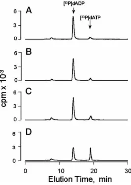
Release of [3H]Adenine-labeled Nucleotides—We loaded the intracellular pool of ATP with [3H]adenine, and the release of 3H-labeled species was quantified by HPLC. Because the
rela-tively large release of [3H]ATP during medium changes (data
not shown and Ref. 9) could mask a potential contribution by CFTR, a protocol was utilized that avoided cell washes. HPLC analysis of (mucosal) medium bathing resting [3
H]adenine-labeled HNE cells demonstrated a small peak of [3H]ADP,
barely distinguishable from background levels (Fig. 1A) (non-incorporated [3H]adenine eluted at 5– 8 min). No increase in
[3H]ATP or [3H]ADP accumulation was observed in forskolin/
isoproterenol-stimulated HNE cells (Fig. 1B). Incubation of cells with ionomycin to induce elevation of intracellular Ca21
resulted in a small accumulation of extracellular [3H]ATP and
greater accumulation of [3H]ADP (Fig. 1C). A marked
accumu-lation of [3H]ATP and [3H]ADP was observed after a medium
(31). In all cases, addition of forskolin/isoproterenol (2–10 min) promoted no release of [3H]adenine-labeled species, but a large
accumulation of 3H-labeled nucleotides was observed after a
medium displacement irrespective of the presence of CFTR (data not shown).
ATP-dependent Conversion of [a-33P]dADP to [a-33P]dATP— The possibility that CFTR mediates the release of ATP from a nucleotide pool not accessed by [3H]adenine (37) was
investi-gated by trapping released ATP with NDPK and [a-33P]dADP.
The NDPK-catalyzed conversion of [a-33P]dADP to
[a-33P]dATP was measured by HPLC as a function of ATP
concentration (Fig. 2). Moreover, since exogenous NDPK activ-ity greatly exceeds endogenous ecto-ATPase activactiv-ity, the assay resulted in a system that effectively locks the g-phosphate of ATP onto the [a-33P]dADP molecule upon release. Fig. 3
illus-trates the conversion of [a-33P]dADP to [a-33P]dATP in the
mucosal HNE cell baths under various conditions. A basal conversion (1663%) of [a-33P]dADP to [a-33P]dATP was
ob-served with cells that had not been treated with agonists (Fig. 2A). Addition of forskolin and isoproterenol (20mMeach) did not result in increased conversion (1464%) of [a-33P]dADP to
[a-33P]dATP, indicating that elevation of cellular cyclic AMP
did not promote the release of ATP (Fig. 2B). However, an ;2-fold greater conversion (26 6 3%) of [a-33P]dADP to
[a-33P]dATP was observed following addition of 1m
M
ionomy-cin, consistent with a Ca21-promoted ATP release (Fig. 2C). A 4-fold increased formation (7166%) of [a-33P]dATP occurred
following mechanical stimulation of the cells (Fig. 2D). The calculated ATP concentrations were 661, 561, 1763, and 97611 nMfor control cells, cAMP-stimulated cells,
ionomycin-treated cells, and mechanically stimulated cells, respectively. This protocol was repeated with immortalized nasal epithe-lial cells derived from either normal or CF mice. The results, summarized in Fig. 4, showed no differences between normal and CF cells under basal or forskolin/isoproterenol-stimulated conditions. The increased [33P]dATP formation observed after
mechanical stimulation was also similar in control and CF mouse cells. The muscarinic-cholinergic agonist carbachol pro-moted a small increase in [33P]dATP formation, although this
was not statistically significant.
The P2Y2Receptor as a Biosensor for ATP—Finally, in an attempt to assay for CFTR-dependent ATP release in the rel-evant physiological environment,i.e.the liquid layer associated with the cell surface, we used the P2Y2receptor as a biosensor
for ATP to investigate the role of CFTR in the activation of P2Y2receptors in intact cells. As such, we took advantage of a
cell line (1321N1 human astrocytoma cells) that is null for expression of P2 receptors (32). First, we achieved a high ex-pression level of P2Y2 receptors by retroviral infection of 1321N1 cells with the cDNA encoding the human P2Y2
recep-tor (32). 1321N1 cells stably expressing the human P2Y2
re-ceptor (HP2U-1321N1 cells) exhibit a marked ATP-stimulated formation of inositol phosphates, whereas control cells are un-responsive (9). Overexpression of the P2Y2receptor in 1321N1
cells resulted in large receptor reserve, and consequently, ATP potency in HP2U-1321N1 cells (EC505180 nM(9)) is increased
30-fold relative to ATP potency observed with the P2Y2
recep-tor natively expressed in airway epithelial cells (38). More important, significant accumulation of inositol phosphates in HP2U-1321N1 cells was detectable at concentrations of ATP as low as 10 nM (see Fig. 6 and Ref. 9). Second, we infected the
HP2U-1321N1 cells with an adenoviral vector containing the CFTR cDNA (AdCFTR) or with adenoviral vectors encoding the
LacZgene (AdLacZ) as a control and assayed for the effect of CFTR expression on the accumulation of inositol phosphates.
FIG. 1.Release of3
H-labeled species from [3
H]adenine-labeled HNE cells.[3H]Adenine-loaded HNE cells were incubated (300ml of
mucosal bath) for 10 min with vehicle (A), 20mMisoproterenol and 20
mMforskolin (B), or 1mMionomycin (C), or they were subjected to a gentle medium displacement by withdrawing and placing back twice 150ml of the mucosal medium (D). The [3H]adenine-labeled species
present in the mucosal medium were analyzed by HPLC as detailed under “Materials and Methods.”
FIG. 2. ATP-dependent conversion of [a-33P]dADP to [a-33P]dATP. Incubations were for 10 min in 0.5 ml of DMEM-H containing 0.5 units/ml NDPK, 10 nM[a-33P]dADP, and the indicated
concentrations of ATP. The formation of [a-33P]dATP was quantified by
HPLC as indicated under “Materials and Methods.” The data are the means6S.E. from four experiments performed in duplicate. TABLE I
Release of51Cr from HNE cells
Mechanical stimulation was applied to [51Cr]Na
2CrO3-labeled HNE
cells by gently pipetting up and down 0.25 ml of the mucosal medium twice. Undisturbed and stimulated cells were incubated for the times indicated, and the radioactivity released into the mucosal medium was quantified. The results are expressed as the means6S.E. from tripli-cate samples, and they are representative of two independent experi-ments performed under similar conditions.
Control Mechanical stimulation
cpm cpm
0 min 775652 752630
1 min 8066124 8506172
5 min 7726209 7306182
10 min 7806234 722675
Fig. 5Ashows a Western blot for CFTR of lysates from AdLacZ-or AdCFTR-infected cells. A band (;180 kDa), revealed by an anti-CFTR antiserum (antiserum 858 (35)), was present in AdCFTR-infected cells, but not in cells infected with theLacZ
vector. A functional cyclic AMP-activated Cl2permeability was demonstrated in AdCFTR-infected HP2U-1321N1 cells, but not in AdLacZ-infected cells, as indicated by forskolin- and isopro-terenol-stimulated36Cl2efflux (Fig. 5B).
Fig. 6 illustrates the effect of CFTR expression and cyclic
AMP-dependent activation on P2Y2 receptor-promoted
accu-mulation of inositol phosphates in HP2U-1321N1 cells. Basal accumulation of inositol phosphates was indistinguishable among control HP2U-1321N1 cells (i.e. cells that were not infected with adenovirus) and AdLacZ- and AdCFTR-infected HP2U-1321N1 cells, indicating that the mere expression of CFTR did not result in basal ATP-mediated activation of P2Y2 receptors. To test the effect of cyclic AMP-activated CFTR, inositol phosphates were measured following addition of for-skolin and isoproterenol. No increase in the total formation of inositol phosphates was observed with any cell type in response to this maneuver (Fig. 6). As a control for the sensitivity of the cells used in this assay, the cells were exposed to 10 nM
exog-enous ATP, and similar 3– 4-fold increases in inositol phos-phate accumulation were observed in the three cell types. We also tested for expression of a Ca21iregulation of ATP release in astrocytoma cells by addition of ionomycin, which resulted in a significant accumulation of inositol phosphates in all three cell types (Fig. 6) that was abolished when hexokinase was included in the assay (data not shown). We have previously shown that wild-type as well as HP2U-1321N1 cells release considerable amounts of ATP into the medium during a me-dium change (9, 32). Consistent with these previous reports, displacement of the medium bathing HP2U-1321N1 cells re-sulted in a marked accumulation of inositol phosphates (Fig. 6) that was sensitive to hexokinase and that was similar in mag-nitude to accumulation stimulated by 100 –300 nM ATP (data
not shown). No difference in response to this maneuver was observed between control cells or cells expressing CFTR (Fig. 6). We conclude from these experiments that CFTR has no role in ATP release, whereas elevation of intracellular calcium and/or mechanical stimulation of HP2U-1321N1 human astro-cytoma cells results in the release of ATP by a CFTR-independ-ent mechanism.
DISCUSSION
This study demonstrates the presence of ATP in the mucosal surface liquid of nonstimulated airway epithelial cells and that accumulation of extracellular ATP increases substantially when cells are subjected to mechanical stimuli. Unlike previous studies in which CFTR was reported to act as an ATP channel
FIG. 3.Conversion of [a-33
P]dADP to [a-33
P]dATP in the me-dium bathing HNE cells.The cells were incubated with vehicle (A), forskolin and isoproterenol (B), or ionomycin (C), or they were subjected to a medium displacement (D) as detailed in the legend of Fig. 1. Incubations were for 10 min in the presence of 0.5 units/ml NDPK and 10 nM[a-33P]dADP, and the resultinga-33P-labeled species were
quan-tified by HPLC as described under “Materials and Methods.” All addi-tions were to the mucosal medium. The results are representative of three experiments performed in duplicate.
FIG. 4.Conversion of [a-33
P]dADP to [a-33
P]dATP in the me-dium bathing mouse nasal epithelial cells.Cells from wild-type (WT;white bars) or CF (black bars) mice were incubated with vehicle, 20mMforskolin and 20mMisoproterenol (forsk/isoprot), or 1 mM car-bachol (carb), or they were subjected to a medium displacement ( medi-um displac). Incubations were for 10 min in the presence of 0.5 units/ml NDPK and 10 nM[a-33P]dADP, and the resultinga-33P-labeled species were quantified by HPLC as descried under “Materials and Methods.” All additions were to the mucosal medium. The results are expressed as the percent conversion of [a-33P]dADP to [a-33P]dATP, and the data represent the means6S.E. from three experiments performed in du-plicate. *,p,0.05.
FIG. 5.Expression of CFTR in HP2U-1321N1 cells.Subconfluent
HP2U-1321N1 cells grown on 12-well (A) or 6-well (B) plastic plates were infected with adenovirus harboring the CFTR (AdCFTR) orLacZ
(AdLacZ) plasmid (31).A, AdLacZ-infected cells, AdCFTR-infected cells, and (control) T84 cells were lysed with urea lysis buffer, and proteins (50 mg/lane) were separated by SDS-polyacrylamide gel electrophoresis and subsequently transferred to a nitrocellulose membrane. CFTR was localized with antiserum 858 (35). B, shown is released 36
Cl from
36Cl-loaded cells. Forskolin and isoproterenol (20 m
Meach) were
(15, 16, 21), we found no evidence involving CFTR in the release of ATP from intact epithelial and non-epithelial cells. The cause of this discrepancy may reside, at least in part, in the methodological approaches.
Previously, studies implicating CFTR as an ATP channel have employed permeabilized cells, membrane patches, and/or repetitive changes of cell media and in general have subjected the cells to stresses not consistent with physiological condi-tions. We have used both classical methods for extracellular detection of ATP,e.g.the luciferase assay, and HPLC separa-tion of3H-labeled nucleotides released from [3
H]adenine-pre-loaded cells as well as newly developed approaches for trapping ATP with NDPK and have adopted conditions in which sam-pling for ATP release was carefully controlled. Although we were able to detect ATP accumulation in the liquids bathing the surface of resting normal HNE cells, we observed no differ-ences after incubating the cells with agents that promoted elevation of intracellular cyclic AMP. Moreover, no differences were found in extracellular ATP accumulation with nasal epi-thelial cells from normal or CF mice under basal conditions or as a function of cyclic AMP pathway stimulation. We have shown here that a marked release of ATP occurred after me-chanical stimulation of airway epithelial cell surfaces by a medium displacement. However, mechanically stimulated re-lease of ATP was of the same magnitude in normal or CF airway epithelial cells as well as in control or CFTR-expressing non-epithelial cells. A CFTR-independent mechanically stimu-lated ATP release was also recently reported with colonic and human airway epithelial cell lines (23).
To directly test the potential regulation of P2Y2receptors by
CFTR, we have used a functional assay in which the P2Y2
receptor acted as a biosensor for releasable ATP. We were able to couple the advantage of the high expression level of recom-binant P2Y2 receptors attainable by retroviral infection of
1321N1 cells with adenovirus-mediated expression of CFTR in these cells to test the effect of CFTR activation on P2Y2
recep-tor-promoted inositol phosphate formation. Our hypothesis was that local accumulation of CFTR-releasable ATP in the un-stirred and consequently difficult to sample liquid layer on the
cell surface would be detected by coexpression of these two recombinant proteins on the surface of 1321N1 cells. Consist-ent with our previous reports showing mechanically promoted release of ATP from 1321N1 cells (9, 36), a sustained accumu-lation of inositol phosphates was observed following a medium displacement (Fig. 6). More important, inositol phosphate ac-cumulation in 1321N1 cells was of the same magnitude regard-less of CFTR expression, indicating that CFTR was not in-volved in mechanically induced ATP release. Although HP2U-1321N1 cells were sensitive to a 10 nM concentration of exogenously added ATP, no effect on inositol phosphates was observed following addition of forskolin and isoproterenol to CFTR-transduced HP2U-1321N1 cells as compared with con-trol cells. Thus, activation of CFTR in 1321N1 cells did not result in accumulation of extracellular ATP in concentrations high enough to promote P2Y2receptor activation. In summary, by functionally expressing both CFTR and P2Y2receptors in a null cell line, we were able to directly test the hypothesis that CFTR regulates the activity of P2Y2receptors. We have found no evidence to support this hypothesis.
The presence of P2Y2receptors in the airway epithelia (32, 38) and their coupling to a Cl2 secretory pathway on the mucosal surface of HNE cells (11) suggest that extracellular accumulation of nucleotides might be important for the regu-lation of ion permeabilities. The recent identification of an ectonucleotidase activity on HNE cells (29) suggests one mech-anism for regulating extracellular accumulation of nucleotides. Our present study indicates that ;20 pmol of ATP/106 cells
accumulate at steady state on the mucosal surface of resting normal HNE cells. This closely resembles the ATP concentra-tion observed in the lumen of normal human airways in vivo
(39). Furthermore, ATP transiently accumulates following me-chanical stimulation of cells in concentrations capable of acti-vating P2Y2receptors. Previously, mechanically induced
non-lytic release of ATP had been described in a variety of tissues. For example, changes in blood fluxes were shown to cause the release of ATP from endothelial cells (10, 40); shear forces promoted ATP release from mouse fibroblasts (6); and ATP release secondary to mechanical stimulation was also described with rat basophilic cells (7), 1321N1 human astrocytoma cells (9, 36), and rat hepatocytes (8). Stretch-promoted ATP release was reported with rabbit urinary bladder epithelial cells (41) and human lung adenocarcinoma Calu-3 cells (40), and ATP release triggered by bath turbulence was observed with T84 cells and with immortalized human tracheal epithelial cell lines (23, 26). Our current study extends these observations to primary human airway epithelial cells. It is not clear whether mechanical stimulation of airway epithelial cells and release across the epithelial surface occursin vivoand what its func-tional significance might be. Mechanical forces applied on the airways by air fluxes may represent a primary mechanism for autocrine and paracrine regulation of electrolyte homeostasis in airway epithelia.
In conclusion, the techniques used in this study for detecting extracellular ATP are thought to closely approximate physio-logical conditions, creating a system in which pertinent and reliable information is generated. We found that ATP is re-leased from each of the studied cell types in response to me-chanical stimulation and/or secondary to elevation of intracel-lular calcium levels. No evidence was found indicating that CFTR was involved in ATP release in airway epithelia from two species. This study suggests that CFTR itself does not directly effect ATP or regulate other possible ATP release pathways in the cells we have studied, but our data do not rule out possible CFTR-dependent regulation of ATP release pathways in other cell types. The occurrence of ATP release independent of
acti-FIG. 6.Effect of CFTR expression on P2Y2receptor-mediated formation of [3H]inositol phosphates.[3H]Inositol-labeled HP2U-1321N1 cells and cells infected with AdLacZ or AdCFTR were incubated for 15 min under the indicated conditions. Forskolin and isoproterenol (cAMP) were added at 20mMfinal concentrations, ATP at 10 nM, and ionomycin (ionom) at 1mM.medium displacindicates withdrawing and gently replacing the medium twice. The results represent the means6
S.E. from four experiments performed in triplicate.WT, wild-type. *,
vation of CFTR raises new possibilities for treatment of CF. For potential therapeutic targeting, it may important to identify, at the molecular level, the mechanism involved in nucleotide re-lease from airway epithelia.
Acknowledgments—We thank J. R. Yankaskas for assistance in ob-taining tissues and L. Brown for editorial assistance.
REFERENCES
1. Dubyak, G. R., and El-Moatassim, C. (1993)Am. J. Physiol.265,C577–C606 2. Fredholm, B. B., Abbracchio, M. P., Burnstock, G., Dubyak, G. R., Harden, T. K., Jacobson, K. A., Schwabe, U., and Williams, M. (1997)Trends Phar-macol. Sci.18,79 – 82
3. Zimmermann, H. (1996)Drug Dev. Res.39,337–352 4. Gordon, J. L. (1986)Biochem. J.233,309 –319
5. Milner, P., Bodin, P., Loesch, A., and Burnstock, G. (1990)Biochem. Biophys. Res. Commun.170,649 – 656
6. Grierson, J. P., and Meldolesi, J. (1995)J. Biol. Chem.270,4451– 4456 7. Osipchuk, Y., and Cahalan, M. (1992)Nature359,241–244
8. Schlosser, S. F., Burgstahler, A. D., and Nathanson, M. H. (1996)Proc. Natl. Acad. Sci. U. S. A.93,9948 –9953
9. Lazarowski, E. R., Watt, W. C., Stutts, M. J., Boucher, R. C., and Harden, T. K. (1995)Br. J. Pharmacol.116,1619 –1627
10. Milner, P., Bodin, P., Loesch, A., and Burnstock, G. (1992)J. Vasc. Res.29,
420 – 425
11. Mason, S. J., Paradiso, A. M., and Boucher, R. C. (1991)Br. J. Pharmacol.103,
1649 –1656
12. Jiang, C., Finkbeiner, W. E., Widdicombe, J. H., McCray, P. B., Jr., and Miller, S. S. (1993)Science262,424 – 427
13. Lethem, M. I., Dowell, M. L., Van Scott, M., Yankaskas, J. R., Egan, T., Boucher, R. C., and Davis, C. W. (1993)Am. J. Respir. Cell Mol. Biol.9,
315–322
14. Davis, C. W., Dowell, M. L., Lethem, M. I., and Van Scott, M. (1992)Am. J. Physiol.262,C1313–C1323
15. Schwiebert, E. M., Egan, M. E., Hwang, T., Fulmer, S. B., Allen, S. S., Cutting, G. R., and Guggino, W. B. (1995)Cell81,1063–1073
16. Reisin, I. L., Prat, A. G., Abraham, E. H., Amara, J. F., Gregory, R. J., Ausiello, D. A., and Cantiello, H. F. (1994)J. Biol. Chem.269,20584 –20591 17. Reddy, M. M., Quinton, P. M., Haws, C., Wine, J. J., Grygorczyk, R.,
Tabcharani, J. A., Hanrahan, J. W., Gunderson, K. L., and Kopito, R. R. (1996)Science271,1876 –1879
18. Li, C., Ramjeesingh, M., and Bear, C. E. (1996) J. Biol. Chem. 271,
11623–11626
19. Grygorczyk, R., Tabcharani, J. A., and Hanrahan, J. W. (1996)J. Membr. Biol.
151,139 –148
20. Pasyk, E. A., and Foskett, J. K. (1997)J. Biol. Chem.272,7746 –7751 21. Prat, A. G., Xiao, Y. F., Ausiello, D. A., and Cantiello, H. F. (1995)Am. J.
Physiol.268,C1552–C1561
22. Takahashi, T., Matsushita, K., Welsh, M. J., and Stokes, J. B. (1994)J. Biol. Chem.269,17853–17857
23. Grygorczyk, R., and Hanrahan, J. W. (1997) Am. J. Physiol. 272,
C1058 –C1066
24. Abraham, E. H., Okunieff, P., Scala, S., Vos, P., Oosterveld, M. J. S., Chen, A. Y., Shrivastav, B., and Guidotti, G. (1997)Science275,1324 –1325 25. Reddy, M. M., Quinton, P. M., Haws, C., Wine, J. J., Grygorczyk, R.,
Tabcharani, J. A., Hanrahan, J. W., Gunderson, K. L., and Kopito, R. R. (1997)Science275,1325
26. Grygorczyk, R., and Hanrahan, J. W. (1997)Science275,1325–1326 27. Devidas, S., and Guggino, W. B. (1997)Curr. Opin. Cell Biol.9,547–552 28. Yankaskas, J. R., Cantrell, C., Kelly, K., and Walstad, D. (1994)Pediatr.
Pulmonol.18,Suppl. 10, 195 (abstr.)
29. Lazarowski, E. R., Paradiso, A. M., Watt, W. C., Harden, T. K., and Boucher, R. C. (1997)Proc. Natl. Acad. Sci. U. S. A.94,2599 –2603
30. Stutts, M. J., Lazarowski, E. R., Paradiso, A. M., and Boucher, R. C. (1995)
Am. J. Physiol.268,C425–C433
31. Stutts, M. J., Gabriel, S. E., Olsen, J. C., Gatzy, J. T., O’Connell, T. L., Price, E. M., and Boucher, R. C. (1993)J. Biol. Chem.268,20653–20658 32. Parr, C. E., Sullivan, D. M., Paradiso, A. M., Lazarowski, E. R., Burch, L. H.,
Olsen, J. C., Erb, L., Weisman, G. A., Boucher, R. C., and Turner, J. T. (1994)Proc. Natl. Acad. Sci. U. S. A.91,3275–3279
33. Grubb, B., Lazarowski, E., Knowles, M., and Boucher, R. (1993)Am. J. Respir. Cell Mol. Biol.8,454 – 460
34. Ratliff, R. L., Weaver, R. H., Lardy, H. A., and Kuby, S. A. (1964)J. Biol. Chem.
239,301–309
35. Sarkadi, B., Bauzon, D., Huckle, W. R., Earp, H. S., Berry, A., Suchindran, H., Price, E. M., Olsen, J. C., Boucher, R. C., and Scarborough, G. A. (1992)
J. Biol. Chem.267,2087–2095
36. Lazarowski, E. R., Homolya, L., Boucher, R. C., and Harden, T. K. (1997)
J. Biol. Chem.272,24348 –24354
37. de Korte, D., Gouwerok, C. W., Fijnheer, R., Pietersz, R. N., and Roos, D. (1990)
Thromb. Haemostasis63,275–278
38. Brown, H. A., Lazarowski, E. R., Boucher, R. C., and Harden, T. K. (1991)Mol. Pharmacol.40,648 – 655
39. Donaldson, S. H., Boucher, R. C., and Knowles, M. R. (1996)Pediatr. Pulmo-nol.22,Suppl. 13, 289 (abstr.)
40. Thomas, S. A., and Hume, R. I. (1990)J. Gen. Physiol.95,569 –590 41. Ferguson, D. R., Kennedy, I., and Burton, T. J. (1997)J. Physiol.(Lond.)505,
![FIG. 6. Effect of CFTR expression on P2Y 2 receptor-mediated formation of [ 3 H]inositol phosphates](https://thumb-us.123doks.com/thumbv2/123dok_us/7935049.2109599/5.907.105.416.81.368/effect-cftr-expression-receptor-mediated-formation-inositol-phosphates.webp)
