As early as 1713, an Italian medical professor by the name of Bernadino Ramazzini recorded the health hazards associated with 52 occupations. In this document he noted that: ‘Almost all who make a living by sifting or measuring grain are short of breath and cachectic and rarely reach old age; in fact, they are very liable to lapse into orthopnoea and finally dropsy’.[1] With this description,
Ramazzini essentially provided the first description of hypersensitivity pneumonitis (HP) with the development of chronic respiratory failure and cor pulmonale. Subsequently in 1932, the first detailed descriptions of HP were made following an outbreak in 10 employees at a company that manufactured railroad ties.[2] These employees, who
were required to strip bark from maple logs, developed symptoms of dyspnoea, cough, night sweats, weight loss and sputum production. It was subsequently discovered that beneath the bark of the maple logs a black sooty dust was present from which a fungus called Cryptostroma corticale was isolated. The chest X-ray and clinical features were compatible with what we now accept as HP.
HP has long been considered an orphan disease, and over the last 10 - 15 years there remains one intriguing question – why do only a small percentage of those who are exposed develop the disease? There is a lack of consensus as to the definition of HP and this is reflected in the different definitions suggested by two major groups of international experts. The National Heart, Lung, and Blood Institute and the Office of Rare Diseases (NHLBI/ORD) workshop report defines HP as ‘a complex health syndrome of varying intensity, clinical presentation, and natural history. HP is the result of an immunologically induced inflammation of the lung parenchyma in response to inhalation exposure to a large variety of antigens’.[3] The
HP Study group defined HP as ‘a pulmonary disease with symptoms of dyspnoea and cough resulting from the inhalation of an antigen to which the patient has been previously sensitised’.[4] More recently,
HP has been accepted as a pulmonary disease consisting of a spectrum of granulomatous, interstitial, bronchiolar and alveolar-filling lung diseases with or without systemic manifestations (fever and weight loss) caused by the inhalation of a wide variety of organic aerosols and low molecular weight (LMW) chemical antigens to which the subject is sensitised and hyper-responsive. Sensitisation
and exposure alone in the absence of symptoms do not define the disease, which is characterised by a lymphocytic alveolitis and granulomatous pneumonitis, usually with improvement or complete recovery if exposure ceases, otherwise it leads to interstitial fibrosis if exposure continues.[5]
Aetiology
A wide group of causative antigens have been described for HP and new sources are continually being recognised (Table 1). The most commonly implicated antigens in the disease process are derived from thermophilic actinomycetes, such as Saccharopolyspora rectivirgula (which causes farmer’s lung) following exposure to mouldy hay, and others that cause Bagassosis and cheese-washer’s lung, as well as antigen derived from protein in the droppings, feathers or serum of birds (such as pigeons, budgerigars, ducks, parrots, etc.) which cause bird-fancier’s lung.[6,7] Increasingly duck and goose down used
in duvets and feather pillows have been implicated as possible causes for chronic HP, often mistakenly labelled as idiopathic pulmonary fibrosis (IPF).[8] Some LMW chemicals such as isocyanates (used in
polyurethane foams, paints and plastics), zinc, inks and dyes can act as haptens to induce HP. It may be useful to consider the implicated antigens in three groups: microbial (fungal, bacterial, mycobacterial), animal proteins (bird feathers, droppings and serum) and LMW chemicals (isocyanates).
Epidemiology
The prevalence of HP varies considerably around the world and is dependent on a number of factors including disease definition, diagnostic methods, occupational exposures (type and intensity), geographic conditions, industrial and agricultural practices, as well as host risk factors. Bird-fancier’s lung is usually the most common form of HP. HP is an uncommon condition that has been shown to make up 4 - 15% of all interstitial lung diseases,[9] and in
Britain the incidence has been found to be 0.9 cases per 100 000 person-years.[10] What remains an enigma is the low proportion of
patients who develop disease despite more widespread exposure, as was demonstrated in French dairy farmers, where only 0.5 - 3.0% developed disease.[11] In South Africa, in a single-centre tertiary
Hypersensitivity pneumonitis (HP) refers to the inflammatory lung condition that arises from inhalational exposure to various (mostly organic) antigens with the development of lymphocytic alveolitis and granulomatous pneumonitis. A myriad of antigens have been implicated and the list continues to grow. The clinical features are varied, implicated antigens protean, and the investigations complementary in making a diagnosis, but the condition remains a difficult one to diagnose, mostly due to a lack of clinical suspicion. In this article we discuss HP, its aetiology, clinical features as well as diagnosis and management.
S Afr Resp J 2015;21(3):70-76. DOI:10.7196/10.2015.v21i3.70
Hypersensitivity pneumonitis
H Khalfey, MB ChB, FCP SA, Cert Pulm SA
Pulmonologist, Life Vincent Pallotti Hospital, Pinelands, Cape Town
referral hospital over a period of 30 years, 40 patients were diagnosed with HP due to bird exposure.[12] In this group there was no gender
difference, 30/40 (75%) were of mixed race and 10/40 (25%) were white subjects, with no black or Asian subjects. The age range at presentation was 13 - 75 years (mean 49.2 (standard deviation 15.4) years). Fifty-five per cent were non-smokers, 20% ex-smokers and 25% were current smokers.
Pathogenesis
While up to 50% of exposed subjects develop sensitisation with positive antibodies, only about 3 -15% of bird fanciers develop HP.[13]
This suggests that the phenotypic expression of disease depends on host and environmental factors. The underlying immune mechanisms implicated in disease development have been demonstrated to be a combination of a Type III hypersensitivity (in acute HP) and Type IV delayed-type hypersensitivity (in chronic HP). A two-hit hypothesis has been postulated where genetic susceptibility or environmental factors serve as the first hit and increase the risk of development of disease after antigen exposure (second hit).[14] There are few studies
on the underlying genetic susceptibility in HP and available data increasingly point to abnormalities in the antigen processing and presentation pathways of the immune system. Polymorphisms associated with HLA-DR and DQ have been associated with increased risk for HP in populations with different genetic backgrounds.[15,16]
PSMB8 is an immunoproteasome catalytic subunit that is important in the generation of peptides presented by MHC class 1 molecules, and the PSMB8 KQ genotype has been found with increased frequency in subjects with HP.[17] One study reported that Mexican patients with
HP had increased expression of alleles Gly-637 and genotypes Asp-637/Gly-637 and Pro661/Pro661 on the TAP1 (transporters associated with antigen processing 1) gene, which may lead to an exacerbated immune response and hence increased susceptibility to developing HP.[18]
There is ongoing research into the immunopathogenesis of HP and we are likely to learn more in the future about the immune tolerance
occurring in most exposed individuals. Regulatory T cells (Treg) may
play an important role in this process, as has been shown in in vitro studies.[19,20]
In addition to underlying genetic susceptibility it has been shown that environmental factors such as exposure to parainfluenza viral infections in mice[21] and pesticide exposure in farmers may increase
susceptibility to development of clinical HP.[22]
The effects of smoking are interesting in that paradoxically smokers appear to have a reduced susceptibility to the development of HP and 95% of HP cases occur in non-smokers.[23] This may be due to a
decrease in the production of specific antibodies to inhaled organic antigens due to various immunosuppressive effects of tobacco smoke. Despite these relative protective effects of smoking, the smokers who do develop HP have lower vital capacities and a poorer 10-year survival in comparison with non-smokers.[24]
Clinical features
Three clinical forms of HP have been described: acute, subacute and chronic. However, it is often difficult to distinguish subacute HP from either acute or chronic as there is often overlap in clinical features, which blurs this distinction.
Acute HP
Patients typically give a history of flu-like symptoms occurring 4 - 8 hours following exposure to antigen and complain of dry cough, dyspnoea, fever, rigors, myalgia and malaise, which reach peak intensity at 12 - 24 hours and usually resolve completely within 48 hours if exposure ceases. These episodes will recur upon subsequent re-exposure to the antigen and are usually more intense. Clinical examination will reveal an acutely ill patient with tachypnoea, tachycardia, bibasal inspiratory crackles on auscultation of the chest and, in severe cases, acute respiratory failure evident on arterial blood gas analysis. Serum precipitating antibodies are usually positive in most cases and chest X-ray will reveal bilateral patchy or diffuse reticulonodular infiltrates mostly in the lower zones, with apical Table 1. Common antigens implicated in HP
Disease Antigen Source of antigen
Microbial – fungal and bacterial
Farmer’s lung (FLD) Bagassosis
Maple bark stripper’s lung Cheese-washer’s lung Hot tub lung
Summer-type pneumonitis
Saccharopolyspora rectivirgula Thermoactinomyces vulgaris Cryptostroma corticale
Penicillium casei, Aspergillus clavatus Mycobacterium avium complex Trichosporon cutaneum
Mouldy hay, grain Mouldy sugarcane Mouldy maple bark Mouldy cheese Hot tub mists
Contaminated old Japanese houses in summer months
Animal proteins
Bird-fancier’s lung (BFL) Animal-handler’s lung
Avian droppings, serum, feathers Rats, gerbils, hamsters
Pigeons, budgerigars, parrots, ducks Urine, serum, fur
Low molecular weight chemicals
Painter’s lung Plastic-worker’s lung
Isocyanates Anhydrides
sparing. High-resolution computerised tomography scanning (HRCT) will typically show multiple small nodules with bilateral areas of ground-glass opacification, pre-dominantly in the bases. Spirometry usually reveals a restrictive pattern with a reduced gas transfer. Both spirometry and radiology may often return to normal within 4 - 6 weeks if antigen exposure ceases.
Subacute HP
This is a difficult entity to distinguish from acute HP and usually occurs due to repeated low-grade exposure to antigens with the development of similar symptoms to acute HP that resolve within 24 hours of exposure, but due to ongoing exposure is associated with repeated bouts over a period of time. These tend to become more severe over time with longer recovery periods, but patients usually feel well inbetween attacks. The chest X-ray may be completely normal in between attacks but usually demonstrates fine nodular infiltrates in the mid to upper zones. The HRCT may show areas of linear fibrosis due to repeated bouts of inflammation with areas of micronodules and mosaic attenuation in the mid to upper zones. Pulmonary function tests usually show mild restriction with or without a reduced gas transfer.
Chronic HP
Chronic HP is the form most commonly seen by pulmonologists, as patients most often present late or acute and subacute forms are not recognised as such. Chronic HP occurs in about 5% of patients with HP[25]
and develops insidiously over months to years in those with ongoing exposure and is usually irreversible. Patients usually present with chronic, progressive cough often with sputum production and progressive dyspnoea. Examination usually reveals tachypnoea with bibasal fine inspiratory crackles. Clubbing may be present and usually represents a poorer prognosis.[26] Chronic respiratory
failure occurs in severe cases leading to cor pulmonale. In chronic HP, precipitating antibodies or specific IgG may be negative if there is time latency between exposure and presentation. The chest X-ray typically shows features of volume loss with bilateral diffuse reticulonodular infiltrates with coarse linear opacities and reticulation in an upper and mid-zone distribution. Multiple areas of fibrosis with patchy ground-glass opacification
and centrilobular nodularity with mosaic attenuation on expiratory views are usual. Honeycombing may also be present, which makes chronic HP difficult to distinguish from IPF. The lung function tests usually demonstrate restriction with reduced lung volumes and a reduced gas transfer. In up to 10%, an obstructive defect may be evident.[27]
Diagnosis
No single diagnostic biomarker or procedure exists to confirm a diagnosis of HP and the clinician is thus faced with a diagnostic dilemma. HP lacks unique features that distinguish it from other interstitial lung diseases, and a diagnosis relies on a high index of suspicion when a history of antigen exposure is obtained, together with a constellation of clinical, radiological, laboratory and pathological findings.
Establish a history of exposure
Careful history-taking is essential in order to elucidate a history of antigen exposure, which is the first critical prerequisite. This is especially difficult due to the fact that exposure may occur both directly and indirectly, such as may occur in bird-fancier’s lung where exposures to birds may occur in parks, railway stations and neighbours’ yards, as well as by birds in roofs of houses. Exposure to mouldy hay in barns is implicated in farmer’s lung. To further add to the difficulties in history taking, avian antigen has been reported to persist in a house for 6 months after removal of the bird.[28]
Imaging
Imaging by chest X-ray and HRCT is not always diagnostic, but adds the most value in reaching a diagnosis. A confident
Table 2. HRCT findings in HP.*
Stage of disease References Sample size Findings†
Acute Cormier et al.[30] N=20 Ground-glass opacities
Micronodules Mosaic perfusion Emphysema Honeycombing Mediastinal lymphadenopathy Subacute Hansell et al.[31] N=17 (including
9 with pigeon breeder’s disease and 4 with Farmer’s lung)
Generalised increase in attenuation of the lung Nodular pattern Reticular pattern Patchy air space opacification Remy-Jardin et al.[32] N=21 (pigeon
breeder’s disease) Micronodular pattern(<5 mm in diameter)
Ground-glass attenuation Emphysematous change Honeycombing Chronic Adler et al.[33] N=16
(antigen = ?) FibrosisGround-glass attenuation Nodules
Remy-Jardin et al.[32] N=24 (pigeon
breeder’s disease) HoneycombingGround-glass attenuation Micronodules
Emphysema *Reproduced with permission from Lacasse.[55]
radiological diagnosis has been shown to be accurate in up to 81%.[29] Radiological features
of each HP subtype have been addressed previously in this article. In acute HP, the following features have been described in descending order of frequency: ground-glass opacities, micronodules, mosaic perfusion, emphysema, honeycombing and mediastinal lymphadenopathy.[30]
These usually have a lower and mid-zone predominance. In subacute HP, the most common features are a micronodular pattern followed by reticulation, patchy airspace opacification and in some cases honeycombing, emphysematous change and fibrosis which tend to have a mid-zone predominance as well.[31,32] In chronic HP,
the predominant HRCT finding is that of fibrosis with ground-glass attenuation, honeycombing and micronodules as well as
emphysema.[32,33] This often spares the bases
but may be predominantly sub-pleural or peribronchovascular.
See Table 2, and Figures 1a and b and Figures 2a and b.
Specific circulating antibodies
These techniques detect IgG to specific antigens and a positive result merely reflects sensitisation and not necessarily disease. A positive result can be obtained in up to 10% of farmers and 40% of pigeon breeders without any evidence of clinical disease. However, a positive test in the correct clinical scenario strongly supports the diagnosis of HP.[34,35]
A negative test, however, does not exclude disease. Positive specific antibodies were shown to be a significant predictor of disease in the HP study.[4] Enzyme immunoassays
have now largely replaced the precipitation techniques and have been shown to be a more reliable technique. The test has demonstrated varying sensitivity and specificity. In acute HP in bird-fancier’s lung disease, antibody titres were markedly increased and showed
high sensitivity and specificity ranging from 75 to 100%, while in chronic HP, titres were only slightly increased with a sensitivity between 27 and 73% and specificity of 45 - 100%.[36] False negatives may also be seen
across the disease spectrum and this depends on the latency period between symptoms and testing, laboratory techniques and panels of antigens tested, etc.
Inhalational provocation test
This involves the inhalation of 2 mL of pigeon dropping extract (PDE 340 μg/mL) through a handheld nebuliser for a maximum period of 10 minutes. A number of parameters are recorded for 24 hours thereafter and a test is regarded as positive if three or more of the following are demonstrated: increased radiological abnormalities, increased A-a gradient >10 mmHg or decrease in transfer factor (TLCO) of >20%, decrease in forced vital capacity (FVC) >15%, increase in peripheral white blood cell count (WCC) of >30%, increase in C-reactive protein (CRP) >1.0 mg/dl, increase in body temperature >1oC and/or development of systemic
manifestations including chills and fatigue, development of respiratory symptoms such as cough and dyspnoea (Table 3).[37,38]
Inhalational provocation testing has been considered to be the ‘gold standard’ by Morell, in whose series the test performed with a sensitivity of 92% and specificity of 100% in patients with bird-fancier’s lung disease.[39]
Although it appears to be a useful test, it lacks standardisation with regards to antigen preparation, administration and monitoring and is not without risk to the patient. It is best conducted in research centres with expertise in the procedure.
Fig. 1a. Postero-anterior (PA) chest radiograph of acute HP due to pigeons showing diffuse ground-glass opacification. Reproduced with permission from Professor Ainslie.[12]
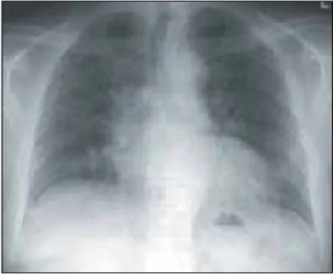
Fig. 1b. PA chest radiograph of patient with chronic HP due to pigeons showing small lungs with diffuse reticulonodular infiltration. Reproduced with permission from Professor Ainslie.[12]
Fig. 2a. Inspiratory HRCT film in patient with acute HP due to pigeons showing patchy ground-glass opacification and multiple small centrilobular nodules. Reproduced with permission from Professor Ainslie.[12]
Fig. 2b. Expiratory HRCT film in patient with acute HP due to pigeons showing patchy gas trapping (mosaic attenuation pattern). Reproduced with permission from Professor Ainslie.[12]
Table 3. Inhalation challenge testing Positive if 3 or more of the following:
Increased radiological abnormalities Increased A-a gradient >10 mmHg or decrease in TLCO of >20%
Decrease in VC of >15%
Increase in peripheral WCC of >30% Increase in CRP >1.0 mg/dL
Increase in body temperature >1oC and/or
development of systemic manifestations including chills and fatigue
Bronchoalveolar lavage (BAL)
Demonstrating BAL lymphocytosis is a highly sensitive method of determining lung inflammation in patients with suspected HP but the test lacks standardisation. BAL lymphocytosis >50% characterises HP and distinguishes it from other conditions, but it may be lower in smokers and those with predominantly fibrotic disease.[5]
Asymptomatic exposed individuals may also have a lymphocytosis that represents a low-grade alveolitis, while other conditions (such as sarcoidosis and organising pneumonia) may also have elevated BAL lymphocytes. Demonstrating a CD4/CD8ratio <1 is suggestive of HP in up to 34%[40] but the ratio
may be as high as that seen in sarcoidosis and is thus not recommended routinely anymore.[41,42]
Biopsy
Biopsy in the form of transbronchial biopsy or surgical lung biopsy is rarely needed for a diagnosis of acute HP. In chronic HP it is usually characterised by a triad of chronic
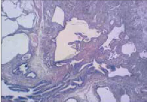
inflammatory infiltrates along small airways (cellular bronchiolitis), diffuse interstitial infiltrates of inflammatory cells (lymphocytes and plasma cells) and scattered non-necrotising granulomas (Figs. 3a and 3b).[43] Granulomas
Fig. 3a. Low-power image of a surgical lung biopsy in a patient with chronic HP showing patchy peribronchiolar inflammatory process.
Fig. 3b. High-power image of a surgical lung biopsy in a patient with chronic HP showing a peribronchiolar granuloma with multinucleated giant cells.
Table 4. Proposed diagnostic criteria for HP for clinical purposes.*
Author Major criteria Minor criteria
Terho[45] 1. Exposure to offending antigens (revealed by history
aerobiological or microbiological investigations of the environment, or measurements of antigen-specific IgG antibodies)
2. Symptoms compatible with HP present and appearing or worsening some hours after antigen exposure
3. Lung infiltrations compatible with HP visible on chest X-ray
1. Basal crepitant rales
2. Impairment of the diffusing capacity (TLCO) 3. Oxygen tension (or saturation) of the arterial blood either decreased at rest, or normal at rest but decreased during exercise
4. Restrictive ventilation defect in the spirometry
5. Histological changes compatible with HP 6. Positive provocation test whether by work
exposure or by controlled inhalation challenge
Richerson et al.[46] 1. The history and physical findings and pulmonary function tests
indicate an interstitial lung disease 2. The X-ray film is consistent
3. There is exposure to a recognised cause 4. There is antibody to that antigen Cormier et al.[47] 1. Appropriate exposure
2. Inspiratory crackles
3. Lymphocytic alveolitis (if BAL is done) 4. Dyspnoea
5. Infiltrates on chest radiographs or HRCT
1. Recurrent febrile episodes 2. Decreased transfer factor (TLCO) 3. Precipitating antibodies to HP antigens 4. Granulomas on lung biopsy (usually not required) 5. Improvement with contact avoidance or
appropriate treatment Schuyler et al.[48] 1. Symptoms compatible with HP
2. Evidence of exposure to appropriate antigen by history or detection in serum and/or BAL fluid antibody
3. Findings compatible with HP on chest radiograph or HRCT 4. BAL fluid lymphocytosis
5. Pulmonary histological changes compatible with HP 6. Positive natural challenge
1. Bibasilar rales 2. Decreased TLCO
3. Arterial hypoxaemia, either at rest or during exercise
occur in varying degrees across studies, and in an epidemiologic survey in Japan, it was demonstrated in 16.7% with BFL, 44.4% with summer-type HP and 60% with isocyanate HP.[44] Surgical lung
biopsy was only helpful in 37% in the HP study.[4]
Diagnostic criteria
The diagnosis of HP remains a difficult one and, as mentioned before, rests on a high index of suspicion in a patient with a history of exposure, with compatible clinical and radiological features complemented by further investigations. A number of diagnostic criteria have been studied and recommended but none have been effectively validated. Most diagnostic criteria essentially correspond with definitions of the disease (Table 4).[45-48] The HP study was a centred,
multi-national study that recruited patients with suspected HP with the aim of developing a predictive tool for the diagnosis of HP.[4] HP was
diagnosed in 199 out of 661 subjects in the study and they identified six significant predictors useful in assisting clinicians to arrive at a more accurate estimate of probability of HP. The following were identified as significant predictors:
The probability of HP in the HP study ranged from 98%, when all 6 predictors were present, to 0% when there were none. The authors
suggested that a probability of >90% is sufficient to rule in and a probability <10% to rule out HP, especially in areas of high and low prevalence respectively. The results of this study are thus useful to assess probability of HP but are not by definition diagnostic criteria. They may be useful in reducing the number of unnecessary invasive procedures such as BAL and biopsy.
Treatment
Complete antigen avoidance is the first and most important step in managing HP. In acute HP, this is usually sufficient to prevent disease progression. The treatment of subacute and chronic HP is usually the same. Evidence-based guidelines are lacking, and the strongest available evidence comes from a randomised, placebo-controlled trial of 36 patients with farmer’s lung disease who were randomised to receive either 40 mg prednisolone daily, tapering over 8 weeks, or a placebo.[49] This study showed no significant improvement in
pulmonary function after 1 month of treatment apart from a small increase in the TLCO and there was no difference in the 5-year outcome. A reasonable recommendation by Selman[50] is to start with
0.5 mg/kg of prednisone for 4 - 6 weeks in patients with chronic HP, followed by a slow wean to a maintenance dose of 10 mg/day, which should eventually be weaned and discontinued in a manner similar to the treatment of sarcoidosis. Given the risks of systemic steroid therapy, it would be appropriate to wean as soon as possible in the event of a lack of response.
Prognosis
The long-term outcome in HP is highly variable and is related to the duration, type and intensity of antigen exposure. In a population-based cohort study, patients with HP had a markedly increased risk of death (hazard ratio 2.98, 95% CI 2.05 - 4.33).[2] The 5-year mortality has
been noted to be 29% and strongly related to the presence of fibrosis and honeycombing on HRCT scan.[51] In Ainslie’s South African series
in bird-fanciers’ lung[12] with a mean follow-up period of 45.6 months,
18% died, with a suggestion that severe restriction on spirometry and a poorer response to steroids may be associated with poor outcome. Traction bronchiectasis and extent of honeycombing have shown to be superior to forced expiratory volume in 1 second (FEV1), FVC
and gas transfer for predicting mortality.[52] Pulmonary hypertension
holds a poorer prognosis and death may occur in up to 20%.[53] As in
IPF, there is an increased prevalence of lung cancer, with a reported prevalence of 10.6%.[54]
Conclusion
HP remains a complex condition that is difficult to diagnose. Probability tools exist which are useful in reaching a diagnosis and may reduce the need for BAL and lung biopsy. The disease is completely reversible if detected early, and a high index of suspicion is required in order to detect and eliminate antigen exposure. Systemic steroids remain the mainstay of treatment but robust studies are lacking. The mechanisms of immune tolerance remain a point of interest and ongoing research.
Acknowledgements
Professor Gillian Ainslie – Pulmonologist, University of Cape Town – for the use of her chest X-ray and HRCT images.
Dr Fabio Crabbia and Dr Ellen Bolding – Histopathologists, Pathcare Cape Town – for the pathology slides.
Professor Yves Lacasse – Centre de Pneumologie, Université Laval, Hôpital Laval, 2725 Chemin Ste-Foy, Ste-Foy, Quebec, G1V 4G5, Canada – for Tables 2 and 4.
References
1. Ramazzini B. Diseases of Workers (Wright WC, trans). New York, NY: Hafner, 1964:243. 2. Towey JW, Sweany HC, Huron WH. Severe bronchial asthma apparently due to fungus spores found in maple bark. JAMA 1932;99(6):453-459. [http://dx.doi.org/10.1001/ jama.1932.02740580021005]
3. Fink JN, Ortega HG, Reynolds HY, et al. Needs and opportunities for research in hypersensitivity pneumonitis. Am J Respir Crit Care Med 2005;171: 792-798. [http:// dx.doi.org/10.1164/rccm.200409-1205WS]
4. Lacasse Y, Selman M, Costabel U, et al. Clinical diagnosis of hypersensitivity pneumonitis. Am J Respir Crit Care Med 2003;168:952-958. [http://dx.doi. org/10.1164/rccm.200301-137OC]
5. Lacasse Y, Girard M, Cormier Y. Recent advances in hypersensitivity pneumonitis. Chest 2012;142:208-217. [http://dx.doi.org/10.1378/chest.11-2479]
6. Bice DE, Salvaggio LE, Hoffman EO, Salvaggio J. Adjuvant properties of Microspolyspora faeni. J Allergy Clin Immunol 1976;55:267-274.
Table 5. Significant predictors of NSCLC
Predictor Odds ratio (OR) 95% confidence interval (CI) Exposure to a known offending
antigen 38.8 (11.6 - 129.6)
Symptoms 4 - 8 hours after
exposure 7.2 (1.8 - 28.6)
Positive precipitating
antibodies 5.3 (2.7 - 10.4)
Inspiratory crackles 4.5 (1.8 - 11.7)
Recurrent episodes of
symptoms 3.3 (1.5 - 7.5)
7. Riley DJ, Saldana M. Pigeon breeder’s lung: Subacute course and the importance of indirect exposure. Am Rev Respir Dis 1973;107:456-460.
8. Morell F, Villar A, Montero MA. Chronic hypersensitivity pneumonitis in patients diagnosed with idiopathic pulmonary fibrosis: A prospective case-cohort study. Lancet Respir Med 2013;1:685-694. [http://dx.doi.org/10.1016/S2213-2600(13)70191-7] 9. Thomeer MJ, Costabe U, Rizzato G, Poletti V, Demedts M. Comparison of registries
of interstitial lung diseases in three European countries. Eur Respir J Suppl 2001;32:114s-118s.
10. Solaymani-Dodaran M, West J, Smith C, Hubbard R. Extrinsic allergic alveolitis: Incidence and mortality in the general population. QJM 2007;100(4):233-237. [http:// dx.doi.org/10.1093/qjmed/hcm008]
11. Dalphin JC, Debieuvre D, Pernet D, et al. Prevalence and risk factors for chronic bronchitis and farmer’s lung in French dairy farmers. Br J Ind Med 1993;50(10):941-944. [http://dx.doi.org/10.1136/oem.50.10.941]
12. Ainslie GM. Birdfancier’s hypersensitivity pneumonitis in South Africa: Clinical features and outcomes. South African Respiratory Journal 2013;19(2):48-53. 13. Patel AM. Hypersensitvity pneumonitis: Current concepts and future questions.
J Allergy Clin Immunol 2001;108:661-670. [http://dx.doi.org/10.1067/ mai.2001.119570]
14. Selman M, Pardo A, King TE Jr. Hypersensitivity pneumonitis: Insights in diagnosis and pathobiology. Am J Respir Crit Care Med 2012;186(4):314-324. [http://dx.doi. org/10.1164/rccm.201203-0513CI]
15. Ando M, Hirayama K, Soda K, Okubo R, Araki S, Sasazuki T. HLA-DQw3 in Japanese
summer-type hypersensitivity pneumonitis induced by Trichosporon cutaneum. Am
Rev Respir Dis 1989;140:948-950. [http://dx.doi.org/10.1164/ajrccm/140.4.948] 16. Camarena A, Juarez A, Mejia M, et al. Major histocompatibility complex and tumor
necrosis factor-alpha polymorphisms in pigeon breeder’s disease. Am J Respir Crit Care Med 2001;163:1528-1533. [http://dx.doi.org/10.1164/ajrccm.163.7.2004023] 17. Camarena A, Aquino-Galvez A, Falfan-Valencia R, et al. PSMB8 (LMP7) but
not PSMB9 (LMP2) gene polymorphisms are associated to pigeon breeder’s hypersensitivity pneumonitis. Respir Med 2010;104:889-894. [http://dx.doi. org/10.1016/j.rmed.2010.01.014]
18. Aquino-Galvez A, Camarena A, Monta-o M, et al. Transporter associated with antigen processing (TAP) 1 gene polymorphisms in patients with hypersensitivity pneumonitis. Exp Mol Pathol 2008;84(2):173-177. [http://dx.doi.org/10.1016/j. yexmp.2008.01.002]
19. Kim JM, Rasmussen JP, Rudensky AY. Regulatory T cells prevent catastrophic autoimmunity throughout the lifespan of mice. Nat Immunol 2007;8:191-197. [http:// dx.doi.org/10.1038/ni1428]
20. Girard M, Israel-Assayag E, Cormier Y. Impaired function of regulatory T-cells in hypersensitivity pneumonitis. Eur Respir J 2011;37:632-639. [http://dx.doi. org/10.1183/09031936.00055210]
21. Cormier Y, Tremblay GM, Fournier M, Israel-Assayag E. Long-term viral
enhancement of lung response to Saccharopolyspora rectivirgula. Am J Respir Crit
Care Med 1994;149:490-494. [http://dx.doi.org/10.1164/ajrccm.149.2.8306051] 22. Hoppin JA, Umbach DM, Kullman GJ, et al. Pesticides and other agricultural factors
associated with self-reported farmer’s lung among farm residents in the Agricultural Health Study. Occup Environ Med 2007;64:334-341. [http://dx.doi.org/10.1136/ oem.2006.028480]
23. Cormier Y, Gagnon L, Berube-Genest F, Fournier M. Extrinsic allergic alveolitis: The influence of cigarette smoking. Am Rev Respir Dis 1988;137:1104-1109. [http:// dx.doi.org/10.1164/ajrccm/137.5.1104]
24. Munakata M, Tanimura K, Ukita H, et al. Smoking promotes insidious and chronic farmer’s lung disease, and deteriorates clinical outcome. Intern Med 1995; 34:966-971. [http://dx.doi.org/10.2169/internalmedicine.34.966]
25. Pozzi E. Extrinsic allergic alveolitis (hypersensitivity pneumonitis). In: Grassi C, Brambella C, Costabel U, et al., eds. Pulmonary Diseases. London: McGraw-Hill International, 1999:289-294.
26. Sansores R, Salas J, Chapela R, Barquin N, Selman M. Clubbing in hypersensitivity pneumonitis: Its prevalence and possible prognostic role. Arch Intern Med 1990;150:1849-1851. [http://dx.doi.org/10.1001/archinte.1990.00390200053010] 27. Selman M, Vargas MH. Airway involvement in hypersensitivity pneumonitis. Curr
Opin Pulm Med 1998;4:9-15. [http://dx.doi.org/10.1097/00063198-199801000-00003] 28. Craig TJ, Hershey J, Engler RJ, Davis W, Carpenter GB, Salata K. Bird antigen
persistence in the home environment after removal of the bird. Ann Allergy 1992;69(6):510-512.
29. Tomiyama N, Müller NL, Johkoh T, et al. Acute parenchymal lung disease in immunocompetent patients: Diagnostic accuracy of high-resolution CT. Am J Roentgenol 2000;174(6):1745-1750. [http://dx.doi.org/10.2214/ajr.174.6.1741745] 30. Cormier Y, Brown M, Worthy S, Racine G, Muller NL. High-resolution computed
tomographic characteristics in acute farmer’s lung and in its follow-up. Eur Respir J 2000;16:56-60. [http://dx.doi.org/10.1034/j.1399-3003.2000.16a10.x]
31. Hansell DM, Moskovic E. High-resolution computed tomography in extrinsic allergic alveolitis. Clin Radiol 1991;43:8-12. [http://dx.doi.org/10.1016/S0009-9260(05)80345-9]
32. Remy-Jardin M, Remy J, Wallaert B, Muller NL. Subacute and chronic bird breeder hypersensitivity pneumonitis: Sequential evaluation with CT and correlation with lung function tests and bronchoalveolar lavage. Radiology 1993;189:111-118. [http:// dx.doi.org/10.1148/radiology.189.1.8372179]
33. Adler BD, Padley SP, Muller NL, Remy-Jardin M, Remy J. Chronic hypersensitivity pneumonitis: High-resolution CT and radiographic features in 16 patients. Radiology 1992;185:91-95. [http://dx.doi.org/10.1148/radiology.185.1.1523340]
34. Cormier Y , Bélanger J , Durand P. Factors influencing the development of serum precipitins to farmer’s lung antigen in Quebec dairy farmers. Thorax 1985;40(2):138-142. [http://dx.doi.org/10.1136/thx.40.2.138]
35. Dalphin JC, Toson B, Monnet E, et al. Farmer’s lung precipitins in Doubs (a department of France): Prevalence and diagnostic value. Allergy 1994;49(9):744-750. [http://dx.doi.org/10.1111/j.1398-9995.1994.tb02097.x]
36. Inase N, Unoura K, Miyazaki Y, Yasui M, Yoshizawa Y. Measurement of bird specific antibody in bird-related hypersensitivity pneumonitis. Nihon Kokyuki Gakkai Zasshi 2011;49(10):717-722.
37. Ramırez-Venegas A, Sansores RH, Perez-Padilla R, Carrillo G, Selman M. Utility of a provocation test for diagnosis of chronic pigeon breeder’s disease. Am J Respir Crit Care Med 1998;158:862-869. [http://dx.doi.org/10.1164/ajrccm.158.3.9710036] 38. Ohtani Y. Inhalation provocation tests in chronic bird fancier’s lung. Chest
2000;118(5):1382-1389. [http://dx.doi.org/10.1378/chest.118.5.1382]
39. Morell F, Roger A, Reyes L, Cruz MJ, Murio C, Mu-oz X. Bird fancier’s lung: A series of 86 patients. Medicine (Baltimore) 2008;87:110-130. [http://dx.doi.org/10.1097/ MD.0b013e31816d1dda]
40. Caillaud DM. Bronchoalveolar lavage in hypersensitivity pneumonitis: A series of 139 patients. Inflamm Allergy Drug Targets 2012;11(1):15-19. [http://dx.doi. org/10.2174/187152812798889330]
41. Ando M , Konishi K , Yoneda R , Tamura M . Difference in the phenotypes of bronchoalveolar lavage lymphocytes in patients with summer-type hypersensitivity pneumonitis, farmer’s lung, ventilation pneumonitis, and bird fancier’s lung: Report of a nationwide epidemiologic study in Japan. J Allergy Clin Immunol 1991; 87(5):1002-1009. [http://dx.doi.org/10.1016/0091-6749(91)90423-l]
42. Wahlström J, Berlin M, Lundgren R, et al . Lung and blood T-cell receptor repertoire in extrinsic allergic alveolitis. Eur Respir J 1997;10(4):772-779.
43. Coleman A. Histologic diagnosis of extrinsic allergic alveolitis. Am J Surg Pathol 1988;12(7):514-518.
44. Yoshizawa Y. Chronic hypersensitivity pneumonitis in Japan: A nationwide epidemiologic survey. J Allergy Clin Immunol 1999;103(2 Pt 1):315-320. [http:// dx.doi.org/10.1016/s0091-6749(99)70507-5]
45. Terho EO. Diagnostic criteria for farmer’s lung disease. Am J Ind Med 1986;10:329.
[http://dx.doi.org/10.1002/ajim.4700100333]
46. Richerson HB, Bernstein IL, Fink JN, et al. Guidelines for the clinical evaluation of hypersensitivity pneumonitis. Report of the Subcommittee on Hypersensitivity Pneumonitis. J Allergy Clin Immunol 1989;84(5):839-844. [http://dx.doi. org/10.1016/0091-6749(89)90349-7]
47. Cormier Y, Lacasse Y. Keys to the diagnosis of hypersensitivity pneumonitis: The role of serum precipitins, lung biopsy, and high-resolution computed tomography. Clin Pulm Med 1996,3(2):72-77. [http://dx.doi.org/10.1097/00045413-199603000-00004] 48. Schuyler M, Cormier Y. The diagnosis of hypersensitivity pneumonitis. Chest
1997;111(3):534-536. [http://dx.doi.org/10.1378/chest.111.3.534]
49. Kokkarinen JI, Tukiainen HO, Terho EO. Effect of corticosteroid treatment on the recovery of pulmonary function in farmer’s lung. Am Rev Respir Dis 1992;145(1):3-5. [http://dx.doi.org/10.1164/ajrccm/145.1.3]
50. Selman M. Hypersensitivity pneumonitis. In: Schwarz M, King TE Jr, eds. Interstitial lung disease, 5th ed. Shelton, CT: People’s Medical Publishing House-USA, 2011:597-625. 51. Pérez-Padilla R. Mortality in Mexican patients with chronic pigeon breeder’s
lung compared with those with usual interstitial pneumonia. Am Rev Respir Dis 1993;148(1):49-53. [http://dx.doi.org/10.1164/ajrccm/148.1.49]
52. Walsh SL, Sverzellati N, Devaraj A, Wells AU, Hansell DM. Chronic hypersensitivity pneumonitis: High resolution computed tomography patterns and pulmonary function indices as prognostic determinants. Eur Radiol 2012;22(8):1672-1679. [http://dx.doi.org/10.1007/s00330-012-2427-0]
53. Koschel DS, Cardoso C, Wiedemann B, Hoffken G, Halank M. Pulmonary hypertension in chronic hypersensitivity pneumonitis. Lung 2012;190:295-302. [http://dx.doi.org/10.1007/s00408-011-9361-9]
54. Kuramochi J. Lung cancer in chronic hypersensitivity pneumonitis. Respiration 2011;82(3):263-267. [http://dx.doi.org/10.1159/000327738]