Original Research Article
Study of histopathological spectrum of gallbladder in
cholecystectomy specimens
Gomathi Srinivasan
1, A. Sagaya Inba Sekar
2*
INTRODUCTION
In India, cholelithiasis is found to be seven times more common in North India than in South India and predominantly affected women. The prevalence of gallstone disease in India is 2 to 29%.1
Gallstones appear to be the most important risk factor being reported in 70-98% cases of gallbladder cancer, a
far higher prevalence than that in age matched general population.1
Cholecystectomy is the most commonly performed surgical procedure for gall bladder diseases. Majority of the cholecystectomies are done for cholelithiasis. Inflammation of gall bladder may be acute, chronic or acute superimposed on chronic. It almost always occurs in association with gallstones.2
1Department ofPathology, 2Department ofSurgery, Govt. Omandurar Medical College, Chennai-2, Tamilnadu, India
Received: 17 November 2018 Accepted: 29 December 2018
*Correspondence: Dr. A. Sagaya Inba Sekar, E-mail: [email protected]
Copyright: © the author(s), publisher and licensee Medip Academy. This is an open-access article distributed under the terms of the Creative Commons Attribution Non-Commercial License, which permits unrestricted non-commercial use, distribution, and reproduction in any medium, provided the original work is properly cited.
ABSTRACT
Background: Cholecystitis is one of the most common indications for abdominal surgery. Routine examination of gallbladder grossly and microscopically shows lot of interesting findings. This study aims to quantify the various outcomes of routine gallbladder examination following cholecystectomy procedure.
Methods: All clinical details and data from case sheet and patient history are collected and analysed for all the patients who underwent cholecystectomy surgery in the Department of General Surgery, Govt. Omandurar Medical College, Chennai, from August 2017 to August 2018. The Study period of this study was from August 2017 to August 2018. Three sections each from neck, body and fundus taken. Tissues were processed in automated tissue processor and paraffin blocks made. Statistical analysis of the data was done.
Results: Total number of specimens received were 36. And among the cases the number of females were 33 and the number of males were 3. The number of cases of calculous cholecystitis were 31 and the number of cases with pigment stones were 26, number of cases with cholesterol stone were 2 and the number of cases with mixed stones were 3. The number of cases of acalculous cholecystitis were 5. The number of cases of cholecystectomy by laproscopy were 30 whereas the number of cases of cholecystectomy by open procedure were 6. Chronic cholecystitis was seen in 34 cases and chronic cholecystitis with stones was seen in 29 cases and chronic cholecystitis without stones was seen in 5 cases and Acute on chronic cholecystitis with pigment stone was seen in 1 case and xanthogranulomatous cholecystitis with pigment stone was seen in 1 case.
Conclusions: The risk factors for developing chronic cholecystitis was seen in female gender. The predominant histomorphological pattern seen in this study group is chronic calculous cholecystitis. And the predominant type of stone found in this study is pigment stone compared to mixed and cholesterol stone.
Keywords: Cholelthiasis, Cholecystitis, Cholecystectomy, Calculous cholecystitis, Mixed stones, Pigment stones
Cholecystitis is one of the most common indications for abdominal surgery. Chronic cholecystitis may be a sequel to repeated bouts of mild to severe acute cholecystitis but in many instances it develops in the apparent absence of antecedent attacks. Routine examination of gallbladder grossly and microscopically shows lot of interesting findings. All gall bladder containing stones should be removed surgically because of the risk of cancer, this being greater than the operative mortality risk.2
This study aims to quantify the various outcomes of routine gallbladder examination following cholecystectomy procedure. The different histopathological patterns which can be seen in gallbladder in chronic cholecystitis and their incidence can be known from this study. The socio-demographic characteristics of the patient can be analysed.
METHODS
The cases were drawn from Clinical Department of the hospital attached to Medical College. The age and sex of the patient, site of biopsy and other relevant clinical data were recorded. Patients of all ages were considered for the present study. All clinical details and data from case sheet and patient history are collected and analysed for all the patients who underwent cholecystectomy surgery in the Department of General Surgery, Govt. Omandurar Medical College, Chennai, from August 2017 to August 2018. The study period of this study was from August 2017 to August 2018. The Study population included, all patients who underwent cholecystectomy in the hospital during the study period.
Clinical details and histopathological data were retrieved from the hospital records All specimens were fixed in 10% formalin. Three sections each from neck, body and fundus taken. Tissues were processed in automated tissue processor and paraffin blocks made, the sections cut into 4mm thickness and stained in Hematoxylin and Eosin stain for studying the general histology. Different histological findings were noted in various layers of cholelithiasis specimens and cholecystitis gallbladder which are compared with each other.
The observations were noted in predesigned proforma and analyzed. In cases with any evidence of gross abnormality, additional sections were taken. Microscopic examination was done to assess the type of histopathological lesions present. Statistical analysis of the data was done. In cases with any suspected growth, irregular mucosa, thickened wall, etc., more sections were taken. Gross and microscopic features of all cases were studied in detail.
Inclusion criteria
All the cases of histopathologically confirmed inflammatory lesions of gall bladder including those
showing metaplastic changes of the gall bladder epithelium, irrespective of age and sex.
Exclusion criteria
All the cases of histopathologically confirmed malignant gallbladder lesions were excluded from this study Autopsy cases were excluded from this study.
There was no inter-observer variability in any of the cases. The consent from the patients was taken in the clinical surgical department before the surgical procedure
RESULTS
Total number of gallbladder specimens received during the study period were 36. Out of 36 number of specimens received the number of female patients were 33 and the number of male patients were -3 (Table 1).
Table 1: Sex distribution in cholecystectomy patients.
Sex Number of specimens
Females 33
Males 3
The female patients were found to be predominantly affected by cholecystitis and cholelithiasis. In this study females patients were found in the Age group between 20-74years and the males patients were found in the age group between 37-44years as shown in (Table 2).
Table 2: Age distribution in both male and female cases. Age group 11-20 21-30 31-40 41- 50 51-60 61- 70 71-80
Females 1 7 10 6 4 4 1 Males 0 0 1 2 0 0 0
Table 3: Various types of cholecystitis and number of cases.
Types of cholecystitis Number of cases
Calculous 31 Acalculous 5
Types of stones that were found in 31 calculous cholecystitis cases
The Number of cases with pigment stones was 26 and the Number of cases with mixed stones was 3 whereas The Number of cases with cholesterol stones was 2 as shown in (Table 4).
Table 4: Various types of stones and number of cases.
Type of stones Number of cases
Pigment 26
Mixed 3
Cholesterol 2
The Figure 1 shows thickened gallbladder wall with multiple black pigment stones. Majority of cases of calculous cholecystitis showed only pigment stones-(84%) as shown in (Figure 1). The Figure 2 shows gallbladder with multiple irregular grey white mixed stones. And few cases with mixed and cholesterol stones were found, being (10%) and (6%) respectively as shown in (Figure 2 and Figure 3). The Figure 3 shows gallbladder with single greyish yellow cholesterol stone.
Figure 1: Pigment gall stones with chronic cholecystitis.
Figure 2: Mixed stone with chronic cholecystitis.
And regarding the surgical procedure followed for these cases, majority of cases of cholecystectomy were done by laproscopy compared to the open procedure.
Figure 3: Cholesterol stone with chronic cholecystitis.
Out of 36 cholecystectomy cases the number of cases of cholecystectomy by laproscopy were 30 (83%) and the number of cases of cholecystectomy by open procedure was 6 (17%) as shown in (Table 5).
Table 5: Various types of surgical procedure and number of cases.
Surgical procedure Number of cases
Laproscopy 30 Open surgical procedure 6
In 36 cholecystectomy cases the following histopathology types were found in the study group.
Table 6: Histopathology types and number of cases.
Histopathological type Number of cases
Chronic cholecystitis 34 Acute on chronic cholecystitis 1 Xanthogranulomatous cholecystitis 1
Figure 4: Chronic cholecystitis.
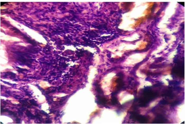
The Figure 5 shows presence of glands within muscle layer called Rokitansky Aschoff sinuses, seen often in chronic cholecystitis which can be mistaken for malignant infiltration. The subepithelium shows infiltration of lymphocytes and plasma cells.
The number of cases of acute on chronic cholecystitis was one in number as shown in (Figures 10,11) and the number of cases of xanthogranulomatous cholecystitis was one in number as shown in (Figures 12,13) and (Table 6).
Figure 5: Chronic cholecystitis with Rokitansky Aschoff sinuses.
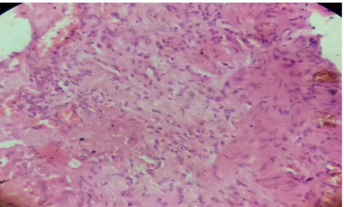
Figure 6: Chronic cholecystitis with lymphoplasmacytic type-low power.
Figure 7: Chronic cholecystitis with lymphoplasmacytic type-high power.
Chronic cholecystitis was the major histopathological type found in majority of cases-(94%) and acute on chronic cholecystitis and xanthogranulomatous cholecystitis were found in (3%) of cases respectively.
The Figure 6 shows focal ulceration of lining epithelium with dense collection of lymphocytes and plasma cells-seen in lymphoplasmacytic type of chronic cholecystitis. Increased number of plasma cells are seen in this type in the subepithelium.
Figure 8: Chronic cholecystitis-follicular type and with hyperplasia of glands-low power.
The Figure 7 shows the same picture as above under higher magnification showing lymphocytes and increased number of plasma cells clearly beneath the lining epithelium.
Figure 9: Chronic cholecystitis-follicular type-high power.
The Figure 8 shows collection of lymphocytes with focal follicle formation in the subepithelium as seen in follicular type of chronic cholecystitis with increase in number of glands. The glandular hyperplasia is commonly seen in chronic cholecystitis.
Figure 10: Acute on chronic cholecystitis with ulceration.
Figure 11: Acute on chronic cholecystitis with extensive ulceration of lining mucosa.
Figure 12: Xanthogranulomatous cholecystitis-low power.
The Figure 10 shows gallbladder wall with complete ulceration of lining epithelium and collection of acute and chronic inflammatory cells consisting of neutrophils and lymphocytes in the subepithelium.
The Figure 11 shows total loss of lining epithelium with acute and chronic inflammatory cells consisting of
extensive infiltration by neutrophils and focal collection of lymphocytes.
Figure 13: Xanthogranulomatous cholecystitis-high power.
The Figure 12 shows collection of foamy histiocytes with focal collection of epithelioid cells and also ulceration of lining epithelium seen in xanthogranulomatous cholecystitis-a type of chronic cholecystitis.
The Figure 13 shows the same picture as above under high power showing foamy histiocytes and focal collection of epithelioid cells clearly. Focal collection of lymphocytes and plasma cells also seen.
In this study the females are more affected in the age group-31 to 40yrs accounting for 30% (Table 2). All 3 male patients of chronic cholecystitis-showed only pigment stones. Histopathology in male cases showed acute on chronic cholecystitis, chronic cholecystitis- clinically diagnosed as empyema-for which open cholecystectomy was done and xanthogranulomatous cholecystitis.
DISCUSSION
More than 95% of biliary tract disease is attributable to cholelithiasis. As chronic cholecystitis is associated with cholelithiasis in more than 90% of cases, the at-risk population is the same as that for gallstones. Supersaturation of bile predisposes to both chronic inflammation and in most instances stone formation.2
Microorganisms usually E. coli and enterococci can be cultured from bile in about one third of cases. Biliary symptoms often emerge following the long-term co-existence of gallstones and low-grade inflammation.
In most instances stones are of mixed combined type. Microscopically mucosa of chronically inflamed gallbladder show varying degrees of mononuclear infiltration and fibrosis. The epithelium may be relatively normal or atrophic or show hyperplastic and metaplastic changes. The metaplasia may be of goblet cell (intestinal type) or pyloric (antral) type, the former being accompanied by the appearance of paneth cells and endocrine cells.
In contrast to the normal glands of the gallbladder neck, the cells of metaplastic glands contain nonsulfated acid mucin and neutral mucin but little sulfated acid mucin. The incidence of metaplastic changes increases with age. Gallbladder may show fibrosis, muscle hypertrophy, encrusted stones and nodular collections of foamy macrophages.2
Irregularly shaped tubular structures are present within the wall in over half of cases. They are lined by columnar or cuboidal epithelium and may contain bile or stones. These are called Rokitansky-Aschoff sinuses represent herniations or diverticula resulting from increased intraluminal pressure. Similar but smaller tubular formations are found in subserosal layer-hepatic side-known as Lushka ducts.
Adenomyoma are exaggerated examples of gallbladder diverticulosis associated with muscular hypertrophy when focal or segmentally present. Occurrence of intra and perineurial invasion can be seen in gallbladder affected by chronic cholecystitis with pyloric gland metaplasia and with segmental adenomyomatous hyperplasia. Morphologic variants of chronic cholecystitis include
• Follicular cholecystitis
• Diffuse lymphoplasmacytic cholecystitis
• Eosinophilic cholecystitis
• Xanthogranulomatous cholecystitis.
Classic study of Lund followed 526 non-operated cases of cholelithiasis and found that one third to one half of patients subsequently developed severe symptoms or complications from disease.2
It was concluded that prophylactic removal of gallbladder containing stones is indicated in all patients who are good surgical risks. Similar conclusions were drawn in National cooperative gallstone study which involved 305 patients. Inflammatory polyps are always associated with chronic cholecystitis, adenomyomatous hyperplasia and adenomyomatous hyperplasia are reactive mucosal changes secondary to inflammation and or lithiasis. The lining cells and neck mucous glands mainly contain sulfated acid mucin and very small amount of non-sulfated acid mucin.2
Tyagi SP et al, studied the morphologic changes of gallbladder in 415 cholecystectomy specimens. Females
were more affected with male to female ratio of 1:6.5. The mean age was 43.6yrs. Most of the cases were seen in 4th and 5th decade. Associated cholelithiasis were
present in 85.3% of cases.3
Chronic cholecystitis was main histological diagnosis. Other lesions were adenomyomatosis, adenomatous hyperplasia, granulomatous cholecystitis, cholesterosis, acute cholecystitis, acute on chronic infection, subacute cholecystitis, carcinoma gallbladder.
Liew LP et al, in Taiwan declared obesity as an important risk factor for gallbladder disease.4 Two prospective
studies by Csendes et al suggested that chronic inflammatory changes could occur prior to appearance of stones.5
Katsika D et al, study shows that overweight and obesity are associated with significantly higher risk of symptomatic gall stone disease.6 Kriska et al, study
showed that physical activity is significantly and inversely related to development of gallbladder disease.7
Pannwitz H et al, concluded that gallstones are infrequently present in nulliparous women. The prevalence of gallstones increased with number of births and with age.8
Study done by Khan MK et al in Bangladesh showed that significantly higher incidence of gallstones found in younger women taking oral contraceptives. The reverse findings were obtained in older age group patients.8
In the study of Kafle S et al gastric metaplasia was present in 33% and intestinal metaplasia in 8%. Maximum positivity among the three mucins was of neutral mucin followed by sulphated and sialomucin. The sulphated mucin positivity with both gastric and intestinal metaplasia yielded significant p value. Neutral mucin yielded significant p value with gastric metaplasia.8
Franco V et al, study showed that Xanthogranulomatous cholecystitis is found with female proponderance in sixth and seventh decade.9
Gupta SC et al, study showed that prevalence of gallstones in gallbladders with metaplastic, dysplastic and neoplastic mucosal changes are significantly higher. Increase in sialomucin with decrease in sulphated mucin was observed from metaplasia to malignancy. Neutral mucin was increased in metaplastic cells but was significantly reduced in neoplastic cells.10
Ganesh IM et al, study shows that sulphated mucins have a great role in gallstone formation than neutral mucins. Sialomucins and sulfomucins play a greater role in cancer progression and metastasis.11
majority of 30 cases (83%) with male to female ratio of 1:14. The gastric and intestinal metaplasia was present in some cases. Certain risk factors for gallstones are well established like female gender and increasing age. Modifiable risk factors are obesity, rapid weight loss and gallbladder stasis. High dietary risk for gallstones is high calorie intake. Protective factors include high fibre intake, vegetable protein, nuts and physical activity. Dittrick et al in their study concluded that the obese patients had an increased incidence of benign gallbladder disease.12
In the present study, there was also a female preponderance with male to female ratio of 1:11. Most of the cases were in their fourth decade of life. The histopathological findings seen were chronic cholecystitis with stones, chronic cholecystis without stones, acute on chronic cholecystitis, xanthogranulomatous cholecystitis.
In the present study xanthogranulomatous cholecystitis has been seen in one male patient only with the age of 44 yrs with pigment stone and carcinoma of gallbladder was not detected. And in the present study acute on chronic cholecystitis has been seen in one male patient only with age of 37 yrs with pigment stone.
CONCLUSION
The risk factors for developing chronic cholecystitis was seen in female gender. The study emphasises the need for histopathology in all specimens of cholecystectomy. Chronic cholecystitis has a wide histomorphological spectrum. Acute on chronic cholecystitis, chronic cholecystitis and xanthogranulomatous cholecystitis are some of them. Most of them are associated with cholelithiasis at all age. The predominant histomorphological pattern seen in this study group is chronic calculous cholecystitis.
And the predominant type of stone found in this study is pigment stone compared to mixed and cholesterol stone. Involvement in younger age group is another finding in this study. Special stains like phenyl hydrazine-PAS for neutral mucin, mild methylation-Alcian blue method for sulphated mucin, mild PAS-technique for sialomucin can be done to categories and quantify metaplastic areas in gallbladder and categorise risk of metaplasia and dysplasia with malignancy of gallbladder can be analysed. If the risk factors for the above changes can be analysed then prevention can be done for malignant conditions of gallbladder.
ACKNOWLEDGEMENTS
Authors would like to thank Dr. Venkatesh and Dr. Anandhi, Department General Surgery, Govt. Omandurar Medical College, Chennai.
Funding: No funding sources Conflict of interest: None declared Ethical approval: Not required
REFERENCES
1. Kumar H, Kini H, Tiwari A. Histological evaluation of 400 cholecystectomy specimens. J Pathol Nepal. 2015 Jan 1;5(10):834-40.
2. Rosai J. Ackermann surgical pathology. 8th ed. Elsevier. 1996:943-963.
3. Tyagi SP, Tyagi N, Maheshwari V, Ashraf SM, Sahoo P. Morphological changes in diseased gall bladder: a study of 415 cholecystectomies at Aligarh. J Ind Med Association. 1992 Jul;90(7):178-81.
4. Liew PL, Wang W, Lee YC, Huang MT, Lin YC, Lee WJ. Gallbladder disease among obese patients in Taiwan. Obesity Surg. 2007 Mar 1;17(3):383-90. 5. Csendes A, Burdiles P, Smok G, Csendes P, Burgos A, Recio M. Histologic findings of gallbladder mucosa in 87 patients with morbid obesity without gallstones compared to 87 control subjects. J Gastrointestinal Surg. 2003 Aug 1;7(4):547-51. 6. Katsika D, Tuvblad C, Einarsson C, Lichtenstein P,
Marschall HU. Body mass index, alcohol, tobacco and symptomatic gallstone disease: a Swedish twin study. J Int Med. 2007 Nov;262(5):581-7.
7. Kriska AM, Laporte RE, Patrick SL, Kuller LH, Orchard TJ. The association of physical activity and diabetic complications in individuals with insulin dependent diabetes-The epidemiology of diabetes complications study-VII. J Cli Epidemiol. 1991:44(11);1207-14.
8. Kafle SU, Sinha AK, Pandey SR. Histomorphology spectrum of gall bladder pathology in cholecystectomy specimens with clinical diagnosis of chronic cholecystitis. J Nepal Med Association. 2013;52(192).
9. Franco V, Aragona F, Genova G, Florena AM, Stella M, Campesi G. Xanthogranulomatous cholecystitis. Histopathological study and classification. Pathol Res Prac. 1990 Jun;186(3):383-90.
10. Gupta SC, Misra V, Singh PA, Roy A, Misra SP, Gupta AK. Gall stones and carcinoma gall bladder. Indian J Pathol Microbiol. 2000 Apr;43(2):147-54. 11. Ganesh IM, Subramani D, Halagowder D. Mucin
glycoarray in gastric and gallbladder epithelia. J Carcinogenesis. 2007;6:10.
12. Dittrick GW, Thompson JS, Campos D, Bremers D, Sudan D. Gallbladder pathology in morbid obesity. Obesity surgery. 2005 Feb 1;15(2):238-42.