AN IN VITRO STUDY OF ANTIMICROBIAL PROPERTIES OF AN ORTHODONTIC SEALANT/ADHESIVE CONTAINING SELENIUM
Michael T. Kelly
A thesis submitted to the faculty at the University of North Carolina at Chapel Hill in partial fulfillment of the requirements for the degree of Master of Science in the School of Dentistry
(Orthodontics).
Chapel Hill 2014
ABSTRACT
Michael T. Kelly: An In Vitro Study of Antimicrobial Properties of an Orthodontic Sealant/Adhesive Containing Selenium
(Under the direction of Lorne D. Koroluk)
Introduction: White spot lesions are a significant risk to patients undergoing orthodontic therapy. Antimicrobial agents are thought to reduce the incidence of white spot lesions due to their ability to kill cariogenic bacteria. The objective of this study was to characterize and quantify the antimicrobial properties of an orthodontic bonding system containing selenium (SeLECT Defense) compared to traditional orthodontic materials.
ACKNOWLEDGEMENTS
TABLE OF CONTENTS
LIST OF TABLES ... vii
LIST OF FIGURES ... viii
WHITE SPOT LESIONS AND ORTHODONTICS ...1
Introduction and Prevalence ...1
Etiology ...3
Treatment ...5
Prevention ...6
Banding and Bonding Materials ...9
Selenium ...15
Conclusion ...20
References ...22
AN IN VITRO STUDY OF ANTIMICROBIAL PROPERTIES OF AN ORTHODONTIC SEALANT/ADHESIVE CONTAINING SELENIUM ...29
Introduction ...29
Methods...32
Agar Diffusion Assay ...32
Direct Contact Inhibition Assay ...33
Statistical Analysis ...34
Discussion ...38
Conclusions ...43
Tables ...44
Figures...46
LIST OF TABLES
Table 1 - Orthodontic materials tested in Agar Diffusion Assay and
Direct Contact Inhibition Assay ...44 Table 2 - Zones of surface growth inhibition (diameter in mm,
mean ± standard deviation) of orthodontic materials against
S. mutans and L. acidophilus using Agar Diffusion Assay ...45 Table 3 - Reduction of colony-forming units of S. mutans and
L. acidophilus inoculated on newly-prepared orthodontic materials
using Direct Contact Inhibition Assay ...45 Table 4 - Reduction of colony-forming units of S. mutans and L.
acidophilus inoculated on orthodontic materials aged 7 days using
LIST OF FIGURES
Figure 1 - White spot lesions following removal of orthodontic appliances ...46 Figure 2 - Orthodontic material disks on WC Agar plates inoculated
with bacteria following 48 hours of aerobic incubation at 37°C
in the presence of 5% carbon dioxide ...47 Figure 3 - L. acidophilus “Reference Plate” in triplicate ...48 Figure 4 - Spot plate in triplicate of L. acidophilus inoculated onto 7-day
WHITE SPOT LESIONS AND ORTHODONTICS
Introduction and Prevalence
A significant percentage of children have poor oral hygiene which can be attributed to a lack of motor skills, supervision, or motivation. Orthodontic appliances tend to increase plaque retention along their gingival margins and may increase periodontal inflammation if oral hygiene is inadequate.1 Although rubber-cup prophylaxis has been shown to be effective in preventing gingival enlargement during orthodontic treatment,2 most patients develop generalized moderate hyperplastic gingivitis within two months of the initiation of orthodontic treatment even in the presence of good oral hygiene.1 Despite the periodontal insult caused by fixed orthodontic appliances, the loss of periodontal attachment has been shown to average 0.1mm or less during orthodontic treatment and is not different between treated and untreated patients.3 Most of the observed changes during orthodontic treatment do not result in permanent damage of the periodontium.1-3
formed by remineralization.5 White spot lesions 75 microns in depth can occur in as little as 4 weeks.6 The decreased mineral content results in an alteration of the optical refractory index of enamel, which makes the white spot lesions appear lighter in color than the surrounding healthy enamel.7 White spot lesions are permanent scars on the enamel, and if left untreated continue to be an esthetic problem for at least five years after treatment.6 A survey has shown that 96% of patients, parents, orthodontists, and general dentists think white spot lesions decrease the esthetics of orthodontically-straightened teeth.8 Although those surveyed agree that patients themselves are most responsible for the occurrence of white spot lesions, orthodontists are troubled by the high incidence of white spot lesion formation in their patients.8
Numerous studies have found that orthodontic patients have significantly more white spot lesions than untreated controls.6,9,10 The incidence of white spot lesion formation during
orthodontic treatment is reported to be as high as 72.9%, while the incidence of cavitated lesions is 2.3%.11 A landmark study by Gorelick, et al. found that 24% of untreated patients had white spot lesions in locations on their teeth where white spot lesions are commonly observed in orthodontically-treated patients.9 Fifty percent of patients who had undergone orthodontic treatment developed at least one white spot lesion, and no difference existed between patients treated with banded versus bonded appliances.9 The authors noted that the labiogingival area of maxillary lateral incisors had the highest incidence of white spot lesions, while the maxillary posterior segment had the lowest.9 No white spots were found on the lingual surface of mandibular incisors and canines after the use of a bonded canine-to-canine retainer.9
brushing twice daily with fluoride toothpaste, flossing, using a fluoride rinse, and using plaque disclosing tablets. Authors concluded that there is an inverse relationship between compliance with oral hygiene instruction and the incidence of white spot lesions, as those with good
compliance had significantly fewer white spot lesions than those with moderate compliance, who in turn had significantly fewer white spot lesions than those with poor compliance.10
Furthermore, treatment duration has been shown to be positively associated with new white spot lesion formation.11
Etiology
White spot lesions are the early manifestations of the caries process. Dental caries is a disease in which bacterial fermentation of sugars in a dental plaque or biofilm results in the production of lactic acid which demineralizes tooth structure.12,13 White spot lesions form when lactic acid produced by the fermentation of carbohydrates diffuse through the porous subsurface enamel.14 Lactic acid very rapidly dissociates to produce free hydrogen ions which dissolve hydroxyapatite, causing the release of calcium and phosphate from the lesion.14 The resulting area of demineralized enamel is termed the incipient carious lesion and can progress to frank cavitation.6 Caries progression is facilitated by aciduric bacteria such as mutans streptococci and lactobacilli, salivary dysfunction, and consumption of sugar.12,13 Protective factors include salivary calcium, phosphate, fluoride, buffers and antibacterial proteins, as well as high salivary flow rate and the use of antibacterial agents.12 There is an ongoing balance between
Treatment
While white spot lesions tend to decrease in surface area and improve in appearance during the first two years after the completion of orthodontic therapy,19 most can benefit from treatment. White spot lesions can be treated with several different modalities, including
remineralization, bleaching, microabrasion, and dental restorations, depending on their severity. Despite the effectiveness of topical fluorides at preventing white spot lesions, the use of high-dose topical fluoride post-orthodontic therapy to treat white spot lesions is contraindicated because it can result in enamel staining due to rapid surface remineralization.7 Remineralization of white spot lesions is best left to nature, and the physiologic levels of ions present in saliva.
Tooth whitening can be effective at masking the presence of white spot lesions. Ideally, healthy enamel will be whitened to a greater degree than white spots, with the effect being to lessen the contrast between the two. A study of 10 patients who underwent in-office bleaching followed by a 2-week regimen of in-home bleaching was conducted.20 Using a colorimeter to assess color, it was determined that although both healthy enamel and white spot lesions were significantly lightened, the healthy enamel was affected more, and the result was to mask the white spots.20 All ten patients reported being satisfied with the outcome.20
A newly-advocated treatment technique involves the microabrasion of the white spot lesions. Microabrasion is performed by using pumice or silicon carbide particles and
significantly improved by treatment with microabrasion, as well as microabrasion with MI PasteTM (GC America, Alsip, IL) treatment.22
A recently developed low-viscosity resin (Icon® by DMG America, Englewood, NJ) has been used to improve the appearance of white spot lesions following orthodontic treatment. The resin infiltrates porous white spot lesions and matches the refractory index of healthy enamel.7 A study of teeth with post-orthodontic white spot lesions treated with Icon® found that 61% of lesions were completely masked, 33% partially masked, and only 6% remained unchanged.23 This product offers a more conservative approach in the treatment of white spot lesions compared to microabrasion, although more data is needed concerning it durability and effectiveness over time.
Prevention
While remineralization, bleaching, microabrasion, and low-viscosity resins can be effective, severe lesions, cavitated lesions, or lesions that are unresponsive to other treatment modalities may benefit from resin composite restorations or porcelain veneers or crowns.7 Due to their potentially high treatment costs, as well as their high prevalence and unaesthetic
appearance, the best approach is to prevent the occurrence of white spot lesions prophylactically. All orthodontic patients are given very specific oral hygiene technique instruction and protocols which typically include the use of fluoride. During orthodontic treatment, the prevention of white spot lesions can be accomplished with topical fluoride. Fluoride inhibits the caries process by several different mechanisms. The fluoride ion is toxic to bacterial cells because it can
fluoride ion can substitute for a hydroxyl group in hydroxyapatite to form fluorapatite. Fluorapatite is less soluble than hydroxyapatite, and is therefore more resistant to acid-attack during the caries process.12 Sound enamel typically contains 20 to 100ppm fluoride content due to fluoride ingestion during tooth development.24 The previously described conversion of hydroxyapatite to fluorapatite by topical fluoride exposure results in enamel’s “fluoride-rich, caries-resistant” outer layer, which contains 1,000-2,000 ppm fluoride.24 Lastly, fluoride promotes remineralization of demineralized enamel by attracting calcium and phosphate ions from saliva to the enamel surface.12 Fluoride levels as low as 0.04ppm can enhance enamel remineralization.12
Topical fluoride, in the forms of toothpaste, mouth rinses, gels, foams, and varnish, are commonly prescribed to the orthodontic patient. Fluoride ions present in drinking water and fluoride-containing products have been shown to reduce caries.12 A systematic review was conducted to determine which topical fluoride formulation is most effective in preventing white spot lesions during orthodontic therapy. The authors concluded that the use of all topical
fluorides in addition to fluoride toothpaste were effective in reducing the incidence of white spot lesions.25 Different preparations of topical fluorides, including stannous fluoride gels, sodium fluoride rinses, and acidulated phosphate fluoride foams, all have the ability to reduce the
incidence of white spot lesions and no single formulation is superior to the others.25 There exists some evidence to suggest that higher concentrations of fluoride ion are more effective in
Some evidence shows that fluoride may not be sufficient to prevent the demineralization of enamel during severe cariogenic insults, and therefore the use of antimicrobial agents such as triclosan, xylitol, or chlorhexidine may be beneficial.12,28 Chlorhexidine is a potent, substantive antimicrobial agent commonly used in the treatment of dental diseases. Daily use of
chlorhexidine rinse for 2 weeks has been shown to kill S. mutans and prevent its recolonization on tooth surfaces for the following three to six months.29 The application of chlorhexidine varnish has been shown to be effective in suppressing oral S. mutans levels for three to seven months after a single application when used within one month of the placement of fixed orthodontic appliances.30 The use of chlorhexidine has its own risks. Prolonged use of
chlorhexidine can result in the alteration of tooth color. Furthermore, there is question whether the presence of chlorhexidine decreases the bond strength of orthodontic brackets. Placement of chlorhexidine varnish immediately prior to bracket placement increases the rate of bond
failure.31,32 Similarly, the use of chlorhexidine rinse immediately prior to orthodontic bonding results in a statistically significant decrease in bond strength compared to the use of
chlorhexidine rinse one week prior to bonding.33 The use of chlorhexidine rinse one week prior to bonding orthodontic brackets did not decrease bond strength.33
MI PasteTM is a new topical agent thought to prevent and repair enamel
PasteTM is effective in preventing white spot lesions, it has not been shown to be able to remineralize existing white spot lesions. A study by Beerens, et al. compared the use of MI PasteTM to normal 1000ppm fluoride tooth paste to treat white spot lesions for 12 weeks following orthodontic treatment.35 The double-blind randomized clinical trial measured the presence of white spot lesions using quantitative light-induced fluorescence. While both resulted in an increase in the mineral content of white spots, there was no statistically significant
difference between MI PasteTM and fluoride toothpaste.35 Furthermore, the size of the lesions did not change.35 It has been concluded that there is no benefit to treating white spot lesions after orthodontic treatment with MI PasteTM alone,22,35 although it has been used in combination with microabrasion to successfully improve the appearance of white spot lesions.22
The success of many of the aforementioned topical products at preventing white spot lesions depends on patient compliance. Unfortunately, patients cannot always be relied upon to adhere to oral hygiene regimens prescribed by their orthodontist. A study of 206 orthodontic patients found only 13% compliance with the daily use of a fluoride mouth rinse.36 The study found that 42% of patients used the fluoride rinse every other day, and the remaining 45% used it less frequently.36 As expected, significantly fewer white spot lesions developed in patients who used the fluoride rinse at least every other day compared to those who used it less frequently.36
Banding and Bonding Materials
surface of a tooth during orthodontic bonding, no enamel is exposed and therefore no enamel is susceptible to acid demineralization. This method of prevention requires the use of sealants containing filler particles for mechanical strength. An in vitro study examined the incidence of white spot lesions on teeth with orthodontic brackets bonded with a filled orthodontic sealant (Pro SealTM by Reliance Orthodontic Products, Itasca, IL) compared to brackets bonded with an unfilled sealant and brackets bonded without a sealant. Each surface was exposed to 15,000 strokes of tooth brushing with non-fluoridated paste, and then cycled for 14 days through demineralization and remineralization processes.27 Microhardness testing found that the filled sealant group had significantly less demineralization than the unfilled sealant group and the no sealant group.27 The authors concluded that filled sealants can be used to prevent white spot lesions in orthodontic patients.27 Despite their effectiveness, filled sealants require replacement as susceptible enamel surfaces become exposed due to toothbrush abrasion of the resin surface. Manufacturers of many commonly used filled orthodontic sealants, including SeLECT
DefenseTM (Element-34 Technologies, Lubbock, TX), Opal SealTM (Opal Orthodontics, South Jordan, UT), and Ortho SoloTM (Ormco, Glendora, CA), recommend reapplying their products every three to six months to maintain a physical barrier on the tooth surface, although Pro SealTM only needs to be reapplied after two years. Although it has been reported that sealants result in enamel loss, cracks and scratches,37 the prevention of white spot lesions has a greater impact on the final esthetic outcome of the dentition. Therefore, the use of sealants should be strongly considered.
The adhesives used to cement fixed orthodontic appliances can also impact the
setting reaction of glass ionomers, fluoride is released from the aluminosilicate glass particles when they contact polyacrylic acid.38 The greatest amount of fluoride is released on the first day with a significant decrease afterwards.38 A split-mouth study compared the incidence of white spot lesion formation on teeth with brackets bonded with glass ionomer cement versus resin composite adhesive.39 At completion of orthodontic treatment, 24% of teeth that had brackets bonded with glass ionomer cement had developed white spot lesions, compared to 40.5% of teeth bonded with resin composite.39 It was also found that for patients who had longer treatment time, white spots were more frequent on teeth with resin composite adhesive.39 The authors concluded that the use of glass ionomer cement for bonding results in fewer white spot lesions than resin composite adhesive in orthodontic patients.39
Despite the reported success of glass ionomer cements in the prevention of white spot lesions, their use in orthodontic bonding is not very common due to their poor mechanical characteristics. Resin-modified glass ionomer cements have improved mechanical properties and adhesive strength compared to traditional glass ionomers.40-42 Similar to traditional glass ionomers, resin-modified glass ionomers have also been shown to be superior at preventing enamel demineralization compared to resin composites adhesives.43 A randomized clinical trial investigated the incidence of demineralization around orthodontic brackets bonded with a resin-modified glass ionomer compared to a resin composite adhesive.44 Microhardness testing concluded that after four weeks, there was significantly less demineralization around brackets bonded with the resin-modified glass ionomer compared to resin composite adhesive.44 These results were supported by a second clinical trial which found that the use of resin-modified glass ionomer cements results in a decreased frequency of caries compared to resin composite
to the orthodontic brackets.45 The study found that there was 21% mineral loss next to brackets bonded with resin-modified glass ionomers compared to 33% next to brackets bonded with resin composite.45 The authors also noted that the microhardness of the enamel under the bracket was the same for both treatment groups, so the observed differences in mineral loss could not be explained by the acid-etching in the resin composite bonding process.45 An in vitro study also found that brackets bonded with resin-modified glass ionomer cement result in white spot lesions of more shallow depth and less mineral loss than resin composite bonding methods.43
It is believed that glass ionomer cements and resin-modified glass ionomer cements are anticariogenic due to their release of fluoride.46,47 While some studies have shown that levels of fluoride in saliva of patient with brackets bonded with glass ionomer cements are not increased,44 others have reported the opposite effect.48 On the first day of orthodontic appliance cementation with glass ionomer, the amount of salivary fluoride is doubled and then rapidly returns to
baseline levels.48 A split-mouth study compared fluoride levels in 48-hour-old plaque next to brackets bonded with glass ionomer cement and resin composite adhesive at days 3, 8, 28, and 180 after appliance placement.49 Results demonstrated that there were significantly higher fluoride amounts in plaque next to brackets bonded with glass ionomer cement at each time point.49 Authors concluded that glass ionomer cements may act as long-term local fluoride-releasing adhesives.49
cements are anticariogenic due to antibacterial properties.47 In vitro studies have confirmed this conclusion in agar diffusion and growth inhibition assays.50
In addition to their anticariogenic properties, resin-modified glass ionomer cements don’t result in changes in tooth surface after fixed appliance removal like resin composite adhesives.51 Despite these potential advantages, most practitioners use resin composites to bond orthodontic brackets due to their superior handling and mechanical properties. Glass ionomers have lower adhesive strength compared to resin composites and therefore are not favored.52
to recharging with fluoride toothpaste.54 Levels of fluoride released after recharging resin-modified glass ionomers and fluoride-releasing composites ranged from 2-35ppm per gram of material.54 While such small amounts of fluoride are probably not effective at inhibiting bacteria in vivo, sub-ppm levels of fluoride are effective at preventing caries by shifting the balance from a state of demineralization to remineralization.55 Similar to glass ionomer cements, in vitro studies have shown that fluoride-releasing resins have antibacterial properties against S. mutans and L. acidophilus.56,57
An in vitro study comparing white spot lesion formation adjacent to orthodontic brackets in an artificial caries solution found that brackets bonded with resin-modified glass ionomer cement and fluoride-releasing resin composite were equally effective at reducing the incidence of white spot lesions compared to a non-fluoride-releasing resin composite adhesive.58 A split-mouth study found that white spot lesions that formed on teeth bonded with fluoride-releasing resin composite adhesive had 48% reduced lesion depth compared to teeth bonded with a non-fluoride-releasing adhesive after 4 weeks.59
poor mechanical characteristics and is therefore not a feasible product.62-66 In vitro studies have shown that a resin composite adhesive that releases the antimicrobial agent benzalkonium chloride has antibacterial activity against S. mutans.67 Unfortunately, the antibacterial effect decreased significantly over time, and concentrations of benzalkonium chloride greater than 0.75% are toxic to human gingival cells.67 The addition of zinc oxide to a resin-modified glass ionomer cement has been shown to increase its antibacterial properties against S. mutans as demonstrated in an agar diffusion assay, but the product is not commercially available.68 Several studies have demonstrated the antibacterial properties of a resin composite containing the
monomer 12-methacryloyloxydodecylpyridinium bromide (MDPB) in vitro against S. mutans and lactobacilli.69,70 Clinical trials of an orthodontic adhesive containing MDPB have
demonstrated a significant decrease in demineralization around orthodontic brackets compared to control adhesives, but no significant beneficial effect on periodontal health has been reported.71,72 Similarly, orthodontic adhesives containing silver nanoparticles have been shown to be
bactericidal.61,73 The slow release of silver ions from carrier materials such as zeolite or silica-gel is used to give many household items such as kitchenware, washing machines, clothes, and toiletries, antibacterial properties.61 Unfortunately, as with those containing MDPB, such resin composite adhesives have poor color stability, handling characteristics, and mechanical
properties.61,69,73
Selenium
A recently developed orthodontic system called “SeLECT DefenseTM” has been released to market. “SeLECT” stands for “Selenium Labeled Extra-Cellular Toxicity.” SeLECT
their resin matrices, while the brackets and elastomers have selenium attached to their surfaces. The manufacturer of SeLECT DefenseTM claims that the selenium in their products catalyzes the formation of localized and short-lived superoxide radicals which results in less plaque formation around orthodontic brackets, improved gingival health and fewer white spot lesions.74 Whereas previous materials containing antibacterial components have displayed poor mechanical
characteristics, SeLECT DefenseTM adhesive has been shown to have clinically acceptable shear bond strength.75
Selenium is element number 34 on the periodic table and is usually classified as a nonmetal, although it has some characteristics of a metal. Selenium is naturally found in the human body, specifically in proteins in the plasma, thyroid, gastrointestinal tract, skin, liver, kidneys and brain.76 Selenium is an active part of the glutathione reductases, which act as antioxidants,77 and many selenoproteins are involved in the immune system.78 Naturally present in plants, the average dietary intake of selenium is 20-300mcg per day.76 Signs of selenium toxicity begin at intake levels of 1,500mcg per day, but much higher doses are tolerable before severe side effects are observed.76 The use of selenium is very common in medicine, and especially in the development of anticancer drugs.76 Selenium has been linked to prevention of atherosclerosis, cancers, arthritis, central nervous system pathologies, and altered function of the immune system.79
Selenium can be covalently linked to solid surfaces and catalyze the formation of
superoxide radicals which are toxic to bacteria and prevent their attachment to a given surface.80 Medical devices, such as hemodialysis catheters or contact lenses, can be coated with organo-selenium to prevent the formation of bacterial biofilms or prevent bacterial growth as
formation of bacterial biofilms on cellulose82 through the reduction of oxygen by organo-selenium to form superoxide radicals which damage bacterial cell walls and DNA.83 Organo-selenium is then reduced by glutathione, which results in the formation of a second superoxide radical.83 The half-life of the radical produced by selenium is 60 nanoseconds and its path length is 35nm.84 While the radical is only toxic to cells in close proximity, formation of a biofilm on a surface coated with selenium can be inhibited.84 It has been demonstrated that concentrations of organo-selenium as low as 0.1% can inhibit bacteria.83
The organo-selenium present in SeLECT DefenseTM is a diselenylmethacrylate with the IUPAC name
demonstrate that bacteria are prevented from adhering to dental materials containing organo-selenium, but do not necessarily suggest that they are killed by contact with organo-selenium. However, this study does provide evidence that dental sealants containing organo-selenium have potential to be effective at preventing white spot lesions when used in the orthodontic patient.
An in vivo study compared SeLECT DefenseTM pit and fissure sealant to UltraSeal XT PlusTM (Ultradent, South Jordan, UT), a commonly used pit and fissure sealant.85 This
randomized, double-blind, split-mouth study measured the clinical retention, caries formation, plaque formation, leakage, and safety of the sealants in 120 adolescents of moderate and severe caries risk.85 The sealants were evaluated every three months for a total of 12 months. Plaque formation and leakage were detected using quantitative light-induced fluorescence.85 The study found that SeLECT DefenseTM sealant had higher retention compared to UltraSeal XT PlusTM (96.2% vs 80.9%) after 12 months and significantly less plaque growth on the sealant.85 No SeLECT DefenseTM sealants displayed any bacterial plaque growth on their surface, which the authors attributed to the antimicrobial nature of SeLECT DefenseTM.85 The authors did note that of the 12% of UltraSeal XT PlusTM sealants that displayed surface plaque growth, all were in the high caries risk group and had poor oral hygiene.85 Neither sealant showed signs of leakage, oral mucosa side effects, or caries.85
An in vitro study using an artificial mouth with simulated tooth brushing investigated the SeLECT DefenseTM orthodontic system. Extracted human teeth bonded with SeLECT
teeth, and plaque accumulation. The simulation lasted 28 days in a carbon dioxide incubator with continuous flow of fluid over the teeth to simulate saliva, and the addition of sucrose every 6 hours to simulate consumption of food.86 The teeth were inoculated with S. mutans and L. casei.86 The formation of white spot lesions was detected using quantitative light-induced fluorescence, transverse microradiography, and polarizing light microscopy, and plaque was measured using quantitative light-induced fluorescence.86 The study found that the SeLECT DefenseTM system significantly prevented the formation of white spot lesions compared to the conventional system with 86% reduction of demineralization based on polarizing light
microscopy.86 SeLECT DefenseTM sealant was able to withstand 28 days of brushing, resulted in a significant decrease in plaque accumulation, and the authors noted that it resulted in a “bluish clouding” of the tooth surface.86
While the data indicate that SeLECT DefenseTM can prevent the formation of white spot lesions, this study does not distinguish between prevention of white spot lesions due to antibacterial properties of SeLECT DefenseTM and prevention due to the presence of a mechanical barrier. The control teeth were bonded with an unfilled sealant, while SeLECT DefenseTM sealant is filled and therefore more likely to resist toothbrush abrasion and provide a constant mechanical barrier against acid demineralization.
DefenseTM sealant compared to uncoated enamel and chlorhexidine.84 Similar to the artificial mouth study, this study did not distinguish between prevention of white spot lesions by mechanical barrier and prevention of white spot lesions due to the antimicrobial properties of SeLECT DefenseTM.
While these studies suggest that SeLECT DefenseTM products have inhibitory effects on the attachment and growth of bacteria,83-86 the bactericidal properties of the materials have not been investigated previously. However, the killing of cariogenic bacteria is not required to prevent the development of white spot lesions in orthodontic patients, while mere inhibition of attachment and growth on the tooth surface would be sufficient. A randomized clinical trial comparing the incidence of white spot lesions in orthodontic patients bonded with SeLECT DefenseTM products versus traditional filled sealants and adhesives would be critical to
investigate these claims. Furthermore, no data exists regarding the effect of SeLECT DefenseTM products on periodontal health.
Conclusion
REFERENCES
1. Zachrisson S, Zachrisson BU. Gingival condition associated with orthodontic treatment. Angle Orthod. 1972;42(1):26-34..
2. Huber SJ, Vernino AR, Nanda RS. Professional prophylaxis and its effect on the periodontium of full-banded orthodontic patients. Am J Orthod Dentofacial Orthop. 1987;91(4):321-327. 3. Alstad S, Zachrisson BU. Longitudinal study of periodontal condition associated with orthodontic treatment in adolescents. Am J Orthod. 1979;76(3):277-286.
4. Summitt JB, Robbins JW, Schwartz RS. Fundamentals of operative dentistry: A contemporary approach. In: 2nd ed. Carol Stream, Illinois: Quintessence; 2001:3.
5. Silverstone LM. Structure of carious enamel, including the early lesion. Oral Sci Rev. 1973;3:100-160.
6. Ogaard B. Prevalence of white spot lesions in 19-year-olds: A study on untreated and orthodontically treated persons 5 years after treatment. Am J Orthod Dentofacial Orthop. 1989;96(5):423-427.
7. Heymann GC, Grauer D. A contemporary review of white spot lesions in orthodontics. J Esthet Restor Dent. 2013;25(2):85-95.
8. Maxfield BJ, Hamdan AM, Tufekci E, Shroff B, Best AM, Lindauer SJ. Development of white spot lesions during orthodontic treatment: Perceptions of patients, parents, orthodontists, and general dentists. Am J Orthod Dentofacial Orthop. 2012;141(3):337-344.
9. Gorelick L, Geiger AM, Gwinnett AJ. Incidence of white spot formation after bonding and banding. Am J Orthod. 1982;81(2):93-98.
10. Hadler-Olsen S, Sandvik K, El-Agroudi MA, Ogaard B. The incidence of caries and white spot lesions in orthodontically treated adolescents with a comprehensive caries prophylactic regimen--a prospective study. Eur J Orthod. 2012;34(5):633-639.
11. Richter AE, Arruda AO, Peters MC, Sohn W. Incidence of caries lesions among patients treated with comprehensive orthodontics. Am J Orthod Dentofacial Orthop. 2011;139(5):657-664.
12. Featherstone JD. The science and practice of caries prevention. J Am Dent Assoc. 2000;131(7):887-899.
14. Featherstone JD, Rodgers BE. Effect of acetic, lactic and other organic acids on the formation of artificial carious lesions. Caries Res. 1981;15(5):377-385.
15. Takahashi N, Nyvad B. The role of bacteria in the caries process: Ecological perspectives. J Dent Res. 2011;90(3):294-303.
16. Scheie AA, Arneberg P, Krogstad O. Effect of orthodontic treatment on prevalence of streptococcus mutans in plaque and saliva. Scand J Dent Res. 1984;92(3):211-217. 17. Arneberg P, Ogaard B, Scheie AA, Rolla G. Selection of streptococcus mutans and lactobacilli in an intra-oral human caries model. J Dent Res. 1984;63(10):1197-1200. 18. Mattingly JA, Sauer GJ, Yancey JM, Arnold RR. Enhancement of streptococcus mutans colonization by direct bonded orthodontic appliances. J Dent Res. 1983;62(12):1209-1211. 19. Al-Khateeb S, Forsberg CM, de Josselin de Jong E, Angmar-Mansson B. A longitudinal laser fluorescence study of white spot lesions in orthodontic patients. Am J Orthod Dentofacial
Orthop. 1998;113(6):595-602.
20. Knosel M, Attin R, Becker K, Attin T. External bleaching effect on the color and luminosity of inactive white-spot lesions after fixed orthodontic appliances. Angle Orthod. 2007;77(4):646-652.
21. Heymann HO, Swift EJ, Ritter AV. Sturdevant’s art and science of operative dentistry. 6th ed. St. Louis: Elsevier; 2013.
22. Pliska BT, Warner GA, Tantbirojn D, Larson BE. Treatment of white spot lesions with ACP paste and microabrasion. Angle Orthod. 2012;82(5):765-769.
23. Kim S, Kim EY, Jeong TS, Kim JW. The evaluation of resin infiltration for masking labial enamel white spot lesions. Int J Paediatr Dent. 2011;21(4):241-248.
24. Robinson C, Kirkham J, Weatherell JA. Fluoride in teeth and bone. In: Fejerskov O,
Ekstrand J, Burt BA, eds. Fluoride in dentistry. Copenhagen, Denmark: Munksgaard; 1996:69--87.
25. Chadwick BL, Roy J, Knox J, Treasure ET. The effect of topical fluorides on decalcification in patients with fixed orthodontic appliances: A systematic review. Am J Orthod Dentofacial Orthop. 2005;128(5):601-6; quiz 670.
26. Todd MA, Staley RN, Kanellis MJ, Donly KJ, Wefel JS. Effect of a fluoride varnish on demineralization adjacent to orthodontic brackets. Am J Orthod Dentofacial Orthop. 1999;116(2):159-167.
28. Rolla G, Ogaard B, Cruz Rde A. Topical application of fluorides on teeth. new concepts of mechanisms of interaction. J Clin Periodontol. 1993;20(2):105-108.
29. Anderson MH, Bales DJ, Omnell KA. Modern management of dental caries: The cutting edge is not the dental bur. J Am Dent Assoc. 1993;124(6):36-44.
30. Sandham HJ, Nadeau L, Phillips HI. The effect of chlorhexidine varnish treatment on salivary mutans streptococcal levels in child orthodontic patients. J Dent Res. 1992;71(1):32-35. 31. Frey C, Yetkiner E, Stawarczyk B, Attin T, Attin R. Effects of different chlorhexidine pretreatments on adhesion of metal brackets in vitro. Head Face Med. 2012;8:36-160X-8-36. 32. Bishara SE, Vonwald L, Zamtua J, Damon PL. Effects of various methods of chlorhexidine application on shear bond strength. Am J Orthod Dentofacial Orthop. 1998;114(2):150-153. 33. Cacciafesta V, Sfondrini MF, Stifanelli P, Scribante A, Klersy C. Effect of chlorhexidine application on shear bond strength of brackets bonded with a resin-modified glass ionomer. Am J Orthod Dentofacial Orthop. 2006;129(2):273-276.
34. Robertson MA, Kau CH, English JD, Lee RP, Powers J, Nguyen JT. MI paste plus to prevent demineralization in orthodontic patients: A prospective randomized controlled trial. Am J Orthod Dentofacial Orthop. 2011;140(5):660-668.
35. Beerens MW, van der Veen MH, van Beek H, ten Cate JM. Effects of casein phosphopeptide amorphous calcium fluoride phosphate paste on white spot lesions and dental plaque after
orthodontic treatment: A 3-month follow-up. Eur J Oral Sci. 2010;118(6):610-617.
36. Geiger AM, Gorelick L, Gwinnett AJ, Benson BJ. Reducing white spot lesions in orthodontic populations with fluoride rinsing. Am J Orthod Dentofacial Orthop. 1992;101(5):403-407. 37. Zachrisson BU, Skogan O, Hoymyhr S. Enamel cracks in debonded, debanded, and orthodontically untreated teeth. Am J Orthod. 1980;77(3):307-319.
38. Cacciafesta V, Sfondrini MF, Tagliani P, Klersy C. In-vitro fluoride release rates from 9 orthodontic bonding adhesives. Am J Orthod Dentofacial Orthop. 2007;132(5):656-662. 39. Marcusson A, Norevall LI, Persson M. White spot reduction when using glass ionomer cement for bonding in orthodontics: A longitudinal and comparative study. Eur J Orthod. 1997;19(3):233-242.
40. Cacciafesta V, Jost-Brinkmann PG, Sussenberger U, Miethke RR. Effects of saliva and water contamination on the enamel shear bond strength of a light-cured glass ionomer cement. Am J Orthod Dentofacial Orthop. 1998;113(4):402-407.
42. Silverman E, Cohen M, Demke RS, Silverman M. A new light-cured glass ionomer cement that bonds brackets to teeth without etching in the presence of saliva. Am J Orthod Dentofacial Orthop. 1995;108(3):231-236.
43. Paschos E, Kleinschrodt T, Clementino-Luedemann T, et al. Effect of different bonding agents on prevention of enamel demineralization around orthodontic brackets. Am J Orthod Dentofacial Orthop. 2009;135(5):603-612.
44. Gorton J, Featherstone JD. In vivo inhibition of demineralization around orthodontic brackets. Am J Orthod Dentofacial Orthop. 2003;123(1):10-14.
45. Pascotto RC, Navarro MF, Capelozza Filho L, Cury JA. In vivo effect of a resin-modified glass ionomer cement on enamel demineralization around orthodontic brackets. Am J Orthod Dentofacial Orthop. 2004;125(1):36-41.
46. Benelli EM, Serra MC, Rodrigues AL,Jr, Cury JA. In situ anticariogenic potential of glass ionomer cement. Caries Res. 1993;27(4):280-284.
47. Hallgren A, Oliveby A, Twetman S. Caries associated microflora in plaque from orthodontic appliances retained with glass ionomer cement. Scand J Dent Res. 1992;100(3):140-143.
48. Hallgren A, Oliveby A, Twetman S. Salivary fluoride concentrations in children with glass ionomer cemented orthodontic appliances. Caries Res. 1990;24(4):239-241.
49. Hallgren A, Oliveby A, Twetman S. Fluoride concentration in plaque adjacent to orthodontic appliances retained with glass ionomer cement. Caries Res. 1993;27(1):51-54.
50. Prati C, Fava F, Di Gioia D, Selighini M, Pashley DH. Antibacterial effectiveness of dentin bonding systems. Dent Mater. 1993;9(6):338-343.
51. Komori A, Ishikawa H. Evaluation of a resin-reinforced glass ionomer cement for use as an orthodontic bonding agent. Angle Orthod. 1997;67(3):189-195.
52. Fajen VB, Duncanson MG,Jr, Nanda RS, Currier GF, Angolkar PV. An in vitro evaluation of bond strength of three glass ionomer cements. Am J Orthod Dentofacial Orthop. 1990;97(4):316-322.
53. Caves GR, Millett DT, Creanor SL, Foye RH, Gilmour WH. Fluoride release from orthodontic band cements-a comparison of two in vitro models. J Dent. 2003;31(1):19-24. 54. Ahn SJ, Lee SJ, Lee DY, Lim BS. Effects of different fluoride recharging protocols on fluoride ion release from various orthodontic adhesives. J Dent. 2011;39(3):196-201.
56. Naorungroj S, Wei HH, Arnold RR, Swift EJ,Jr, Walter R. Antibacterial surface properties of fluoride-containing resin-based sealants. J Dent. 2010;38(5):387-391.
57. Matalon S, Slutzky H, Weiss EI. Antibacterial properties of 4 orthodontic cements. Am J Orthod Dentofacial Orthop. 2005;127(1):56-63.
58. Wilson RM, Donly KJ. Demineralization around orthodontic brackets bonded with resin-modified glass ionomer cement and fluoride-releasing resin composite. Pediatr Dent.
2001;23(3):255-259.
59. Ogaard B, Rezk-Lega F, Ruben J, Arends J. Cariostatic effect and fluoride release from a visible light-curing adhesive for bonding of orthodontic brackets. Am J Orthod Dentofacial Orthop. 1992;101(4):303-307.
60. Weitman RT, Eames WB. Plaque accumulation on composite surfaces after various finishing procedures. J Am Dent Assoc. 1975;91(1):101-106.
61. Imazato S. Antibacterial properties of resin composites and dentin bonding systems. Dent Mater. 2003;19(6):449-457.
62. Anusavice KJ, Zhang NZ, Shen C. Controlled release of chlorhexidine from UDMA-TEGDMA resin. J Dent Res. 2006;85(10):950-954.
63. Hiraishi N, Yiu CK, King NM, Tay FR, Pashley DH. Chlorhexidine release and water sorption characteristics of chlorhexidine-incorporated hydrophobic/hydrophilic resins. Dent Mater. 2008;24(10):1391-1399.
64. Leung D, Spratt DA, Pratten J, Gulabivala K, Mordan NJ, Young AM. Chlorhexidine-releasing methacrylate dental composite materials. Biomaterials. 2005;26(34):7145-7153. 65. Cadenaro M, Pashley DH, Marchesi G, et al. Influence of chlorhexidine on the degree of conversion and E-modulus of experimental adhesive blends. Dent Mater. 2009;25(10):1269-1274.
66. Ferracane JL. Resin composite--state of the art. Dent Mater. 2011;27(1):29-38. 67. Saito K, Hayakawa T, Kawabata R, Meguro D, Kasai K. In vitro antibacterial and
cytotoxicity assessments of an orthodontic bonding agent containing benzalkonium chloride. Angle Orthod. 2009;79(2):331-337.
68. Spencer CG, Campbell PM, Buschang PH, Cai J, Honeyman AL. Antimicrobial effects of zinc oxide in an orthodontic bonding agent. Angle Orthod. 2009;79(2):317-322.
70. Imazato S, Kuramoto A, Takahashi Y, Ebisu S, Peters MC. In vitro antibacterial effects of the dentin primer of clearfil protect bond. Dent Mater. 2006;22(6):527-532.
71. Uysal T, Amasyali M, Ozcan S, Koyuturk AE, Sagdic D. Effect of antibacterial monomer-containing adhesive on enamel demineralization around orthodontic brackets: An in-vivo study. Am J Orthod Dentofacial Orthop. 2011;139(5):650-656.
72. Amasyali M, Enhos S, Uysal T, Saygun I, Kilic A, Bedir O. Effect of a self-etching adhesive containing an antibacterial monomer on clinical periodontal parameters and subgingival
microbiologic composition in orthodontic patients. Am J Orthod Dentofacial Orthop. 2011;140(4):e147-53.
73. Ahn SJ, Lee SJ, Kook JK, Lim BS. Experimental antimicrobial orthodontic adhesives using nanofillers and silver nanoparticles. Dent Mater. 2009;25(2):206-213.
74. White LW. Defeating the scourge of orthodontic therapy – plaque. Orthodontic Practice. ;2(3).
75. Machicek SL, McGrory KR, English JD, et al. Shear bond strengths of a selenium containing and a conventional light cured adhesive for orthodontic bonding. Tex Dent J.
2011;128(12):1261-1267.
76. Patrick L. Selenium biochemistry and cancer: A review of the literature. Altern Med Rev. 2004;9(3):239-258.
77. Behne D, Kyriakopoulos A. Mammalian selenium-containing proteins. Annu Rev Nutr. 2001;21:453-473.
78. Combs GF,Jr. Impact of selenium and cancer-prevention findings on the nutrition-health paradigm. Nutr Cancer. 2001;40(1):6-11.
79. Rayman MP. The importance of selenium to human health. Lancet. 2000;356(9225):233-241. 80. Mathews SM, Spallholz JE, Grimson MJ, Dubielzig RR, Gray T, Reid TW. Prevention of bacterial colonization of contact lenses with covalently attached selenium and effects on the rabbit cornea. Cornea. 2006;25(7):806-814.
81. Tran PL, Lowry N, Campbell T, et al. An organoselenium compound inhibits staphylococcus aureus biofilms on hemodialysis catheters in vivo. Antimicrob Agents Chemother.
2012;56(2):972-978.
83. Tran P, Hamood A, Mosley T, et al. Organo-selenium-containing dental sealant inhibits bacterial biofilm. J Dent Res. 2013;92(5):461-466.
84. Amaechi B. Comparative study of products for prevention of demineralization around orthodontic brackets. Accepted for publication in Am J Orthod Dentofacial Orthop In press. 85. Amaechi B. Clinical trial of a new pit and fissure sealant for preventing dental decay in the permanent teeth of children and adolescents. Unpublished Report. September 2011.
86. Amaechi B. Investigation of the antimicrobial effect of SeLectDefense primer and sealant: In vitro study. Accepted for publication in J Am Dent Assoc In press.
AN IN VITRO STUDY OF ANTIMICROBIAL PROPERTIES OF AN ORTHODONTIC SEALANT/ADHESIVE CONTAINING SELENIUM
Introduction
A significant percentage of children have poor oral hygiene which can be attributed to a lack of motor skills, supervision, or motivation. Orthodontic appliances tend to increase plaque retention along their gingival margins and may increase periodontal damage 1 and the
development of white spot lesions, which are defined as subsurface porosities or
demineralizations of the enamel surface with a chalky white appearance (Fig 1).2 White spot lesions result from cariogenic plaque accumulation located gingival to orthodontic brackets.3 The bacteria most often implicated in the development of caries are mutans streptococci and species of lactobacillus.4 White spot lesions can occur within 4 weeks 5, and if left untreated, can progress to frank cavitation. The incidence of white spot lesion formation during orthodontic treatment is reported to be as high as 72.9%.6 Furthermore, 96% of patients, parents,
orthodontists, and general dentists think white spot lesions decrease the esthetics of orthodontically-straightened teeth.5
White spot lesions can be treated with several different modalities. Remineralization of white spot lesions can be accomplished with topical fluoride. A newly-advocated treatment technique involves the microabrasion of white spot lesions and application of casein
lesions that become cavitated require a dental restoration; moderate to severe white spot lesions in the esthetic zone may require composite restorations, porcelain veneers, or crowns.
Due to their high prevalence, unaesthetic appearance, and potentially high treatment costs, the best approach is to prevent the occurrence of white spot lesions prophylactically. All orthodontic patients are given very specific oral hygiene technique instruction and protocols. Topical fluoride, in the forms of toothpaste, mouth rinses, gels, foams, and varnish, are
commonly prescribed to the orthodontic patient. While fluoride varnish has been shown to result in 50% less demineralization 8, no specific formulation of topical fluoride delivery has been found to be superior to others.9 Chlorhexidine rinses are also known anti-caries agents used in orthodontic patients, and the prophylactic use of casein phosphopeptide-amorphous calcium phosphate is thought to be protective as well.10 While these agents can be effective in preventing white spot lesions, they are all applied at intervals and do not provide constant protection against cariogenic bacteria during the entire course of orthodontic treatment.
Orthodontic sealants and primers form a mechanical barrier to prevent acid attack on enamel. Filled sealants have been shown to significantly reduce enamel demineralization compared to unfilled sealants, surfaces treated with fluoride varnish, and untreated controls.11 Despite their effectiveness, filled sealants require replacement as susceptible enamel surfaces become exposed due to toothbrush abrasion. The adhesives used to cement orthodontic
reported.14 The use of glass ionomers to bond orthodontic brackets and bands is also favored due to the high fluoride release of the material, which has been shown to be bactericidal.15 In vivo studies have shown that orthodontic brackets bonded with glass ionomer cement had fewer white spot lesions when compared to brackets bonded with resin composites.16 Resin-modified glass ionomers have improved mechanical properties compared to traditional glass ionomers and have also been shown to be superior at preventing enamel demineralization compared to resin
composites.17 Despite these potential advantages, most practitioners use resin composites to bond orthodontic brackets due to their superior handling and mechanical properties.
A recently developed orthodontic system called “SeLECT Defense” consists of a filled resin sealant, resin composite bracket adhesive, orthodontic band cement, orthodontic brackets, and elastomeric ligatures. The adhesives contain selenium within their resin matrices, while the brackets and elastomers have selenium attached to their surfaces. Selenium can be covalently linked to solid surfaces and catalyze the formation of superoxide radicals which prevent bacterial attachment to a given surface.18 Medical devices, such as hemodialysis catheters or contact lenses, can be coated with organo-selenium to prevent formation of bacterial biofilms or prevent bacterial growth.19
There exist claims that SeLECT Defense products result in less plaque formation around orthodontic brackets, improved gingival health and fewer white spot lesions due to their
spot lesions formed on enamel surfaces coated with SeLECT Defense sealant compared to uncoated enamel, as well as enamel coated with chlorhexidine.23 However, these studies did not distinguish between prevention of white spot lesions by mechanical barrier and antimicrobial properties of SeLECT Defense. To date, no study has characterized the antimicrobial properties of SeLECT Defense compared to other commonly used orthodontic products. The aim of this study was to characterize and compare the antimicrobial properties of SeLECT Defense orthodontic sealant, adhesive, and band cement to other orthodontic bonding and banding materials.
Methods
Agar Diffusion Assay
An Agar Diffusion Assay was used to determine inhibition of growth of S. mutans strain ATCC 10449 (serotype c) and L. acidophilus strain ATCC 4356 by the orthodontic materials listed in Table 1. Each bacterial strain was cultured at 37°C under anaerobic conditions.
Bacterial suspensions were prepared to 0.5 MacFarland Standard. Wilkins-Chalgren agar plates were evenly inoculated with either S. mutans or L. acidophilus with a cotton swab using aseptic technique.
The disks were placed immediately onto freshly inoculated plates using aseptic
technique. A specimen of each of the four orthodontic sealants was placed on an agar plate in an arbitrary position along with a blank paper disk in the center to serve as a control. A specimen of each of the seven orthodontic adhesives and band cements was placed on an agar plate in an arbitrary position. The agar plates were incubated aerobically at 37°C in the presence of 5% carbon dioxide for 24 to 48 hours to allow sufficient growth of the bacteria.
Following incubation, the diameters of the zone of inhibition of bacterial growth around each disk were measured in millimeters using digital calipers. The Agar Diffusion Assay was conducted in triplicate per orthodontic material for each bacterial species, and the assay was repeated three times.
Direct Contact Inhibition Assay
A Direct Contact Inhibition assay was used to determine the bactericidal properties of each of the orthodontic materials listed in Table 1. Strains of S. mutans strain ATCC 10449 (serotype c) and L. acidophilus strain ATCC 4356 were cultured at 37°C under anaerobic conditions. Bacterial suspensions were prepared to 0.5 MacFarland Standard. The S. mutans suspension was diluted 1:2 with Wilkins-Chalgren broth. The L. acidophilus suspension was not diluted further.
for inoculation with S. mutans, and a second plate was designated for inoculation with L. acidophilus. Each plate contained triplicates of each orthodontic material.
A volume of 10µL of bacterial suspension was deposited on the surface of the orthodontic material in the bottom of each well. The plates were incubated at 37°C under aerobic conditions in the presence of 5% carbon dioxide for one hour to allow sufficient time for all bacteria in the 10µL volume to gravitate to the surface of the orthodontic material. Following incubation, 90µL of Wilkins-Chalgren broth was added to each well, and the plates were agitated for 15 seconds using an automated vortex.
A series consisting of five ten-fold dilutions was produced from the control wells of the S. mutans and L. acidophilus plates. Spots of 7µL of each dilution were inoculated on blood agar plates designated as “Reference Plate.” Additional blood agar plates were inoculated with 7µL spots of bacterial suspension from each well of the S. mutans and L. acidophilus plates. All blood agar plates were incubated at 37°C under aerobic conditions in the presence of 5% carbon dioxide. After 24 hours, the reference plates were used to quantify the reduction in number of colony-forming units (CFU) from each well of the microtiter plate.
The Direct Contact Inhibition Assay was performed twice by inoculating plates immediately upon preparation of the orthodontic materials, and twice after allowing the orthodontic materials to age for 7 days in the microtiter plates at room temperature.
Statistical analysis
existed between the mean diameter of zones of inhibition. The Mantel Haenszel test was then used to determine if there were statistically significant differences between the diameters of the zones of inhibition of SeLECT Defense products compared to other products within the material type. The Mantel Haenszel row mean score test was used for the Direct Contact Inhibition Assay to assess whether, within each general type of material (sealant, adhesive, or band
cement), differences exist between the observed reductions of colony-forming units. The Mantel Haenszel test was then used to determine if there were statistically significant differences
between the reductions of colony-forming units of SeLECT Defense products compared to other products within the material type. The Mantel Haenszel test was used because of concerns about the dispersion of the data, even though means and medians were similar. P <0.05 was
considered significant.
Results
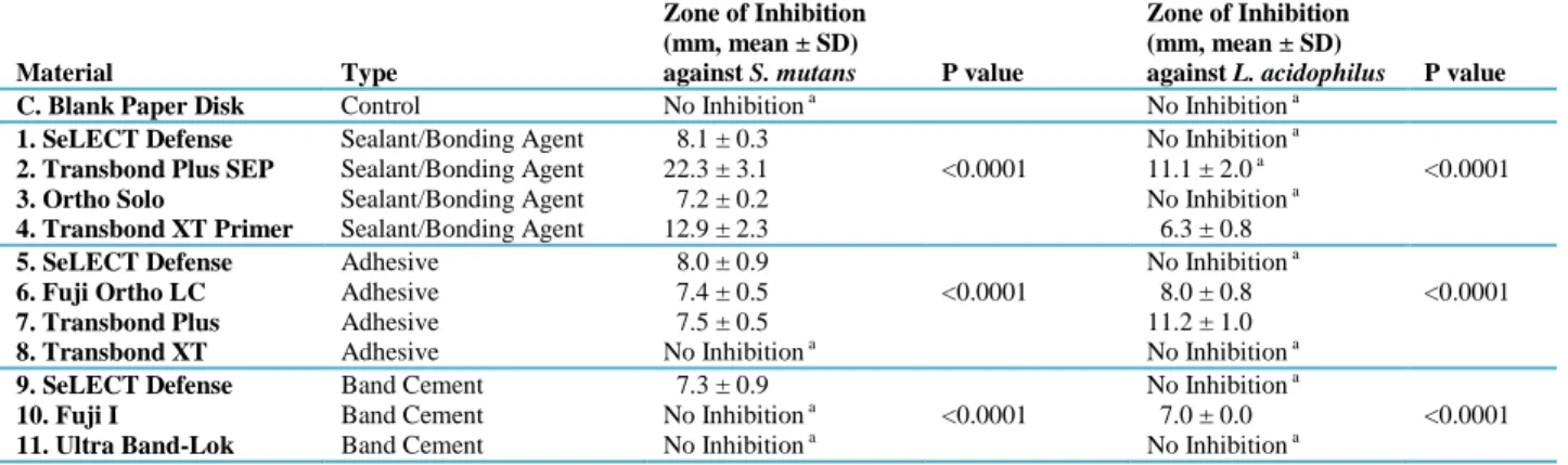
Following 24 to 48 hours of incubation, a bacterial “lawn” had grown on the Agar Diffusion Assay plates (Fig 2). Disks of specific orthodontic materials reproducibly resulted in circular zones marked by an absence of bacterial growth, termed “zones of inhibition” (Fig 2). Diameters of the zones of inhibition in the Agar Diffusion Assay are listed in Table 2.
The differences in means of the zones of inhibition produced by SeLECT Defense adhesive compared to Fuji Ortho LC and Transbond Plus were not statistically significant. SeLECT Defense band cement resulted in a zone of inhibition against S. mutans, while Fuji I and Ultra Band-Lok did not (P<0.0001).
Transbond Plus SEP resulted in a zone of inhibition against L. acidophilus, while SeLECT Defense sealant and Ortho Solo did not (P<0.0001). One of the ten Transbond XT Primer samples resulted in an 8.5mm diameter zone of inhibition on an L. acidophilus plate. Fuji Ortho LC and Transbond Plus resulted in zones of inhibition against L. acidophilus, while
SeLECT Defense adhesive and Transbond XT did not (P<0.0001). Fuji I band cement was the only band cement to result in a zone of inhibition against L. acidophilus, while SeLECT Defense band cement and Ultra Band-Lok did not (P<0.0001).
Of newly-prepared orthodontic materials samples, Transbond Plus SEP and Ortho Solo sealants resulted in clinically significant reduction of S. mutans CFU, while SeLECT Defense sealant and Transbond XT did not (Table 3) (P<0.0001). No adhesives or band cements resulted in a clinically significant reduction in S. mutans CFU, though the clinically insignificant
reduction of S. mutans CFU produced by Fuji Ortho LC was significantly different than
SeLECT Defense adhesive (P = 0.001). Fresh samples of ACT fluoride mouth rinse and Peridex chlorhexidine mouth rinse also resulted in clinically significant reduction of S. mutans. Fuji Ortho LC and Transbond Plus adhesives, Fuji I and Ultra Band-lok band cements, and Duraphat fluoride varnish each demonstrated clinically insignificant reduction of CFU when S. mutans was inoculated immediately following their preparation.
Of newly-prepared orthodontic materials samples, Transbond Plus SEP and Ortho Solo sealants resulted in clinically significant reduction of L. acidophilus CFU, while SeLECT Defense sealant and Transbond XT did not (P<0.0001). No adhesives or band cements resulted in a clinically significant reduction in L. acidophilus CFU, though the clinically insignificant reduction of L. acidophilus CFU produced by Fuji I was significantly different than SeLECT Defense adhesive (P = 0.019). Fresh samples of ACT fluoride mouth rinse and Peridex chlorhexidine mouth rinse also resulted in clinically significant reduction of L. acidophilus.
reduction of S. mutans CFU, though only Fuji Ortho LC’s reduction was statistically different than its SeLECT Defense counterpart (P=0.002).
Discussion
Previous studies by Amaechi found that SeLECT Defense products prevent the formation of white spot lesions.22,23 However, these studies did not distinguish between prevention of white spot lesions due to antibacterial properties of SeLECT Defense and prevention due to the presence of a mechanical barrier, as filled sealants were not used on the control teeth. While the study by Tran et al. reported the antibacterial properties of SeLECT Defense sealant 21, there was no comparison made to other commonly used orthodontic sealants, and the inhibition was not characterized as bacteriostatic or bactericidal. The current study is the first study to characterize the antimicrobial properties of SeLECT Defense and compare them to other commonly used orthodontic products.
The zones of inhibition of the tested materials vary considerably in diameter (Table 2), yet their magnitudes may be of little clinical significance. A more appropriate interpretation of the results may be to consider the presence or absence of a zone of inhibition because white spot demineralization occurs at the enamel surface in direct contact with a cariogenic bacterial plaque. The formation of a zone of inhibition in this assay suggests that these materials have the potential to prevent bacterial growth on a tooth surface in vivo, while the magnitude of the diameter
cannot be directly related to clinical events.
A zone of inhibition indicates that an antimicrobial agent diffuses from the sample disk into the surrounding agar to either kill or inhibit bacterial growth on the surface of the plate. The proposed antimicrobial mechanism of action of the SeLECT Defense products is selenium’s ability to catalyze the formation of superoxide radicals which are bactericidal in nature.18 During phases of bacterial growth, radical ions have the opportunity to irreversibly damage DNA as it is replicated. A material that produces radical ions which kill bacterial cells in this way would be considered bactericidal. There also exist radical-mediated pathways which inhibit metabolic enzyme function to prevent bacterial growth. Such a material that does not reduce the number of colony-forming units, but prevents any measurable bacterial growth would be considered
Superoxide radicals are, by definition, short-lived due to their highly reactive nature. Previous studies have reported that these radicals have a half-life of 60 nanoseconds.23 They are limited to the selenium-coated surfaces of SeLECT Defense products, and do not leach out into the oral environment beyond 35 nanometers.23 The Agar Diffusion Assay used in this study is not sensitive enough to detect zones of inhibition of such small magnitude, yet SeLECT Defense products resulted in measureable zones of inhibition. The data suggest that SeLECT Defense products have antimicrobial properties, but the assay cannot determine if the antibacterial agent is a selenium-catalyzed superoxide radical.
The proposed antimicrobial agents present in the other tested materials are listed in Table 1, but are also not confirmed by these assays. For example, Transbond Plus SEP consistently displayed antimicrobial properties across all assays. While it is reported to be fluoride-releasing, it also contains concentrated phosphoric acid which is meant to demineralize the enamel surface of a tooth to facilitate bonding an orthodontic bracket. In a clinical setting, the acid in the self-etching primer is naturally controlled and buffered by the ions released during demineralization of tooth structure. In the assays used in this study, no buffering agent was present, and likely the robust antimicrobial property of this product was due to the presence of the acid. Transbond XT Primer displayed antimicrobial properties in the Agar Diffusion Assay, despite its lack of
acidophilus, yet Ortho Solo sealant, which also releases fluoride, did not. Such findings may suggest that a threshold amount of fluoride release is necessary to result in a zone of inhibition for a given bacterial species.
The Agar Diffusion Assay demonstrated that all four sealants tested have antimicrobial properties. This is an encouraging finding because sealants can be used to protect the entire facial surface of the tooth when bonding orthodontic brackets. While a smaller percentage of the orthodontic adhesives and band cements demonstrated zones of inhibition, their antimicrobial properties may be of less clinical significance compared to sealants because white spot lesions form around orthodontic brackets more frequently than underneath them (Fig 1).
The Direct Contact Inhibition Assay did not provide evidence that selenium-containing orthodontic materials have bactericidal properties. The assay provided one hour to allow
bacteria in the inoculum to settle via gravity to the surface of the orthodontic material, come into contact with an antimicrobial agent, and be killed. As opposed to the Agar Diffusion Assay, which cannot distinguish bacterial killing from mere growth inhibition, the Direct Contact Inhibition Assay provides evidence of bactericidal properties. If bacteria were killed or irreversibly inhibited upon contact with the orthodontic material, no viable bacteria would remain in the inoculum when spot-plated on agar (Fig 4, material #2). Bacteria that were unaffected or merely inhibited while in contact with the orthodontic material would be able to form colonies when removed from the presence of orthodontic materials in the microtiter plate well, and spotted on the agar (Fig 4, materials #1, 3, and 4).
in CFU in the Direct Contact Inhibition Assay. This assay only demonstrated clinically significant reduction of CFU by Transbond Plus SEP, Ortho Solo sealant, ACT fluoride rinse, and Peridex chlorhexidine rinse. Any reduction of CFU less than 99.9% was deemed
insignificant. It is possible that other materials tested in this assay are capable of clinically significant reductions in bacterial CFU, but require longer than one hour of contact for sufficient killing to occur. However, previous studies have shown that bacterial exposure to orthodontic cements for one hour was sufficient to result in a significant inhibition of bacterial growth.24
Of the nine orthodontic materials that demonstrated positive zones of inhibition against S. mutans or L. acidophilus, in the Agar Diffusion Assay, only two (Ortho Solo and Transbond Plus SEP) can be characterized as bactericidal by the Direct Contact Inhibition Assay. The other seven materials are at a minimum bacteriostatic because their presence prevents bacterial growth (Fig 2). Clinically, bacteriostatic and bactericidal materials would equally prevent white spot lesions, because white spot formation is dependent on the growth of bacterial plaque and subsequent production of lactic acid, not simply the presence of cariogenic bacteria.
When the Direct Contact Inhibition Assay was repeated with orthodontic materials samples that were aged 7 days prior to inoculation, Ortho Solo sealant did not retain its ability to kill S. mutans or L. acidophilus. This may suggest that the antibacterial agent present in this product is volatile in nature, and was not sufficiently present in the sample after one week of aging.
inoculated with S. mutans. It is important to consider that L. acidophilus is most commonly implicated in deep carious lesions, while S. mutans may be present in incipient caries, such as white spot lesions.26 S. mutans has a variety of virulence factors that contribute to their ability to initiate the caries process to produce white spot lesions, while L. acidophilus utilizes its ability to thrive in the low pH environment of existing carious lesions. Many authors suggest that without the initiation of the white spot lesion by S. mutans, L. acidophilus will not be present.26
Therefore, it is of primary importance that antimicrobial orthodontic materials, such as SeLECT Defense, are effective against S. mutans, and secondarily effective against L. acidophilus.
While this study demonstrates that many orthodontic bonding materials, including those containing Selenium, possess antimicrobial properties, it is important to emphasize that this is a short-term, in vitro study testing bacterial species in isolation. Caries is a chronic, multifactorial disease that occurs in a complex environment in which many bacterial species coexist and interact. Furthermore, it is unclear whether or not these antimicrobial properties are maintained throughout the course of orthodontic treatment. The data does not provide evidence that any of the tested bonding materials reduce the incidence of white spot lesions during orthodontic treatment, for which a randomized, controlled clinical trial would be needed. The data does suggest, however, that many orthodontic bonding materials, including those containing
Selenium, have the potential to prevent white spot lesions due to their antimicrobial properties as demonstrated in this study.
Conclusions
Contact Inhibition Assay demonstrated that orthodontic bonding materials, though not SeLECT Defense products, possess bactericidal properties. At this time, the antibacterial properties of SeLECT Defense products against S. mutans can be characterized as bacteriostatic and not bactericidal. A randomized clinical trial is needed to determine if the incidence of white spot lesion formation during orthodontic therapy can be decreased by using antimicrobial orthodontic sealants, adhesives, and band cements containing selenium, such as SeLECT Defense.
Tables
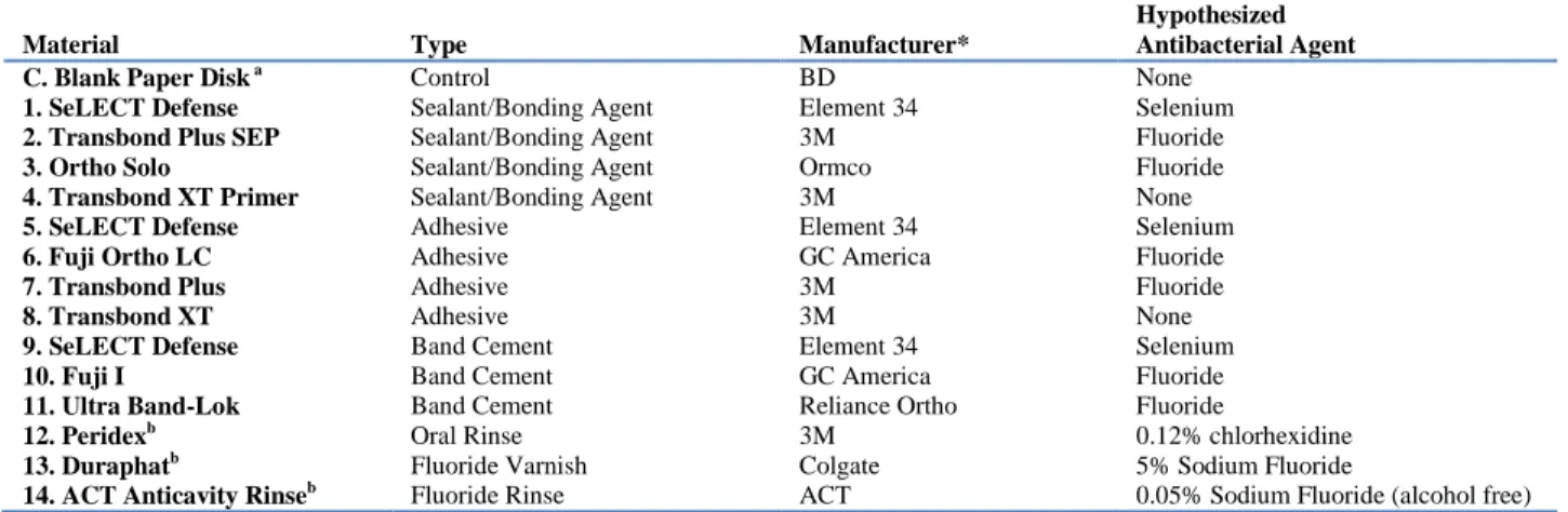
Table 1 - Orthodontic materials tested in Agar Diffusion Assay and Direct Contact Inhibition Assay.
Material Type Manufacturer*
Hypothesized Antibacterial Agent
C. Blank Paper Disk a Control BD None
1. SeLECT Defense Sealant/Bonding Agent Element 34 Selenium
2. Transbond Plus SEP Sealant/Bonding Agent 3M Fluoride
3. Ortho Solo Sealant/Bonding Agent Ormco Fluoride
4. Transbond XT Primer Sealant/Bonding Agent 3M None
5. SeLECT Defense Adhesive Element 34 Selenium
6. Fuji Ortho LC Adhesive GC America Fluoride
7. Transbond Plus Adhesive 3M Fluoride
8. Transbond XT Adhesive 3M None
9. SeLECT Defense Band Cement Element 34 Selenium
10. Fuji I Band Cement GC America Fluoride
11. Ultra Band-Lok Band Cement Reliance Ortho Fluoride
12. Peridexb Oral Rinse 3M 0.12% chlorhexidine
13. Duraphatb Fluoride Varnish Colgate 5% Sodium Fluoride
14. ACT Anticavity Rinseb Fluoride Rinse ACT 0.05% Sodium Fluoride (alcohol free)
a
Blank paper disk served as a control in the Agar Diffusion Assay only. An empty microtiter plate well served as a control in the Direct Contact Inhibition Assay.
b
Material tested in Direct Contact Inhibition Assay only.