15
© 2017 by the Serbian Biological Society How to cite this article: Savin M, Hadžibulić E, Damjanović T, Santrić V, Stanković S. Association of I/D angiotensin-converting enzyme genotype with erythropoietin stimulation in kidney failure. Arch Biol Sci. 2017;69(1):15-22.
Association of I/D angiotensin-converting enzyme genotype with erythropoietin
stimulation in kidney failure
Marina Savin1,*, Edvin Hadžibulić2,#, Tatjana Damnjanović3, Veljko Santrić4 and Sanja Stanković5
1 Clinic of Nephrology, Clinical Centre of Serbia, University Belgrade School of Medicine, Belgrade, Serbia 2 General Hospital of Novi Pazar, Hemodialysis Unit, Novi Pazar, Serbia
3 Institute of Human Genetics, University Belgrade School of Medicine, Belgrade, Serbia
4 Clinic of Urology, Clinical Centre of Serbia, University Belgrade School of Medicine, Belgrade, Serbia 5 Institute of Biochemistry, Clinical Center of Serbia, Belgrade, Serbia
*Corresponding authors: [email protected]; [email protected]
Received: March 3, 2016; Revised: March 31, 2016; Accepted: April 5, 2016; Published online: May 18, 2016
Abstract: Angiotensin-converting enzyme (ACE)-gene polymorphism is a possible predisposing factor of erythropoietin response under hypoxic conditions. However, it is not completely clear whether the ACE insertion/deletion (I/D) genotype has an impact on anemia in patients with permanent kidney failure. A 9-month prospective trial was conducted on 53 patients on hemodialysis aimed at determining the beneficial effect of oral vs intravenous iron in anemia management with recombinant human erythropoietin (rHuEpo), and identifying a possible association of the ACE gene I/D polymorphism with the response to rHuEpo. Patients were randomly allocated to receive 50-100 mg daily of ferrous gluconate orally (N=26) or intravenously every two weeks (N=27), together with rHuEpo-beta (200 IU/kg) subcutaneously, to achieve a hemoglobin increase to 105 g/L; subsequently the rHuEpo dose was adjusted at one or two week intervals. In 34 patients who regularly received ACE-inhibitor (ACEi) medication, genotyping for ACE-gene I/D polymorphism was performed using PCR, gel analysis and appropriate restriction digestion. After prolonged rHuEpo treatment, 24.5% of patients attained the targeted 9th-month hemoglobin concentration (105 g/L). Of these, 6/26 of patients received elemental iron orally and 7/27 received it intravenously. We observed an association between homozygous DD (deletion) of the ACE gene and a remarkable early increase in blood hemoglobin (p=0.028), erythrocyte count (p=0.020) and hematocrit (p=0.043) after reduction of the dose of rHuEpo (F=3.95; p=0.029), irrespective of the iron repletion mode (p=0.960). This is the first report on DD genotype as a linkage marker for the optimization of rHuEpo dose for anemia management in hemodialysis patients.
Key words:hemodialysis; hemoglobin; oral iron; IV iron; epoetin beta; ACE I/D genotype
INTRODUCTION
Angiotensin II is a regulator of the erythropoietin basal level (under physiological conditions) [1]. Angioten-sin II acts as a growth factor that directly stimulates the proliferation of erythroid progenitors in the bone marrow [2]. A worsening of chronic kidney disease (CKD)-associated anemia by treatment with ACE inhibitors and angiotensin II type 1 receptor block-ers is well documented [3,4]. Angiotensin-converting enzyme (ACE)-gene polymorphism is a possible pre-disposing factor of erythropoietin response under hy-poxic conditions. Jeong et al. [5] discovered an associa-tion between the presence of DD-ACE genotype and a lower resistance to recombinant human erythropoietin
(rHuEpo) treatment in CKD patients on peritoneal dialysis. It is not well understood whether the ACE I/D genotype has an impact on anemia management with rHuEpo and iron repletion in patients with permanent kidney failure on regular hemodialysis (HD).
in rHuEpo control of renal anemia is the benefit of continuous iron administration via the dialysate fluid escaping the intestinal iron transport during the he-modialysis procedure [7].
Progress in the HD procedure has significantly im-proved gastrointestinal tolerance to oral iron supple-ments, allowing daily iron repletion for the period of rHuEpo treatment. Oral iron supplementation regulates iron metabolism in a more physiological manner than intermittent supplementation with a high dose of IV ferrous. As regards control of hemoglobin concentra-tions in renal anemia, there are no data related to the possible concerns about the route of iron supplemen-tation and the potential impact of ACE I/D polymor-phism on rHuEpo treatment. The aim of our study was to compare the efficiency of oral vs intravenous iron supplementation in CKD patients undergoing hemo-dialysis, and the inherited factor ACE I/D genotype.
MATERIALS AND METHODS
Subjects
Patients suffering from CKD-related anemia (blood hemoglobin <95 g/L) that were on a regular hemo-dialysis program with dose adequacy assessed by Kt/V (urea)>1.2, were enrolled in the study. The main inclusion criteria were an iron-binding capac-ity (transferrin saturation (TSAT)), calculated as sFe/ TIBCx100>20% that was confirmed after a month-long monitoring, and whether a prior intravenous (IV) iron treatment was required. The patients were randomly allocated into two treatment groups: one group received rHuEpo-beta and 50-100 mg daily of ferrous gluconate orally (Tot Hema, Innotech), the other group received rHuEpo and intravenous fer-rous gluconate (Ferrlecit, Sinofi) of 100 mg every two weeks, according to standard KDIGO recommenda-tion for anemia management in CKD patients [6]. All patients received the rHuEpo-beta (Recormon, Roche) in a starting dose of 200 IU/kg, administered subcuta-neously three times per week, to achieve a hemoglo-bin increase up to 105 g/L, which was subsequently adjusted at intervals of one or two weeks.
Additional inclusion criteria were: ≥18 years of age, dialysis vintage of at least 3 months, hemodialysis
ad-equacy according to Kt/V(urea)>1.2, absence of hepa-titis virus and HIV infection, malignancy, other active infection or elevated inflammation parameters. Exclu-sion criteria were: non-CKD-related anemia, a history of hepatitis B or C, pregnant or nursing women, blood transfusion within the previous 3 months, sustained ferritin ≥800 ng/mL for longer than 3 months, new malignancy, iron overload (hemochromatosis) or seri-ous disturbances in the utilization of iron (allergies to iron preparations, chronic vomiting), decompensated liver cirrhosis or active hepatitis, active acute or chron-ic infections, frequent need of blood transfusions, un-treated vitamin B12 or folate deficiency.
Study protocol
Epoetin beta (200 IU/kg) was administered subcuta-neously at the end of the HD procedure three times per week until an increment of 1 g/dL of hemoglo-bin was achieved. Administration was continued, but with a 25% reduction in the dose of epoetin in order to maintain a therapeutic target hemoglobin maximum of 110 g/L, which was monitored over the period of one month. Iron supplementation with an oral iron prepa-ration containing ferrous gluconate (quantity equiva-lent to 50 mg iron) was prescribed 1-2 times daily, while IV iron supplementation with a sodium ferric gluconate complex of 100 mg was applied once in two weeks after the hemodialysis. This study protocol of iron repletion and erythropoietin stimulation and in association with ACE-I/D genotype was approved the Ethical Commit-tee of the Belgrade School of Medicine, University of Belgrade, and the Ethical Committee of the General hospitals of Novi Pazar and Kraljevo. The informed consent from all the examined patients was obtained.
Biochemical investigation
Treatment regimen efficiency was evaluated by moni-toring the respective hematological and iron metab-olism parameters that were regularly measured in a three-month period. Trial safety parameters included blood leukocytes, platelet count, serum urea and po-tassium, together with systolic/diastolic pressure.
ACE I/D genotyping
Blood samples were obtained in EDTA tubes and DNA was isolated by the salting-out method [8]. The I/D polymorphism of ACE, an accepted marker for renin-angiotensin system (RAS) activity, was determined us-ing the polymerase chain reaction (PCR) with primers for detection of ACE deletion (D) insertion/(I) poly-morphisms, as follows: forward: 5’- CTG GAC ACC ACT CCC ATC CTT TCT -3’, reverse: 5’- GAT GTG GCC ATC ACA TTC GTC AGA T-3’ (Metabion). Half a μl of each primer was solved in a final volume of 25 μl, containing 25 mM MgCl2, 10mM of each dNTP, 10xB (8500 mM KCl, 100 mM TRIS HCl, pH 8.3, 15 mM MgCl2, 0.01% gelatin) and 1 unit of Taq polymerase. PCR was performed with 5 min of initial denaturation at 95°C, followed by 35 cycles of denaturation for 1 min at 94°C, annealing at 63°C for 1 min, extension at 72°C for 1 min and final extension for 10 min at 72°C, using 2720 Thermal cycler (Applied Biosystems). After the PCR, the samples were separated by 8% polyacrylamide gel electrophoresis (Sigma), stained with SYBR Safe (In-vitrogene), and photographed. Gel analysis showed an amplification band of 477 bp in the presence of the insertion in samples with the II genotype, bands of 477 and 190 bp in samples with ID genotype and a band of 190 bp in the absence of the insertion in samples with the DD genotype. To exclude incorrect DD genotyp-ing in samples of ACE DD genotype, the results were confirmed with repeated PCR analysis.
Statistical analysis
Statistical analysis was performed using SPSS 18.0. The patients were randomly separated into two treat-ment groups as described above. The respective vari-ables were presented as the means±SD, with p<0.05 considered as significant. The iron treatment efficacy was evaluated by time-course changes of hematologi-cal and iron metabolism parameters based on the comparison of variable measurements conducted at
3-month check points during a 9-month trial period for both iron treatment groups (oral vs. IV) and ACE-gene I/D polymorphism in a ACE-general linear model used for analysis of variance (ANOVA) by repeated measures’ calculation. To examine the mean differ-ences in related variables with normal distribution between groups, a 2-sample t-test was applied.
RESULTS
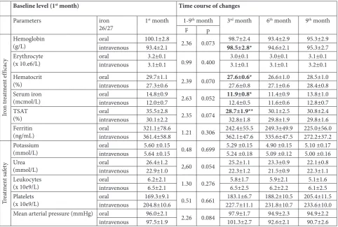
The treatment protocol and general characteristics of 53 patients enrolled in the prospective trial of rHuEpo in the management of anemia associated with chronic hemodialysis, and randomly allocated to oral vs. IV iron supplementation, are shown in Table 1. Patients entered the study with blood transferrin saturation (TSAT) >20%, to determine the effect of iron repletion on the response to rHuEpo and to identify the ACE-gene I/D contribution to rHuEpo dose optimization. Baseline hematological characteristics along with iron metabolism parameters were at similar levels in the two defined cohorts, with the exception of the serum iron value, which was lower in patients selected for IV ferrous gluconate administration (p=0.021) (Table 2). Patient response implied the equal efficiency and safety of the oral vs. IV route of iron application for prolonged rHuEpo control of renal anemia (Table 2). Approximately half of the patients in both iron supple-mental groups maintained a hemoglobin concentra-tion >95 g/L, with 24.5% of patients able to reach the target 9th-month outcome hemoglobin concentration in the blood of 105 g/L, i.e. 6/26 (23.1%) of patients
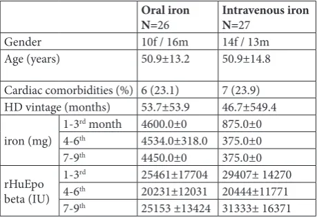
Table 1. Summary of the study protocol.
Oral iron
N=26 Intravenous ironN=27
Gender 10f / 16m 14f / 13m
Age (years) 50.9±13.2 50.9±14.8
Cardiac comorbidities (%) 6 (23.1) 7 (23.9)
HD vintage (months) 53.7±53.9 46.7±549.4
iron (mg) 1-3
rd month 4600.0±0 875.0±0
4-6th 4534.0±318.0 375.0±0
7-9th 4450.0±0 375.0±0
rHuEpo beta (IU)
1-3rd 25461±17704 29407± 14270
4-6th 20231±12031 20444±11771
7-9th 25153 ±13424 31333± 16371
administered oral elemental iron, and 7/27 (25.9%) administered IV iron. Monitoring of the hematologi-cal status revealed proportionate response patterns to both iron regimens after the 3rd month, which were mirrored by the time-course changes in the hemoglo-bin levels (Table 2, Fig. 1).
Significant benefits of iron supplementation to the hematological status and rHuEpo efficacy in renal anemia management were observed during the first 3 months of the treatment (starting from the time of randomization), while the hemoglobin level was aug-mented relative to the iron supply. The concentration of hemoglobin increased with IV iron administration (p=0.026), while the serum iron and TSAT remained stable. During the same period, in the group under-going the oral iron administration regimen, the he-moglobin concentration did not change, while the serum iron (p=0.018), TSAT (p=0.009) and storage iron ferritin declined considerably (Table 2, Fig. 1).
During the first 3 months, the amount of rHuEpo was proportionately reduced in both iron administration groups (Table 1, Fig. 2). The validated early discrep-ancy in the hemoglobin response to iron supply can be explained by the presence of other factors that trig-gered and supported erythropoiesis.
Patients did not show serious adverse reactions to ferrous treatment and regularly used medications. Gender, diabetes, cardiac comorbidities (coronary dis-ease and cardiac insufficiency) or underlying kidney disease did not elicit a response to rHuEpo. Regarding the well-known suppressor effect of antihypertensive ACE inhibitors on hematopoiesis and the lower hemo-globin level observed in our patients on ACE-inhibitor medication at 3rd month (97.2±13.2, and 106.2±12.2 g/l, respectively, p=0.077), subsequent analysis included subgroup of 34 patients on standard ACE-inhibitor treatment during the entire study period (with 17 pa-tients each receiving iron either orally or IV) to
deter-Table 2. Baseline values of parameters and treatment efficacy and safety.
Baseline level (1st month) Time course of changes
Parameters iron
26/27 1
st month 1-9th month 3rd month 6th month 9th month
F p
Ir
on t
re
at
m
en
t ef
fic
ac
y
Hemoglobin
(g/L) oralintravenous 100.1±2.893.4±2.1 2.36 0.073 98.5±2.8*98.7±2.4 93.4±2.994.6±2.1 95.3±2.995.3±2.7
Erythrocyte
(x 10.e6/L) oralintravenous 3.2±0.13.1±0.1 0.99 0.400 3.0±0.13.1±0.1 3.0±0.13.1±0.1 3.1±0.13.2±0.1
Hematocrit
(%) oralintravenous 29.7±1.127.3±0.6 2.39 0.070 27.6±0.6*27.6±0.8 26.6±1.027.1±0.6 28.5±1.028.4±0.8
Serum iron
(mcmol/L) oralintravenous 14.8±0.912.0±0.7 2.63 0.052 11.9±0.8*12.4±0.5 11.4±0.911.6±0.6 13.8±1.012.8±0.7
TSAT
(%) oralintravenous 35.5±2.830.1±2.2 2.35 0.074 28.7±1.9**32.8±1.8 30.1±2.529.8±1.9 30.8±2.429.8±1.6
Ferritin
(ng/mL) oralintravenous 321.1±78.6361.4±58.8 1.21 0.306 242.4±55.5362.1±47.6 249.3±49.9335.6±47.5 225.0±56.0272.2±37.2
Tr
ea
tm
en
t s
af
et
y
Potassium
(mmol/L) oralintravenous 5.60 ±0.155.64 ±0.15 0.48 0.699 5.29 ±0.155.24 ±0.18 4.90 ±0.155.09 ±0.12 5.10 ±0.175.00 ±0.16 Urea
(mmol/L) oralintravenous 26.4±1.222.9±1.0 2,60 0.054 25.2±1.122.3±1.2 23.3±0.921.5±0.9 22.1±0.822.3±1.1
Leukocytes
(x 10e9/L) oralintravenous 6.2±2.16.5±2.1 1.30 0.276 5.8±1.76.5±2.5 5.9±2.16.2±2.2 5.1±1.66.1±2.5
Platelets
(x 10e9/L) oralintravenous 204.8±10.6169.3±9.1 0.51 0.661 227.7±11.1183.1±6.7 188.2±10.5231.8±10.7 205.4±11.5233.6±10.0
Mean arterial pressure (mmHg) oral 96.0±2.1
2.26 0.084 97.9±1.7 94.9±2.3 94.9±2.2
intravenous 97.5±1.9 101.3±2.7 92.6±2.1 90.7±2.6
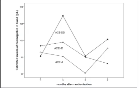
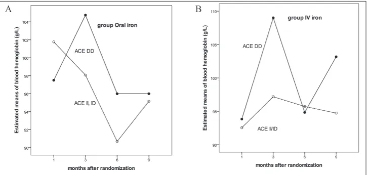
mine the possible impact of ACE genotype on an early hemoglobin increase induced by rHuEpo stimulation. The remaining 19 patients (10 on IV iron and 9 on oral iron) out of the whole group included in the study received either temporarily ACE inhibitor or started with medication after the 3rd month (5 patients received ACEi between the 3rd and the 6th months, and 3 pa-tients were supplied with ACEi medication after the 5th month of the study), as required to maintain the mean arterial pressure within a normal range, and 11 out of 53 patients with the mean arterial pressure below 105 mmHg did not receive antihypertensive drugs over the study period. By analyzing the possible impact of ACE genotype DD, DI or II on early hemoglobin response to rHuEpo, an association was observed between homo-zygous DD (deletion) of the ACE gene and a remark-able early increase in blood hemoglobin concentration (p=0.028), erythrocyte count (p=0.020) and hematocrit (p=0.043). Carriers of the ID or II genotypes main-tained the hematological parameters irrespective of the mode of iron repletion (Firon x ACEgenotype =0.003; p=0.960) (Table 3A, Figs. 1 and 3A,B). There was a significant reduction in the dose of rHuEpo in patients with the DD genotype in the 2nd month of the study (F
1st vs. 2nd month = 3.95; p=0.029) (Table 3B, Fig. 2).
During the next three months, the supplemen-tation with rHuEpo was significantly reduced (Fig. 2); the hemoglobin concentration was significantly decreased only in patients with the DD-ACE geno-type. In these patients, the mean arterial systolic and diastolic pressure decreased. The patients with ei-ther ID or II genotype were not as receptive to rapid changes in rHuEpo dosage and their hemoglobin lev-els remained much more stable during the same time (Figs. 1 and 2).
DISCUSSION
Herein are presented the results of a randomized 9-month trial examining the hematological response to rHuEpo (beta) administration with respect to the route of iron supplementation (oral vs. intravenous), and I/D-ACE gene polymorphism. It was demonstrat-ed that about one-fifth of the 53 hemodialysis patients enrolled attained the set blood hemoglobin concentra-tion of 105 g/L with comparable success of both oral elemental iron and intravenous (IV) iron repletion. The elevation of iron transport parameters (serum iron and total iron binding capacity (TIBC)) was as-sociated with a proportional increase in hematological parameters following the 3rd month of treatment. The significant increase in the first 3 months in hemoglo-bin level in patients on intravenous iron and in
ACE-Fig. 1. Time-course changes of blood hemoglobin concentration
with respect to the I/D-ACE genotype. The curves illustrate the estimated hemoglobin concentration at each time point relative to angiotensin-converting enzyme (ACE) I/D genotype carri-ers, by the general linear model for repeated measures analysis. Curve II – genotype carriers hemoglobin means±SE (g/L) at 1st month = 96.4±4.74; 3rd month=95.2±4.6; 6th month=90.2±4.8; 9th month= 97.6±5.0; Curve ID – genotype carriers – hemoglobin means±SE (g/L) 1st month = 98.4±4.0; 3rd month=99.4±3.9; 6th
month=95. 1±4.8; 9th month= 93.1±4.2; Curve DD – genotype
carriers – hemoglobin means±SE (g/L): 1st month = 95.3±4.7; 3rd month=107.3±4.6; 6th month=95.3±4.8; 9th month= 100.3±5.0.
Fig. 2. Time-course of changes in monthly dosage of rHuEpo with
DD genotype carriers (p=0.026, and p=0.028, respec-tively) can be considered as two independent events. It seems that the DD carriers were much prone to an oscillation in hemoglobin levels with rapid changes in rHuEpo stimulation dosage than DI or DD carriers.
Several trials have previously reported the im-proved effect of oral administration on iron levels in dialysis patients [9,10]. In accordance with our results, forty-six patients treated with epoetin, and grouped randomly to receive different oral iron preparations, maintained the target hematocrit during a 6-month period [11], with the hemoglobin concentrations and iron indices maintained, rather than increased.
Impaired dietary iron absorption, in addition to impaired iron release from body storage and lack of regular daily intake of iron, are established limiting factors in oral iron supplementation [12]. The high adequacy of hemodialysis with kT/V (urea)>1.2,
which is the standard measure of optimal hemodialy-sis in the last decade [6], enables satisfactory clearance of uremic toxins and substantially improved gastroin-testinal tolerance to iron supplements. This was not the case in the past when an attempt to introduce oral iron in hemodialysis failed, and oral iron repletion was applied to CKD patients prior to chronic dialysis [6,13]. Although IV administration of iron provides rapid iron repletion and is superior to oral iron ad-ministration in many circumstances, long-term IV treatment, intermittently applied at high dosage, may contribute to increased mortality in the hemodialysis population, due to deleterious side effects and dis-ruption of the physiological pattern of iron metabo-lism. Intravenous iron may cause oxidative stress and endothelial injury due to high single and cumulative ferrous doses. rHuEpo treatment per se induces many serious adverse events, such as elevated systolic blood pressure. Optimization of the rHuEpo and iron reple-tion dose is of crucial importance for renal anemia management and attenuation of the risk of chronic hemodialysis. ACE I/D genotyping provides impor-tant information about the inherent potency of the he-matological response to rHuEpo. We determined that the DD genotype requires a lower amount of rHuEpo in the first months of application, although treatment with an ACE inhibitor can limit this effect.
There are no literature data evaluating the associa-tion between ACE genotypes and iron replacement therapy in the control of anemia by rHuEpo in patients on regular hemodialysis. It wasrecognizedmore than two decades agothat ACE gene polymorphism on chromosome 23(17q), characterized by the deletion/
Table 3A. ACE I/D genotype and hematological parameters.
Baseline levels Time course of changes
Parameters I/D-ACE
genotype (n) 1
st month 1→9th month 3rd month 6th month 9th month
F p
Hemoglobin (g/L)
II (10) 96.4±4.6
1.92 0.086
95.2±4.7 90.2±3.8 97.6±5.6
ID (14) 98.4±4.0 99.4±4.3 95.0±4.0 93.1±3.9
DD (10) 95.3±4.9 107.3±3.5** 95.3±5.8 100.3±4.7
Erythrocyte (x 10e6/L)
II 3.2 ±0.2
2.13 0.057
3.0 ±0.1 3.0 ±0.2 3.3 ±0.2
ID 3.2 ±0.1 3.1 ±0.1 3.0 ±0.1 3.0 ±0.1
DD 3.1 ±0.2 3.4 ±0.1* 3.1 ±0.2 3.3 ±0.2
Hematocrit (%) II ID 28.9±41.529.2±2.3 1.75 0.118 26.8±1.227.6±1.8 26.1±1.226.5±1.4 29.2±1.927.2±1.4
DD 28.1±1.4 30.7±1.0* 27.3±1.9 29.9±1.5
Parameters are expressed as means ± SE; asterisks represent significance levels: *p<0.05, **p<0.005 of the difference between prior and subsequent checkpoint values over the study period
Table 3B. Monthly dosage of rHuEpo with respect to the
I/D-ACE genotype.
Month II ID DD
1 13200±2254 10143±1561 11800±2723
2 13400±2130 10286±1814 6800±1665*
3 9200±1982 5857±1266 4600±1462
4 8600±2171 4714±1179 2600±1400
5 6600±1431 6143±1248 4800±1794
6 10400±1627 9429±1670 8600±2023
7 10000±1032 7286±1477 8200±1698
8 9400±1863 7857±1747 9600±2544
9 7400±2212 10143±2272 9600±2344
insertion of 287-base pairs, could have an important impact on ACE activity. ACE gene polymorphism is a functional polymorphism that is linked to several other important polymorphisms in the ACE gene [14]. The ACE I/D genotype is now an accepted marker for renin-angiotensin system (RAS) activity. ACE and its novel homolog, angiotensin converting enzyme 2 (ACE2), are two key enzymes involved in the synthesis of bioactive components of the RAS [15]. Individuals with the DD genotype produce increased angiotensin II and present higher angiotensin II activity. Angioten-sin II concentration may be around 1000 times higher in the kidney than in the peripheral blood outside the kidney in CKD. High RAS activity is a mechanism for the progression of CKD in the advanced stage, fol-lowed by low erythropoietin production and serious anemia[15,16]. Individuals from the general popula-tion with the DD genotype are at high risk of perma-nent loss of kidney function [17]; DD poses a risk of CKD progression in transplant kidneys, as well [18].
Increased activity of ACE and angiotensin II may have a beneficial effect on erythropoiesis, in a direct manner. The addition of angiotensin II to cultured kid-ney peritubular fibroblasts induces the production of endogenous epoetin, as has been reported in patients on hemodialysis [19]. Angiotensin II is an important additional stimulator of erythropoiesis, and as a growth factor it can assist in epoetin augmentation of the ery-throid pool [4]. Two clinical trials have revealed an increased requirement for epoetin at the end stage of kidney failure in patients with II or ID genotypes on peritoneal dialysis [20,21]; one trial included hemo-dialysis patients [5]. We observed that the DD-ACE genotype is more receptive to rapid decrease of rHuEpo dose than individuals with ID and II genotypes, as a result of a very rapid decline in hemoglobin concentra-tion. Consequently, a decrease in the dosage of epoetin in D allele carriers will subsequently require additional supplementation with rHuEpo and iron. Recently, in a cross-sectional study on a large group of patients, Kiss
Fig. 3. Time-course of changes in blood hemoglobin concentration with respect to the I/D-ACE genotype
at al. [22] reported that hemodialysis patients with DD genotype on ACE-inhibitor therapy had lower hemo-globin concentrations and a higher erythropoietin re-sistance index than patients with II genotype.
To conclude, although small numbers of patients were included in the randomized study of iron repletion and ACE-I/D genotype association with erythropoietin stimulation, an important impact of ACE-I/D genotype on hematological regulation was observed in patients on hemodialysis. Our investigation suggests that the genetic ACE typing can provide valuable information regarding the optimal dosage of rHuEpo and iron sup-plementation via oral or intravenous administration.
Authors’ contribution: All authors contributed equally to this
work.
Conflict of interest disclosure: The authors declare that there is
no conflict of interest.
REFERENCES
1. Kim YC, Mungunsukh O, McCart EA, Roehrich PJ, Yee DK, Day RM. Mechanism of erythropoietin regulation by angio-tensin II. Mol Pharmacol. 2014; 85(6):898-908.
2. Vlahakos DV, Marathias KP, Madias NE. The role of the renin-angiotensin system in the regulation of erythropoiesis. Am J Kidney Dis. 2010;56(3):558-65.
3. Iodice C, Balletta MM, Minutolo R, Giannattasio P, Tuccillo S, Bellizzi V, D’Amora M, Rinaldi G, Signoriello G, Conte G, De Nicola L. Maximal suppression of renin-angiotensin system in nonproliferative glomerulonephritis. Kidney Int. 2003;63(6):2214-21.
4. Jacobsen P, Andersen S, Jensen BR, Parving HH. Additive effect of ACE inhibition and angiotensin II receptor block-ade in type I diabetic patients with diabetic nephropathy. J Am Soc Nephrol. 2003;14(4):992-9.
5. Jeong K, Lee T, Ihm C, Lee S, Moon J. Polymorphisms in two genes, IL-1b and ACE, are associated with erythropoietin resistance in Korean patients on maintenance hemodialysis. Exp Mol Med. 2008;40(2):161-6.
6. KDIGO clinical practice guideline for anemia in chronic kidney disease. Kidney Int Suppl. 2012;2:279-335.
7. Tsuchida A, Paudyal B, Paudyal P, Ishii Y, Hiromura K, Nojima Y, Komai M. Effectiveness of oral iron to manage anemia in long-term hemodialysis patients with the use of ultrapure dialysate. Exp Therap Med. 2010;1:777-81. 8. Miller SA, Dykes DD, Polesky HF. A simple salting out
procedure for extracting DNA from human nucleated cells. Nucleic Acids Res. 1988;16 (3):1215.
9. Macdougall IC, Tucker B, Thompson J, Tomson CR, Baker LR, Raine AE. A randomized controlled study of iron sup-plementation in patients treated with erythropoietin. Kidney Int. 1996;50:1694-9.
10. Fudin R, Jaichenko J, Shostak A, Bennett M, Gotloib L. Cor-rection of uremic iron deficiency anemia in hemodialyzed patients: a prospective study. Nephron. 1998;79:299-305. 11. Wingard RL, Paeker RA, Nuhad I, Hakim RM. Efficacy of
oral iron therapy in patients receiving recombinant human erythropoietin. Am J Kidney Dis. 1995;35(3):433-9. 12. Babitt JL, Lin HY. Molecular mechanisms of hepcidin
regu-lation: implications for the anemia of CKD. Am J Kidney Dis. 2010;55:726-41.
13. National Kidney Foundation. K/DOQI clinical practice guidelines and clinical practice recommendations for anemia in chronic kidney disease. Am J Kidney Dis.2006;47:S11-S145.
14. Rigat B, Hubert C, Alhenc-Gelas F, Cambien F, Corvol P, Soubrier F. An insertion/deletion polymorphism in the angiotensin I-converting enzyme gene accounting for half the variance of serum enzyme levels. J Clin Invest. 1990;86(4):1343-6.
15. Seikaly MG, Arant BS, Seney FD. Endogenous angiotensin concentrations in specific intrarenal fluid compartments of the rat. J Clin Invest. 1990;86(4):1352-7.
16. Ingelfinger JR, Dzau VJ. Molecular biology of renal injury: Emphasis on the role of the renin-angiotensin system. J Am Soc Nephrol. 1991;2:S2-S20.
17. Zhou TB, Yin SS, Qin YH. Association between angiotensin-converting enzyme insertion/deletion gene polymorphism and end-stage renal disease susceptibility. J Renin Angioten-sin Aldosterone Syst. 2014;15(1):22-31.
18. Savin M, Petronic V. The significance of angiotensin-con-verting enzyme inhibitors genotype for ACEi response in patients with chronic allograft nephropathy. Serb Arch Medicine. 2005; 133:194-8.
19. Vlahakos DV, Balodimos C, Papachristopoulos V, Vassila-kos P, Hinari E, Vlachojannis JG. Renin-angiotensin system stimulates erythropoietin secretion in chronic hemodialysis patients. Clin Nephrol. 1995;43(1):53-9.
20. Varagunam M, McCloskey DJ, Sinnott PJ, Raftery MJ,
Yaqoob MM.Angiotensin-converting enzyme gene
poly-morphism and erythropoietin requirement. Perit Dial Int 2003;23(2):111-5.
21. Sharples EJ, Varagunam M, Sinnott PJ, McCloskey DJ, Raftery MJ, Yaqoob MM. The effect of proinflammatory cytokine gene and angiotensin-converting enzyme poly-morphisms on erythropoietin requirements in patients on continuous ambulatory peritoneal dialysis. Perit Dial Int. 2006;26(1):64-8.