285
© 2017 by the Serbian Biological Society How to cite this article: Jordanova M, Rebok K, Spasova A, Roganovic-Zafirova D. Gill lesions in Prespa barbel (Barbus prespensis) inhabiting a polluted area of Lake Prespa. Arch Biol Sci. 2017;69(2):285-9.
Gill lesions in Prespa barbel (
Barbus prespensis
) inhabiting a polluted area of Lake Prespa
Maja Jordanova*, Katerina Rebok, Adriana Spasova and Danica Roganovic-Zafirova
Laboratory of Histology and Embryology, Institute of Biology, Faculty of Natural Sciences and Mathematics, Ss. Cyril & Methodius University, Arhimedova 3, 1000 Skopje, Republic of Macedonia.
*Corresponding author: [email protected]
Received: March 9, 2016; Revised: April 9, 2016; Accepted: April 10, 2016; Published online: October 17, 2016
Abstract: The study focused on gill lesions observed in Prespa barbel individuals caught on the north littoral site of Lake Prespa in the area of the Golema River confluence, which is under agricultural, industrial and urban pollution pressure. Microscopic analysis revealed a series of circulatory, regressive, progressive and, to a less extent, inflammatory changes of which the most prominent were: telangiectasia, necrosis of respiratory epithelium accompanied by collapsed secondary lamellae, necrosis and proliferation of interlamellar and lamellar respiratory epithelium, and parasite infection. These results indicate that the reiterated gill lesions were probably due to the double influence of the toxic effects of a polluted aquatic environment and opportunistic infections, which indicates that the application of mitigating measures is urgently needed in order to protect the Prespa Lake ecosystems.
Key words: gill; histopathology; pollution; barbel; Lake Prespa
INTRODUCTION
Prespa barbel (Barbus prespensis Karaman, 1924) is one of four endemic species found in Lake Prespa and is considered an endangered species [1]. There are numerous potential factors that may threaten Pre-spa barbel, including overfishing, the introduction of non-native species, stream and lakeshore habitat destruction, increasing water eutrophication, etc. [1]. Lake Prespa is estimated to be under considerable ag-ricultural, urban and industrial pollution pressure [2]. The water quality of the lake is particularly affected by run off of agricultural chemicals, considering that most of the near shore ground is under or chards and other croplands. According to Grupce [3], Lake Prespa is also suffering from a phosphorus overload of about 6.4 tons per year, which leads to the rapid eutrophi-cation of this once oligotrophic lake. However, there is no evidence to date concerning the exposure and health impact of water pollution on Prespa barbel or other fish species in the lake.
It is well known that toxicants can induce different gill lesions in fishes [4-10]. Toxic substances in aquatic environments can alter the chloride and mucous cells situated in the gill epithelium [11]. Parasitic
infec-tions can also provoke gill lesions [12]. Therefore, the primary objective in this study was to examine the histopathological alterations in Prespa barbel gills, the vulnerable respiratory and ion-balance organs highly exposed to the environment. A quantitative histopathological analysis approach was employed in attempt to establish to what extent the registered gill lesions are induced by pollution-born contaminants.
MATERIALS AND METHODS
Fish sampling and tissue proceedings
μm resin embedded semi-thin sections, which were subsequently stained by hematoxylin and eosin (H&E) and 1% toluidine blue staining, respectively.
Histopathology analysis
The registered histopathological changes were quanti-fied by the standardized protocol according to Bernet et al [13]. Briefly, gill lesions classified into 4 reaction patterns (circulatory disturbance, regressive changes, progressive changes and inflammations) were scored (value range 1-6) depending of the degree and the ex-tent of the alterations. Every score value was multiplied by an importance factor reflecting the pathological sig-nificance of the lesion. The sum of all quantified le-sions from the same reaction pattern gave its reaction index: IGC for circulatory disturbances, IGR for regressive changes, IGP for progressive changes and IGI for inflam-mations. The protocol was modified by introducing an additional variable – the relative value of infection index (Youdin index, YI). The severity of parasite infec-tions and candidiasis-like infection was also scored in the range 1-6 according to the abundance of registered infection in the respiratory epithelium. The sum of all reaction indexes gave the total index (IG). Particular attention was paid to the number, distribution and morphology of mucous and chloride cells in the gill epithelium. Mucous and chloride cell counting was performed on semi-thin sections and expressed as the number of cells per interlamellar space.
Statistical analysis
The calculations of the indices were performed in Ex-cel, and graph plotting in Statistics 6.0 for Windows. For correlation analyses, Pearson’s correlation coef-ficient was estimated.
RESULTS
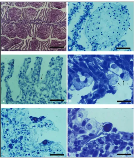
The microscopic structure of almost all investigated Prespa barbel individuals collected from Lake Prespa (Fig.1) revealed the presence of some type of lesion in all fish, with variable severity. In the range of cir-culatory disturbances, the most remarkable findings were the telangiectasia or aneurysms in the secondary lamellae (Fig. 2A), which were recorded in 86% of
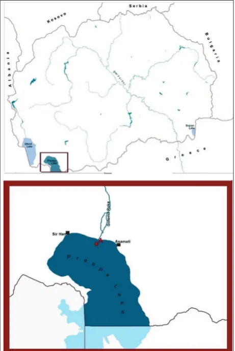
Fig 1. Map of Lake Prespa with the sampling site (A).
the fish. Focal necrosis of the respiratory epithelium associated with full destruction of the secondary la-mellae was also a very common finding and occurred in 71% of the fish. This change was recognized as a mild, long-term process, leading to respiratory epi-thelia dilatation and necrosis (Fig. 2B) in 57% of the fish. Proliferation of interlamellar surfaces (Fig. 2C) and the large number of chloride cells, both on the la-mellar surface and interlala-mellar space, were regularly registered in the 70% of investigated barbel gills. In 66% of the fish, necrotic changes in the chloride cells occurred (Fig. 2D).
manifesting as a frequent occurrence of budding yeast blastocysts in the cytoplasm or intercellular spaces of the gill epithelium and other gill structures (Fig. 2E).
Taking into consideration the abovementioned observations, a quantitative estimation of histo-pathological changes was made and the results are presented in Fig 3. As can be seen, the histopatho-logical condition expressed by the total index [13] appeared to be under the dominant influence of re-gressive changes with high and statistical significant correlation (r=0.916, p<0.0001). Progressive changes, mostly found as a proliferation of interlamellar re-spiratory epithelia (sometimes leading to gill fusion), evidently contributed to a smaller degree to the overall
gill pathology. Circulatory disturbances, despite their prominence, are in slight correlation with the total index and just below statistical significance (r=0.328; p=0.055). The observed inflammatory changes were not in correlation with any other parameters and had no statistically significant influence on the total his-topathology index.
It is interesting to note that the observed infec-tions are not correlated with progressive and regres-sive histopathological changes found in Prespa barbel gills. There is only a slight correlation with circulatory disturbances, just below statistical significance, but having statistical significant influence on the total histopathological index (r=0.644; p<0.001).
Cell counting showed a prominent proliferation of chloride cells in the investigated barbel gills with a median value of 3.31 cells per interlamellar space. The increased number of these cells was accompanied by remarkable necrotic changes (Fig. 2D). Mucous cells, on the other hand, showed no noticeable proliferation – 0.25 cells per interlamellar space. The occurrence of both type of cells showed no correlation with the other gill changes.
DISCUSSION
Prespa barbel is a near-bottom-living fish that is more closely exposed to sediments than other fish species in the lake. The histopathology survey of the gills
re-Fig. 2. Light micrograph of Prespa barbel gill. A – Extensive telan-giectasia. Paraffin section H&E. Scale bar=100μm. B – Enlarged secondary lamellae which is full with erythrocytes, exhibiting necrosis of the respiratory epithelium (arrow). Semi-thin sec-tion, toluidine blue. Scale bar=20μm. C – Fused lamellae and local proliferation of the interlamellar epithelium (arrow). Paraffin sec-tion; toluidine blue. Scale bar=20μm. D – Group of chloride cells with necrotic changes (arrow): pyknotic nucleus and vacuoles in the cytoplasm. Semi-thin section; toluidine blue. Scale bar=20μm.
E – Interlamellar space with proliferating epithelia with eosinophil granulocytes (arrows). On the epithelium surface Trichodina sp. can be noted (stars). F – Fungal infection (arrow). Semi-thin sec-tion; toluidine blue. Scale bar=20μm.
vealed several pathological conditions and parasitic infection. Prespa barbel gills seem to be most severely impacted by proliferative changes, mostly observed as respiratory epithelial proliferation and lamellar fu-sion. Despite their highly significant impact on gill pathology, regressive changes are present with the least severity. It is likely that these changes are caused by one or more toxic contaminants present in the water, although some contribution of associated infective pathology cannot be completely ruled out. There are many studies in the scientific literature providing evi-dence of lamellar and respiratory epithelial prolifera-tion and hyperplasia, as well as the various necrotic changes induced by organic toxicants, including heavy metals [4-10]. The Prespa barbel seems to be most severely impacted by circulatory changes, described as secondary gill lamellae telangiectasia. The toxico-pathic etiology of this circulatory disturbance cannot be ruled out, as many water contaminants are reported to cause similar gill aneurysms [4,10,14], which can also be due to infection [12].
Histopathological investigation of barbel gills showed several parasitic and fungal infections. Para-site infection is comparable with the severity of circu-latory changes, which suggests that the lesions could be infection-induced. Candidiasis-like infection was much more prominent, and this type of infection hasnot been described in fish yet, neither is its pa-thology well understood. According our analyses, the only gill lesion that may be linked to yeast infection is the aneurism in the secondary lamellae, even though no strongly significant evidence of it was provided in our study. Lee et al [15-16] found a remarkable formation of aneurismal hematomas within the gill lamellae in Japanese eel caused by birnavirus and herpesvirus infection in the lamellar space. A similar mechanism may underlie the aneurysm observed in the yeast-infected Prespa barbel gills. Yeasts are usu-ally opportunistic pathogens, so their occurrence in Prespa barbel gills could imply that the fish are in an immunocompromised state. Further investigation of the candidiasis-like infection pathology in Prespa bar-bel, and its eventual link to the observed circulatory disturbance in the gill are warranted.
The mass occurrence of chloride cells and fre-quent degenerative changes in their morphology recorded on the gills of Prespa barbel could be
con-nected with negative environmental effects. Micro-scopic analyses show the proliferation of chloride cells accompanied by both cytoplasm hypertrophy and necrosis. Chloride cells are involved in maintain-ing the ionic and acid base balance in freshwater fish [17]. Experimental and field exposure to acid water was reported to result in chloride cell hyperplasia in several freshwater fish, such as fathead minnow, yel-low perch, pearl dace and rainbow trout [5-6,18]. The necrotic changes in chloride cells observed in Prespa barbel gills in this study are in accordance with similar changes induced experimentally in fathead minnow and yellow perch by exposure to extremely low pH [5] or acid water + Al treatment [18]. Chloride cell proliferation provides a compensating mechanism for increasing net ion uptake when fish are exposed to conditions that promote abnormally high ion losses. In the presence of toxic metals or other toxicants, this change is associated with chloride cell loss and higher turnover [18-19]. This situation possibly exists with Prespa barbel, indicating some kind of osmotic stress exposure. The possibility of fish exposure to sediment-eluted acidified microenvironments containing addi-tional toxic contaminants should be explored.
Stoffet et al. [19] found that the non-ionic deter-gent nonylphenol, a known environmental xenoestro-gen, induces chloride cell activity and proliferation in rainbow trout. The authors speculated that the reason for this effect may be a nonylphenol-generated de-mand for calcium as a result of induced synthesis of the calcium-rich yolk protein, vitellogenin. There is a possibility that the increased number of chloride cells in Prespa barbel is related to xenoestrogen-induced vitellogenesis, a process recorded in this and other fish species found in Lake Prespa [20].
environmentally induced lesions in Prespa barbel gills, which warrants further work on health status estima-tion on a broader range level.
Authors’ contribution: The study was designed by DRZ, sample collection, preparation of slides and analyses were carried out by AS and KR. Preparation of the manuscript, interpretation of the results and discussion were performed by MJ and DRZ.
Conflict of interest disclosure: The authors declare that they have no competing interest.
REFERENCES
1. Crivelli A, Malakou M, Castsadorakis G, Rosecchi E. The Prespa barbel, Barbus prespensis, a fish species endemic to Prespa Lake (North-Western Greece). Folia Zool. 1996;45(suppl.1):21-32.
2. Spirovska S, Trajkovski V, Nonkulovski K. How to save our Prespa Lake. In: Gusevska D, Naumovski T, Mitic V, Vel-kova-Jordanovska L, Stojanovski S, Trajanovski S, editors. 1stSymposium for protection of natural lakes in Republic of Macedonia: Proceedings; 2007 May 31 – Jun 06; Ohrid, Makedonia. Ohrid : Ministry of education and science of the Republic of Macedonia; 2007. p. 58-64.
3. GrupceLj. Autochthonous and allochthonous quantities of phosphorus in Prespa Lake waters. International Symposium towards integrated conservation and sustainable develop-ment of transboundary Macro and Micro Prespa Lakes. Proceedings; 1997 Oct 24 – 26; Korcha, Albania. Korcha: Ministry of education and science of the Republic of Mace-donia and Ministry of education and science of the Republic of Albania; 1997 p.68-72.
4. Khan RA, Kicerniuk J. Histopathological effects of crude oil on Atlantic cod following chronic exposure. Can J Zool. 1984;62:2038-43.
5. Leino RL, McCornick H. Morphological and morpho-metrical changes in chloride cells of the gills of Pimephales promelas after chronic exposure to acid water. Cell Tissue Res. 1984;236:121-8.
6. Leino RL, Wilkinson P, Anderson JG. Histopathological changes in the gills of pearl dace, Semotilus margarita, and fathead minnows, Pimephales promelas, from experimentally acidified Canadian lakes. Can J Fish Aquat Sci. 1987;44:126-34. 7. Leino RL, McCornick HJ. Response of juvenile largemouth
bass to different pH and aluminium levels at overwinter-ing temperatures: effects on gill morphology, electrolyte balance, scale calcium, liver glycogen and depot fat. Can J Zool. 1992;71:531-43.
8. Munvich AG, Jones RT, Kane AS, Anderson RS, Reim-scheussel R. Effects of chronic copper exposure on the macrophage chemiluminescent response and gill histology in godfish (Carassiusauratus L.). Fish Shellfish Immunol. 1995;5:251-64.
9. Authman MMN, Ibrahim SA, El-Kasheif MA, Gaber HS. Heavy Metals Pollution and Their Effects on Gills and Liver of the Nile Catfish Inhabiting El-Rahawy Drain, Egypt. Glob Vet. 2013;10(2):103-15.
10. Barisic J, Dragun Z, Ramani S, Filipovic-Marjic V, Nes-reteKrasnici; Coz-Rakovac R; Kostov V, Rebok K, Jordanova M. Evaluation of histopathological alterations in the gills of Vardar chub (Squalus vardarensis Karaman) as an indicator of river pollution. Exotoxycol Environ. Safe. 2015;118:158-66. 11. Khan RA, Payne JF. Multidisciplinary approach using several
biomarkers, including a parasite, as indicators of pollution: a case study from a paper mill in Newfoundland. Parasitol. 1997;39:183-8.
12. Powel, MD, Harris JO, Carson J, Hill JV. Effects of gill abra-sion and experimental infection with Tenacibaculum mariti-mum on the respiratory physiology of Atlantic salmon Salmo salar affected by amoebic gill disease. DAO. 2005;63(2-3):169-74.
13. Bernet D, Schmidt H, Meier W, Burkhardt-Holm P, Wahli T. Histopathology in fish: proposal for a protocol to assess aquatic pollution. J Fish Dis. 1999;22:25-34.
14. Syassina IG, Sokolovsky AS. Assessment of the condition of plaice from Sivuchya Bight (Peter the Great Bay, Sea of Japan) by histopathological indices. BiolMorya Vladivostok. 2001;27(2):102-9.
15. Lee NS, Kobayashi J, Miyazaki T. Gill filament necrosis in farmed Japanese eels, Anguila japonica (Temminck & Schlegel), infected with Herpesvirus anguillae. J Fish Dis. 1999;22:457-63.
16. Lee NS, Nomura Y, Miyazaki T. Gill lamellar pillar cell necrosis, a new birnavirus disease in Japanese eels. Dis Aquat Org. 1999;37:13-21.
17. Evans DH. Ionic exchange mechanisms in fish gills. Comp Biochem Physiol. 1975;51A:491-5.
18. Leino RL, McCornick H. Morphological and morpho-metrical changes in chloride cells of the gills of Pimephales promelas after chronic exposure to acid water. Cell Tissue Res. 1984;236:121-28.
19. Stoffel MH, Wahli T, Friess AE, Burkhard-Holm P. Exposure of rainbow trout (Oncorhynchus mykiss) to nonylphenol is associated with an increased chloride cell fractional surface area. Schweizer Arch Tierh. 2000;142(5):263-7.