433
© 2018 by the Serbian Biological Society How to cite this article: Mohammadali F, Abroun S, Atashi A. Combined mild hypoxia and bone marrow mesenchymal stem cells improve expansion and
HOXB4 gene expression of human cord blood CD34+ stem cells. Arch Biol Sci. 2018;70(3):433-41.
Combined mild hypoxia and bone marrow mesenchymal stem cells improve expansion
and
HOXB4
gene expression of human cord blood CD34+ stem cells
Fatemeh Mohammadali1, Saeid Abroun1,* and Amir Atashi1,2
1Department of Hematology and Blood Banking, Faculty of Medical Sciences, Tarbiat Modares University, Tehran, Iran 2Stem Cell and Tissue Engineering Research Center, Shahroud University of Medical Sciences, Shahroud, Iran
*Corresponding author: [email protected]
Received: October 28, 2017; Revised: December 21, 2017; Accepted: January 14, 2018; Published online: January 26, 2018
Abstract: Cord blood (CB) is a rich source of hematopoietic stem cells (HSC). It has been used successfully to treat a variety of hematological and non-hematological disorders. Beside the advantages of CB, its main disadvantages are the limited number of stem cells available for transplantation and delayed engraftment. Identifying strategies to enhance expansion and self-renewal of HSCs can improve transplantation efficiency. The aim of this study was to examine different culture conditions on ex vivo expansion and homeobox protein Hox-B4 gene (HOXB4) expression in cord blood CD34+ stem cells.
Human cord blood CD34+ HSC were cultured in serum-free medium supplemented with cytokines with and without a
feeder layer in normoxia (21% O2) and mild hypoxia (5% O2).At the end of 7 days of culture, the highest number of total nucleated cells (TNC), CD34+ cells, colony forming units (CFUs) and HOXB4 mRNA were observed in a co-culture of
HSC with a bone marrow mesenchymal stem cell(MSC) feeder layer at 5% O2.We concluded that the combination of bone marrow (BM)-MSC and mild hypoxia (5% O2) not only improved HSC expansion but also enhanced HOXB4 gene
expres-sion as a self-renewal marker of HSC, and better mimicked the niche microenvironment conditions.
Key words: cord blood; hematopoietic stem cells; CD34+ cells; mesenchymal stem cells; hypoxia; HOXB4
Abbreviations: CB – cord blood; HSC – hematopoietic stem cells; HSPC − hematopoietic stem/progenitor cells; HOXB4 – homeobox protein Hox-B4; TNC –total nucleated cells; CFU – colony forming unit; MSC – mesenchymal stem cells; BM – bone marrow; SCF –stem cell factor; FLT3L – FMS-like tyrosine kinase 3 ligand; TPO –thrombopoietin; IL – interleukin; M-CSF – macrophage-colony stimulating factor; HIF-1 – hypoxia inducible factor-1; SDF1 – stromal cell-derived factor 1; MNC – mono-nuclear cells; FBS – fetal bovine serum; IMDM – Iscove’s modified Dulbecco’s medium; DMEM – Dulbecco’s modified Eagle’s medium; PBS – phosphate-buffered solution; ROS –reactive oxygen species; GEMM – granulocyte/erythroid/macrophage/ megakaryocyte, GM – granulocyte/monocyte, G – granulocyte, M – monocyte, E – erythroid (E); BFU – burst forming unit
INTRODUCTION
Stem cells are known for their ability to remain undif-ferentiated and their capacity to undergo self-renewal, which allows them to proliferate during fetal develop-ment and to be maintained throughout adult life [1-4]. There are different sources of HSC. One important source of HSC is CB. Human umbilical CB contains CD34+ hematopoietic stem/progenitor cells (HSPC) with a high proliferative potential [5].
Beside the advantages of CB, including wide-spread availability and decreased ability to induce im-munological reactivity against the patient, CB also has disadvantages, include the low and sometimes
limit-ing number of cells collected in one unit, which can be less than optimal for engraftment in many adults and higher weight children, and the relatively slower engraftment time of neutrophils and platelets [6-8].
In vivo, HSC are maintained and regulated by a specific microenvironment referred to as a “niche” in the BM. Multiple cellular types, soluble and mem-brane- bound factors and extracellular matrix com-ponents form this niche [11]. MSC in stem cell niches play an essential role in quiescence, proliferation and differentiation of HSC [12].Studies have shown that MSC secrete at the base state 6, 7, 8, IL-11,IL-12, IL-14, IL-15, M-CSF, Flt-3L and SCF [13].
Stem cell niches are located in regions away from blood vessels. This provides a low O2 tension envi-ronment that enhances stem cell proliferation to the detriment of their differentiation potential [14]. Sev-eral studies in murine and human HSC demonstrated that a culture of HSC at 21% O2 leads to the rapid ex-haustion of their stem cell potential. Culture in anoxic conditions (0.1-1% O2) promotes the maintenance and return of HSC to quiescence in G0 [15]. Cultures at higher O2 levels (3-5%) maintain cell proliferation [16-18]. The cellular mechanisms by which human hematopoietic progenitors and stem cells respond to hypoxia have not been characterized, but it has been accepted that HIF-1 protein [19] is rapidly degraded under normoxic conditions and is stabilized under low O2 levels (<5%).
Several molecules have been proposed to control the self-renewal of HSC. Among these molecules, the homeobox family of proteins, HOXB4 are expressed in most primitive hematopoietic stem cells. A physiologi-cal approach suggests that an environment associated with MSC and low O2 concentration would be the most favorable for ex vivo expansion and self-renewal of HSC. In this study, we examined the feasibility of combining serum-free media, BM-MSC and mild hypoxia (5% O2) to establish a novel culture system for the ex vivo expansion of HSC and expression of
HOXB4 as a stem cell self-renewal marker.
MATERIALS AND METHODS
CB collection and CD34+ cell purification
CB samples were collected from 4-5 different full-term deliveries from umbilical cord blood after obtaining the parents’ informed written consent for each experi-ment (n=3). CB units were obtained from the Iranian
Blood Transfusion Organization. Briefly, up on deliv-ery of the baby the cord was doubly clamped and tran-sected, and blood was collected in special collection bags containing citrate-phosphate dextrose. After col-lection, the samples were stored at room temperature (15-25°C) and processed within 12 h. Research was carried out according to the guidelines approved by the Ethics Committee at Tarbiat Modares University (reference number 4506).Red cells were precipitated with 6% hydroxyethyl starch (Grifols, Spain). MNC were isolated by Ficoll-Hypaque density gradient centrifugation (Lymphodex, inno-Train Diagnostic, Germany). CB-CD34+ cells were purified from MNC using a MACS CD34+ cell isolation kit following the manufacturer’s instructions (Miltenyi Biotec, USA CD34 MicroBead Kit). CD34+ cell purity was evalu-ated by flow cytometry analysis using an FITC-human CD34 antibody. Nonspecific reactions were excluded using isotype controls.
Feeder layer preparation
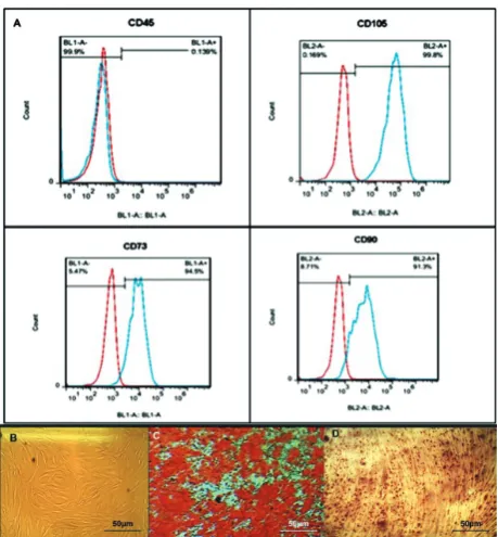
BM-MSC were a kind gift from Dr. M. Soleimani (Stem Cells and Tissue Engineering Department, Stem Cell Technology, Tehran, Iran.). Three BM-MSC samples from different healthy donors were cultured in DMEM+10% FBS (Gibco, Grand Island, NY, USA) supplemented with streptomycin (0.025 mg/mL) and penicillin (0.025 U/mL; Gibco BRL), at 37°C and 5% CO2 in a humidified atmosphere. On reaching 80% confluency, cells were trypsinized and replated at a density of about 104 cells/cm2. Cells were expanded for 2 to 4 passages. Phenotypic characterization and differentiation into osteoblasts and adipocytes were carried out. MSC were characterized by flow cyto-metric analysis using monoclonal antibodies against CD105, CD73, CD90 and CD45.MSC were positive for CD105, CD73, CD90 and negative for CD45.In order to be used as feeder layers, cells were seeded on 24-well plates (1×10⁴ cells/well) and grown until 70-80% confluency.
Coculture with a feeder layer
cytokines: Flt3-L (50 ng/mL, ORF Genetics, Iceland), SCF(50 ng/mL, ORF Genetics, Iceland) and TPO (50 ng/mL, Peprotech, Rocky Hill, NJ, USA). In this study, the cells harvested at 2-4 passages were used as BM-MSC. All experiments were cultured at 37°C, 5% CO2 in 5% O2 (two-gas incubator) and 21% O2 for 7 days. The culture conditions for the present study were clas-sified into six groups as follows: (i) cytokine group: CD34+ cells cultured in serum-free medium in the presence of cytokines in 21% O2; (ii) feeder group: CD34+ cells cultured in the serum-free medium di-rectly on the confluent MSC feeder layer without cy-tokines in 21% O2; (iii) feeder+cytokine group: CD34+ cells cultured in the serum-free medium on a confluent MSC feeder layer and in the presence of cytokines in 21% O2; (iv) cytokine group: CD34+ cells cultured in serum-free medium in the presence of cytokines in 5% O2; (v) feeder group: CD34+ cells cultured in serum-free medium directly on the confluent MSC feeder layer without cytokines in 5% O2; (vi) feeder+cytokine group: CD34+ cells cultured in serum-free medium on a confluent MSC feeder layer and in the presence of cytokines in 5% O2.HSC were additionally cultured in Stemline II serum-free medium without any cytokines and the MSC layer as the control group. After 7 days, the co-cultures were harvested, and viable cell counts were taken using trypan blue dye exclusion. The total number of nucleated cells was counted.
Flow cytometric analysis
Expanded CD34+ cells from each culture condition were collected and resuspended in PBS. After label-ing the cell suspension with monoclonal antibod-ies (CD34-FITC, BD Pharmingen™, USA), the cells were incubated for 30-45 min on ice. The expansion-fold of CD34+ cells was calculated by comparing the TNC×CD34+ percentage obtained after 7 days with the TNC×CD34+ percentage before culture initiation. Data analysis was performed using FlowJo analytical software (Tree Star, Inc., Ashland, OR).
Colony-forming cell (CFC) assay
One×103freshly isolated CD34+ or expanded cells in 6 different culture conditions were harvested on day 7 and cultured on 12-well plates in
cytokine-supple-mented methylcellulose media (MethoCult H4434 classic with cytokine, Stem Cell Technologies, Cana-da) with 2% FBS in IMDM as indicated in the manu-facturer’s user instructions to enumerate the CFC. CFU colonies consisting of 50 cells were counted after 14 days of incubation at 37°C under humidified condi-tions in 5% CO2. The experiments were performed in duplicate in three independent experiments. The total CFU fold change was calculated by dividing the num-ber of colonies obtained per 103cultured cells by the number of the colonies obtained at day 0, and mul-tiplying by the fold increase in total number of cells.
RNA extraction and cDNA synthesis
Cell RNA was extracted using TRIzol (Sigma, Ger-many). Total RNAs were reverse transcribed using the cDNA kit (Primescript RT reagent kit perfect real time, Takara) according to the manufacturer’s instruc-tions. Each cDNA sample was run in duplicate for target (HOXB4) or HPRT endogenous control.
Real-time PCR
Quantitative reverse transcriptase (q-PCR) was per-formed using 1μL total cDNA in 10μL reaction vol-ume with 0.3 μM of each primer and 5 μL of real-time master mix (Amplicon, Denmark).Thermal cycling was initiated at 95°C for 15 min followed by 40 cy-cles of PCR (95°C, 20 s). The annealing temperature was 61°C. Quantification of qRT-PCR signals was performed using the delta-delta Ct method which calculates the relative changes in gene expression of the target gene normalized to the HPRT endog-enous control. The values obtained were represented as the relative quantity of mRNA level variations. The sequence of genesisas follows: HOXB4: forward: 5ʹ-TGCAAAGAGCCCGTCGT-3΄; reverse: 5΄-GGC-GTAATTGGGGTTTACCG-3΄.HPRT: forward: 5ʹ-CCTGGCGTCGTGATTAGTG-3΄ and reverse: 5ʹ-TCAGTCCTGTCCATAATTAGTCC-3΄
Statistical analysis
were assessed by either the Student t-test or ANOVA. Comparison of the data between normoxia and hy-poxia was done using an independent t-test. One-way ANOVA was used to calculate the significance within groups. Statistics were calculated with using Graph Pad Prism 6 software. The value <0.05 were consid-ered statistically significant.
RESULTS
Total nucleated cell expansion
To assess whether various culture conditions could ac-celerate cell expansion (Fig. 1), we analyzed the count increase of cells after culture as defined by the condi-tions in different experimental groups. The trypan blue exclusion test demonstrated >85% viable cells both at 21% and 5% O2 in different culture conditions. The initial cell count was 1×10⁴, and after 7 days culture in normoxia, the mean TNC count was 75600±4900 in the cytokine culture without the MSC feeder layer, 41540±2660 in the coculture system without cyto-kines and 112000±3361 in the co-culture system with cytokines. In hypoxic cultures, the mean TNC count was 89000±6151 in the cytokine culture without the MSC feeder layer, 48700±3161 in the co-culture sys-tem without cytokines, and 144080±8518 in the co-culture system with cytokines. In all co-co-cultures, TNC increased significantly compared to the feeder-free cul-ture conditions (n=3, P<0.05). In all of the groups in hypoxic conditions compared to normoxic conditions, TNC increased significantly (P<0.05) (Fig. 2).
CD34+ cell expansion
As CD34+ is often used as a surrogate marker for HSC, the enumeration of CD34+ cells has been used to quantify the progenitor and stem cell contents.CD34+ cell purity, evaluated by flow cytometry analysis us-ing an FITC-human CD34+ antibody, was >90% in all cases on the first day of purification. Fig. 3 shows the fold change of CD34+ cells in different culture condi-tions after 7 days. The mean fold change of CD34+ cells was 1.96±0.15 in the cytokine culture without the MSC feeder layer, 1.04±0.08 in the co-culture system without cytokines, and 2.98±0.2 in the co-culture sys-tem with cytokines in 21% O2. In the hypoxic culture,
the mean fold change in CD34+ cells was 2.45±0.15 in the cytokine culture without the MSC feeder layer, 1.32±0.15 in the co-culture system without cytokines, and 4.91±0.23 in the co-culture system with cytokines. The mean fold change in CD34+ cells on day 7 in the co-culture system with cytokines was higher than in the cytokine culture without the MSC feeder layer and the co-culture system without cytokines (N=3, P<0.05). Our results showed that both the co-culture and the hypoxic environment had a significant effect on the final CD34+ cell output, and that the collective impact of these two factors was remarkable.
In the co-culture systems without cytokines, TNC and CD34+ cell numbers increased 5- and 1.5-fold, respectively, and cell viability remained 90% after 7 days. In both hypoxia and normoxia cultures, the TNC count and CD34+ fold change were higher in the cytokine groups when compared to the feeder groups (Figs. 2 and 3).
Colony-forming assay
The colony-forming unit ability of CB-CD34+ cells was investigated by performing a clonogenic assay in methylcellulose-based cultures (14 days) under dif-ferent culture conditions that expanded for 7 days, and with fresh CD34+ cells. The results showed that the expanded cells in all culture conditions possessed the ability to produce clonogenic progenitor cells: CFU-GEMM,-GM, -G, -M, -E and -BFU-E, similar to fresh CD34+ cells. The total fold increase in clono-genic potential was calculated by dividing the num-ber of colonies obtained per 103 cultured cells under the different culture conditions by the number of the colonies obtained at day 0 and multiplying by the fold increase in total number of cells.
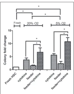
The mean CFU fold change of different culture conditions compared to fresh CD34+cells was 3.7±0.7 in the cytokine culture without the MSC feeder lay-er,1.6±0.2 in the co-culture system without cytokines, and 6.7±1.6 in the co-culture system with cytokines in
normoxia. In 5% O2, the mean CFU fold changes were as follows: 5.6±0.5 in the cytokine culture without the MSC feeder layer, 2.3±0.8 in the co-culture system without cytokines, and 10.8±1.5 in the co-culture sys-tem with cytokines.
Cultures in the presence of the feeder layer and cytokines were capable of maintaining a higher clono-genic potential (P<0.05). There was higher colony fold change in hypoxia compared to normoxia (P<0.05). The data are shown in Fig. 4 and the morphology of the colonies is presented in Fig. 5.
HOXB4 gene expression
The expression of HOXB4 in fresh CD34+ cells and expanded cells after 7 days of growth in all culture conditions was evaluated by RT-PCR. The mean fold change ratio of HOXB4 mRNA expression in the expanded cells to fresh CD34+cells in normoxia was 0.5±0.1 in the cytokine culture without the MSC feeder layer, 0.8±0.1 in the co-culture system without
Fig. 2. Total nucleated cell count in dif-ferent culture conditions. One×104 CD34+ cells/well cultured in Stemline II serum-free media supplemented with the combi-nation of Flt3-L(50 ng/mL), SCF (50 ng/ mL) and TPO (50 ng/mL) in 5% O2 and 21% O2, for 7 days with/without the bone marrow MSC feeder layer. After 7 days, the TNC was counted. Data represents the mean±SD from three independent experiments. Error bars represent SD, significant values are *p<0.05 (two-tailed Student’s t test).
Fig. 3. CD34+ cell fold change in differ-ent culture conditions. One×105 expanded CD34+cells from different culture condi-tions evaluated by labeling the cells with antibody conjugated to FITC by flow cytometry. The fold-expansion of CD34+ cells was calculated by comparing the TNC×CD34+% obtained after 7 days with the TNC×CD34+% before culture initiation. Data represent the means±SD from three independent experiments. Er-ror bars represent SD, significant values are*p<0.05 (two-tailed Student’s t test).
cytokines, and 1.36±0.12 in the co-culture system with cytokines. In hypoxic culture, the mean fold change ratio was 0.9±0.1 in the cytokine culture without the MSC feeder layer, 1.03±0.15 in the co-culture system without cytokines, and 1.83±0.15 in the co-culture system with cytokines.
We observed that in cytokine groups, either in normoxia or hypoxia, HOXB4 expression decreased rapidly, but in the feeder groups without cytokines,
HOXB4 expression was better maintained. The highest
HOXB4 levels were detected in the feeder+cytokine
groups. The results showed that HOXB4 gene expres-sion was sensitive to the oxygen level and the presence of the MSC feeder layer (Fig. 6) (N=3, P<0.05).
DISCUSSION
The major focus in HSC expansion is still the defini-tion of optimal culture condidefini-tions as regards hemato-poietic growth factor combinations, co-cultures with feeder cells and other stem cell niche requirements. Our experiments were carried out in serum-free culture medium because in clinical use, serum-free
culture medium has been proposed to avoid some concerns, such as the risk of infection by viruses or prions, variation between individual batches [20], the presence of inhibitors or stimulating factors of hema-topoiesis, and finally, the use of serum free-media pro-vides biochemically defined culture conditions [21].
The best results in HSC expansion have been ob-tained by researchers using early-acting factors, such as SCF, FLT3L and TPO. Although the application of cytokine-supplemented media results in the fast and effective expansion of hematopoietic cells, rapid expansion is accompanied by differentiation, resulting in depletion of the stem cell pool [22].
Interaction between stem cells and their niches can modulate HSC functions in vitro [23].A more nat-ural stem cell proliferation was achieved by culturing the stem cells on a layer of MSC feeder cells [24,25]. We showed that a serum-free culture system using human BM MSC-derived feeder layers supplemented with a cytokine cocktail of SCF, TPO and FLT3L, al-lowed for a higher expansion of CD34+ HSC from CB. The mean fold changes of TNC and CD34+ cells in the cytokine culture with MSC were higher than in the cytokine culture without MSC. Consistent with our results, co-culture with MSC maintained HSC with a primitive immune phenotype (CD34+CD38- or CD133+CD38-) [26], and ex vivo contact with stromal components of the hematopoietic microenvironment preserved HSC [27,28].
Fig. 5. Morphology of colonies cultured 14 days in MethoCult H4434 with cytokine. A – BFU-E, B – CFU-GEMM, C– CFU-E, D – CFU-GM, E – CFU-G, F – CFU-M (scale bars in A-F:50μm).
In the co-culture system without cytokines, TNC and CD34+cell numbers were increased up to 5- and 1.5-fold, respectively, but cell viability was more than 90%.MSC as a feeder layer without the addition of any cytokines maintained HSC expansion at a low but constant level for 7 days. In keeping with our results, Amirizadeh et al. [29] also showed that the proliferation capacity of HSC in recombinant cytokine culture with a MSC feeder layer was higher than in a cytokine culture without MSC and in a co-culture system without cytokines. It seems that BM-MSC se-creted cytokines and growth or survival signals that were transferred to the HSC via adhesive molecules through the modulation of cytokine- and growth factor-dependent signals.
Several publications support the fundamental role of low O2 concentrations in the regulation of stem cell homeostasis (similar to those in bone marrow). It is generally believed that the hypoxic niche protects stem cells from oxidative stress, and thus, the HSC present in such niches contain low in vivo ROS levels [30].
The relationship between the cycle/quiescence bal-ance for HSC and physiological O2 tension (<5%) in their environment has been reported both in vitro and
in vivo [31]; for these reasons we choose 5% O2 tension.
A higher TNC count and CD34+ fold change in mild hypoxia (5% O2 tension) demonstrated that mild hypoxia stimulated the expansion of HSC. Recently, it was reported that cultivation of human CD34+, 38- HSC in hypoxic conditions prior to transplanta-tions maintains and to some extent improves their engraftment to immune-compromised mice [32,33]. Our results showed that expanded cells in all culture conditions possessed the ability to produce clonogen-ic progenitor cells similar to fresh CD34+ cells. The colony-forming potential depended on the number of HSC, and our results showed that the number of CFC in HSC cultured on the MSC feeder layer in the presence of cytokines at 5% O2 was higher than other culture conditions. We choose HOXB4 gene expres-sion as a self-renewal marker of HSC because HOXB4
is probably one of the most important regulators of HSC self-renewal [34]. It is expressed in the stem cell fraction of the bone marrow and subsequently down-regulated during differentiation in humans [35] and mice [36]. Several studies showed that HOXB4
in-duces ex vivo expansion of HSC and in vivo HOXB4
overexpression accelerated stem cell regeneration and led to a marked competitive repopulating advantage [37-39].Our results showed that despite the higher expansion of HSC in the cytokine group, HOXB4 ex-pression was lower in this group when compared to the feeder group and the feeder+cytokine group. Sev-eral studies have reported that cytokines such as GM-CSF, IL-3, SCF and TPO increased murine and human HSC proliferation with a rapid increase in the level of ROS in cells [40].It seems that MSC co-culture at-tenuated the side effects of ROS and better-preserved HSC self-renewal. We showed higher HSC expansion and expression of HOXB4 in hypoxia (5% O2 tension). Our study is in accordance with previous studies that reported that hypoxia is beneficial in maintaining the self-renewal properties of HSC [41,42].
CONCLUSIONS
con-tribute to the development of more efficient culture systems for ex vivo expansion of CB HSC for cellular therapy that resemble the in vivo microenvironment in which these cells reside.
Acknowledgments: This paper is based on research undertaken for a PhD thesis and was supported by the Deputy of Research Affairs and Faculty of Medical Sciences at Tarbiat Modares Uni-versity. We would like to thank the Royan Stem Cell Technology Company.
Author contributions:FM performed the experiments, analyzed the data and wrote the manuscript.SA and AA contributed to the experimental design and manuscript editing. All authors reviewed and approved the final manuscript.
Funding: This paper includes part of a thesis approved by Tarbiat Modares University.
Conflict of interest disclosure: The authors declare that they have no conflict of interest disclosure.
REFERENCES
1. Wang LD, Wagers AJ. Dynamic niches in the origination and differentiation of hematopoietic stem cells. Nat Rev Mol Cell Biol. 2011;12:643-55.
2. Mendelson A, Frenette PS. Hematopoietic stem cell niche maintenance during homeostasis and regeneration. Nat Med. 2014;20:833-46.
3. Lilly AJ, Johnson WE, Bunce CM. The hematopoietic stem cell niche: new insights into the mechanisms regu-lating hematopoietic stem cell behavior. Stem Cells Int. 2011;2011:274.
4. Shizuru JA, Negrin RS, Weissman IL. Hematopoietic stem and progenitor cells: clinical and preclinical regeneration of the hemato-lymphoid system. Annu Rev Med. 2005;56:509-38.
5. Hordyjewska A, Popiołek Ł, Horecka A. Characteristics of hematopoietic stem cells of umbilical cord blood. Cytotech-nology. 2015;67(3):387-96.
6. Jacobson CA, Turki AT, McDonough SM, Stevenson KE, Kim HT, Kao G, Herrera MI, Reynolds CG, Alyea EP, Ho VT, Koreth J, Armand P, Chen YB, Ballen K, Soiffer RJ, Antin JH, Cutler CS, Ritz J. Immune reconstitution after double umbilical cord blood stem cell transplantation: com-parison with unrelated peripheral blood stem cell transplan-tation. Biol Blood Marrow Transplant. 2012;18:565-74. 7. Majhail NS, Brunstein CG, Tomblyn M, Thomas AJ, Miller
JS, Arora M, Kaufman DS, Burns LJ, Slungaard A, McGlave PB, Wagner JE, Weisdorf DJ. Reduced-intensity allogeneic transplant in patients older than 55 years: unrelated umbili-cal cord blood is safe and effective for patients without a matched related donor. Biol Blood Marrow Transplant. 2008;14:282-9.
8. Brunstein CG, Gutman JA, Weisdorf DJ, Woolfrey AE, Defor TE, Gooley TA, Verneris MR, Appelbaum FR, Wagner JE,
Delaney C. Allogeneic hematopoietic cell transplantation for hematologic malignancy: relative risks and benefits of double umbilical cord blood. Blood. 2010;116:4693-9. 9. Mehta RS, Rezvani K, Olson A, Oran B, Hosing C, Shah N,
Parmar S, Armitage S, Shpall E..Novel Techniques for Ex Vivo Expansion of Cord Blood: Clinical Trials. Front Med. 2015;2:89.
10. Tanavde VM, Malehorn MT, Lumkul R , Gao Z, Wingard J, Garrett ES, Civin CI. Human stem-progenitor cells from neonatal cord blood have greater hematopoietic expansion capacity than those from mobilized adult blood. Exp Hema-tol. 2002;30:816-23.
11. Mendez-Ferrer S. Mesenchymal and hematopoietic stem cells form a unique bone marrow niche. Nature. 2010; 466(7308):829-34.
12. Jing D, Fonseca A-V, Alakel N, Fierro FA, Muller K, Born-hauser M, Ehninger G, Corbeil D, Ordemann R. Hematopoi-etic stem cells in co-culture with mesenchymal stromal cells - modeling the niche compartments in vitro. Haematologica. 2010;95(4):542-50.
13. Majumdar MK, Thiede MA, Mosca JD, Moorman M, Ger-son SL.Phenotypic and functional compariGer-son of cultures of marrow-derived mesenchymal stem cells (MSCs) and stromal cells. J Cell Physiol. 1998;176:57-66.
14. Dellatore SM, Garcia AS, Miller WM. Mimicking stem cell niches to increase stem cell expansion. Curr Opin Biotech-nol. 2008;19(5):534-40
15. Hermitte F, Brunet de la Grange P, Belloc F, Praloran V, Iva-novic Z. Very low O2 concentration (0.1%) favors G0 return of dividing CD34+ cells. Stem Cells. 2006;24:65.
16. Cipolleschi MG, Dello Sbarba P, Olivotto M. The role of hypoxia in the maintenance of hematopoietic stem cells. Blood. 1993;82(7):2031-7.
17. Cipolleschi MG, Rovida E, Ivanovic Z, Praloran V, Olivotto M, Dello Sbarba P. The expansion of murine bone marrow cells preincubated in hypoxia as an in vitro indicator of their marrow-repopulating ability. Leukemia. 2000;14:735-9. 18. Ivanović Z, Bartolozzi B, Bernabei PA, Cipoleschi MG,
Rovida E, Milenkovic P, Praloran V, DeloSbarba P. Incuba-tion of murine bone marrow cells in hypoxia ensures the maintenance of marrow-repopulating activity together with the expansion of committed progenitors. Br J Haema. 2000;108:424.
19. Jiang BH, Semenza GL, Bauer C, Marti HH. Hypoxia-induc-ible factor 1 levels vary exponentially over a physiologically rel-evant range of O2 tension. Am J Physiol. 1996;271:C1172-80. 20. Möbest D, Mertelsmann R, Henschler R. Ex vivo expansion
of CD34+ blood progenitor cells in serum-free medium. Biotechnol Bioeng. 1998;60:341-7.
21. Lebkowski JS, Schain LR, Okarma TB. Serum-free culture of hematopoietic stem cells: a review. Stem Cells. 1995;13:607-12. 22. Boitano AE, Wang J, Romeo R, Bouchez LC, Parker AE, Sut-ton SE, Walker JR, Flaveny CA, Perdew GH, Denison MS, Schultz PG, Cooke MP. Aryl hydrocarbon receptor antago-nists promote the expansion of human hematopoietic stem cells. Science. 2010;329:1345-8.
24. Goncalves R, Lobato da Silva C, Cabral JM, Zanjani ED, Almeida-Porada G. A Stro-1(+) human universal stromal feeder layer to expand/maintain human bone marrow hema-topoietic stem/progenitor cells in a serum-free culture sys-tem. Exp Hematol. 2006;34:1353-9.
25. Zhang Y, Chai C, Jiang XS, Teoh SH, Leong KW. Co-culture of umbilical cord blood CD34 + cells with human mesen-chymal stem cells. Tissue Eng. 2006;12:2161-70.
26. Walenda T, Bork S, Horn P, Wein F, Saffrich R, Diehlmann A, Eckstein V, Ho AD, Wagner W. Coculture with mesen-chymal stromal cells increases proliferation and mainte-nance of hematopoietic progenitor cells. J Cell Mol Med. 2010;14(1‐2):337-50.
27. Breems DA, Blokland EA, Siebel KE, Mayen AE, Engels LJ, Ploemacher RE. Stroma-contact prevents loss of hema-topoietic stem cell quality during ex vivo expansion of CD34+ mobilized peripheral blood stem cells. Blood. 1998;91(1):111-7.
28. Chute JP, Saini AA, Chute DJ, Wells MR, Clark WB, Harlan DM, Park J, Stull MK, Civin C, Davis TA. Ex vivo culture with human brain endothelial cells increases the SCID-repopulating capacity of adult human bone marrow. Blood. 2002;100:4433-9.
29. Amirizadeh N, Oodi A, Nikougoftar M, Soltanpour MS. Expression and promoter methylation changes of the P15INK4b during ex vivo cord blood CD34+ cell expan-sion following co-culture with mesenchymal stromal cells. Hematology. 2013;18.5:260-8.
30. Jang YY, Sharkis SJ. A low level of reactive oxygen species selects for primitive hematopoietic stem cells that may reside in the low-oxygenic niche. Blood. 2007;110:3056-63. 31. Guitart AV, Hammoud M, Dello Sbarba P, Ivanovic Z,
Pral-oran V. Slow-cycling/quiescence balance of hematopoietic stem cells is related to physiological gradient of oxygen. Exp Hematol. 2010;38(10):847-51.
32. Danet GH, Pan Y, Luongo JL, Bonnet DA, Simon MC. Expansion of human SCIDrepopulating cells under hypoxic conditions. J Clin Invest. 2003;112:126-35.
33. Shima H, Takubo K, Iwasaki H, Yoshihara H, Gomeia Y, Hosokawa K, Arai F, Takahash T Suda T. Reconstitution activity of hypoxic cultured human cord blood CD34-pos-itive cells in NOG mice. Biochem Biophys Res Commun. 2009;378:467-72.
34. Zhang Y, Gao Y. Novel chemical attempts at ex vivo hemato-poietic stem cell expansion. Int J Hematol. 2016;103:519-29. 35. Sauvageau G, Lansdorp PM, Eaves CJ, Hogge DE, Dragowska WH, Reid DS, Largman C, Lawrence HJ, Humphries RK. Differential expression of homeobox genes in functionally distinct CD34+ subpopulations of human bone marrow cells. Proc Natl Acad Sci USA. 1994;91:12223-7.
36. Pineault N, Helgason CD, Lawrence HJ, Humphries RK. Differential expression of Hox, Meis1, and Pbx1 genes in primitive cells throughout murine hematopoietic ontogeny. Exp Hematol. 2002;30:49-57.
37. Antonchuk J, Sauvageau G, Humphries RK. HOXB4 over-expression mediates very rapid stem cell regeneration and competitive hematopoietic repopulation. Exp Hematol. 2001;29:1125-34.
38. Thorsteinsdottir U, Sauvageau G, Humphries RK. Enhanced in vivo regenerative potential of HOXB4-transduced hema-topoietic stem cells with regulation of their pool size. Blood. 1999;94(8):2605-12.
39. Antonchuk J, Sauvageau G, Humphries RK. HOXB4-induced expansion of adult hematopoietic stem cells ex vivo. Cell. 2002;109:39-45.
40. Kumar S, Geiger H. HSC Niche Biology and HSC Expansion Ex Vivo. Trends Mol Med. 2017;23(9):799-819.
41. IvanovićZ, Bartolozzi B, Bernabei PA, Cipolleschi MG, Rovida E, Milenković P, Praloran V, Dello Sbarba P. Incuba-tion of murine bone marrow cells in hypoxia ensures the maintenance of marrow repopulating ability together with the expansion of committed progenitors. Br J Haematol. 2000;108: 424-9.