A GENOMIC APPROACH TO THE IDENTITY, CLONAL DIVERSITY, AND CLINICAL IMPORT OF TUMOR-INFILTRATING LYMPHOCYTES IN HUMAN CANCERS
Michael D. Iglesia
A dissertation submitted to the faculty at the University of North Carolina at Chapel Hill in partial fulfillment of the requirements for the degree of Doctor of Philosophy in the Curriculum in Genetics and Molecular
Biology in the School of Medicine.
Chapel Hill 2015
;
j
iii
ABSTRACTMichael D. Iglesia: A Genomic Approach to the Identity, Clonal Diversity, and Clinical Import of Tumor-infiltrating Lymphocytes in Human Cancers
(Under the direction of Charles M. Perou)
Immune infiltration in solid tumors has emerged as an important aspect of cancer biology. In particular, the presence of tumor-infiltrating lymphocytes (TILs) within the tumor microenvironment is associated with improved outcomes and response to treatment in a wide variety of tumor types. These beneficial TIL infiltrates, comprising both B-cells and T-cells, may represent the successful targeting of tumor-associated antigens by the immune system leading to direct tumor cell killing. The success of immune checkpoint inhibitors has shown that this immune response may be manipulated to induce durable responses in several tumor types, and has strengthened the notion that an antigen-directed TIL response may occur in some cancers. However, the precise patient and tumor characteristics associated with these beneficial TIL responses are unclear.
In this work, a genomic, mRNA-sequencing approach was taken to address TIL infiltration in diverse human tumor types, with a focus on breast cancer. Associations between signatures of TIL infiltration, B-cell TIL clonal diversity, genomic subtype, and other clinical correlates were identified, and show that significant B-cell TIL infiltrates typically represent less clonally diverse populations.
Furthermore, in subtype-specific mouse models of breast cancer, T-cell and B-cell TIL clonal diversity are directly assessed through immune repertoire sequencing and a mouse model of claudin-low breast cancer is shown to elicit a recurrent, clonally-restricted T- and B-cell TIL response.
iv
E
v
ACKNOWLEDGEMENTS
The work presented here is the product of not only my efforts, but also the careful thought and collaboration of many talented scientists. In particular, the work presented here represents an extensive collaboration between the laboratories of Drs. Chuck Perou and Jon Serody, and I am thankful for their mentorship and the immunological expertise of Dr. Ben Vincent and other members of the Serody lab, without which this project would not be possible. This research, especially Chapters 2 and 4, was originally planned out through this collaboration, and the mentorship and oversight of Chuck and Jon as Ben and I made progress on it has continued to grow into an exciting and wide-reaching collaboration in breast cancer immunogenomics. This experience of multi-disciplinary team science, as well as the individual mentorship of Jon and Chuck and collaboration with Ben, has been instrumental in the accomplishment of the work presented here and in my development as a scientist.
Chapter 2 of this dissertation was originally published in Clinical Cancer Research (and is included here as permitted in AACR Journals Permissions and Reprints policy,
http://www.aacrjournals.org/site/misc/permissions.xhtml) with myself and Dr. Ben Vincent as co-first authors (1).
Iglesia MD, Vincent BG, Parker JS, Hoadley KA, Carey LA, Perou CM, et al. Prognostic B-cell signatures using mRNA-seq in patients with subtype-specific breast and ovarian cancer. Clin Cancer Res.
2014;20:3818-29.
Chapters 2 and 3 involved extensive use of data from The Cancer Genome Atlas; I was fortunate to rely on Dr. Katie Hoadley for her understanding of the genomic and clinical data and how to obtain them.
E
vi
Index as in Figure 3.6, is the result of considerable assistance from Dr. Joel Parker. Indeed, it’s fair to say that nearly every analysis in this dissertation represents at least one conversation with Joel.
The mouse work included in Chapter 4 of this dissertation was accomplished through the
assistance of Dr. Jerry Usary in the Perou lab and Dr. Nick Taylor in the Serody lab. The assistance of the Mouse Phase I Unit and the UNC Flow Cytometry and High-Throughput Sequencing core facilities were also instrumental in carrying out this work.
I am also grateful to my committee; their guidance and mentorship has been instrumental in directing the course of my graduate studies and in producing the work included in this dissertation. Their input has continually challenged me as a scientist and broadened my understanding of cancer research and of my own project.
I performed this work as a student in the UNC MD-PhD program, and both the program
leadership and my fellow MD-PhD students have contributed greatly to my development as a physician-scientist. Furthermore, through the MD-PhD program I was able to participate in a two-year longitudinal advanced practice selective course. Working with my clinical mentor, Dr. Carey Anders, not only advanced my clinical training but also afforded me the opportunity to think critically about the clinical management of patients with breast cancer and provided me exposure to that dynamic synergy of research and medicine that defines the physician-scientist track.
The continual mentorship, collaboration, and insight of every member of the Perou lab has been critical in my development as a scientist and in the production of the work included in this dissertation. The entirety of this work is due to collaboration and critique from my labmates. Not least among my sources of mentorship in the Perou lab has been my mentor, Dr. Chuck Perou. His guidance of my work and confidence in me has allowed me to develop into a more independent, thorough, and data-driven scientist, and his mentorship has prepared me well for a career in cancer genomics research.
E
vii
TABLE OF CONTENTS
LIST OF FIGURES ... viii
LIST OF TABLES ... ix
LIST OF ABBREVIATIONS... x
CHAPTER 1 – INTRODUCTION ... 1
CHAPTER 2 – PROGNOSTIC B-CELL SIGNATURES USING mRNA-Seq IN PATIENTS WITH SUBTYPE-SPECIFIC BREAST AND OVARIAN CANCER ... 9
Introduction... 9
Methods... 12
Results ... 16
Discussion ... 34
CHAPTER 3 – THE PROGNOSTIC IMPACT OF TUMOR IMMUNE INFILTRATION ACROSS DIVERSE SOLID TUMOR TYPES ... 38
Introduction... 38
Methods... 41
Results ... 44
Discussion ... 59
CHAPTER 4 – MOUSE MODELS OF BREAST CANCER ELICIT CONSISTENT, SUBTYPE SPECIFIC IgH AND TCRβ CLONAL RESTRICTION ... 63
Introduction... 63
Methods... 66
Results ... 68
Discussion ... 79
CHAPTER 5 – DISCUSSION... 82
APPENDIX 1: GENE SIGNATURES ... 88
APPENDIX 2: SURVIVAL ANALYSES ... 93
E
viii
LIST OF FIGURES
Figure 2.1 – Derivation of Immune Signatures through Unsupervised Hierarchical Clustering ... 17
Figure 2.2 – Immune Signature Expression by Subtype ... 18
Figure 2.3 – Signature Correlation ... 22
Figure 2.4 – BCR Segment Expression ... 25
Figure 2.5 – Prognostic Value of BCR Gene Segment Expression ... 26
Figure 2.6 – Example K-M Plots of BCR Segment Expression ... 27
Figure 2.7 – Schema for Estimating BCR Diversity ... 29
Figure 2.8 – BCR V Segment Diversity vs. Expression ... 30
Figure 2.9 – Schema for Estimating Enrichment of SHM ... 32
Figure 2.10 – SHM Enrichment by Subtype ... 33
Figure 3.1 – Immune Signature Expression Clusters by Subtype Across Tumor Types ... 46
Figure 3.2 – Immune Signature Expression by Tumor Type and COCA Subtype ... 47
Figure 3.3 – Immune Signature Correlation by Tumor Type ... 48
Figure 3.4 – BCR Gene Segment Expression Clustering ... 56
Figure 3.5 – BCR Expression and Diversity by Tumor Type and COCA Subtype ... 57
Figure 3.6 – Restriction Index: Schema and Results ... 58
Figure 4.1 – Sample Preparation Schema ... 69
Figure 4.2 – Reads by Repertoire Library ... 70
Figure 4.3 – Repertoire Entropy ... 72
Figure 4.4 – Matched Spleen Clonotype Abundance ... 73
Figure 4.5 – Clonotype Sharing ... 75
Figure 4.6 – CD4+/CD8+ Clonotype Mapping ... 76
E
ix
LIST OF TABLES
Table 2.1 – Univariate Survival Analysis of Immune Signatures ... 20
Table 2.2 – Multivariate Survival Analysis of Immune Signatures ... 21
Table 3.1 – Immune Signature Univariate Surviva by Tumor Type ... 50
Table 3.2 – Immune Signature Univariate Survival by COCA Subtype ... 51
E
x
LIST OF ABBREVIATIONS
APC Antigen-presenting Cell BCR B-Cell Receptor
BLCA Urothelial Bladder Carcinoma BRCA Breast Ductal Carcinoma
CDR3 Complementarity-determining Region 3 COCA Cluster of Clusters Assignments CRC Colorectal Adenocarcinoma GBM Glioblastoma Multiforme
HNSC Head and Neck Squamous Cell Carcinoma IgH Immunoglobulin Heavy Chain
KIRC Clear Cell Renal Cell Carcinoma LUAD Lung Adenocarcinoma
LUSC Lung Squamous Cell Carcinoma MDSC Myeloid-derived Suppressor Cell OV Serous Ovarian Adenocarcinoma RI Restriction Index
SHM Somatic Hypermutation SKCM Cutaneous Melanoma
TAM Tumor-associated Macrophage TCR T-Cell Receptor
E
1
CHAPTER 1 – INTRODUCTION Breast Cancer Subtypes
Human breast cancer is now understood as a multitude of diseases rather than as one disease entity. These groups, or subtypes, of breast cancer vary widely in almost every regard: by morphology, response to treatment, clinical outcome, potential for angiogenesis and metastasis, mutational load and spectrum, DNA copy number, and biological pathways driving tumor development. These breast cancer subtypes were identified through analysis of global gene expression patterns, which defined at least five subtypes: luminal A, luminal B, HER2-enriched, basal-like, and claudin-low, as well as a normal-like group (2-6). Of these subtypes, the basal-like group has been the focus of extensive research due to its high rate of relapse, poor overall survival, and large degree of overlap with the “triple-negative” class of breast tumors (7, 8). Clinically triple-negative breast tumors are defined by the lack of expression of the estrogen receptor, progesterone receptor, and HER2, and thus therapies targeting these molecules and their related pathways are not useful for patients with triple-negative tumors (9). Due to the lack of good drug targets and the high rates of relapse for patients with basal-like breast cancer, this disease presents a unique opportunity for novel therapeutic approaches in breast cancer.
Tumor-infiltrating Lymphocytes in Breast Cancer
E
2
immune infiltrate and its prognostic impact may be instrumental in bringing about new therapies for this clinically challenging tumor subtype.
Breast Cancer Immune Infiltration: CD8+ T-Cells
The component of the breast tumor immune infiltrate that is most intuitively promising is the CD8+ subset of T-cell TILs. Several studies have shown that breast tumor infiltrates are phenotypically diverse, but that CD8+ TILs are often the most abundant TIL type (18-20). Furthermore, CD8+ T-cells have specifically been shown to be associated with improved breast cancer prognosis, again highlighting basal-like breast cancer as the subtype where this association is most clearly observed (13, 14, 21). It is appealing and intuitive to imagine that many CD8+ T-cell TILs are functioning as cytotoxic effectors targeting tumor cells. Recent work has shown that tumors with greater cytolytic activity are associated with a greater mutational load, expression of tumor necrosis genes and endogenous retroviruses, and mutations in antigen-presenting and innate immune sensing, which bolsters the idea that CD8+ TILs may be directly attacking tumor cells in an antigen-specific manner (22). It is important to note that analysis of CD8+ cytotoxic T-cells may be complicated by the presence of natural killer (NK) cells, which may also express a rearranged T-cell receptor (TCR) and contribute to direct cell killing by cytolytic granule release (23). NK cells are not as well understood in breast cancer as CD8+ T-cells, but tumor-infiltrating NK cells are present in breast cancer and have been implicated in contributing to the anti-tumor immune response (24, 25).
Breast Cancer Immune Infiltration: CD4 + T-Cells
While CD8+ T-cells certainly play a large role in the breast cancer-associated immune response, CD4+ T “helper” cells are likely to also be important. It has been shown that CD8+ T-cells in the tumor microenvironment do not function properly in the absence of adequate CD4+ cells (26, 27). The polarization of CD4+ T helper cells is important for understanding their role in the tumor
microenvironment, with IFN-γ-producing Th1 cells acting together with CD8+ T-cells, mature dendritic cells, M1 macrophages, NK cells, B-cells, and plasma cells in the anti-tumor response, while Th2 cells producing IL-4 and IL-5 act alongside regulatory T-cells (Treg), regulatory B-cells (Breg), M2 macrophages,
;
3
microenvironment, both in breast cancer and in other tumor types. Current research suggests that both pro-tumor and anti-tumor subsets of Th17 TILs may exist, and furthermore that Th17 TILs may retain the ability to differentiate into either Th1 cells or Treg (31, 32).
Breast Cancer Immune Infiltration: Regulatory T-Cells
Regulatory T-cells, or Treg, represent in some ways the opposite end of the TIL spectrum from
CD8+ T-cells. Prevalence of CD4+CD25+FOXP3+ Treg in breast tumors is correlated with worse prognosis,
and has been shown to decrease the proliferation, cytolytic granule production, and cell killing of tumor-infiltrating CD8+ T-cells (30, 33). Thus, Treg play an important, and potentially dominant, role in inhibiting
anti-tumor immunity. As Treg act to suppress the anti-tumor immune response, it seems likely that Treg are
actively recruited by the tumor. Indeed, current work suggests that Treg are recruited to breast tumors via
a CCR4/CCL22 interaction, and that within the tumor they are activated, potentially by exposure to tumor-associated antigens (30). In addition to inhibiting CD8+ TIL function, Treg may directly inhibit naïve T-cells,
and are also known to decrease the ability of antigen-presenting cells (APCs) to present tumor antigens by downregulating the costimulatory molecules CD80 and CD86 (34). The full mechanisms by which Treg
contribute to the immunosuppressive tumor microenvironment, both in breast cancer and other tumor types, is not fully understood.
Breast Cancer Immune Infiltration: B-Cells
While T-cell TILs have been the most extensively studied and are likely a very influential component of the tumor immune response, B-cell TILs are also present and active within breast tumors. B-cell TILs may compose up to 40% of the tumor immune infiltrate in some tumors, and have been estimated as being present in about 25% of breast tumors (35-37). Several closely related TIL
B-;
4
cell TILs are playing within the breast tumor microenvironment. One possibility is that B-cell TILs are mediating a direct, antigen-specific anti-tumor immune response via production of antibodies against tumor-associated antigens. At least some of the cytotoxic activity of the anti-HER2 monoclonal antibody trastuzumab is mediated through antibody-dependent cellular cytotoxicity (ADCC), lending credence to the idea that naturally arising anti-tumor antibodies may also constitute an effective anti-tumor response (42-45). Indeed, several studies have identified auto-antibodies from patients with breast cancer, including antibodies against TP53 and improperly processed β-actin; however, no clear association between specific auto-antibodies and patient outcomes has been established (35, 46-51). However, several studies of small numbers of breast cancers have demonstrated B-cell TILs that are clonally expanded and have undergone class switching and somatic hypermutation, evidence of activation through antigen binding (46, 47, 52-55). Previous work by our group has also identified that high expression of immunoglobulin genes in basal-like and HER2-enriched breast cancer is associated with reduced antibody sequence diversity, further suggesting an oligoclonal anti-tumor antibody response (1). However, B-cell TILs may also serve primarily to potentiate the anti-tumor T-cell response. B-cell TILs may differentiate into either effector or regulatory B-cells, which in turn either potentiate or inhibit CD4+ T helper cell immune responses (56). Additionally, B-cells may present antigens to both CD4+ and CD8+ T-cells, making it possible that B-cell TILs function as local APCs (57, 58).
Breast Cancer Immune Infiltration: Tumor-associated Macrophages and Myeloid-derived Suppressor Cells
While TILs have garnered attention as important contributors to the tumor immune
microenvironment, tumor-associated macrophages and other myeloid-derived cells are also critical in shaping the immune milieu in breast cancer and other solid tumors. TAMs are prevalent in many breast tumors (59). Like T helper TILs, cytokine signaling leads to polarization of TAMs into an anti-tumor M1 phenotype that promotes the Th1 effector response or a pro-tumor M2 phenotype associated with metastasis, angiogenesis, and an inflammatory wound-healing response (60-63). As part of the immunosuppressive microenvironment cultivated by breast tumors and other tumor types, TAMs are largely polarized to the M2 phenotype (60, 64). In this role, TAMs in breast cancer contribute to
;
5
prepare tissue microenvironment for seeding by breast cancer cells, further contributing to metastatic potential (70, 71). Aside from TAMs, myeloid-derived suppressor cells (MDSCs) represent a distinct myeloid-derived subset that plays a major role in breast tumor immune infiltration. MDSCs can act through several immunosuppressive mechanisms, including secretion of inflammatory pro-Th2 cytokines, sequestration of arginine and cysteine, and generation of reactive oxygen species and nitric oxide(72, 73). Production of reactive oxygen species by MDSCs impairs CD8+ T-cell antigen recognition; MDSCs may also prevent antigen recognition by nitration of the TCR, which prevents MHC complex binding (72, 74-76). In one study of women with HER2- stage II-III breast cancer, patients with fewer MDSCs detected in peripheral blood at baseline and prior to the last cycle of chemotherapy were more likely to achieve pathologic complete response to chemotherapy, further supporting the role of MDSCs in tumor progression (77).
Tumor Immune Infiltration in Other Solid Tumor Types
The role of the immune system in breast cancer is similar to the role various tumor-infiltrating immune cells play in a wide spectrum of solid tumor types. The picture emerging from studies of tumor immune infiltration in diverse tumor types and subtypes is that some tumor types are highly immunogenic and are infiltrated with CD8+ and Th1 T-cells, NK cells, B-cells, and/or M1 macrophages that correlate with improved clinical course, but these anti-tumor immune responses generally do not lead to
spontaneous tumor regression due to immunosuppressive signals from tumor cells, Th2 TILs, regulatory T- and B-cell TILs, M2 TAMs, and MDSCs. Tumor types including melanoma, non-small cell lung cancer, renal cell carcinoma, ovarian cancer, bladder cancer, head and neck cancer, and others have
demonstrated this paradigm of conflicting pro- and anti-tumor immune responses (64, 78-85). There are variations between tumor types in the level and makeup of pro-tumor and anti-tumor immune infiltration: in melanoma, spontaneous tumor regression has been documented, whereas in other tumor types including many renal cell carcinomas, immunosuppressive MDSCs and regulatory B-cells dominate and minimize the role of effector immune cells (79, 86, 87).
Immune Checkpoint Inhibitors as Cancer Immunotherapy
;
6
antibody and the first immune checkpoint inhibitor approved for use in cancer, has shown impressive efficacy in unresectable or metastatic melanoma: a pooled retrospective analysis of trials demonstrated a 3-year survival of 22% in such cases, including many heavily pre-treated patients (88). CTLA-4 works by a number of mechanisms to inhibit effector T-cells; among these, it inhibits signaling through the TCR by outcompeting the co-stimulatory molecule CD28 for its usual binding partner B27 and also induces cell cycle arrest in T-cells (89-93). Both ipilumumab and tremelimumab, another anti-CTLA-4 monoclonal antibody, have shown durable responses in a subset of patients (94-96). Other successful immune checkpoint inhibitors target the PD-1/PDL-1 axis, with nivolumab and pembrolizumab both being anti-PD-1 monoclonal antibodies. PD-anti-PD-1 is expressed on mature T-cells following activation and in tumors is believed to primarily act on T-cells during the effector phase, later than CTLA-4 (97, 98). In addition, PD-1 is expressed on a variety of immune cell types, including B-cells and NK cells, and effects on those cell types may contribute to its function in cancer (97, 99). PD-1 acts by binding two different ligands, PDL-1 and PDL-2. Surface expression of PDL-1 has been observed in a variety of cell types, including T-cells, B-cells, endothelial cells, pancreatic islet cells, and keratinocytes, and is also highly expressed in many tumor types (98). PDL-2 is primarily expressed on APCs, including macrophages, mast cells, dendritic cells, and B-cells (98). PD-1 binds to either PDL-1 or PDL-2 as a costimulatory molecule upon binding of the TCR to antigen loaded on the major histocompatibility complex (MHC), and subsequently sends downstream signals within the T-cell to inhibit T-cell proliferation, cytokine production, and cytolytic function, and to impair T-cell survival (100). Thus, PD-1 contributes to effector T-cell exhaustion; this role has been observed both in chronic viral infection and in the tumor microenvironment (100). Interestingly, PD-1 signaling in Treg leads to improved immunosuppressive activity in those Treg, furthering the inhibition
;
7
and induces the durable responses that are now understood to be characteristic of immune checkpoint inhibition (106, 107). Further research into immune checkpoint inhibitors is needed to examine
combinations with cytotoxic chemotherapy, kinase inhibitors, and radiotherapy, as well as to expand trials to greater numbers of patients in a wide variety of potentially immunogenic tumor types.
Predicting Immunogenicity in Tumors
The success of immune therapies across many cancer types has led to greater interest in the common features that distinguish immunogenic tumors. Because immunogenic tumors are often characterized by a dense lymphocytic infiltrate that improves survival, the notion that tumor-associated antigens drive the development of the anti-tumor immune response is appealing. Several potential sources of tumor antigens have been proposed, including mutations in existing genes (such as TP53), gene products overexpressed in cancer (such as HER2), viral peptides (such as HPV genes), and the so-called cancer-testis antigens normally expressed only in immune-privileged sites (such as MAGE) (22). Several studies have identified peptides in one or more of these categories that may be acting as tumor antigens (108-113). If the immune response associated with improved survival in many cancer types is driven by T-cell and/or B-cell binding to tumor-associated antigens, this holds promise for cancer therapeutic strategies aimed not only at potentiating the existing immune response, but also at direct targeting of tumor-associated antigens. Because lymphocytes binding antigen through the TCR or B-cell receptor (BCR) subsequently undergo clonal expansion, the question of the antigen-directedness of TILs may be addressed by assessing the clonal diversity of TILs: in a tumor-associated antigen-driven
response, the TIL population is expected to be clonally restricted, with one or more dominant clones able to bind antigens. Historically, the extreme level of diversity generated by TCR and BCR sequences has prevented thorough study of lymphocyte population clonality (114). However, recent advances in next generation sequencing have led to the development of immune repertoire sequencing, a collection of related methods for direct next-generation sequencing of recombined BCR and TCR chains (115, 116). Initial repertoire sequencing studies in cancer have shown clonal expansion of public TCR sequences in Treg in the TC-1 murine tumor model, as well as nonspecific T-cell proliferation leading to an expanded
TCR repertoire in human melanoma patients treated with tremelimumab (117, 118). As tumor
;
8
;
9
CHAPTER 2 – PROGNOSTIC B-CELL SIGNATURES USING mRNA-Seq IN PATIENTS WITH SUBTYPE-SPECIFIC BREAST AND OVARIAN CANCER
Introduction
The role of tumor-infiltrating lymphocytes (TILs) in breast cancer is not fully understood, although multiple studies have shown an association between the presence of TILs and an improved prognosis (10-14). TILs in breast tumors are predominantly cytotoxic (CD8+) T-cells, and the proportion of CD8+ T-cells may be prognostic (13, 14, 19, 119, 120). In contrast, TILs of the regulatory T-cell phenotype (CD4+CD25+FoxP3+ Treg) are associated with poorer outcomes in breast cancer (121, 122). The role of
B-cell TILs in human breast cancer is not as clear as that of T-B-cell TILs. Using gene expression profiling, our group and others have shown that gene signatures representing B-cells, plasmablasts, plasma cells and immunoglobulin predicted favorable clinical outcome in ER+ and ER- breast tumors(15, 16, 38-40). In this manuscript, these are referred to as B-cell signatures; while plasmablasts and plasma cells are known to infiltrate some breast tumors, we use the term “B-cell TIL” here to refer to any TIL in the B-cell lineage. The presence of B-cell TILs as assessed by immunohistochemistry (IHC) has also been shown to be an independent prognostic feature in breast cancer (41). Studies of small numbers of breast tumors have shown the B-cell response in these tumors to be clonally expanded, with evidence of having undergone class switching and somatic hypermutation (46, 47, 52-55). This strongly suggested that in some breast tumors there may be a clonally restricted, antigen-directed B-cell anti-tumor response. Several studies have identified auto-antibodies in breast cancer patients, including antibodies against improperly processed β-actin in some medullary breast cancers, although the association between such auto-antibodies and patient survival is unclear (35, 46, 47). Together, these findings provide evidence that B-cell TILs may be important in affecting breast cancer biology and progression.
;
10
claudin-low, as well as a normal-like group (2-6). The prognostic value of both T- and B-cell TILs may be restricted to a subset of highly immune-infiltrated breast tumors (15). Basal-like breast tumors, in
particular, appear to have beneficial TILs (16, 123). Multiple groups have identified signatures of lymphocyte-related gene expression that are overrepresented in basal-like breast tumors and predict better survival(15, 16); in contrast, luminal A breast tumors show low levels of lymphocytic infiltrate(123).
Comprehensive genomic profiling of multiple tumor types in TCGA has shown there is a strong similarity between basal-like breast cancer and serous ovarian cancer (2). These two tumor types exhibit a similar mutational spectrum and share many of the same driver events (i.e. TP53 loss, RB1 loss, c-MYC gain, etc.). Like basal-like breast cancer, many ovarian tumors are rich in TILs. Analysis of TCGA serous ovarian cancer gene expression identified four genomic subtypes: mesenchymal, proliferative,
differentiated, and immunoreactive(124). The immunoreactive subtype, in particular, showed high expression of T-cell chemokine ligands and lymphocyte-related genes. Furthermore, a number of studies have shown the presence of T- and B-cell TILs is a positive prognostic feature in ovarian cancer (14, 125-127). As in breast cancer, the precise role of B-cell TILs is less understood than that of T-cell TILs. These data suggest that, like basal-like breast cancer, serous ovarian cancer may be a likely candidate for identifying a productive anti-tumor T-cell and/or B-cell TIL response.
If there is an effective, subtype-specific antitumor response mediated by B-cell TILs, this presents the possibility of specific immunogenic epitopes that could promote development of a subtype-specific antibody response. While some studies have identified antigen-directed TIL clones in breast tumors (46, 47, 52, 53, 55), currently the degree to which TILs are antigen-directed is unknown. The development of a mature B-cell response following antigen stimulation depends on a number of
;
11
㡠ҫ
12
MethodsData Sets
The breast cancer data set used for all analyses except the survival analysis of gene expression signatures was the TCGA data set of 819 mRNA-seq samples, comprising 728 breast tumors and 91 normal breast samples (see TCGA Data Portal at https://tcga-data.nci.nih.gov/tcga/, CGHub at https://cghub.ucsc.edu/). This 728 sample set is an extension of the 480 tumors previously profiled by microarray(2), but these 480 plus 350 new samples have all been assayed by mRNA-seq using Illumina 2x50bp sequencing as described by the TCGA in an evaluation of lung squamous samples(131). Gene expression values were represented as RSEM (RNA-seq by Expectancy-Maximization) data normalized within-sample to the upper quartile of total reads as previously described (131). These data and further details about data processing are available at the TCGA Data portal under the V2_MapSpliceRSEM workflow
(https://tcga-data.nci.nih.gov/tcgafiles/ftp_auth/distro_ftpusers/anonymous/tumor/brca/cgcc/unc.edu/illuminahiseq_rna seqv2/rnaseqv2/unc.edu_BRCA.IlluminaHiSeq_RNASeqV2.mage-tab.1.6.0/DESCRIPTION.txt). Genomic subtype was assigned within the set of 728 mRNA-seq samples using the PAM50 assay (132). The training set of breast samples used in the PAM50 assay is 50% clinically ER+, therefore the mRNA-seq data were normalized to reflect the training set
(https://genome.unc.edu/pubsup/breastGEO/Guide%20to%20Intrinsic%20Subtyping%209-6-10.pdf). Based on clinical data taken from the TCGA Data portal on September, 2012
(https://tcga-data.nci.nih.gov/tcga/), of the 728 samples, 157 were ER-, 535 were ER+, 2 were ER-indeterminate, 29 did not have ER status assays performed, and 5 did not have available data, indicating that 77% of the mRNA-seq samples were ER+. To normalize the data similar to the PAM50 training dataset, in which 50% of samples are ER+, all 157 ER- samples were selected, as well as 157 randomly selected ER+ samples. The median gene expression for the PAM50 intrinsic gene list was calculated based on this subset of samples. To perform platform correction for mRNA-seq, these median values were then subtracted from all 728 samples prior to running the PAM50 assay as previously described(132)
㡠ҫ
13
microarray-based gene expression data set of 855 breast tumors with published intrinsic subtype calls (140 basal-like, 90 claudin-low, 144 HER2-enriched, 243 luminal A, 162 luminal B, 76 normal-like) and clinical data (combined data from the following data sets: GSE2034, GSE12276, GSE2603, and the NKI295 (microarray-pubs.stanford.edu/wound_NKI/Clinical_Data_Supplement.xls)) (133). Survival analyses of BCR segment expression, however, used the TCGA mRNA-seq data set. For all analyses of ovarian cancer, we used the TCGA serous ovarian cancer mRNA-seq data set, which, like the breast cancer data set, represents new mRNA-seq data, again using Illumina 2 x 50bp sequencing, on a subset of the 500 cases from the TCGA ovarian project (124). This mRNA-seq data set consists of 266 tumors with follow-up data (https://tcga-data.nci.nih.gov/tcga/, https://cghub.ucsc.edu/).
Gene Expression Signatures and Survival Analyses
Immune gene expression signatures were established using unsupervised hierarchical clustering of mRNA-seq expression data for 728 breast tumor samples. Gene dendrogram nodes corresponding to genes characteristically expressed in specific immune cell types were identified and validated through DAVID functional annotation clustering and IPA (Ingenuity®Systems, www.ingenuity.com)(134, 135). Gene lists for all five signatures are included in Appendix 1. Additional lymphocyte gene signatures were obtained from published studies: IGG_Cluster(38), B_Cell(136), and B_Cell_60gene(39) are B-cell signatures, and T_Cell(136), CD8(136), LCK(137), and TNBC_T-Cell(16) are T-cell signatures, with the CD8 signature specifically representing CD8+ T-cells.
Survival analyses were performed by Kaplan-Meier analysis and log-rank testing, and hazard ratios were derived from the Cox proportional hazards model. For analysis of the prognostic value of BCR segment expression, samples were divided into high and low expression groups of equal number for Kaplan-Meier analysis and log-rank testing. To evaluate the prognostic value of gene expression signatures, the Cox proportional hazards model was used with each signature tested as a continuous variable. Multivariate Cox proportional hazards models were used to test the prognostic value of individual gene expression signatures when combined with the other clinical and genomic variables.
;
14
gene segments. The number of significant (p < 0.05) p values was calculated from this set and 95% confidence intervals were calculated through bootstrap resampling.
B-Cell Receptor Diversity
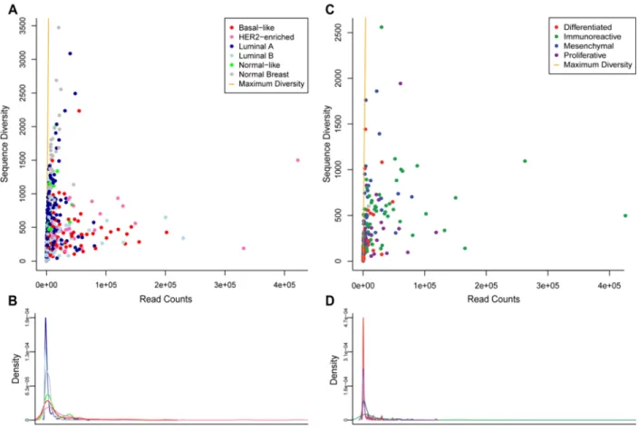
The method for estimating sequence diversity of a BCR gene segment for an individual sample/tumor using paired-end mRNA-seq data is outlined in Figure 2.7. Read pairs mapping to the EntrezGene genomic location (www.ncbi.nlm.nih.gov/entrez/query.fcgi?db=gene)(138) of a given BCR gene segment were identified (mapped by MapSplice(139)). The sequence of these read pairs was compared to the hg19 reference genome to identify non-reference bases. The genomic position and nucleotide identity of all non-reference bases was identified for each read pair. Each observed pattern of non-reference bases was then assigned a score representing the number of read-pairs containing exactly that pattern of non-reference bases. This set of observed patterns and their corresponding count was used to calculate the effective number of species, which is a diversity function isomorphic to Shannon entropy, as described by Jost et al.(140) .
De novo Assembly and Somatic Hypermutation Analysis
De novo assembly of BCR variable (V) gene segments from paired-end mRNA-seq reads was performed using the Assembly-Based Re-Aligner (ABRA) algorithm (Mose et al., manuscript in
preparation). To generate ABRA contigs, unmapped reads and reads mapping to a BCR variable region of interest were first split into overlapping k-mers where k=31. K-mers that were comprised exclusively of non-ambiguous bases with quality score > 20 were assembled into a de Bruijn graph. K-mers with fewer than 100 observations were then pruned from the graph. The graph was then traversed to produce all possible contigs. This set of contigs was used for somatic hypermutation analyses.
To determine if the sequence of a BCR variable gene segment was consistent with somatic hypermutation, the reference sequence for that gene segment was first established by Smith-Waterman alignment to each IMGT® (IMGT®, the international ImMunoGeneTics information system®
㡠ҫ
15
non-mutated (i.e., reference and non-reference) bases were counted again within CDRs. Chi-square testing was used to determine if the distribution of mutated bases was consistent with the mutation pattern expected in SHM; chi-square testing was conducted separately for the whole V segment sequence and for CDRs.
;
16
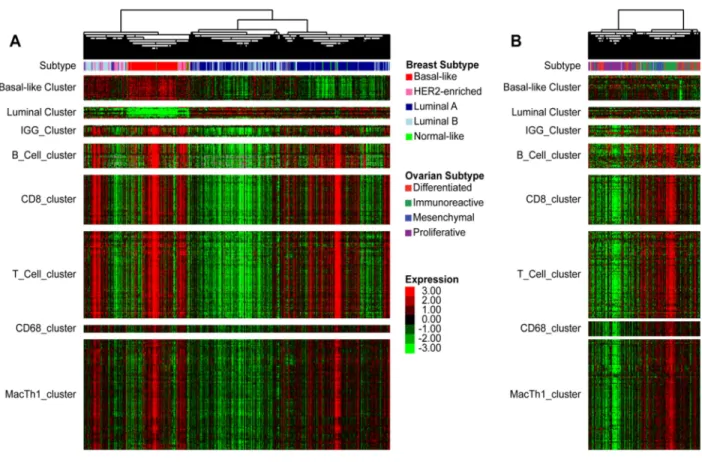
ResultsB-cell gene expression signatures are prognostic in breast and ovarian cancer.
Increased expression of B-cell gene signatures has been shown to be favorably prognostic in breast cancer (38-40). To explore the role of B-cells and other lymphocyte cell types in the different intrinsic subtypes of breast cancer, immune cell associated genomic signatures were newly derived from unsupervised clustering of mRNA-seq data from 728 TCGA breast cancer samples (Figure 2.1A). Gene dendrogram nodes containing characteristic lymphocyte genes were selected as potential gene
17
芀Ҩ
18
眀ڢ
19
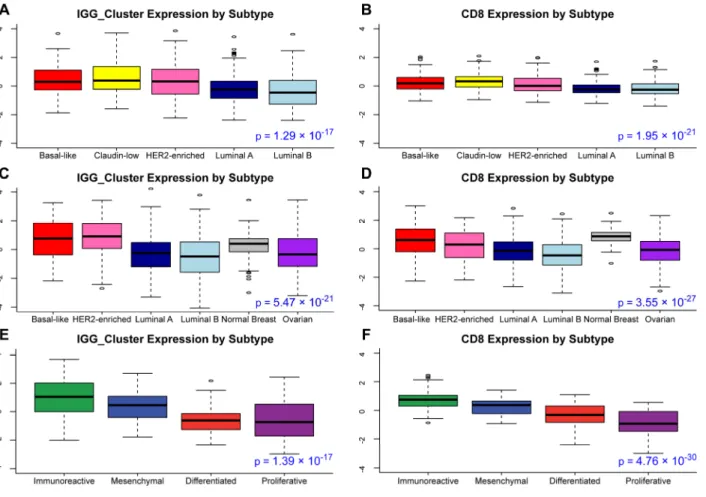
We next performed a similar analysis on TCGA ovarian cancer data. Overall B-cell gene signature expression was increased in immunoreactive ovarian tumors (Figure 2.2E-F). Several B-cell gene signatures were prognostic for progression-free survival in the immunoreactive ovarian tumor subtype, which was not true for the other subtypes (Table 2.1). T-cell signatures (15, 16, 137) were also evaluated, and showed a similar pattern of expression and prognostic value (Fig 2B, D). In multivariate survival analysis of individual immune signatures with other clinical and genomic features in breast cancer, most B-cell and T-cell signatures remained significantly prognostic (Table 2.2).
眀ڢ
22
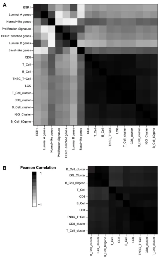
Figure 2.3
Signatures of lymphocyte infiltration in breast tumors are highly芀Ҩ
23
Specific B-cell receptor gene segment expression is prognostic in basal-like breast cancer.
Next, we wished to determine if the B-cell gene signature found in patients with basal-like breast cancer was consistent with an antigen-specific response. Other groups have shown clonal expansion and somatic hypermutation in breast B-cell TILs, suggesting an antigen-directed response in those samples (46, 52, 55). Actively responding antigen-specific B-cell populations are characterized by clonal
expansion; thus, we expect B-cell clonal expansion in patients where an effective, antigen-directed anti-tumor response is occurring. Because the clonal diversity of a B-cell population can be inferred by the diversity of the BCRs they express, there should be a prognostic benefit in samples with increased expression of specific BCR gene segments (i.e. immunoglobulin heavy chain and light chain variable, joining, diversity, and constant region segments). It has been shown that the BCR protein from breast cancer TILs is mainly produced by plasma cells, not B-cells (40). Here, we will continue to use the term “B-cell” to refer to the heterogeneous group of BCR-producing cells in the B-cell lineage.
7
24
眀ڢ
25
Figure 2.4
Expression level of all BCR segments across breast cancer subtypes and ovarian眀ڢ
26
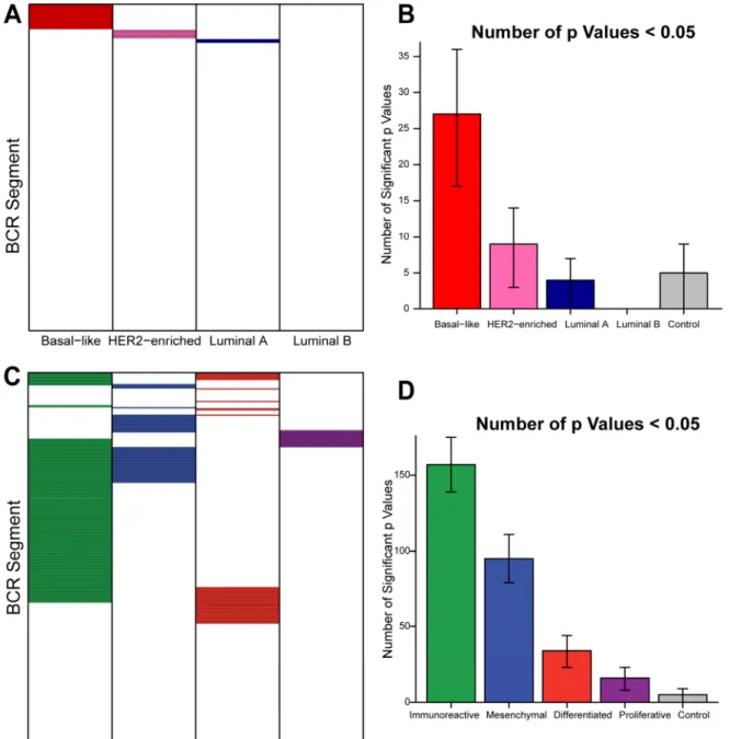
Figure 2.5
Expression level of BCR segments is preferentially predictive of improved overallsurvival in basal-like breast cancer and progression-free survival in immunoreactive ovarian cancer. A, C Grid of prognostic value of all BCR segments (colored cells represent positively prognostic
芀Ҩ
27
Figure 2.6
Kaplan-Meier plots, with log-rank p value, of overall or progression-free survival in7
28
Since B-cells undergo somatic hypermutation following antigen stimulation in the germinal center reaction, reads mapping to each germline BCR gene segment are expected to contain many
7
29
Figure 2.7
Method of calculating BCR sequence diversity from⁰Ҩ
30
Figure 2.8
A Basal-like, HER2-enriched, and luminal B tumors show high expression of믠Ҩ
31
Analysis of somatic hypermutation patterns in mRNA-seq data.
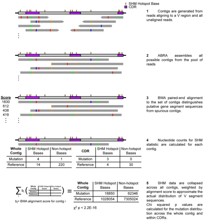
Somatic hypermutation in BCR gene segments is characterized by mutations that favor defined local sequence regional “hotspots” and CDRs, due to bias in the enzymatic activity that facilitates the mutation process(130). In order to evaluate the degree of somatic hypermutation represented in our data, we made use of the novel de novo assembly algorithm ABRA to assemble unique contigs from reads that map to each BCR variable (V) segment locus, followed by analysis of the contigs for the presence or absence of SHM. These contigs allowed us to analyze SHM mutation patterns across a V segment or its CDRs, rather than interrogating each mRNA-seq read pair separately. An overview of this method is given as Figure 2.9.
We applied our method of analyzing somatic hypermutation in mRNA-seq data to the TCGA breast and ovarian data sets. For the top 10 most highly expressed BCR V gene segments in breast or ovarian tumors in our data sets, the basal-like and HER2-enriched breast subtypes were enriched for tumors with V gene segments consistent with SHM (Figure 2.10). Immunoreactive ovarian tumors showed a high proportion of segments with mutation patterns suggestive of SHM, but it was not significantly higher than the proportion observed in other ovarian subtypes. The presence of SHM sequence
걐ҩ
32
Figure 2.9
Method of calculating likelihood of enrichment for somatic hypermutation from걐ҩ
33
Figure 2.10
Percent of assembled contigs significantly enriched for mutation patterns consistent걐ҩ
34
DiscussionWe define here four characteristics of an active, antigen-driven, anti-tumor B-cell response that can be identified from mRNA-seq data, namely: 1) increased expression and prognostic value of B-cell gene signatures, 2) increased expression and prognostic value of BCR gene segments, 3) decreased diversity of highly expressed BCR gene segments, and 4) mutation patterns consistent with BCR somatic hypermutation. All four conditions were found in basal-like breast cancers, and three of these conditions were found for immunoreactive ovarian tumors and HER2-enriched breast tumors. These findings support the hypothesis that a productive B-cell-driven endogenous anti-tumor response may be generated in many basal-like breast and immunoreactive ovarian carcinomas. To our knowledge this represents the first inference of BCR repertoire characteristics from mRNA-seq data.
Investigations into the anti-cancer adaptive immune response have largely been focused on T-cells. Accordingly, current cancer immunotherapy is directed at modifying the T-cell immune response through modulating targets like CTLA-4 and PDL-1. In this work, we show that the presence of tumor-infiltrating B-cells correlated with overall and progression-free survival suggesting that B-cells play an important role in anti-tumor immunity. We do show that the expression of B-cell genes was highly correlated with the expression of T-cell genes. By further demonstrating that in specific breast and ovarian cancer subtypes B-cell TILs are clonally expanded and enriched for somatic hypermutation, we provide evidence that B-cell TILs are not merely a surrogate marker for an anti-tumor T-cell response. While it is technically possible that previously expanded B-cell clones may be trafficked to the tumor independent of their antigen binding capability, previous studies showing clonal evolution within breast tumors make this unlikely(47, 52, 53, 55), as does the association with specific tumor subtypes. Tumor antigen-directed B-cell responses, which we suggest are present in many basal-like breast and
immunoreactive ovarian tumors, may provide a novel way to clinically target these tumor types.
덠ҩ
35
to evaluate B-cell TILs to investigate if and how anti-cancer immunotherapies may modulate the B-cell compartment.
As more immunomodulatory treatments become available for cancer therapy, one critical issue is the identification of the specific cancer patients who may benefit from such therapy. This work highlights the subtype association of clonally restricted B-cell responses. Previous studies in ovarian cancer have been mixed as to the importance of B-cell TILs, perhaps because of the heterogeneity of the B-cell response across the subtypes of ovarian cancer. Milne, et al. highlighted the high-grade serous histologic subtype as being selectively associated with TILs predictive of disease-specific survival (127). Here, we further identify the immunoreactive genomic subtype of serous ovarian cancer as containing prognostic TILs. Among basal-like breast tumors, it is interesting to note that patients with high B-cell infiltration as assessed by gene signatures were also significantly younger than other patients with basal-like breast cancer (data not shown), corroborating previous work highlighting this group(152).
Other investigations of B-cell TILs in breast cancer have found the survival benefit associated with B-cell gene expression to be dependent on proliferation (39, 40, 150). However, we do not see this association in our data. While the basal-like and HER2-enriched subtypes are both highly proliferative, we observed no survival benefit for B-cell TILs in luminal B tumors, which are also characterized by high proliferation. Furthermore, likelihood ratio testing conditioning on clinical variables and genomic subtype (data not shown) demonstrated that proliferation did not significantly increase the predictive ability of the model in breast cancer.
This work again underscores the similarity between basal-like breast and ovarian tumors.
Previous genomic studies have established that serous ovarian tumors resemble basal-like breast cancer in terms of their mutational profiles and DNA copy number changes (2). Here we show that this similarity extends to the immune component of the tumor microenvironment. In terms of the immune response, basal-like breast cancer bears more similarity to ovarian immunoreactive cancer than to the luminal A breast cancer subtype. This adds further weight to the notion that the therapeutic approach to basal-like breast and ovarian cancer could be similar.
HER2
-36
enriched subtypes, TIL abundance was not associated with a survival benefit within claudin-low tumors (Table 2.1). This could potentially be due to different immunosuppressive mechanisms within the tumor microenvironment, or it is possible that claudin-low tumors elicit a nonspecific inflammatory response in contrast with other high-immune breast tumors. We were unable to assess the BCR sequence diversity of claudin-low TILs as very few (fewer than ten) claudin-low tumors have been identified within the TCGA data set. If TILs within claudin-low tumors are not productive or antigen-directed, misclassification of these tumors may limit the effectiveness of immunomodulatory treatments within triple-negative breast cancers.
There are several standard approaches to analyzing the adaptive immune response present in the tumor microenvironment and tumor-draining lymph node. Immunohistochemistry and
immunofluorescence can be performed, although specific antibodies often require frozen tissue; similarly, flow cytometry can be performed on frozen tissue. One of the benefits of the approach described here is the potential to perform the analysis on formalin-fixed paraffin embedded tissue as methods for mRNA-seq from these samples have been demonstrated(154), and will continue to improve and become
standardized. As this is available on a substantially greater number of patients compared to frozen tissue, this approach could allow for a much larger group of patients to be analyzed for the presence of adaptive immune signatures. Indeed, there are a large and growing number of human tissue samples with
available mRNA-seq data; through the methods described here, these samples may be analyzed for antigen-directed B-cell responses.
Given our data that the presence of B-cells in the tumor microenvironment in patients with basal-like and HER2-enriched breast cancers and immunoreactive ovarian cancer is predictive of outcome, the role that endogenous B-cells play at these sites of tumor growth is a critical question. Plasma cells could generate anti-tumor antibodies that could be important in the early control of the growth of breast cancer cells, but which may ultimately become lost during tumor progression. Alternatively, B-cells may function as antigen-presenting cells to activate tumor-specific T-cells, which in turn may be inhibited via
뎰ҩ
37
The most difficult breast tumors to treat clinically are often of the “triple-negative” class, defined as such by the lack of cell-surface expression of estrogen receptor, progesterone receptor, and HER2 (9). The majority (60-80%) of triple-negative tumors are basal-like (7), and thus the basal-like subtype
represents a critical target for the development of novel therapeutics. The presence of BCR
-38
CHAPTER 3 – THE PROGNOSTIC IMPACT OF TUMOR IMMUNE INFILTRATION ACROSS DIVERSE SOLID TUMOR TYPES
Introduction
Many solid tumors are associated with a substantial immune infiltrate. This immune infiltration represents a complex and important aspect of the tumor microenvironment, and may comprise many distinct cell types, including various phenotypes of T and B infiltrating lymphocytes (TIL), tumor-associated macrophages (TAM), natural killer (NK) cells, and myeloid-derived suppressor cells (MDSCs). The complex interplay between solid tumors and the host immune system has been widely studied, but is in many cases still poorly understood. In a number of distinct tumor types, the presence of TILs has been demonstrated as an important prognostic factor (10, 78-84). For example, the presence of CD8+ T-cell TILs is a good prognostic factor in many tumor types including melanoma, colorectal, breast, ovarian, and non-small cell lung cancer, and in several cases it has been demonstrated that these CD8+ TILs are able to specifically kill tumor cells (155). Th1 T helper cells and B-cell TILs have also been implicated in the anti-tumor immune response (56, 85). Solid tumors classically are understood to cultivate an
immunosuppressive microenvironment to combat the anti-tumor host immune response through anergy and exhaustion of TILs and induction of a pro-tumor, inflammatory wound-healing response (156). This is accomplished through cytokines such as IL-8 and IL-12 released by tumor and stromal cells, and the recruitment of M2 macrophages, regulatory T-cells (Treg), and MDSCs (64, 157, 158). This tumor immune
regulatory response has been put forth as an explanation for the inability of the anti-tumor adaptive immune response to cause tumor regression. This is supported by studies showing that the presence of Treg, TAMs, and/or MDSCs predict worse outcomes in diverse tumor types including melanoma, renal cell
됀ҩ
39
Next-generation sequencing and large-scale genomics have become critical to our understanding of human cancers. Through efforts such as The Cancer Genome Atlas (TCGA), genomic data on a wide variety of platforms has become available. In many tumor types these data have been used to derive genomic subtypes that complement previous approaches to categorizing human tumors. By combining diverse data types, groupings both within and across tumor types have emerged (159). This deepens our understanding of distinctions within tumor types, such as in the identification of a subtype of
glioblastomas driven by methylation (i.e. G-CIMP), and also highlights significant similarities between previously distinct tumor types, such as in the identification of the “squamous” genomic subtype, which combines lung, head and neck, bladder, and some breast tumors into a single group (159, 160). Describing tumors along genomic lines may refine clinical practice in a way that more closely reflects tumor biology.
Cancer immunotherapy has been extensively pursued as an alternative or complement to cytotoxic chemotherapy and radiotherapy, but has recently shown great promise in a number of tumor types on its own. Inhibition of the immune checkpoint proteins CTLA-4, PD-1, and PDL-1 reduces the ability of the tumor microenvironment to suppress the host anti-tumor immune response (94, 102, 104). This leads to a decrease in the functionality of Treg TILs and proliferation of CD8
+
TILs. The immune checkpoint inhibitors ipilumumab (anti-CTLA-4), nivolumab and pembrolizumab (anti-PD-1), and various early-stage PDL-1 inhibitors such as MPDL3280A have shown clinical responses in melanoma, bladder, ovarian, renal cell, and non-small cell lung cancers (95, 102, 104, 105, 107). Interestingly, both nivolumab and ipilumimab have resulted in durable responses long after discontinuing treatment in studies involving melanoma, non-small cell lung cancer, and renal cell carcinoma, suggesting an ongoing anti-tumor immune response (101-103). As immune checkpoint inhibitors and other immune-targeted therapies gain ground in diverse tumor types, a key question is the identification of tumor characteristics that predict response to these agents. Evidence in melanoma and bladder cancer suggests that responders may already harbor clonally restricted, anti-tumor TILs that are able to take effect after tumor
眀ڢ
40
41
MethodsData Sets
The data set used for all analyses comprised mRNA-seq data from 3485 TCGA tumors (see TCGA Data Portal at https://tcga-data.nci.nih.gov/tcga/, CGHub at https://cghub.ucsc.edu/), which was originally described in Hoadley, et al. 2014 with the following modifications (159). All tumor samples from Hoadley, et al. were used, except for 161 AML samples, which were removed. Data for 329 melanoma samples were added to the set. Data for all melanoma samples represent the mRNA-seq data set used in the TCGA melanoma manuscript (TCGA, Cell, In Press). In total, this data set includes 118 urothelial bladder carcinomas (BLCA), 821 breast ductal carcinomas (BRCA), 250 colorectal adenocarcinomas (CRC, 179 colon tumors and 71 rectal tumors), 120 glioblastoma multiforme tumors (GBM), 301 head and neck squamous cell carcinomas (HNSC), 461 clear cell renal cell carcinomas (KIRC), 267 lung
adenocarcinomas (LUAD), 237 lung squamous cell carcinomas (LUSC), 240 serous ovarian adenocarcinomas (OV), 329 cutaneous melanomas (SKCM), and 341 uterine corpus endometrial carcinomas (UCEC). All samples were assayed by mRNA-seq using Illumina 2x50bp sequencing as described by the TCGA Research Network (131). Gene expression values were represented as RSEM (RNA-seq by Expectancy-Maximization) data normalized within-sample to the upper quartile of total reads as previously described (37). These data and further details about data processing are available at the TCGA Data portal under the V2_MapSpliceRSEM workflow
(https://tcga-data.nci.nih.gov/tcgafiles/ftp_auth/distro_ftpusers/anonymous/tumor/brca/cgcc/unc.edu/illuminahiseq_rna seqv2/rnaseqv2/unc.edu_BRCA.IlluminaHiSeq_RNASeqV2.mage-tab.1.6.0/DESCRIPTION.txt). Genomic subtypes within tissue types and Cluster of Clusters Assignments (COCA) subtypes were obtained from previous publications of those TCGA tissue types (2, 131, 160-165). No COCA subtype was assigned to melanoma samples, and thus melanoma was assigned to its own group in COCA subtype analyses due to existing data showing its distinctive features distinguishing it from the other included tumor types (166).
Gene Signatures and Survival Analyses
-42
Multiple signatures were identified for each cell type to reduce bias from individual gene lists. IGG_Cluster (38), B_Cell (136), B_Cells (167), B_Cell_cluster (1), B_Cell_60gene (39), and
TNBC_B_Cell (16) are B/plasma cell signatures. T_Cell (136), T_Cells (167), T_Cell_cluster (1), CD8 (136), CD8_cluster (1), LCK (137), and TNBC_T-Cell (16) are T-cell signatures, with the CD8 and CD8_cluster signatures specifically representing CD8+ T-cells. MacTh1_cluster (1), CD68_cluster (1), Mac_CSF1 (168), and Macrophages (167) are all macrophage/monocyte signatures.
Univariate survival analyses were performed by Cox proportional hazards modeling with each signature tested as a continuous variable. Multivariable Cox proportional hazards models were used to evaluate the prognostic value of individual gene expression signatures when combined with tumor type/subtype information and clinical variables. All multivariable survival analyses included one gene expression signature as well as patient age and pathologic tumor (T), node (N), and metastasis (M) stages. Pathologic T, N, and M stages were binned into T1 vs. T >1, N0 vs. N>0, and M0 vs. M >0 groups to reduce the number of variables in each model and standardize across tumor types. All survival
analyses were performed in R version 2.13.1 using the “survival” package (169, 170).
BCR Restriction Index
볐ҩ
43
앐ҩ
44
ResultsSignatures of Immune Infiltration are Coordinately Expressed in Human Cancer
Increased expression of gene signatures representing TILs has been shown to be favorably prognostic in many tumor types, including breast, colorectal, ovarian, non-small cell lung cancer, and melanoma. To clarify the role of tumor-infiltrating immune cells, gene signatures associated with distinct immune cell types were evaluated in a diverse data set of 3485 tumors representing 12 distinct tumor types (see methods). Signatures were chosen that were previously published as corresponding to tumor-infiltrating immune cells in at least one solid tumor type. Several gene signatures were used for each cell type studied (T-cells, CD8+ T-cells, B/plasma cells, and tumor-associated macrophages), which served to mitigate the effects of differences in gene list constituency and/or the technical effects seen when
translating a microarray-derived gene list for application to our mRNA-seq data set. A heat map of a subset of these signatures across all 3485 samples ordered by tissue type and genomic subtype shows high expression across gene signatures in particular cancer types and subtypes (Figure 3.1). Immune signature expression is highest in the basal bladder, basal-like and HER2-enriched breast,
immunoreactive ovarian, and immune melanoma subtypes, as well as in the head and neck squamous, clear cell renal cell, lung adenocarcinoma, squamous cell lung carcinoma tissue types (Figure 3.1). Within tissue types, immune signature expression varied widely by subtype. While only the ovarian
immunoreactive and melanoma immune subtypes were defined with immune infiltration as a key feature, immune infiltration often segregated by tumor subtype. Similarly the LUAD-enriched, squamous, breast basal-like, and KIRC COCA (Cluster of Cluster Analysis genomic classification) subtypes show high expression of immune signatures. Signature scores across tissue types and COCA subtypes are
summarized in Figure 3.2. Within and across all tumor types, all immune gene signatures that were tested showed very high correlations, with the average Pearson correlation > 0.63 for all tumor tissue of origin or COCA types (Figure 3.3). The lowest inter-signature correlation was among glioblastoma samples; all glioblastoma samples showed low expression of lymphocyte-related gene signatures and high expression of signatures related to macrophages and other myeloid-derived cell types. This is likely due to
배ҭ
45
48
잠ҩ
49
Signatures of Immune Infiltration are Prognostically Important in Diverse Human Cancers
Univariate survival analyses for immune signature expression within tissue of origin groups or COCA genomically-defined subtypes are summarized in Tables 3.1-3.2 (hazard ratios are provided in Appendix 2, Supplementary Tables 1-2). Number of events and median follow-up times varied
52
쁀ҩ
54
Prognostic Immune Infiltrates are Associated with Reduced Tumor-infiltrating B-Cell Clonality
Previous investigations of TIL infiltration have suggested that many tumors with prevalent TILs contain clonally restricted T- or B-cell infiltrates (46, 52, 118). These TIL infiltrates may potentially be targeting tumor antigens, which is consistent with results showing a prognostic benefit for TILs in these tumor types. As previously described, mRNA-seq data can be used to estimate relative clonal diversity of cell populations (1). Somatic hypermutation introduces many point mutations in the sequence of the B-cell receptor (BCR) of B-B-cells undergoing the germinal center reaction. These point mutations, combined with the identity of the variable (V), diversity (D) , and joining (J) segments of the BCR, are likely unique to a B-cell clone within an individual. Different patterns of non-reference bases identified in mRNA-seq reads mapping to BCR V gene segments represent different B-cell clones, so enumerating those patterns across highly expressed V gene segments provides an estimate of clonal diversity in a B-cell population. Because this method relies on mutations generated by SHM, it cannot be readily applied to TCR segment reads. For this analysis, BCR V gene segment expression and diversity values were calculated for all samples. Figure 3.4 illustrates BCR gene segment expression across all tissue types and COCA
subtypes, highlighting the same tissue types identified by gene signature analysis. While BCR expression is able to distinguish certain samples and tumor types as high expressers of BCR segments, expression was high across multiple BCR gene segments in such samples. This could potentially represent
쩰ҩ
55
배ҭ
56
섰ҩ
57
쯠ҩ
58
배ҭ
59
DiscussionHere we highlight several aspects of an effective, potentially antigen-driven immune response, as determined from mRNA-seq data, in diverse solid human tumor types. Patients with apparently beneficial tumor immune responses had high levels of immune signature expression for several cell types including cytotoxic and helper T-cells, B/plasma cells, and macrophages/monocytes. High expression of these immune signatures predicted survival across distinct tumor types. Furthermore, among these highly immune infiltrated tumors, a subset contained a highly expressed population of BCRs with low sequence diversity, which also predicted improved survival in these patients. Together, these findings highlight a subset of tumors among the squamous/basal bladder, basal-like and HER2-enriched breast,
immunoreactive ovarian, and immune melanoma subtypes, as well as a subset of tumors among head and neck, lung adenocarcinoma, and lung squamous cell carcinoma, that appear to have a beneficial, antigen-directed immune response.
Tumors in this data set were analyzed both by tissue of origin and genomic COCA subtype, as well as by genomic subtype defined within the tissue of origin. By considering each of these classification schemes, it is possible to identify classifications that are relevant for the role of immune infiltration in these tumor types. The squamous COCA subtype, for example, groups together some bladder, head and neck, and lung tumors with a common set of genomic features. This set of tumors is also consistently high for immune signature expression, and groups together tumors and tumor types with prognostic immune infiltrates. Similarly, the breast basal-like COCA subtype, which also shows some squamous-like genomic characteristics, also separates out breast tumors that often harbor prognostic immune infiltrates. The fact that these subtypes were defined comprehensively by global gene expression, protein levels, copy number miRNA, and DNA methylation demonstrates that these subtype-defining features may be responsible for the immunogenic phenotype shared among these tumors.
술ҩ
60
to suppose that the tumor types and subtypes highlighted above as having high immune signature expression and restricted B-cell TIL infiltrates may also represent the groups in which immune checkpoint inhibition is most promising.
Most investigations into the role of TILs in human tumors have been focused on T-cells. Many studies have shown that the presence of T-cell TILs is associated with response to immune checkpoint inhibition and survival in diverse tumor types (13, 88, 105, 173). Here, we demonstrate that in all tumor types analyzed in this work, B-cell gene expression, including expression of BCR gene segments, was highly correlated with the expression of T-cell genes. The high correlation of immune signature
expression across distinct immune cell types illustrates that the tumor immune infiltrate is diverse and complex, and supports the idea of an important supportive role for B-cell TILs and some types of TAM in the anti-tumor immune response. Cox proportional hazards modeling of BCR expression and diversity demonstrates that BCR restriction adds significantly to the prognostic ability of the model beyond BCR expression or other B-cell gene expression (although this finding will need to be further validated in test sets of independent data). The additional prognostic value of BCR restriction indicates an important role for B-cell TILs in the anti-tumor immune response. It is unclear whether that role includes the production of anti-tumor antibodies to mediate cell killing, or if B-cell TILs primarily function by supporting an effective T-cell response. Previous work has shown that PD-1 blockade enhances the activation, proliferation, and cytokine production of human B-cells after TLR stimulation (151). Further work is needed to clarify the role of B-cell TILs in the clinical efficacy of immune checkpoint inhibition.
Overall, for most of the tumor types tested, immune infiltrates portended an improved outcome; however, there were three tumor types which are exceptions to this rule. Thus, it should not be assumed that immune cells play a simple, one-dimensional role across all tumor anatomic sites. One notable tumor type in which immune signature expression consistently did not predict survival in this work was colorectal adenocarcinoma. There are many studies indicating a beneficial and prognostic role for TILs, including helper and cytotoxic T-cells and potentially Treg, in human colorectal cancer (174-176). The failure to
쵀ҩ
61
cancer. Similarly, poor survival data in breast and endometrial cancer limited survival predictions as well, resulting in inconsistent prognostic value among immune signatures.
An interesting biological outlier tumor type in our results was renal cell carcinoma, which scored consistently high for expression of immune signatures. However, unlike other highly immune-infiltrated tumor types in this analysis, high expression of T-cell signatures did not predict good outcomes in renal cell carcinoma, and high expression of B-cell signatures consistently predicted poor prognosis. To examine the role of B-cells in renal cell carcinoma more carefully, we generated a restriction index score solely within this tumor type and found that it was almost precisely the opposite of the restriction index developed on other tumor types and applied to renal cell samples (r2 = 0.938, slope of line of best fit = -3.44; data not shown). This suggests that it is specifically the presence of a clonally restricted B-cell infiltrate that predicts poor prognosis within renal cell carcinomas. Previous work has suggested that this tumor type may contain abundant Breg; a clinical trial was conducted in which the anti-CD20 monoclonal
antibody Rituximab was used to deplete B-cells from renal cell tumors (86). The failure of that trial to show clinical benefit may have been largely due to the low CD20 expression of Breg and the resulting
failure of Rituximab to affect that population (87). The presence of B-cell TILs associated with poor outcome in renal cell carcinoma could also be explained by other mechanisms: B-cell tolerization and anergy due to other immunoregulatory cell types likely plays a role. It is also possible that this association may be partly artifactual due to an association between TILs and higher tumor grade, which in turn predicts worse outcomes, in this tumor type. However, the successes of anti-PD-1 treatment and, earlier, of IL-2 therapy in renal cell carcinoma, shows that a beneficial immune response is possible in this tumor type. Thus, renal cell carcinoma remains an immunogenic tumor type in which further study is certainly needed.