ENDOCRINE DISRUPTING POTENTIAL OF ENVIRONMENTAL CHEMICALS CHARACTERIZED BY HIGH-THROUGHPUT SCREENING
Daniel M. Rotroff
A dissertation submitted to the faculty of the University of North Carolina at Chapel Hill in partial fulfillment of the requirements for the degree of Doctor of Philosophy in the Department of Environmental Sciences and Engineering.
Chapel Hill 2013
Approved by:
Ivan Rusyn, M.D. Ph.D. David J. Dix, Ph.D
Thomas Knudsen, Ph.D.
Fred Wright, Ph.D.
ABSTRACT
DANIEL M. ROTROFF: Endocrine Disrupting Potential of Environmental Chemicals Characterized by High-Throughput Screening
(Under the direction of Dr. David J. Dix)
ACKOWLEDGEMENTS
First and foremost I would like to express my deep gratitude to my research advisor, Dr. David Dix, Ph.D. Without his support and expertise this would not have been possible. His academic and professional guidance has been invaluable, and will continue to be so in all of my future endeavors. I also owe a great deal of appreciation to my academic advisor, Dr. Ivan Rusyn, M.D. Ph.D., who made sure that I stayed on target throughout this entire process, despite my occasional efforts to test certain boundaries. I would like to express my appreciation to the additional members of my committee, Dr. Thomas Knudsen, Ph.D., Dr. Fred Wright, Ph.D., and Dr. Rebecca Fry, Ph.D. Their diverse knowledge and experience pushed me to be a better student and a better researcher.
I owe a great deal of gratitude to Dr. Richard Judson, Ph.D., who kept his door open to me and spent many hours helping me formulate new and innovative ideas. His amazing work ethic sets a high standard, and through his support and mentorship I have benefitted greatly. Lastly, I would like to show my gratefulness to my colleagues, collaborators, and managers at the U.S. Environmental Protection Agency National Center for Computational Toxicology. Their continued effort to push science forward and willingness to share from their vast knowledge and their distinct backgrounds are what have made this experience so significant.
TABLE OF CONTENTS
LIST OF TABLES ...x
LIST OF FIGURES ... xi
LIST OF ABBREVIATIONS ... xiii
Chapter I. INTRODUCTION ...1
Endocrine Physiology ... 1
Human Endocrine Disruption and Pathology ... 3
Environmental Impact of Endocrine Disruption ... 6
Endocrine Disruptor Screening ... 8
Computational Toxicology and Alternatives to Traditional Test Methods ... 10
Estrogen Signaling Pathways in Predictive Toxicology ... 13
Predictive Modeling of Endocrine Disruptors ... 17
References ... 24
II. USING IN VITRO HIGH THROUGHPUT SCREENING ASSAYS TO IDENTIFY POTENTIAL ENDOCRINE-DISRUPTING CHEMICALS ...32
Introduction ... 32
Methods ... 36
Results ... 41
Discussion ... 44
III. REAL-TIME GROWTH KINETICS MEASURING HORMONE MIMICRY FOR 1816 UNIQUE TOXCAST CHEMICALS IN T-47D HUMAN DUCTAL
CARCINOMA CELLS ...64
Introduction ... 64
Methods ... 66
Results ... 74
Discussion ... 78
Tables ... 84
Figures ... 89
References ... 96
IV. ENDOCRINE TESTING IN THE 21ST CENTURY: USING IN VITRO ASSAYS TO PREDICT ESTROGEN RECEPTOR SIGNALING RESPONSES...101
Methods ... 103
Discussion ... 118
Tables ... 124
Figures ... 126
References ... 137
V. GENERAL DISCUSSION ...141
Advances in In Vitro Technology ... 141
Phenotypic Anchoring ... 143
Combining In Vitro Data to Test for In Vivo Effects ... 145
Limitations ... 148
References ... 152
VI. FUTURE DIRECTIONS ...155
LIST OF TABLES
Table
2.1 Summary of Endocrine Related HTS Assays ………. 50 2.2 Summary of Endocrine Literature Survey ……….. 51 2.3 Chemical Results from Overlapping HTS-E and Guideline Reports ……
52 2.4 Chemical Chemical Results from Overlapping HTS-A and Guideline
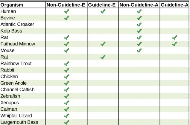
Reports ………... 53 2.5 Test Species in Guideline and Non-Guideline In Vitro and In Vivo
Studies ………. 54
3.1 ER Reference Chemicals for Sensitivity and Specificity Analysis ……….
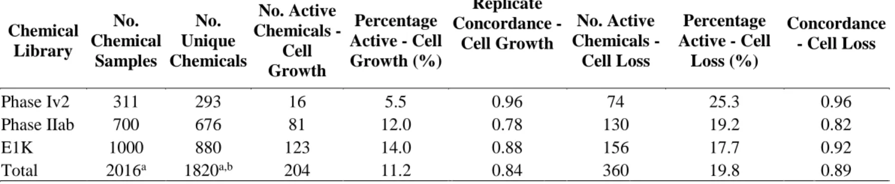
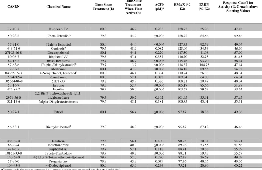
84 3.2 Chemical Library Results Summary and Replicate Concordance ……….. 85 3.3 25 Most Potent and Efficacious Compounds in the Cell Growth Assay at
80 h ……….. 86
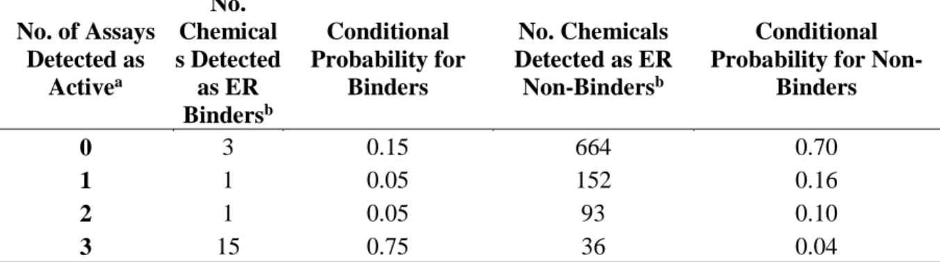
3.4 Paraben Compounds – T-47D Cell Growth Potency and Efficacy at 80 h.. 87 3.5 Estrogen Receptor Binding Conditional Probabilities ……… 87 3.6 Logistic Regression Model Performance……….
88 4.1 ER In Vitro Assay Annotation and Model Grouping……….….. 124 4.2 15 Chemicals with Highest ER Interaction Scores ……….
LIST OF FIGURES
Figure
1.1 Schematic representation of the HPG, HPA, and HPT axes……... 23
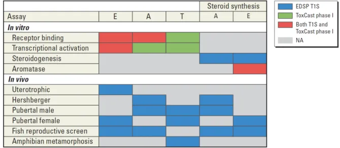
2.1 Overlap between EDSP T1S assays and ToxCast phase I assays by
endocrine MOAs ………. 55
2.2 Illustration of the balanced optimization model used to analyze
predictive capacity of endocrine-related ToxCast assays ……….. 56 2.3 Forest plot illustrating the performance —as measured by sensitivity,
specificity, and BA—of ToxCast endocrine-related assays for predicting
outcomes captured in EDSP/OECD guideline studies ………. 57 2.4 Forest plot illustrating the performance —as measured by sensitivity,
specificity, and BA—of ToxCast endocrine-related assays for predicting
outcomes captured in non-guideline endocrine studies ………. 58 3.1 RTCA Cell Growth Assay …………... 89 3.2 Example of Growth Curve Translation to Concentration Response Curve.
90 3.3 Representative growth curves of various test compounds measuring
changes in normalized cell index over time…...……….
91 3.4 Comparison of Cell Growth Results with Additional ER Assays ……… 92 3.5 Reciever operating characteristic plot for uterotrophic comparisons ……
93 3.6 Reciever operating characteristic plot for ER reference chemical
comparisons ……… 94
3.7 Combination Treatment of Electron Transport Chain Inhibitors with ER
Figure
4.1 ER Signaling Pathway Overview (Overlayed with ToxCast
Assays)………..………... 126
4.2 Graphical Representation of the Logical Structure of the Model………… 127
4.3 Composite Curve Example ………..…………...
128 4.4 Heatmap of Composite Scores ………...………
139 4.5 Impact of Linear Transformation on the Distributions of
R2……….………
130 4.6 Agonist Model Stability …………...……….. 131
4.7 Agonist/Antagonist Volcano Plot ………... 132 4.8 ERα/ERβ Volcano Plot ………...…… 133
4.9 Uterotrophic Analysis ……….
134 4.10 Reference Chemical Spectrum ………... 135
LIST OF ABBREVIATIONS
AC50 – Activity concentration at 50% efficacy AF-1 – Activation function 1
AF-2 – Activation function 2
Agonist_CS – Agonist Composite Score AhR – Aryl hydrocarbon receptor
Antagonist_CS – Antagonist Composite Score AR – Androgen receptor
BA – Balanced accuracy
bER – Bovine estrogen receptor
Binding_CS – Binding Composite Score
CASRN – Chemical Abstracts Service Registry Number CDK – Cyclin dependent kinase
CI – Cell index
CRH – Corticotropin releasing hormone CYP19 – Aromatase cytochrome P-450 DBD – DNA Binding Domain
DBP – Dibutyl Phthalate DES - Diethylstilbestrol
E2 - 17β-estradiol
EDC – Endocrine disrupting chemical
EDSP – Endocrine Disrupter Screening Program EMAX- Maximum efficacy
EPA –Environmental Protection Agency ER – Estrogen receptor
ERE – Estrogen response element
ERES – Estrogen Receptor Expert System FBS – Fetal bovine serum
FDA – Food and Drug Administration
FFDCA – Federal Food, Drug and Cosmetic Act FN – False negative
FP – False positive
FQPA – Food Quality and Protection Act FSH - Follicle stimulating hormone
GHRH – Growth hormone-releasing hormone GnRH - Gonadotropin-releasing hormone GR – Glucocortocoid receptor
HPT – Hypothalamic-pituitary-thalamic HPV – High production volume
HTS – High Throughput Screening
HTS-A – High Throughput Screening-Androgen HTS-E – High Throughput Screening-Estrogen HTS-S – High Throughput Screening-Steroidogenesis HTS-T – High Throughput Screening-Thyroid
LBD – Ligand binding domain
LEC – Lowest effective concentration LH - Lutenizing hormone
LOC – Level of concern
mERa – Murine estrogen receptor-α
MOA – Mode-of-Action NCI – Normalized cell index NR – Nuclear receptor
OECD – Organization for Economic Co-operation and Development OR – Odds ratio
PCA – Protein complementation assay PMID – PubMed Identifier
PR – Progesterone receptor
QSAR – Quantitative structure-activity relationship r – Correlation coefficient
RTU-reporter transcription unit SDWA – Safe Drinking Water Act
SERM – Selective estrogen receptor modulator SGK-1 – Serum/glucocortocoid regulated kinase-1 SMA – Simple moving average
T1S – Tier 1 Screening T2S – Tier 2 Screening T3 - Triiodothyronine T4 - Thyroxine
TDS – Testicular Dysgenesis Syndrome TN – True negative
TP – True positive
TPO – Thyroid peroxidase
CHAPTER 1
INTRODUCTION Endocrine Physiology
The endocrine system consists of glands that secrete hormones that regulate and influence almost every organ and cell type. These hormones are secreted into the blood stream and activate signaling cascades to regulate and maintain a diverse set of biological functions. The various functions include glucose metabolism, sexual differentiation, bone density, blood pressure, and digestion (Cooper and Kavlock 1997; Dupont et al. 2000; Lodish et al. 2009; de Mello et al. 2011; Ng et al. 2001).
The endocrine system is commonly described in three axes: the hypothalamic-pituitary-gonadal (HPG) axis, the hypothalamic-pituitary-adrenal (HPA) axis, and the hypothalamic-pituitary-thyroid (HPT) axis (Figure 1.1). These physiological axes represent three different feedback systems and involve multiple, highly interrelated, signaling
pathways occurring between key organs to maintain homeostasis of bodily functions. The ultimate control center, the hypothalamus, secretes gonadotropin-releasing hormone (GnRH), growth hormone-releasing hormone (GHRH), thyrotropin-releasing hormone (TRH),
Gonadotropins are a group of anterior pituitary hormones that include follicle-stimulating hormone (FSH) and luteinizing hormone (LH) (Figure 1.1a) (Fox 2004). These peptide hormones, along with the steroid hormones estrogen and progesterone, control the ovarian cycle in females (Fox 2004). In males, FSH stimulates primary spermatocytes to undergo meiosis and LH triggers testosterone production (Walker and Cheng 2005). Gonadal stimulation by FSH induces the release of the peptide factor ‘inhibin’, which in turn acts back on the anterior pituitary to inhibit FSH production. Increases in serum concentration of sex steroids (estrogen, testosterone) suppress GnRH secretion by the hypothalamus and FSH and LH secretion by the anterior pituitary gland. The coordination of these hormones in both positive and feedback circuits is necessary for proper homeostasis of the HPG axis (Fox 2004).
The HPT axis is primarily involved in regulating growth, metabolism and
development (Figure 1.1b) (Zoeller et al. 2007). Thyroid follicular cells, transport iodine and secrete triiodothyronine (T3) and thyroxine (T4) via thyroid peroxidase (TPO),when
stimulated by TSH from the anterior pituitary. The main role of TSH is to free T3 and T4 from thyroglobulin so they can be released into the blood stream (Zoeller et al. 2007). T3 is critical to fetal neurodevelopment, and postnatally helps to regulate metabolism and cardiac output (Bernal, 1984; Oppenheimer and Schwartz, 1997). T3 and T4 work synergistically with growth hormone (GH) and consequently (an inhibitor of GH) to further down-regulate TSH secretion from the anterior pituitary (Figure 1.1b) (Zoeller et al. 2007).
is the adrenal cortex. Its outer zone (zona glomerulosa), middle zone (zona fasciculate), and inner zone (zona reticularis) produce different hormones in response to ACTH stimulation and all three are synthesized from cholesterol. Although not entirely distinct from one
another, the zona glomerulosa primarily secretes mineralocorticoids such as aldosterone (Fox 2004). Aldosterone regulates blood pressure through renal retention of sodium and water (Brewster and Perazella 2004). The zona fasciculata and reticularis are responsible for synthesizing the glucocorticoids cortisol and corticosterone, which upregulate
gluconeogenesis and lypolysis for bioenergy production (Fox 2004). The two innermost zones are also responsible for synthesizing androstenedione, a precursor molecule to both testosterone and estrogen. The enzymes 17β-hydroxysteroid-dehydrogenase and aromatase (CYP19) convert androstenedione to testosterone and estradiol, respectively. These
enzymatic conversions typically occur in the testis and ovary, respectively; however, aromatase is also expressed in non-gonadal tissues such as fat, which can further promote estradiol synthesis (Simpson and Davis 2001). Increased serum concentrations of these hormones have the ability to down-regulate CRH release from the hypothalamus and ACTH from the anterior pituitary (Figure 1.1c).
Human Endocrine Disruption and Pathology
highly interrelated, treating one type of endocrine deficiency can have unintended
consequences on another endocrine process. For example, hyperaldosteronism can be a result of adrenal hyperplasia or adrenal carcinoma and can cause increased water and sodium retention resulting in subsequent hypertension (Calhoun et al. 2002; Lindsay and Atkinson 2009). One treatment option for hyperaldosteronism is to administer a mineralocorticoid antagonist, such as spironolactone; however, this drug is a weak androgen receptor (AR) and glucocortocoid receptor (GR) antagonist that also displaces testosterone and estradiol from sex hormone binding globulin. Manifestations of this disruption in males is include
gynecomastia and testicular atrophy, and in females altered menstruation (Lubbos HG 1998; de Souza et al. 2010).
Epidemiological evidence suggests an increasing incidence of poor semen quality, testicular cancer, and other male reproductive disorders including undescended testis (cryptorchidism) and urethral fusion defects (hypospadias) (Auger et al. 1995; Carlsen et al. 1992; Skakkebaek et al. 2001). Cryptorchidism and hypospadias are common human malformations with an incidence in liveborn infants of 2-9% and 0.2-1%, respectively (Toppari et al. 2010). The collective term for developmental defects of the male tract is Testicular Dysgenesis Syndrome (TDS). TDS is diagnosed by the co-occurrence of poor semen quality, testicular germ line tumors, hypospadias, and/or cryptorchidism in various combinations (Skakkebaek et al. 2001). Studies have demonstrated associations of increased incidences of TDS with exposure to certain environmental chemicals (Toppari et al. 2010).
women to lower the risk of miscarriage (Palmlund et al. 1993). Approximately 5 million women were prescribed DES during pregnancy in the United States alone (Palmlund et al. 1993). DES not only failed as a therapeutic for spontaneous pregnancy loss but also led to premature labor and neonatal death. Some females exposed in utero displayed increased incidences of cervical and vaginal clear cell adenocarcinoma, vaginal adenosis, and genital tract abnormalities (Palmlund et al. 1993). Males exposed in utero demonstrated increased incidences of cryptorchidism, hypospadias, infertility, poor semen quality, and rete testis adenocarcinoma among other disorders (Toppari et al. 2010).
Some environmental chemicals have been shown to interfere with male reproductive development through endocrine mechanisms. Vinclozolin is one example. This fungicide was used to treat many types of berries, grapes, and other crops (U.S. EPA 2000). Current
exposures to vinclozolin are considered to be very low after the voluntary termination of uses and import tolerances (U.S. EPA 1998). Its antiandrogenic properties are well characterized in both in vitro and in vivo rodent bioassays. Vinclozolin is a weak AR antagonist but is quickly metabolized in serum to active metabolites, M1 and M2 (Sierra-Santoyo et al. 2012). Numerous studies demonstrated the ability of vinclozolin to compete with testosterone and DHT for AR (Fang et al. 2003; Kojima et al. 2003; Scippo et al. 2004). In vitro
transactivation assays have also demonstrated that M1 and M2 can downregulate
testing vinclozolin for antiandrogenic and TDS phenotypes have been conducted using animal studies, a mode-of-action framework created to determine the relevance of these animal effects for human risk assessment identified the vinclozolin-induced malformations of the male reproductive tract as being highly plausible in humans (Kavlock and Cummings 2005).
Environmental Impact of Endocrine Disruption
Endocrine disruption not only poses a risk to human health, but also poses a significant risk to environmental health (Caliman and Gavrilescu 2009; Kiesecker et al. 2001). Furthermore, the environment often serves as a sentinel for potential human health hazards (Caliman and Gavrilescu 2009). Pesticide runoff or drift from crops, as well as pharmaceutical presence in wastewater have potential for significant adverse health effects on wildlife (Sparling and Fellers 2009; Tavera-Mendoza et al. 2002). An active area of research for the impact of environmental chemicals on wildlife is the significant global decline of amphibians (Hof et al. 2011).
suggests that decreases in the secretion of LH is the primary cause of testicular consequences.
Atrazine is not believed to directly affect LH, because LH was induced in atrazine-treated rats administered GnRH. This implies an alternate neuroendocrine mechanism (Cooper et al. 1996, 2000). A study conducted by Shafer et al. supported this hypothesis by showing that atrazine and cyanazine non-competitively inhibited the binding of a
benzodiazepine to GABAA receptors (Shafer et al. 1999). Furthermore, GABAA receptors
have been shown to be important regulators of GnRH release (Terasawa 1998). Leydig cells respond to LH by synthesizing testosterone, which can then be converted to
dihydrotestosterone (DHT) by 5α-reductase. Additional data suggests that atrazine can inhibit the conversion of testosterone to DHT by 5α-reductase (Simic et al. 1991). Although the effects on 5α-reductase may not be the primary mechanism for alterations in
spermatogenesis, the important role DHT plays in the development and maintenance of tissues such as the prostate and seminal vesicles may exacerbate the effects from LH suppression (Cai et al. 1994).
The U.S. EPA currently estimates the aquatic ecosystem level of concern (LOC) for atrazine as approximately 10 ppb (10 µg/L) over a 60-day period (U.S. EPA 2009a).
on time to metamorphosis or mortality; however, effects at all doses higher than .01 ppb produced multiple gonads or were hermaphrodites with multiple testes and ovaries, and displayed decreased plasma testosterone.
Environmental exposures of EDC’s that play a role in the decline of wildlife
amphibian populations may also be relevant for human health. Cragin et al. (2011) provided epidemiological evidence of statistically significant trends in reports of irregular menstrual cycle lengths in areas with high levels of atrazine drinking water contamination versus women residing in areas with low levels of atrazine contamination. In order to maintain healthy wildlife areas and prevent adverse human health effects from environmental
exposures to EDC’s, it is critical that a complete characterization of the potential interactions pharmaceuticals and environmental chemicals could have on healthy endocrine physiology is performed.
Endocrine Disruptor Screening
Environmental chemicals such as bisphenol-A, phthalates, and parabens have exhibited potential to interfere with the endocrine system and are nearly ubiquitous in the environment. These compounds have commercial applications in food and drink packaging, polycarbonate plastics, detergents, polystyrene tubes, cosmetics, and shampoos (Calafat et al. 2005; Smith et al. 2012; Sonnenschein and Soto 1998; U.S. HHS 2008). The potential for adverse health and environmental effects due to EDC exposure has lead to increased public awareness of safe products and increased pressure for political and regulatory action.
1937 (Ballentine 1981). The incident occurred after the drug, sulfanilamide, was exposed with diethylene glycol. Subsequently, Congress passed the FFDCA and provided authority to the Food and Drug Administration (FDA) to regulate safety requirements for food, drugs and cosmetics (Ballentine 1981). In 1996, the Food Quality Protection Act (FQPA) and the Safe Drinking Water Act (SDWA) were passed. These two acts amended the FFDCA, and required that the U.S. EPA determine whether certain substances may have an effect in humans similar to a naturally occurring estrogen, or other endocrine related effects (21 U.S.C. 346a 1996; U.S. Public Law 1996). To address these requirements, the EPA formed the Endocrine Disruptor Screening Program (EDSP) (www.epa.gov/endo/) (U.S. EPA 2012a).
EDSP requires that chemical manufacturers provide data from specific in vitro and in vivo assays while following strict guidelines to insure testing is performed consistently and reliably. EDSP consists of two tiers of testing. The first is composed of in vitro and in vivo assays designed to measure effects on estrogenic, androgenic, thyroid and steroidogenesis modes of action (MOA). A weight-of-evidence approach has been proposed as a decision making tool for determining what compounds will go from tier 1 (T1S) to tier 2 (T2S). The selection of assays in T2S has not been finalized, but the assays under consideration include more resource intensive assays such as the mammalian two-generation, avian two-generation, and others (U.S. EPA 2012a).
will be selected for T2S testing or what assays will be included in T2S testing remain to be seen.
In an effort to increase throughput, the EPA’s EDSP21 project is aiming to use high-throughput in vitro and in silico approaches to prioritize which chemicals should be tested in T1S first (U.S. EPA 2011a, 2012b). Goals of the EDSP21 work plan include replacing certain resource intensive T1S assays within the next five years and full replacement of T1S with high-throughput in vitro and in silico assays as a long-term goal (U.S. EPA 2011a). In order to meet these goals, methods for validating in vitro assays, and models that combine in vitro assays to optimize for predictive capabilities must be developed.
The U.S. EPA National Center for Computational Toxicology’s ToxCastTM program
and the cross-agency Tox21 program are currently screening chemicals in hundreds of high-throughput in vitro assays, some of which are endocrine related (Kavlock et al. 2012). The current ToxCast and Tox21 chemical libraries represent approximately 17% and 53% of the EDSP chemical universe, respectively. Successful predictive models have been developed using the assays and chemicals from these projects (Judson et al. 2012; Martin et al. 2011; Sipes et al. 2011). Furthermore, tools for prioritizing chemicals have also been developed using these data sets (Reif et al. 2010). Therefore, it stands to reason that these programs are uniquely poised to develop predictive models for endocrine related targets that would be valuable in augmenting the EDSP screening effort.
Computational Toxicology and Alternatives to Traditional Test Methods
thousands of chemicals are now being manufactured, and due to the backlog of chemicals in regulatory testing, the majority of chemicals have yet to undergo full toxicological evaluation (Judson et al. 2009). Due to ethical limitations, toxicologists have always had to rely on extrapolating toxicities observed in animals to potential adverse health effects in humans with an equivalent exposure. Because animals are intrinsically different than humans, this process can be quite uncertain. Furthermore, deciding on an appropriate animal model or reconciling contradictory results between animal models can further pose a challenge for decision makers.
Today, toxicology finds itself in the midst of a paradigm shift. Traditionally, the primary source of data relating chemical exposures to adverse outcomes in humans came from in vivo animal models. Study designs using mostly rats, mice, and rabbits are used for testing chemicals for carcinogenic, reproductive, developmental, chronic, and acute health effects (Knudsen et al. 2009; Martin et al. 2009b, 2009a). In addition to the difficulty of extrapolating human health effects from animal tests, these studies can require up to two years and $2 million for an individual chemical. Although these studies offer the advantage of an intact organism to test for toxicity, they often require high doses and overt effects before a chemically induced effect can be observed.
because transcriptional activation/suppression is thought to be one of the earliest cellular changes in an adverse response. The ability to sensitively measure these changes can allow for experiments to be conducted at more realistic exposure levels than many traditional toxicology tests, which rely on measurements of overt toxicity from high doses (Nuwaysir et al. 1999). However, this technology comes with certain disadvantages and challenges. One of the disadvantages is that not all genes perturbed after a chemical exposure may be involved in the toxicity pathway, leading to results that can be difficult to interpret (Green et al. 2007). Furthermore, it is difficult to distinguish the primary genes regulated by a chemical versus the less-specific secondary or tertiary gene changes and these results can vary by assay technology and laboratory practice (Beyer et al. 2007).
Many studies have attempted to use in vitro assay data to demonstrate associations with human and rodent adverse health effects using a wide variety of methods and
The ToxCast program and the cross-agency Tox21 program are currently evaluating the use of high-throughput in vitro assays to prioritize chemicals for testing in more
traditional toxicology bioassays (Judson et al. 2010; Kavlock et al. 2012). The enormous influx of data provided by these technologies on thousands of chemicals has many
advantages, but also comes with certain challenges. Not the least of which is the integration of these molecular initiating events with new and existing in vivo data, and relating these effects to relevant human outcomes (Collins et al., 2008; Kavlock et al., 2009; Judson et al., 2010).
In an attempt to identify informative endocrine-related in vitro assays, many studies demonstrate their utility by comparing the results to known reference compounds or compare the results to other in vitro assay results (Kloas et al. 1999; Soto et al. 1995). However, until now no resource for comparing a large number of chemicals for in vitro to in vivo endpoints was available for endocrine-specific targets. The present body of work develops a library of quantitative in vivo and in vitro data from a wide variety of endocrine studies, including validated study designs (as described in Chapter 2). This dataset provides the first resource for phenotypically anchoring endocrine related assays for a large set of chemicals and the following work described in Chapter 2, tests whether HTS assays, relevant for key endocrine targets, can classify chemicals consistently with guideline endocrine studies captured in this dataset.
Estrogen Signaling Pathways in Predictive Toxicology
estrogens, 17β-estradiol and estrone, by the aromatase enzyme (CYP19A1). Aromatase is present in a wide variety of tissues including ovaries, placenta, brain, and adipose tissue (Simpson and Davis 2001; Simpson et al. 1994). Circulating estrogen can bind and modulate the ER which in part regulates sexual differentiation, bone density, fertility, cell proliferation, and inflammatory responses (Harnish 2006; Kobayashi et al. 1996; Krege et al. 1998).
Although there are still many unanswered questions regarding the ER signaling pathway, the information gained from understanding the relationship between chemical behaviors and endogenous hormone structure has led to the development of successful pharmaceutical compounds(Gabriel and Jatoi 2012; O’Regan and Jordan 2001). These drugs are most often used to treat various forms of ER-responsive cancers, although they can also be used to treat individuals with hormone insufficiency as well. The success of these chemotherapeutics has been dampened by discovery of side-effects, such as increased incidences of endometrial cancer after tamoxifen treatment (Fisher et al. 1994). These discoveries demonstrate the complexities of ER signaling, and contributed to the understanding that some chemicals that target ER have opposite behaviors in different tissues. These compounds are called selective estrogen receptor modulators (SERM) (Lewis and Jordan 2005; Tee et al. 2004).
between the two subtypes. Therefore, many ligands that can bind both receptor subtypes may do so with varying affinities or cellular consequences. Furthermore, ERα and ERβ are
expressed at different basal levels in different tissues. With only a 17% overlap in the genes regulated by both receptors (Tee et al. 2004), this expression implies differential biological effects to either receptor subtype. Finally, many different cofactors can bind to further regulate these differential responses and themselves vary in cellular concentration and cellular expression (Shanle and Xu 2011). As such, it is not surprising that slight variants in chemical structure can still engage the ligand binding domain (LBD) but invoke distinct responses.
Steroid hormones produced by the adrenal cortex and the gonads are synthesized from a cholesterol precursor. For that reason, steroid hormones share a similar structure with three 6-carbon rings and one 5-carbon ring (Fox 2004). Xenobiotic compounds known to target ER, such as DES, genestein, ICI 182,780, o,p’-DDT, p,p’-DDT, and tamoxifen, have structural similarity to 17β-estradiol (they all have at least three 6-carbon rings). However, these compounds differ from one another by potency and pharmacological activity as agonists, antagonists, or both. Part of this differential activity may be due to the presence of side-chains or functional groups that protrude from the ligand binding pocket (Wu et al. 2005). An AF-2 H12 motif typically wraps around the ligand inside of the binding pocket, and the side-chain or functional group present on a particular chemical structure can
side-chain that allows H12 to more closely associate with the LBD (Wu et al. 2005). Many estrogens are structurally similar; however, small changes in chemical structure can drastically alter the phenotypic response.
Alterations in receptor mediated activity are responsible for the different phenotypic profiles observed with various estrogenic compounds. Characteristically, SERMs exhibit ‘partial agonist’ ER activity. These compounds compete for the ligand binding domain and will result in a reduction in overall signal by ER (Zhu 2005). However, if no full agonist is present, these SERM compounds will cause an overall increase in signal by ER. For example, tamoxifen given to premenopausal women can accelerate osteoporosis by
competing with endogenous 17β-estradiol. However, tamoxifen can help prevent bone loss in postmenopausal since endogenous levels of 17β-estradiol are greatly reduced (Dardes et al. 2002).
Another contributor to the differential responses caused by xenoestrogens results from the ratio of ERα/ERβ. Tissues with high ERα/ERβ ratios, such as breast tissue,
proliferate when exposed to estrogens. Other tissues, such as the colon, have a low ERα/ERβ
ratio and experience suppressed growth when exposed to estrogens (Fiorelli et al. 1999). Endocrine disrupting compounds that operate through receptor-mediated mechanism(s) tend to have common chemical structure characteristics. The knowledge of structural
Predictive Modeling of Endocrine Disruptors
With the growing number of manufactured chemicals, there is an increasing need for high-throughput and computational techniques to test chemicals for their potential to cause adverse health effects. Traditional regulatory toxicology tests are too expensive and are too slow to effectively screen all necessary chemicals. Biotechnology has improved significantly in the past 15 years, and in vitro assays are now capable of testing many human molecular targets with known toxicological implications for thousands of chemicals (Kavlock et al. 2012). These assays must be qualified as to their predictive capabilities in terms of sensitivity (identifying positives) and specificity (differentiating positives from negatives) in order to be applicable for chemical prioritization in lieu of a traditional low-throughput regulatory paradigm.
In order to successfully predict and model biological endpoints, data is needed to anchor and validate toxicities predicted by computational toxicology. Although a recent transformation for the field of regulatory toxicology, high-throughput assays and predictive models have been used extensively in drug screening (Bibette 2012). For example, based on rodent 28-day repeat-dose studies using 15 male Sprague–Dawley rats treated with known renal toxicants, Fielden et al. (2005) demonstrated a toxicogenomic signature of 35 genes that correctly classified 76% of compounds that caused renal tubular degeneration. As with any in vivo bioassay or in vitro assay, there are distinct advantages and limitations that must be considered when extending a classification schema into a predictive signature. Genomic perturbations may classify compounds correctly, but the target may not be directly related to the toxicological endpoint. The gene may be activated or deactivated by a secondary
Quantitative structure activity relationship (QSAR) models that make use of chemical structure to predict biological outcomes have historically shown marginal success for some outcomes but become less effective for toxicities of reproduction and development. One reason for this may be a limited capacity to reproduce the entire cellular and mechanistic complexity of developmental and reproductive processes. An integrative approach with QSAR and in vitro assays may, however, improve certain predictions of in vivo adverse effects, including those relevant for reproductive and developmental toxicity (Sedykh et al. 2011; Zhu et al. 2008). Because the endocrine system is highly complex and interconnected with many different targets capable of affecting steroid hormone production and function, it is not plausible that any single assay would accurately predict the endocrine disrupting potential across a large and diverse chemical landscape. One method of addressing these limitations is to use an integrative approach of developing models composed of multiple assays for each endocrine MOA. The work presented in this dissertation aims to combine multiple in vitro assays into a model capable of detecting compounds that will interfere with the estrogen MOA.Ultimately combining multiple models across each relevant endocrine MOA may provide the greatest opportunity for comprehensively detecting endocrine disrupting chemicals.
screening assay in H295R cells measuring effects on estrogen and androgen synthesis is already included in the EDSP T1S, but in a 24 well format (U.S. EPA 2012a) thus limiting this to a medium throughput platform. The ToxCast program is currently exploring methods for a high-throughput version of this assay.
For detecting chemicals with the potential to interact with ER, the EPA has developed the estrogen receptor QSAR/rule-based expert system (ERES). ERES uses
structural characteristics, ER binding affinity and fish vitellogenin as indicators of estrogenic activity, along with certain decision making criteria (U.S. EPA 2009b). Although this
approach may be useful for prioritizing or screening chemicals for estrogenic activity, there are several shortcomings highlighted by the 2009 EPA Scientific Advisory Panel. 1) The model was constructed using pesticide inert ingredients and antimicrobial pesticides that may not be applicable to other chemical domains. 2) The ER binding assay does not distinguish agonist from antagonist, or ERα from ERβ activity. 3) There are examples of non-receptor mediated estrogenic activity that may go undetected in this model. 4) Lastly, there was no automatic approach to deriving decision rules (U.S. EPA 2009b).
One method for combining in vitro assays to predict reproductive toxicants was explored by the 6th European Union’s ReProTect project (Schenk et al. 2010). This study
50% response (AC50) was calculated and ranked in comparison to reference chemical responses in each assay. The entire study was conducted in a double-blind fashion, and the chemicals were only revealed after running the model. Effect types were split into three groups (female fertility, male fertility, and developmental toxicity). Overall, the 10 chemicals in the 3 groups resulted in 26/30 correct classifications. The results indicate that for this small set of chemicals, the rankings are highly correlated with their known toxicological profile (Schenk et al. 2010). This study successfully demonstrated that models based on combining in vitro assays can correctly classify compounds that interfere with complex mechanisms of reproduction and fetal development in vivo.
In order for the long-term goal of replacing in vivo assays with in vitro and in silico based approaches to be successful, a method for deriving human oral equivalent doses based on activity concentrations will be needed. A computational approach for this was developed in Rotroff et al. (2010) and was later applied to a larger chemical library in Wetmore et al. (2012). This approach uses human serum albumin and metabolic clearance to derive
A wide variety of methods and technologies exist for developing predictive models. If modeling efforts are to succeed for endocrine screening, data to anchor predictions will not only need to be high quality, but will need to exist for enough chemicals to determine the predictive capability of the model. This can be challenging since there are limited chemicals for which testing has been completed relative to the numbers screened in high-throughput efforts. Previous studies investigating a chemical response in multiple ER assays compare the assay responses equally and usually only take into account potency measurements (e.g. averaging AC50 values) (Kloas et al. 1999; Schenk et al. 2010).
The methods outlined in this dissertation are novel in that they characterize
endocrine-related in vitro assays by their entire concentration response, and take into account potency, efficacy, and uncertainty (as described in Chapter 4). Furthermore, the assays were previously characterized to determine causes for false positives and false negatives, and then combined in a manner that minimizes the impact of assay discrepancies (as described in Chapters 3 and 4). Strong reference chemicals are detected by most in vitro assays; however, accounting for sources of false positives and false negatives can help to address
alter cell growth kinetics through steroid hormone pathways in T47D cancer cells to test the hypothesis that compounds that activate ER transcription in the Attagene transactivation assay represent compounds upregulating the ER pathway through ligand binding and will result in compound-induced cellular changes in T47D growth kinetics. Finally, Chapter 4 aims to develop a biologically-based model capable of determining estrogenic potential based on in vitro measurements from multiple components of the estrogenic pathway. This study tests the hypothesis that estrogen related disruption of reproductive health by
2
3
Figures
Figure 1.1 (A) A schematic representation of the HPG axis. The hypothalamus triggers a series of hormones resulting in sex steroid production from the gonads. Increased serum levels inhibit the further production of sex steroids. (B) A schematic representation of the HPT axis. The hypothalamus, anterior pituitary, and thyroid gland regulate thyroid hormone (T3/T4) production. (C) The HPA axis is responsible for regulating a wide variety of hormones including corticosteroids, mineralocorticoids. The hypothalamus triggers a series of hormones resulting in sex steroid production from the gonads.
Hypothalamus
Anterior Pituitary
FSH LH
Gonads SteroidsSex Inhibin
Sperm Production or Ovulation GnRH Hypothalamus Anterior Pituitary TSH Thyroid Gland Somatostatin
Metabolic Increase, Heart Rate, Growth and Development
TRH T3/T4 Hypothalamus Anterior Pituitary ACTH Adrenal Cortex Blood-glucose levels, metabolism CRH Corticosteroids
Mineralocorticoids Sex Steroids
Blood pressure Male reproductive tissues, converted to estrogen in
A. HPG axis B.
HPT axis
C.
References
Abarikwu SO, Adesiyan AC, Oyeloja TO, Oyeyemi MO, Farombi EO. 2010. Changes in sperm characteristics and induction of oxidative stress in the testis and epididymis of
experimental rats by a herbicide, atrazine. Archives of environmental contamination and toxicology 58: 874–882.
Auger J, Kunstmann JM, Czyglik F, Jouannet P. 1995. Decline in semen quality among fertile men in Paris during the past 20 years. New England Journal of Medicine 332: 281–285. Ballentine C. 1981. Taste of raspberries, taste of death: the 1937 elixir sulfanilamide incident.
FDA Consumer magazine.
Benigni R, Bossa C, Giuliani A, Tcheremenskaia O. 2010. Exploring in vitro/in vivo correlation: Lessons learned from analyzing phase I results of the US EPA’s ToxCast project. Journal of Environmental Science and Health, Part C 28: 272–286.
Beyer RP, Fry RC, Lasarev MR, McConnachie LA, Meira LB, Palmer VS, et al. 2007.
Multicenter study of acetaminophen hepatotoxicity reveals the importance of biological endpoints in genomic analyses. Toxicological sciences 99: 326–337.
Bibette J. 2012. Gaining confidence in high-throughput screening. PNAS 109:649–650; doi:10.1073/pnas.1119350109.
Breen MS, Breen M, Terasaki N, Yamazaki M, Conolly RB. 2010. Computational model of steroidogenesis in human H295R cells to predict biochemical response to endocrine-active chemicals: model development for metyrapone. Environmental health perspectives 118: 265.
Brewster UC, Perazella MA. 2004. The renin-angiotensin-aldosterone system and the kidney: effects on kidney disease. The American journal of medicine 116: 263.
Cai LQ, Fratianni CM, Gautier T, Imperato-McGinley J. 1994. Dihydrotestosterone regulation of semen in male pseudohermaphrodites with 5 alpha-reductase-2 deficiency. Journal of Clinical Endocrinology & Metabolism 79: 409–414.
Calafat AM, Kuklenyik Z, Reidy JA, Caudill SP, Ekong J, Needham LL. 2005. Urinary Concentrations of Bisphenol A and 4-Nonylphenol in a Human Reference Population. Environ Health Perspect 113:391–395; doi:10.1289/ehp.7534.
Calhoun DA, Nishizaka MK, Zaman MA, Thakkar RB, Weissmann P. 2002.
Carlsen E, Giwercman A, Keiding N, Skakkebæk NE. 1992. Evidence for decreasing quality of semen during past 50 years. British medical journal 305: 609–613.
Cooper RL, Kavlock RJ. 1997. Endocrine disruptors and reproductive development: a weight-of-evidence overview. Journal of Endocrinology 152: 159–166.
Cooper RL, Stoker TE, Goldman JM, Parrish MB, Tyrey L. 1996. Effect of atrazine on ovarian function in the rat. Reproductive Toxicology 10: 257–264.
Cooper RL, Stoker TE, Tyrey L, Goldman JM, McElroy WK. 2000. Atrazine disrupts the hypothalamic control of pituitary-ovarian function. Toxicological sciences 53: 297–307. Cragin LA, Kesner JS, Bachand AM, Barr DB, Meadows JW, Krieg EF, et al. 2011. Menstrual
cycle characteristics and reproductive hormone levels in women exposed to atrazine in drinking water. Environmental research.
Dardes RC, Schafer JM, Pearce ST, Osipo C, Chen B, Jordan VC. 2002. Regulation of estrogen target genes and growth by selective estrogen-receptor modulators in endometrial cancer cells. Gynecologic oncology 85: 498–506.
Dupont S, Krust A, Gansmuller A, Dierich A, Chambon P, Mark M. 2000. Effect of single and compound knockouts of estrogen receptors alpha (ERalpha) and beta (ERbeta) on mouse reproductive phenotypes. Development 127: 4277–4291.
Fang H, Tong W, Branham WS, Moland CL, Stacy L, Hong H, et al. 2003. Study of 202 natural, synthetic, and environmental chemicals for binding to the androgen receptor. Chemical research in toxicology 16: 1338–1358.
Fielden MR, Eynon BP, Natsoulis G, Jarnagin K, Banas D, Kolaja KL. 2005. A gene expression signature that predicts the future onset of drug-induced renal tubular toxicity. Toxicologic pathology 33: 675–683.
Fiorelli G, Picariello L, Martineti V, Tonelli F, Brandi ML. 1999. Functional estrogen receptor β
in colon cancer cells. Biochemical and biophysical research communications 261: 521– 527.
Fisher B, Costantino JP, Redmond CK, Fisher ER, Wickerham DL, Cronin WM. 1994. Endometrial Cancer in Tamoxifen-Treated Breast Cancer Patients: Findings From the National Surgical Adjuvant Breast and Bowel Project (NSABP) B-14. JNCI J Natl Cancer Inst 86:527–537; doi:10.1093/jnci/86.7.527.
Fox SI. 2004. Human Physiology. 9th ed. Mcgraw-Hill (Tx).
Gabriel EM, Jatoi I. 2012. Breast cancer chemoprevention. Expert Review of Anticancer Therapy 12:223–228; doi:10.1586/era.11.206.
Gray LE, Ostby JS, Kelce WR. 1994. Developmental effects of an environmental antiandrogen: the fungicide vinclozolin alters sex differentiation of the male rat. Toxicology and applied pharmacology 129: 46–52.
Green ML, Singh AV, Zhang Y, Nemeth KA, Sulik KK, Knudsen TB. 2007. Reprogramming of genetic networks during initiation of the Fetal Alcohol Syndrome. Developmental Dynamics 236:613–631; doi:10.1002/dvdy.21048.
Harnish DC. 2006. Estrogen receptor ligands in the control of pathogenic inflammation. Current opinion in investigational drugs (London, England: 2000) 7: 997.
Hayes T, Haston K, Tsui M, Hoang A, Haeffele C, Vonk A. 2003. Atrazine-induced
hermaphroditism at 0.1 ppb in American leopard frogs (Rana pipiens): laboratory and field evidence. Environmental Health Perspectives 111: 568.
Hayes TB, Collins A, Lee M, Mendoza M, Noriega N, Stuart AA, et al. 2002. Hermaphroditic, demasculinized frogs after exposure to the herbicide atrazine at low ecologically relevant doses. Proceedings of the National Academy of Sciences 99: 5476–5480.
Hays SM, Aylward LL. 2011. Interpreting human biomonitoring data in a public health risk context using Biomonitoring Equivalents. International journal of hygiene and environmental health.
Hof C, Araújo MB, Jetz W, Rahbek C. 2011. Additive threats from pathogens, climate and land-use change for global amphibian diversity. Nature.
Jessop DS. 1999. Review: Central non-glucocorticoid inhibitors of the hypothalamo-pituitary-adrenal axis. J Endocrinol 160:169–180; doi:10.1677/joe.0.1600169.
Judson R, Richard A, Dix DJ, Houck K, Martin M, Kavlock R, et al. 2009. The Toxicity Data Landscape for Environmental Chemicals. Environ Health Perspect 117:685–695; doi:10.1289/ehp.0800168.
Judson RS, Houck KA, Kavlock RJ, Knudsen TB, Martin MT, Mortensen HM, et al. 2010. In Vitro Screening of Environmental Chemicals for Targeted Testing Prioritization: The ToxCast Project. Environ Health Perspect 118:485–492; doi:10.1289/ehp.0901392. Judson RS, Mortensen HM, Shah I, Knudsen TB, Elloumi F. 2012. Using pathway modules as
targets for assay development in xenobiotic screening. Molecular BioSystems 8: 531– 542.
Kavlock R, Cummings A. 2005. Mode of action: Inhibition of androgen receptor function-Vinclozolin-induced malformations in reproductive development. CRC Critical Reviews in Toxicology 35: 721–726.
Kiesecker JM, Blaustein AR, Belden LK. 2001. Complex causes of amphibian population declines. Nature 410:681–684; doi:10.1038/35070552.
Kloas W, Lutz I, Einspanier R. 1999. Amphibians as a model to study endocrine disruptors: II. Estrogenic activity of environmental chemicals in vitro and in vivo. The Science of the total environment 225: 59.
Kniewald J, Jakominić M, Tomljenović A, \vSimić B, Romac P, Vrane\vsić Ð, et al. 2000. Disorders of male rat reproductive tract under the influence of atrazine. Journal of applied toxicology 20: 61–68.
Knol BW. 1991. Stress and the endocrine hypothalamus-pituitary-testis system: A review. Veterinary Quarterly 13: 104–114.
Knudsen TB, Martin MT, Kavlock RJ, Judson RS, Dix DJ, Singh AV. 2009. Profiling the activity of environmental chemicals in prenatal developmental toxicity studies using the US EPA’s ToxRefDB. Reproductive toxicology (Elmsford, NY) 28: 209.
Kobayashi S, Inoue S, Hosoi T, Ouchi Y, Shiraki M, Orimo H. 1996. Association of bone mineral density with polymorphism of the estrogen receptor gene. Journal of Bone and Mineral Research 11: 306–311.
Kojima H, Iida M, Katsura E, Kanetoshi A, Hori Y, Kobayashi K. 2003. Effects of a diphenyl ether-type herbicide, chlornitrofen, and its amino derivative on androgen and estrogen receptor activities. Environmental health perspectives 111: 497.
Krege JH, Hodgin JB, Couse JF, Enmark E, Warner M, Mahler JF, et al. 1998. Generation and reproductive phenotypes of mice lacking estrogen receptor β. Proceedings of the National Academy of Sciences 95: 15677–15682.
Lewis JS, Jordan VC. 2005. Selective estrogen receptor modulators (SERMs): Mechanisms of anticarcinogenesis and drug resistance. Mutation Research/Fundamental and Molecular Mechanisms of Mutagenesis 591:247–263; doi:10.1016/j.mrfmmm.2005.02.028.
Lindsay JR, Atkinson AB. 2009. 49 Adrenal Causes of Cushing’s Syndrome. Clinical Endocrine Oncology 378.
Lodish MB, Sinaii N, Patronas N, Batista DL, Keil M, Samuel J, et al. 2009. Blood pressure in pediatric patients with Cushing syndrome. Journal of Clinical Endocrinology &
Metabolism 94: 2002–2008.
Martin MT, Judson RS, Reif DM, Kavlock RJ, Dix DJ. 2009a. Profiling Chemicals Based on Chronic Toxicity Results from the U.S. EPA ToxRef Database. Environ Health Perspect 117:392–399; doi:10.1289/ehp.0800074.
Martin MT, Knudsen TB, Reif DM, Houck KA, Judson RS, Kavlock RJ, et al. 2011. Predictive model of rat reproductive toxicity from ToxCast high throughput screening. Biology of reproduction 85: 327–339.
Martin MT, Mendez E, Corum DG, Judson RS, Kavlock RJ, Rotroff DM, et al. 2009b. Profiling the reproductive toxicity of chemicals from multigeneration studies in the toxicity reference database. Toxicological Sciences 110: 181–190.
Mello WG de, Morais SRL de, Dornelles RCM, Kagohara Elias LL, Antunes-Rodrigues J, Bedran de Castro JC. 2011. Effects of neonatal castration and androgenization on sexual dimorphism in bone, leptin and corticosterone secretion. Bone.
Ng PC, Lam CWK, Fok TF, Lee CH, Ma KC, Chan IHS, et al. 2001. Refractory hypotension in preterm infants with adrenocortical insufficiency. Archives of Disease in Childhood-Fetal and Neonatal Edition 84: F122–F124.
Nuwaysir EF, Bittner M, Trent J, Barrett JC, Afshari CA. 1999. Microarrays and toxicology: The advent of toxicogenomics. Molecular Carcinogenesis 24: 153–159.
O’Regan RM, Jordan VC. 2001. Tamoxifen to raloxifene and beyond. Seminars in Oncology 28:260–273; doi:10.1016/S0093-7754(01)90119-8.
Palmlund I, Apfel R, Buitendijk S, Cabau A, Forsberg JG. 1993. Effects of diethylstilbestrol (DES) medication during pregnancy: report from a symposium at the 10th international congress of ISPOG. Journal of Psychosomatic Obstetrics & Gynecology 14: 71–89. Reif DM, Martin MT, Tan SW, Houck KA, Judson RS, Richard AM, et al. 2010. Endocrine
Profiling and Prioritization of Environmental Chemicals Using ToxCast Data. Environmental Health Perspectives 118: 1714–1720.
Rotroff DM, Wetmore BA, Dix DJ, Ferguson SS, Clewell HJ, Houck KA, et al. 2010. Incorporating human dosimetry and exposure into high-throughput in vitro toxicity screening. Toxicological Sciences 117: 348–358.
Schenk B, Weimer M, Bremer S, Der Burg B Van, Cortvrindt R, Freyberger A, et al. 2010. The ReProTect Feasibility Study, a novel comprehensive in vitro approach to detect
reproductive toxicants. Reproductive toxicology 30: 200–218.
Sedykh A, Zhu H, Tang H, Zhang L, Richard A, Rusyn I, et al. 2011. Use of in Vitro HTS-Derived Concentration-Response Data as Biological Descriptors Improves the Accuracy of QSAR Models of in Vivo Toxicity. Environmental health perspectives 119: 364–370. Shafer TJ, Ward TR, Meacham CA, Cooper RL. 1999. Effects of the chlorotriazine herbicide,
cyanazine, on GABAA receptors in cortical tissue from rat brain. Toxicology 142:57–68; doi:10.1016/S0300-483X(99)00133-X.
Shanle EK, Xu W. 2011. Endocrine Disrupting Chemicals Targeting Estrogen Receptor Signaling: Identification and Mechanisms of Action. Chem. Res. Toxicol. 24:6–19; doi:10.1021/tx100231n.
Sierra-Santoyo A, Angeles-Soto E, Lourdes López-González M de, Harrison RA, Hughes MF. 2012. In vitro metabolism of the anti-androgenic fungicide vinclozolin by rat liver microsomes. Archives of toxicology 1–9.
Simic B, Kniewald Z, Kniewald J, Davies JE. 1991. Reversibility of the inhibitory effect of atrazine and lindane on cytosol 5. alpha.-dihydrotestosterone receptor complex formation in rat prostate. Bulletin of Environmental Contamination and Toxicology;(United States) 46.
Simpson ER, Davis SR. 2001. Minireview: aromatase and the regulation of estrogen biosynthesis—some new perspectives. Endocrinology 142: 4589–4594.
Simpson ER, Mahendroo MS, Means GD, Kilgore MW, Hinshelwood MM, Graham-Lorence S, et al. 1994. Aromatase cytochrome P450, the enzyme responsible for estrogen
biosynthesis. Endocrine reviews 15: 342–355.
Sipes NS, Martin MT, Reif DM, Kleinstreuer NC, Judson RS, Singh AV, et al. 2011. Predictive models of prenatal developmental toxicity from ToxCast high-throughput screening data. Toxicological Sciences 124: 109–127.
Skakkebaek NE, Rajpert-De Meyts E, Main KM. 2001. Testicular dysgenesis syndrome: an increasingly common developmental disorder with environmental aspects: Opinion. Human reproduction 16: 972–978.
Smith KW, Braun JM, Williams PL, Ehrlich S, Correia KF, Calafat AM, et al. 2012. Predictors and Variability of Urinary Paraben Concentrations in Men and Women, Including before and during Pregnancy. Environmental health perspectives.
Solomon KR, Baker DB, Richards RP, Dixon KR, Klaine SJ, Point TW La, et al. 2009.
Ecological risk assessment of atrazine in North American surface waters. Environmental Toxicology and Chemistry 15: 31–76.
Soto AM, Sonnenschein C, Chung KL, Fernandez MF, Olea N, Serrano FO. 1995. The E-SCREEN assay as a tool to identify estrogens: an update on estrogenic environmental pollutants. Environmental health perspectives 103: 113.
Souza F de, Muxfeldt E, Fiszman R, Salles G. 2010. Efficacy of spironolactone therapy in patients with true resistant hypertension. Hypertension 55: 147–152.
Sparling DW, Fellers GM. 2009. Toxicity of two insecticides to California, USA, anurans and its relevance to declining amphibian populations. Environmental Toxicology and Chemistry 28: 1696–1703.
Stoker TE, Laws SC, Guidici DL, Cooper RL. 2000. The effect of atrazine on puberty in male Wistar rats: an evaluation in the protocol for the assessment of pubertal development and thyroid function. Toxicological Sciences 58: 50–59.
Tavera-Mendoza L, Ruby S, Brousseau P, Fournier M, Cyr D, Marcogliese D. 2002. Response of the amphibian tadpole Xenopus laevis to atrazine during sexual differentiation of the ovary. Environmental Toxicology and Chemistry 21: 1264–1267.
Tee MK, Rogatsky I, Tzagarakis-Foster C, Cvoro A, An J, Christy RJ, et al. 2004. Estradiol and Selective Estrogen Receptor Modulators Differentially Regulate Target Genes with Estrogen Receptors α and β. Mol. Biol. Cell 15:1262–1272; doi:10.1091/mbc.E03-06-0360.
Terasawa E. 1998. Cellular Mechanism of Pulsatile LHRH Release1. General and comparative endocrinology 112: 283–295.
Thomas RS, Black MB, Li L, Healy E, Chu T-M, Bao W, et al. 2012. A Comprehensive Statistical Analysis of Predicting In Vivo Hazard Using High-Throughput In Vitro Screening. Toxicological Sciences 128: 398–417.
Thomas RS, Rank DR, Penn SG, Zastrow GM, Hayes KR, Pande K, et al. 2001. Identification of toxicologically predictive gene sets using cDNA microarrays. Molecular Pharmacology 60: 1189–1194.
Toppari J, Virtanen HE, Main KM, Skakkebaek NE. 2010. Cryptorchidism and hypospadias as a sign of testicular dysgenesis syndrome (TDS): environmental connection. Birth Defects Research Part A: Clinical and Molecular Teratology 88: 910–919.
U.S. EPA. 2012a. Endocrine Disruptor Screening Program.
U.S. EPA. 2011a. Endocrine Disruptor Screening Program for the 21st Century: EDSP21 Work Plan.
U.S. EPA. 2011b. Re-evaluation of the Human Health Effects of Atrazine: A review of Non-Cancer Effects, Drinking Water Monitoring Frequency and Non-Cancer Epidemiology. FIFRA Scientific Advisory Panel Meeting. July 2011.
U.S. EPA. 2009a. The Ecological Significance of Atrazine Effects on Primary Producers in Surface Water Streams in the Corn and Sorghum Growing Region of the United States (part II).
U.S. EPA. 2009b. The Use of Structure Activity Relationships of Estrogen Binding Affinity to Support Priorization of Pesticide Inert Ingredients and Antimicrobial Pesticides for Screening and Testing.
U.S. EPA. 2012b. U.S. Environmental Protection Agency Endocrine Disruptor Screening Program Comprehensive Management Plan.
U.S. EPA. 1998. Vinclozolin; Voluntary Termination of Uses.
U.S. HHS. 2008. NTP-CERHR Monograph on the Potential Human Reproductive and Developmental Effects of Bisphenol-A.
Vinggaard AM, Jacobsen H, Metzdorff SB, Andersen HR, Nellemann C. 2005. Antiandrogenic effects in short-term in vivo studies of the fungicide fenarimol. Toxicology 207: 21–34. Walker WH, Cheng J. 2005. FSH and testosterone signaling in Sertoli cells. Reproduction 130:
15–28.
Wetmore BA, Wambaugh JF, Ferguson SS, Sochaski MA, Rotroff DM, Freeman K, et al. 2012. Integration of dosimetry, exposure, and high-throughput screening data in chemical toxicity assessment. Toxicological sciences 125: 157–174.
Wu Y-L, Yang X, Ren Z, McDonnell DP, Norris JD, Willson TM, et al. 2005. Structural basis for an unexpected mode of SERM-mediated ER antagonism. Molecular cell 18: 413–424. Zhu BT. 2005. Mechanistic explanation for the unique pharmacologic properties of receptor
partial agonists. Biomedicine & Pharmacotherapy 59:76–89; doi:10.1016/j.biopha.2005.01.010.
Zhu H, Rusyn I, Richard A, Tropsha A. 2008. Use of cell viability assay data improves the prediction accuracy of conventional quantitative structure–activity relationship models of animal carcinogenicity. Environmental health perspectives 116: 506.
CHAPTER 2
USING IN VITRO HIGH THROUGHPUT SCREENING ASSAYS TO IDENTIFY POTENTIAL ENDOCRINE-DISRUPTING CHEMICALS1
Introduction
Endocrine hormones regulate a diverse set of physiological responses, some of which include sexual dimorphism, reproductive capacity, glucose metabolism, and blood pressure (Cooper and Kavlock 1997; Dupont et al. 2000; Lodish et al. 2009; de Mello et al. 2011; Ng et al. 2001). The many types of responses regulated by hormones makes them of particular concern for disruption by xenobiotics (Ankley and Giesy 1998; Colborn and Clement 1992; Soto and Sonnenschein 2010; Tilghman et al. 2010).Endocrine disruption can lead to many adverse consequences, some of which include altered reproductive performance and hormonally mediated cancers (Birnbaum and Fenton 2003; Kavlock et al. 1996; Soto and Sonnenschein 2010; Spencer et al. 2012). Endocrine disruption can also have adverse effects on the fetus or newborn because of the delicate balance of hormones required during critical developmental windows (Bigsby et al. 1999; Chandrasekar et al. 2011; Cooper and Kavlock 1997; Mahoney and Padmanabhan 2010). For example, studies have demonstrated that thyroid hormone insufficiency during pregnancy may lead to adverse neurological outcomes in children (Haddow et al. 1999).
The Federal Food, Drug, and Cosmetic Act (FFDCA , 1996), as amended by the Food Quality Protection Act (FQPA, 1996), and the Safe Drinking Water Act Amendments (SDWA, 1996), requires the U.S. Environmental Protection Agency (EPA) to determine whether certain substances may have an effect in humans similar to that produced by a naturally occurring estrogen, or other such endocrine effects (FFDCA , 1996). In response, the U.S. EPA formed the Endocrine Disruptor Screening Program (EDSP) (U.S. EPA 2012a). The EDSP is a two-tiered program that requires chemical manufacturers to submit or generate data on a suite of both in vivo and in vitro assays. The first phase of EDSP assays are designated as the Tier 1 screening (T1S) battery (U.S. EPA 2012b). These tests identify chemicals with the potential to interact with endocrine pathways or mechanisms, and focus on disruption of estrogen, androgen, and thyroid hormone pathways. Based on a weight-of-evidence approach, chemicals showing positive activity in T1S assays could then be subject to more complex Tier 2 tests (U.S. EPA 2012a). The European Commission is continuing the implementation of the European Union’s Community Strategy for Endocrine Disrupters, which includes the establishment of a priority list of substances for further evaluation and assay development and validation (European
Commission 2012). In addition, the European Commission is working toward defining specific criteria to identify endocrine disruptors within a legislative framework, drawing on current scientific opinion (Kortenkamp et al. 2011).
for EDSP T1S on the basis of exposure potential or registration status. Because of fiscal and time constraints, the U.S. EPA is considering using endocrine-related in vitro high throughput
screening (HTS) assays and in silico models to prioritize chemicals for testing in T1S (U.S. EPA 2011a). There has been a significant improvement in HTS technologies since the U.S. EPA began work on developing and implementing the EDSP. In 2007, the National Research Council Report Toxicity Testing in the 21st Century: A Vision and a Strategy (National Research Council 2007) acknowledged these advances and recommended that the agency develop a strategy to use modern molecular-based screening methods to reduce, and ultimately replace, the reliance on whole-animal toxicity testing. The U.S. EPA’s ToxCast program (U.S. EPA 2012d), and the U.S. government’s cross-agency Tox21 program (U.S. EPA 2012c) are using HTS assays and
hypothesis that if a chemical activates the estrogen or androgen receptor in vitro, estrogen-and androgen-related effects will occur in in vivo bioassays. Ideally, HTS tests should be highly reproducible and yield a minimal number of false-positive (specificity) and false-negative (sensitivity) chemicals.
Previous studies have suggested the use of HTS assays for identifying endocrine disrupt-ing potential. For example, the ReProTect project developed within the 6th European Framework Program tested 14 in vitro assays using 10 prototype compounds to determine feasibility for a reproductive screening program (Schenk et al. 2010). Those in vitro assays were grouped into three segments of the reproductive cycle: endocrine disruption, fertility, and embryonic development. The results of ReProTect showed, at least for the 10 prototype chemicals, that appropriate in vitro assay selection can effectively group compounds based on known reproduc-tive toxicity (Schenk et al. 2010).
Methods
Chemical Selection
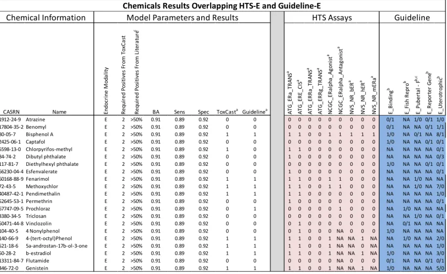
In this study we used data from the ToxCast Phase I chemical library, containing data for 309 unique chemical structures (U.S. EPA 2012e). Most of these chemicals are either current or former active ingredients in food-use pesticides that were designed to be bioactive, or they are industrial chemicals that are environmentally relevant. Details of the chemical library were reported by Judson et al. ( 2009). Data on an additional 23 reference chemicals were included that were tested in a separate study (Judson et al. 2010), 17 of which were not in the ToxCast Phase I library. CAS registry numbers (CASRN) for the ToxCast Phase 1 chemicals and the additional 17 chemicals are available online in Supplemental_File_1.csv (Rotroff et al. 2013).
Guideline and Non-Guideline Endocrine Assays
Data from guideline endocrine-related in vitro and in vivo studies were extracted from EDSP Tier 1 validation reports from the U.S. EPA EDSP web site (U.S. EPA 2012a). Non-guideline studies were obtained from open literature by querying PubMed
a bioassay, studies measuring bioaccumulation). Studies that identified their methods as following the Organisation for Economic Co-operation and Development (OECD) guidelines (Kanno et al. 2001, 2003; OECD 1999, 2001, 2003, 2007) or EDSP protocols were grouped together with EDSP T1S data for the guideline analysis. When available, PubMed identifiers (PMID) were used as unique annotations for each report. For the few instances when no PMID was available or for each EDSP T1S validation report, a unique identifying number was gen-erated. The citation information for all documents used in the analysis is available online in Supplemental_File_2.txt (Rotroff et al. 2013).
Guideline endocrine-related assays gathered from EDSP validation reports and OECD guideline studies were categorized according to whether they tested estrogen-, androgen-, steroidogenesis-, or thyroid-related MOAs (guideline-E, guideline-A, guideline-S, guideline-T, respectively). Additional information captured included study type (e.g., amphibian
metamorphosis, reporter gene), assay type (e.g., serum levels, organ weight), species, strain, cell type, target, and whether or not it was an EDSP/OECD guideline study. Chemical potency [e.g., concentration at half-maximum activity (AC50), lowest effective concentration] for a given end point was captured as it was represented in the study report along with the maximum
active and inactive, respectively. We combined all guideline and non-guideline literature studies so as to have a single hit value for each study–chemical–MOA combination. Data that were con-flicting or otherwise unclear were included in the data table but annotated as such, and removed from analyses. The data obtained from guideline endocrine-related studies and other non-guideline literature reports are available online Supplemental_File_3.csv (Rotroff et al. 2013).
ToxCast In Vitro Assays
HTS competitive binding, enzyme inhibition, and reporter gene assays representing estrogen-, androgen-, steroidogenesis-, or thyroid-related end points (HTS-E, HTS-A, HTS-S, HTS-T, respectively) were selected as a subset of the > 500 HTS assays generated by the
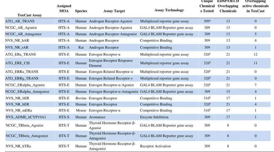
ToxCast program (ToxCastDB v.17; U.S. EPA, 2012e) [see Supplemental_File_1.csv (Rotroff et al. 2013)]. The details and a description of each assay are reported in Table 2.1.
For chemicals that produced a statistically significant and concentration-dependent response in a given assay, the AC50 was recorded. The criteria for determining the activity of a compound are assay platform. The data were then dichotomized so that if an AC50 was present for a given chemical end point concentration, a 1 was reported; if no response was observed, a 0 was reported. Chemicals tested in triplicate for quality control purposes were designated 1 or 0 on a majority basis. Chemicals that were run in duplicate with at least one sample producing an AC50 were designated as a 1.
Model Development
assays, we used disjunctive logic employing varied weight-of-evidence thresholds to determine optimal predictive performance. This model tested variable thresholds for the HTS ToxCast assay results represented as unweighted binary data, while the guideline or non-guideline endocrine-related assay results remained static. Initially, the model began with a threshold criterion of one positive ToxCast HTS assay out of the total number of ToxCast HTS assays for a chemical to be considered to perturb a given MOA. Once calculated, the model was then re-run with increasing increments of one assay until all ToxCast HTS assays for a given endocrine MOA were required to be positive for a chemical to be considered to perturb the given MOA. As the threshold for a positive call was increased, a larger weight of evidence was required for a chemical to be considered a “hit” for perturbing the given endocrine MOA. An exception was made for guideline pubertal studies and the ToxCast NVS_NR_hAR assay. Guideline pubertal studies test for effects that can arise through multiple different endocrine-related pathways. For this reason, if a chemical was considered positive in the pubertal assay and the result conflicted with other guideline studies (e.g., receptor binding, reporter gene), the pubertal assay was not included in the weight of evidence. The ToxCast NVS_NR_hAR assay is a human androgen receptor binding assay in the LNCaP prostatic cell line. The androgen receptor in this cell line is known to bind to steroid hormones other than androgens (Veldscholte et al. 1992). For this reason, if a compound was negative in all other HTS-Aassays, the result for the NVS_NR_hAR assay was not included in the weight-of-evidence.