MOLECULAR APPROACHES FOR CONTROLLING RNA STABILITY
Erin Katherine Borchardt
A dissertation submitted to the faculty at the University of North Carolina at Chapel Hill in partial fulfillment of the requirements for the degree of Doctor of Philosophy in the
Curriculum in Genetics and Molecular Biology in the School of Medicine.
Chapel Hill 2016
© 2016
ABSTRACT
Erin Katherine Borchardt: Molecular Approaches for Controlling RNA Stability (Under the direction of Aravind Asokan)
Nature utilizes a number of methods for regulating gene expression via
ACKNOWLEDGEMENTS
I am profoundly grateful to have been able to work in the supportive environment of the Asokan laboratory for my graduate studies. As my advisor, Dr. Aravind Asokan allowed me the intellectual freedom to explore new research areas for the lab while guiding my development as a scientist. Aravind has been consistently approachable, encouraging, enthusiastic, and honest in his mentoring. I have learned so much from him, within science and beyond, and I feel lucky to be able to count him among my friends.
Lavanya Rao, Leonidas Vandoros, Michael Huang, Ryan Fogg, Joey Heider, Rebecca Reardon, and Rita Meganck. I owe all members of the Asokan laboratory thanks not only for their scientific support but also for making the lab an open and friendly environment. I will always treasure our times, inside the lab and out, teeming with laughter and full conversation.
I am also fortunate to have had the support and guidance of a wonderful thesis committee in Drs. William F. Marzluff, Alain Laederach, Dale Ramsden, and Brian Kuhlman. Dr. Marzluff and Dr. Laederach introduced me to the incredibly talented and diverse RNA community present in the Triangle area. This RNA community has been welcoming and I am privileged to have had the opportunity to learn from them.
I became inspired to pursue a career in molecular biology through working in the laboratory of Dr. Lisa Stubbs and through the iGEM competition during my
undergraduate years at the University of Illinois at Urbana-Champaign. It was in these environments that I first learned basic laboratory techniques and scientific approaches. For this, I owe many thanks to Dr. Lisa Stubbs, Dr. Christopher Rao, Dr. Yong-Su Jin, Younguk (Calvin) Sun, Dina Leiding, and Andrea Skinner.
Lastly, I am grateful for the support of my family and friends outside of North Carolina. This includes my parents, Kathy and Mike Borchardt, my brothers, Jason, Brett, and Elliott Borchardt, my grandparents Lois Borchardt and Ted Rogachuk, and my closest, lifelong friend, Selynn Hinkle.
TABLE OF CONTENTS
LIST OF FIGURES ...ix
LIST OF ABBREVIATIONS ...xi
CHAPTER 1: INTRODUCTION ... 1
1.1 CRISPR Systems ... 1
1.2 Pre-crRNA Processing by Csy4 ... 5
1.3 CRISPR Components as Tools ... 7
1.4 Sequence Elements Involved in RNA Stability ... 10
1.5 Circular RNAs ... 11
1.6 Adeno-Associated Virus-Mediated Gene Delivery and RNA Therapeutics ... 15
CHAPTER 2: CONTROLLING MRNA STABILITY AND TRANSLATION WITH THE CRISPR ENDORIBONUCLEASE CYS4 ... 20
2.1 Overview ... 20
2.2 Introduction ... 21
2.3 Materials and Methods ... 24
2.4 Results and Discussion ... 29
CHAPTER 3: INDUCING CIRCULAR RNA FORMATION USING THE CRISPR ENDORIBONUCLEASE CSY4 ... 47
3.2 Introduction ... 48
3.3 Materials and Methods ... 50
3.4 Results and Discussion ... 54
CHAPTER 4: IN VIVO DELIVERY OF TRANSLATABLE CIRCULAR RNA CASSETTES USING RECOMBINANT AAV VECTORS ... 66
4.1 Overview ... 66
4.2 Introduction ... 67
4.3 Methods ... 70
4.4 Results and Discussion ... 74
CHAPTER 5: SYNOPSIS AND FUTURE DIRECTIONS ... 85
5.1 Summary ... 85
5.2 Further Evaluation and Regulation of Csy4 in Mammalian Cells ... 86
5.3 Future Applications and Engineering of Csy4 ... 88
5.4 Improving Current CircRNA Expression Cassettes ... 90
5.5 CircRNAs Containing an EMCV IRES are Efficiently Translated in Mice ... 94
5.6 Future Evaluation of rAAV CircRNA Expression Cassettes ... 95
5.7 Final Remarks ... 95
LIST OF FIGURES
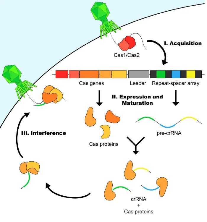
Figure 1: General schematic of CRISPR mediated immunity ... 17
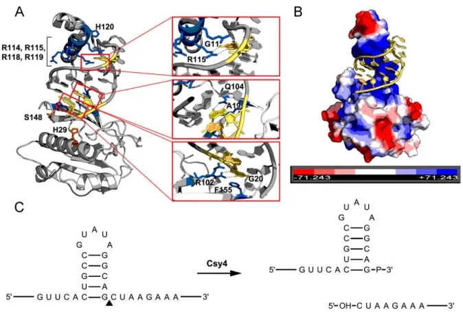
Figure 2: Characteristics the Csy4-hairpin interaction. ... 18
Figure 3: Genomic map of AAV ... 19
Figure 4: Csy4-mediated knockdown of 5' UTR-hairpin (HP) and ATG-HP constructs ... 39
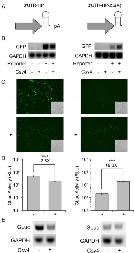
Figure 5: Effect of placing the HP in the 3’UTR on Csy4-mediated knockdown ... 41
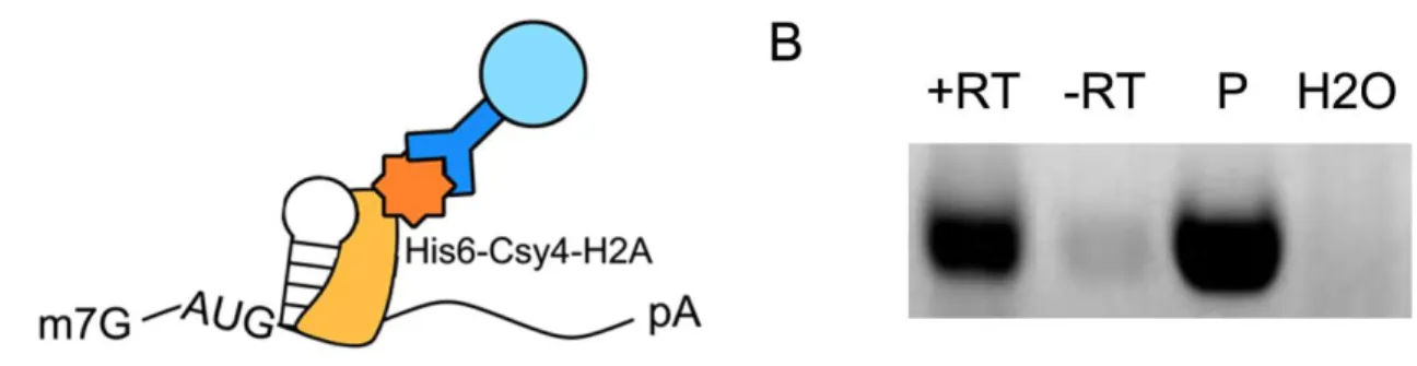
Figure 6: Cys4 interacts with the substrate hairpin in human cells. ... 43
Figure 7: Csy4 binding and cleavage are necessary for regulation of transgene expression. ... 44
Figure 8: Selective processing by Csy4 is essential for rescue of 3'UTR-HP-Δp(A) constructs. ... 45
Figure 9: Potential events outlining Csy4-mediated processing of 3'UTR-HP-Δp(A) mRNA. ... 46
Figure 10: Schematic of circGFP-CD splicing patterns and reporter outputs. ... 60
Figure 11: Csy4 induces circGFP-CD reporter expression to switch from dsRed to GFP. ... 61
Figure 12: Detection of circGFP-CD expression products. ... 63
Figure 13: Accumulation of circRNA and linear splicing products over time. ... 65
Figure 15: rAAV mediated delivery of circGFP expression cassette in mice. ... 82
Figure 16: Molecular characterization of circGFP expression products in mice. ... 83
Figure 17: In vitro expression of circRNA reporters from viral vectors. ... 97
LIST OF ABBREVIATIONS
AAT Alpha-1-antitrypsin AAV Adeno-associated virus Cas CRISPR associated CBA Chicken beta actin cDNA complementary DNA
cGFP Coral green fluorescent protein CIP Calf intestinal phosphatase circGFP-CD circGFP-Csy4 dependent circRNA Circular RNA
CRISPR Clustered regularly interspaced short palindromic repeats crRNA CRISPR RNA
DAB 3,3’-Diaminobenzidine DNA Deoxyribonucleic acid
EDTA Ethylenediaminetretraacetic acid EIciRNA Exon-intron circular RNA
EMCV encephalomyocarditis virus
ENE Expression and nuclear retention element ESF Engineered splicing factor
FGF-2 Fibroblast Growth Factor-2 GFP Green fluorescent protein GLuc Gaussia luciferase
HEK293 Human embryonic kidney 293
HIPK3 Homeodomain interacting protein kinase 3 HIV Human immunodeficiency virus
HSV-TK Herpes simplex virus thymidine kinase
IGF2BP1 Insulin-like growth factor 2 mRNA binding protein 1 IRES Internal ribosome entry site
ITR Inverted terminal repeat Kd Binding affinity
KSHV Kaposi’s sarcoma associated herpesvirus LTR Long terminal repeat
MALAT1 Metastasis associated lung adenocarcinoma transcript 1 MascRNA MALAT1-associated small cytoplasmic RNA
MBL Muscleblind
MEN Multiple endocrine neoplasia
miRNA MicroRNA
PEI Polyethylenimine
Pre-crRNA Precursor CRISPR RNA PUF Pumilio/fem-2 binding factor
qPCR quantitative polymerase chain reaction
qRT-PCR quantitative reverse transcription polymerase chain reaction rAAV Recombinant Adeno-Associated Virus
RBS Ribosome binding site RNA Ribonucleic acid
RT PCR Reverse transcription polymerase chain reaction shRNA Short hairpin RNA
TBST Tris buffered saline with Tween 20 tracrRNA trans-activating CRISPR RNA tricRNA tRNA intronic circular RNA tRNA Transfer RNA
UTR Untranslated region VPR Viral protein R
CHAPTER 1: INTRODUCTION
1.1 CRISPR Systems
Prokaryotic organisms regularly encounter foreign genetic elements which pose a threat to their survival. As such, ~45% of bacteria and ~83% of archaea utilize the Clustered Regularly Interspaced Short Palindromic Repeats (CRISPR) system as a mechanism of defense against these invading nucleic acids (Grissa et al., 2007).
CRISPR systems neutralize foreign nucleic acid using a combination of proteins and RNA elements encoded within CRISPR loci. These loci are typically characterized by the presence of a repeat-spacer-array, a leader sequence, and a series of CRISPR-associated (Cas) protein encoding genes. Within the repeat-spacer array, short
CRISPR systems have been divided into classes, types, and subtypes, discussed below.
CRISPR Classification:
CRISPR systems are functionally categorized into two classes based on their complement of Cas proteins (Makarova et al., 2015). CRISPR classes are divided into five types denoted by roman numerals, which are further separated into 16 subtypes denoted by alphabetical letters (Makarova et al., 2015). All functional CRISPR systems require the presence of both Cas1 and Cas2, which are essential for the adaptation stage of CRISPR immunity. Accessory Cas proteins provide the basis for classification and specify the mechanistic differences of immunity between each CRISPR subtype. Class 1 CRISPR systems are characterized by a multi-subunit protein complex of Cas proteins which is utilized to carry out target neutralization. Conversely, Class 2 CRISPR systems utilize a single Cas protein to mediate CRISPR immunity.
CRISPR Adaptation:
Initial studies exploring CRISPR immunity recognized that spacer sequences were identical to sequences present in phage and plasmids (Bolotin et al., 2005; Mojica et al., 2005; Pourcel et al., 2005). In addition, studies demonstrated that phage-resistant
Streptococcus thermophilus could be generated by challenging the bacteria with
these spacer sequences were subsequently deleted from the bacteria, their acquired resistance was lost (Barrangou et al., 2007b). Together, these data determined a relationship between the spacer sequence and CRISPR-mediated immunity. In particular, this study established the fundamental adaptability of the CRISPR system, demonstrating that new, immunizing spacers can be acquired upon exposure to a pathogen.
The acquisition (or adaptation) phase of CRISPR immunity requires Cas1 and Cas2 to incorporate a small segment of foreign nucleic acid sequence as a new spacer in the repeat-spacer array (Nunez et al., 2015; Yosef et al., 2012) (Figure 1). Integration of a new spacer into the repeat-spacer array always occurs at the leader proximal end of the array (Barrangou et al., 2007b). The original sequence of the spacer, found in the foreign nucleic acid, is known as the protospacer. A protospacer adjacent motif (PAM) immediately flanking the protospacer is utilized by the adaptation machinery for
recognizing new potential spacer sequences (Heler et al., 2015).
Interestingly, CRISPR systems also appear to be primed for acquisition of new spacers against previously encountered targets. Such primed acquisition occurs when a particular target shares partial homology with a spacer that is already present in the repeat-spacer array (Datsenko et al., 2012; Swarts et al., 2012). This may represent a mechanism for combatting bacteriophage that escape from CRISPR targeting by mutating their protospacers (Datsenko et al., 2012).
CRISPR Expression and Maturation:
array is transcribed into a single, long precursor transcript (pre-crRNA) (Figure 1). Transcription of the pre-crRNA is driven by a promoter in the leader sequence (Pul et al., 2010). The pre-crRNA is subsequently processed by Cas proteins into small CRISPR RNAs (crRNAs) such that each crRNA contains a single spacer and a partial repeat handle. The pre-crRNA processing step of the CRISPR pathway varies between CRISPR types and subtypes. For example, in the Class 1 Type-IF CRISPR system of
Pseudomonas aeruginosa, the task of crRNA biogenesis falls to Csy4 (Cas6f) (Cady
and O'Toole, 2011; Haurwitz et al., 2010). However, in Class 2 type II CRISPR systems, the combined efforts of Cas9, cellular Ribonuclease III, and an additional small RNA (trans-activating CRISPR RNA, or tracrRNA) are responsible for pre-crRNA processing (Karvelis et al., 2013).
CRISPR Interference:
During the interference phase, Cas nucleases are guided to their targets by crRNAs in a sequence specific manner (Figure 1). An important checkpoint in
distinguishing target from self occurs in most CRISPR systems during the interference stage. In this step, the interference complex interrogates the sequence adjacent to the protospacer for a PAM. If the PAM is present, the Cas nuclease is licensed to cleave the target, rendering the invading nucleic acid inactive (Sternberg et al., 2014;
Szczelkun et al., 2014). Verification of the PAM sequence prevents targeting of the genomic CRISPR locus, which contains crRNA binding sites but lacks a PAM.
between CRISPR subtypes, both in sequence and location relative to the protospacer (5' or 3') (Shah et al., 2013). Further, in Class 1 systems, the interference complex is formed by crRNA interaction with a multi-subunit Cas protein complex. Target cleavage is carried out by Cas3 in Class 1 Type I systems while Cas10 carries out this task in Class 1 Type III systems (Brouns et al., 2008; Hatoum-Aslan et al., 2014; Samai et al., 2015; Sinkunas et al., 2011; Westra et al., 2012). In comparison, Class 2 systems utilize a single Cas protein bound to crRNA to form the effector complex (Makarova et al., 2015). In Class 2 Type II systems, this task falls to Cas9. (Barrangou et al., 2007b; Garneau et al., 2010b; Jinek et al., 2012; Sapranauskas et al., 2011).
1.2 Pre-crRNA Processing by Csy4
Csy4 (Cas6f) is a CRISPR endoribonuclease member of Class 1 Type IF CRISPR systems. Functioning in the expression stage of CRISPR immunity, Csy4 cleaves the pre-crRNA at regular intervals to generate small crRNAs (Cady and O'Toole, 2011; Haurwitz et al., 2010). In the P. aeruginosa Type I-F CRISPR system, each repeat in the repeat-spacer array consists of 28 nucleotides. Sixteen of these nucleotides form a short hairpin which serves as the substrate for Csy4 binding and cleavage. Csy4-mediated cleavage occurs at the 3' base of each hairpin stem,
interference stage of the CRISPR pathway (Sternberg et al., 2012; Wiedenheft et al., 2011).
Csy4 and the target hairpin form a high affinity interaction (Kd ≈ 50 pM) which is dependent on both sequence- and structure-specific contacts (Haurwitz et al., 2010; Sternberg et al., 2012). A positively charged, arginine-rich helix docks into the major groove of the hairpin stem (Haurwitz et al., 2010) (Figure 2A, B). Within the arginine-rich helix, Arg114, Arg115, Arg118, Arg119 and His120 make contact with phosphate
groups in the RNA hairpin (Haurwitz et al., 2010) (Figure 2A). When a subset of these arginine residues are mutated, the capacity of Csy4 to bind the substrate hairpin is greatly reduced (Sternberg et al., 2012). Nucleotide-specific contacts are specified by hydrogen bonding between Arg102 and G20, Gln104 and A19, and Arg115 and G11 (Haurwitz et al., 2010) (Figure 2A). Lastly, Phe155 interacts with, and positions G20 in the enzyme active site via base-stacking interactions (Haurwitz et al., 2010; Sternberg et al., 2012) (Figure 2A).
The Csy4 active site relies on two critical amino acids for catalysis, Ser148 and His29 (Figure 2A). Mutation of either of these residues to cysteine and alanine
respectively renders Csy4 inactive (Haurwitz et al., 2010). Subsequent studies
1.3 CRISPR Components as Tools
CRISPR systems have provided the basis for a breadth of new technologies ranging from bacterial genotyping to genome engineering (Selle and Barrangou, 2015; Wright et al., 2016). Much of the original focus on CRISPR was aimed at developing tools for application in the dairy industry, which relies on valuable bacterial starter cultures. Initial studies sought new techniques for vaccination of bacteria against bacteriophages and drove many seminal CRISPR findings (Barrangou et al., 2007b; Deveau et al., 2008; Horvath et al., 2008). As such, a number of CRISPR tools support the study and maintenance of bacteria commonly used in the food industry. For
example, the consistent integration of new spacers at the leader proximal side of the repeat-spacer array records a sequential history of exposure to foreign nucleic acid (Barrangou et al., 2007b). This trait has been exploited for the genotyping of various bacteria (Grissa et al., 2009; Vergnaud et al., 2007).
Perhaps the most widely appreciated CRISPR-based tool is Cas9, an RNA guided nuclease present in all Class 2 type-II CRISPR systems (Makarova et al., 2015). During the interference phase, Cas9 is guided to foreign DNA targets through the
specificity of a crRNA. Cas9’s interaction with the crRNA is bridged by an additional small RNA called the trans-activating crRNA (tracrRNA) (Deltcheva et al., 2011). The Cas9-crRNA-tracrRNA complex is guided to foreign DNA through the sequence
region of interest. Consequently, Cas9 can be guided to cleave any sequence, provided the appropriate PAM is present.
Cas9 is an attractive tool for genome engineering, and a myriad of other
applications, due to the ease of which it can be programmed. Initial applications of Cas9 focused on genome editing but have since expanded to include functions such as RNA-guided RNA imaging and transcriptional control (Gilbert et al., 2013; Mali et al., 2013a; Nelles et al., 2016). Furthermore, Cas9 seems to function in nearly every organism tested, ranging from bacteria to human cell lines (Cong et al., 2013; DiCarlo et al., 2013; Friedland et al., 2013; Gratz et al., 2013; Hwang et al., 2013; Jiang et al., 2013; Jinek et al., 2013; Li et al., 2013; Mali et al., 2013b; Nekrasov et al., 2013; Wang et al., 2013).
interference from sequences that may affect gRNA, function, stability, or localization. Further, multiple gRNA can be expressed from a single promoter if Csy4 hairpins are placed between each gRNA, allowing multiplexed Cas9 applications (Nissim et al., 2014; Tsai et al., 2014).
Csy4 has also formed the basis of a number of other tools and techniques. Inactivation of Csy4 catalytic activity through a H29A mutation has allowed for Csy4-mediated purification of hairpin-tagged RNA transcripts (Lee et al., 2013; Salvail-Lacoste et al., 2013). The purified transcripts can subsequently be analyzed for identification of proteins associated with a transcript of interest (Lee et al., 2013). Additionally, Csy4 has been adapted for gene regulation and processing RNA transcripts encoding multiple genes in Escherichia coli, Bacillus subtilis, and
Saccharomyces cerevisiae (Qi et al., 2012). Csy4 was also utilized in regulating
translation of prokaryotic gene expression (Du et al., 2016a). In this context, Csy4 hairpins separated a ribosome binding site (RBS) and open reading frame (ORF) from an upstream cis-repressive RNA. The cis-repressive RNA functions by hybridizing with, and masking the RBS, preventing ribosomal loading and translation. Upon
co-expression with Csy4, the cis-repressive RNA is cleaved away, exposing the RBS, and allowing translation (Du et al., 2016a). Csy4 has also been used to regulate viral
Two new applications of Csy4 are delineated in this dissertation. Chapter 2 details the development of Csy4 as a tool for regulating the stability and translation of transgene-derived RNA. Chapter 3 describes the utilization of Csy4 for inducing engineered circular RNA formation.
1.4 Sequence Elements Involved in RNA Stability
RNA stability is tightly controlled in cells using a wide range of methods. Of note, are 3' end stabilizing sequences, which protect the 3' ends of RNAs from exonucleolytic degradation. The majority of mammalian cellular mRNAs are terminated at the 3' end with a poly(A) tail. The poly(A) tail ensures mRNA stability through recruitment of the poly(A) binding protein Pab1p (Coller et al., 1998). Interestingly, not all eukaryotic mRNAs possess a poly(A) tail, and these RNAs must ensure stability by distinct
mechanisms. For example, replication-dependent histone mRNAs possess a 3' terminal stem loop structure in place of a poly(A) tail. The persistence of these mRNAs is
influenced by a set of proteins interacting with the stem loop and stem loop binding protein (SLBP) to promote either maturation or degradation in a cell cycle dependent manner (Marzluff et al., 2008).
In addition, triple helical elements have demonstrated the capacity to mediate 3' RNA end stabilization. Triple helix elements have been identified thus far in the 3' ends of non-coding and viral RNAs. The MALAT1 (metastasis-associated lung
cleavage event to remove a tRNA-like structure called the mascRNA
(MALAT1-associated small cytoplasmic RNA). The resulting 3' end of the MALAT1 transcript folds into a triple helical structure which is resistant to exonucleolytic degradation (Brown et al., 2012; Wilusz et al., 2012). MALAT1 triple helix-mediated stability can be conferred to an RNA of interest by simply placing the 3' end of the MALAT1 transcript at the 3' end of the RNA. Surprisingly, when placed downstream of an ORF lacking a
poly-adenylation signal, the MALAT1 3' end is also capable of supporting protein translation (Wilusz et al., 2012).
Viruses, such as Kaposi’s sarcoma-associated herpesvirus (KSHV) also make use of triple helix elements. In the KSHV polyadenylated nuclear (PAN) RNA, a triple helix is formed within an expression and nuclear retention element (ENE). The ENE contains a U-rich loop which sequesters and protects the PAN RNA poly(A) tail (Conrad et al., 2006; Mitton-Fry et al., 2010). The resulting triple helical structure stabilizes the PAN RNA and protects it from exonucleolytic degradation (Conrad et al., 2006).
Structure-based bioinformatics analysis has since identified similar ENEs in a range of viral genomes (Tycowski et al., 2012). The occurrence of ENEs in a diverse set of viral genomes highlights the utility and value of this stabilizing structure.
1.5 Circular RNAs
Circular RNAs (circRNAs) are highly stable, covalently closed RNAs. Typically, circRNAs are generated through backsplicing, a process in which a downstream
(Zaphiropoulos, 1996). Another class of circRNAs, tRNA intronic circular RNAs (tricRNAs), are generated using tRNA maturation machinery (Lu et al., 2015).
Components of the canonical splicing machinery and splicing signals have been identified as factors involved in circRNA formation (Starke et al., 2015). However, circRNA biogenesis can be facilitated by a number of both trans- and cis-acting factors. For instance, circularization of a subset of circRNAs is regulated by the alternative splicing factor, Quaking, during the human epithelial to mesenchymal transition (Conn et al., 2015). Similarly, the muscleblind (MBL) protein is involved in circularization of the MBL circRNA. This observation might suggest an autoregulatory role for MBL in which excess of MBL favors the circularization of its flanked exon. This would in turn reduce the amount of linear, MBL encoding transcript (Ashwal-Fluss et al., 2014). Furthermore, circMBL itself contains MBL binding sites and is capable of binding MBL protein,
contributing to the potential autoregulatory feedback loop (Ashwal-Fluss et al., 2014). Cis-acting factors are also involved in circRNA biogenesis. Of recent interest are inverted repeat sequences (e.g. Alu repeats in humans) which have been found flanking exons known to circularize. These inverted repeat sequences are essential for the circularization of the intervening exon(s) (Ashwal-Fluss et al., 2014; Ivanov et al., 2015; Jeck et al., 2013; Kramer et al., 2015; Liang and Wilusz, 2014; Zhang et al., 2014). Circularization mediated by inverted repeats is thought to occur through a mechanism in which base pairing brings splice sites in close proximity to facilitate backsplicing.
Regardless of their mechanism of biogenesis, circRNA’s are markedly more stable than their linear counterparts. The lack of exposed 3' and 5' ends likely renders circRNAs resistant to a number of cellular exonucleases. Consequently, this inherent stability leads to sustained persistence of circRNAs (Jeck et al., 2013; Lasda and Parker, 2014; Liang and Wilusz, 2014). This is exemplified by the median half-life of circRNAs, which is at least 2.5X longer than that of their linear counterparts (Enuka et al., 2015).
Deep sequencing studies have revealed differences in the abundance of various circRNAs, which are dictated by tissue, cell type, and developmental stage (Memczak et al., 2013). CircRNAs are known to be highly abundant in the mammalian brain
compared to other tissues. In particular, they appear to be enriched at the synapses and synthesized during neuronal differentiation and development (Rybak-Wolf et al., 2015; You et al., 2015). Synaptic localization of certain circRNAs also suggests that they may be specifically targeted to different sites in the cell, as their linear counterparts are localized in the cytoplasm (Rybak-Wolf et al., 2015). This suggests possible specific roles for circRNAs at the synapse. Though they are often most highly expressed in the brain, circRNAs are also expressed in non-neuronal tissues. For example, the
ZKSCAN1 circRNA is also detected in the liver while the HIPK3 circRNA is detected in the kidney, heart, lung, thyroid, and uterus. (Liang and Wilusz, 2014).
Despite their abundance, the functions for most circRNAs remains to be
determined (Ebbesen et al., 2015). The roles of a small number of circRNAs have been proposed, but it is likely that there are a wide variety of possible functions. One
splicing regulation. This regulation would be carried out by affecting exon skipping, due to the mutually exclusive relationship between circular and linear splicing of an exon (Ashwal-Fluss et al., 2014). Alternatively, some circRNAs such as circHIPK3, ciRS-7 (Cdr1as) and the Sry circRNA act as microRNA (miRNA) sponges (Hansen et al., 2013; Memczak et al., 2013; Zheng et al., 2016). However, most circRNAs are not enriched for miRNA binding sites and this is unlikely to be a general function of circRNAs (Guo et al., 2014). As described above, circMbl has been proposed as a sponge for MBL
protein, and some circRNAs containing introns (EIciRNAs) have been implicated in transcription regulation (Ashwal-Fluss et al., 2014; Li et al., 2015). Additionally,
circFoxo3 appears to bind the cell cycle regulators CDK2 (cyclin dependent kinase 2) and p21 to repress cell cycle progression (Du et al., 2016b). CircRNAs have also been proposed as templates for retrotranscription and reinsertion into the genome to
generate pseudogenes (Dong et al., 2016).
Although most circRNAs arise from protein-coding exons, endogenously
1.6 Adeno-Associated Virus-Mediated Gene Delivery and RNA Therapeutics
Adeno-Associated Virus (AAV) is a helper-dependent parvovirus with a 4.7 kb single stranded DNA genome. AAV utilizes three promoters to express a series of proteins though overlapping reading frames in Rep and Cap genes. The genome is flanked on either end by inverted terminal repeat (ITR) sequences which are essential for genome packaging into the viral capsid (Figure 3A). The ITRs are the only cis-elements required for genome packaging. As such, AAV can be converted into a recombinant vector for gene therapy by replacing the intervening sequence with an expression cassette of interest (Figure 3B).
As a gene therapy vector, recombinant AAV (rAAV) is particularly valuable due to long-term transgene expression, lack of pathogenicity, and inability to replicate
independently (Samulski and Muzyczka, 2014). Further, the tropism of a number of serotypes is well defined and can be engineered through capsid manipulations (Asokan et al., 2012; Pulicherla et al., 2011a; Shen et al., 2013b). rAAV can be generated and purified through a triple plasmid transfection protocol in HEK293 cells, and can package any ITR-flanked cassette less than 4.7 kb in size (Xiao et al., 1998). These features make rAAV an ideal vector for delivery of therapeutic genes in mice and potentially higher order mammals. In particular, the well-studied tropism of various AAV serotypes allows for direct targeting of specific tissues and cell types. This prevents exposing the entire organism to the therapeutic in situations where it might not be necessary or may be detrimental.
treatments for factor IX deficiency (hemophilia B), lysosomal storage diseases, and alpha-1-antitrypsin (AAT) deficiency, among others (Brimble et al., 2016; Hinderer et al., 2015; Samulski and Muzyczka, 2014; Wozniak et al., 2015). rAAV-mediated delivery of Cas9 and gRNA has shown promise for treatment of Duchenne’s muscular dystrophy (Long et al., 2016; Nelson et al., 2016; Tabebordbar et al., 2016). Additionally, rAAV-based treatment developed for Leber’s congenital amaurosis, a form of blindness, has been successful (Cideciyan et al., 2008; Maguire et al., 2008). An rAAV-delivered treatment for lipoprotein lipase deficiency (Glybera) has been approved in Europe, and represents the first approved rAAV therapeutic in the West (Bryant et al., 2013; Gaudet et al., 2010).
rAAV vectors have been successfully used for the expression of RNA
Figure 1: General schematic of CRISPR mediated immunity. Upon encountering foreign nucleic acid, such as from a phage genome, Cas1 and Cas2 mediate
CHAPTER 2: CONTROLLING MRNA STABILITY AND TRANSLATION WITH THE
CRISPR ENDORIBONUCLEASE CYS41
2.1 Overview
The bacterial CRISPR endoribonuclease, Csy4 has recently been described as a potential RNA processing tool. Csy4 recognizes substrate RNA through a specific 28 nucleotide hairpin sequence and cleaves at the 3' end of the stem. To further explore applicability in mammalian cells, we introduced this hairpin at various locations in mRNAs derived from reporter transgenes and systematically evaluated the effects of Csy4-mediated processing on transgene expression. Placing the hairpin in the 5' untranslated region (UTR) or immediately after the start codon resulted in efficient degradation of target mRNA by Csy4 and knockdown of transgene expression by 20 to 40-fold. When the hairpin was incorporated in the 3' UTR prior to the poly(A) signal, the mRNA was cleaved, but only a modest decrease in transgene expression (~2.5 fold) was observed. In the absence of a poly(A) tail, Csy4 rescued the target mRNA substrate from degradation, resulting in protein expression, which suggests that the cleaved mRNA was successfully translated. In contrast, neither catalytically-inactive (H29A) nor binding-deficient (R115A/R119A) Csy4 mutants were able to exert any of the above-described effects. Generation of a similar 3' end by RNase P-mediated cleavage was unable to rescue transgene expression independent of Csy4. These
results support the idea that the selective generation of the Csy4/hairpin complex resulting from cleavage of target mRNA might serve as a functional poly(A)/Poly-A Binding protein (PABP) surrogate, stabilizing the mRNA and supporting translation. Although the exact mechanism(s) remain to be determined, our studies expand the potential utility of CRISPR nucleases as tools for controlling mRNA stability and translation
2.2 Introduction
Endogenous control of gene expression is achieved by regulating transcription, processing, translation and/or degradation of mRNA through a myriad of genetic elements. Artificial control of gene expression, on the other hand, requires the development of small molecule, protein or RNA-based tools and is essential for
advancing synthetic biology and gene therapies. Post-transcriptional regulation of gene expression has been achieved by engineering RNA, for instance by employing
riboswitches that can not only be exploited to gain insight into endogenous RNA
approach, controlling RNA stability and translation is a key aspect underlying these gene regulatory strategies.
Structural studies have provided further mechanistic insight into pre-crRNA processing enzymes (Hochstrasser and Doudna, 2014). Despite their functional similarity, these enzymes display minimal primary sequence homology. Likewise, the sequences which they process also differ both in sequence and structure, with Cas5d, Cas6e, and Csy4 associated repeat elements containing hairpin structures and Cas6 targeting a predicted unstructured sequence (Kunin et al., 2007). To carry out target neutralization, Csy4 remains bound to the processed crRNA and associates with
additional Cas proteins, Csy1, Csy2, and Csy3 for target recognition (Wiedenheft et al., 2011). This complex is guided to target DNA based on sequence complementarity provided by the crRNA. In Class 1 Type-I CRISPR systems, Cas3 is then recruited to cleave and degrade the target DNA, neutralizing the invader (Sinkunas et al., 2013; Westra et al., 2012).
been utilized in the isolation of RNA-interacting proteins and has the potential to help analyze the protein-associations of diverse transcripts (Lee et al., 2013). In the current study, we systematically evaluated the ability of Csy4 to exercise post-transcriptional control of transgene expression in mammalian cells. Specifically, we investigate the positional effects of Csy4 mediated-cleavage in the 5' untranslated region (UTR), coding sequence, and 3'UTR of transcripts. Surprisingly, we find that the Csy4 processing of the 3' ends of mRNA supports translation and stabilizes the mRNA in lieu of a poly(A) signal.
2.3 Materials and Methods
Plasmids. The Csy4 gene was amplified from P. aerugionsa strain UCBPP-PA14 genomic DNA and cloned under the control of the chicken beta-actin (CBA) promoter using the following primers: Csy4wt Forward 5'- ATC GTC TAG AAT GGA CCA CTA CCT CGA CAT TCG CTT GC-3' and Csy4wt Reverse 5'- CGA TGC GGC CGC TCA GAA CCA GGG AAC GAA ACC TCC TTT GC-3' (IDT DNA Technologies). P.
aeruginosa UCBPP-PA14 genomic DNA was kindly provided by Dr. Matthew Wolfgang
(University of North Carolina at Chapel Hill). Csy4-H29A was amplified from pHMGWA-Pa14Csy4H29A (Addgene plasmid #41092) which was provided as a gift from Dr. Jennifer Doudna (Haurwitz et al., 2010). Csy4-H29A was cloned under the control of the CBA reporter. Csy4 R115A/R119A was synthesized as a gBlock ® (IDT DNA
Technologies) and cloned under the control of the CBA promoter.
(HP) in different locations as follows. (i) Reporter cassettes containing the HP after the start codon (ATG-HP) were generated using overlap extension PCR with primers that generated two fragments containing a single, in-frame Csy4 HP repeat sequence (5'-AGTTCACTGCCGTATAGGCAGCTAAGAAAT-3') in the 3' or 5' end. The two fragments were then gel purified and combined in equimolar quantities (40ng each) in consecutive PCR reactions without primers (35 cycles) and with flanking primers (30 additional cycles) to obtain the ATG-HP construct. The latter PCR products were then cloned under the control of the CBA promoter. (ii) The 5'UTR-HP-GFP was constructed by overlap extension PCR in a similar fashion, while the 5'UTR-HP-GLuc was synthesized as a gBlock ® (IDT DNA Technologies) and cloned under the control of the CBA
promoter. (iii) 3'UTR-HP reporters and poly-A deleted 3'UTR-HP (3'UTR-HP-Δp(A)) reporters were synthesized from gBlocks® and cloned as described earlier. The cGFP-mMALAT1-3' reporter was generously provided by Dr. Jeremy Wilusz (University of Pennsylvania). A partial Csy4 hairpin (5'-GTT CAC TGC CGT ATA GGC AG-3') and mascRNA were synthesized as a gblock ® (IDT DNA Technologies) and cloned in place of the mMALAT1 3' UTR in the cGFP-mMALAT1-3' reporter to generate cGFP-HP-masc-Δp(A). Similarly, a second gblock ® (IDT DNA Technologies) was synthesized containing the partial Csy4 hairpin and mascRNA separated by 10 nucleotides (5'-CTA AAC GCG T-3') and cloned as described earlier, to generate cGFP-HP10-masc-Δp(A).
Cell culture. HEK293 cells were cultured in Dulbecco’s Modified Eagle’s Medium
Penicillin/Streptomycin and 2.5 μg/mL amphotericin B (Sigma-Aldrich) and maintained at 37˚C and 5% CO2.
Transfection and luciferase reporter assays. Equimolar amounts (totaling 500ng) of three plasmids (different HP GLuc reporters, Csy4 and a control plasmid containing the tdTomato reporter driven by the CBA promoter) were transfected into HEK293 cells using PEI Max seeded at a density of 5x10⁴ per well in a 24 well plate. Media (50 μl) was collected from each well at different time intervals and diluted (1:100) before assessing luciferase reporter activity. For measuring GLuc activity, native
coelenterazine (Nanolight) was dissolved in methanol to a concentration of 1 mg/mL and diluted (1:200) in 600 mM NaCl-Tris-EDTA buffer, following which, 50 μl of the substrate solution was added to 50 μl of collected media. Luminometric analysis was carried out using a Perkin Elmer Victor 3 ® plate reader.
Fluorescence microscopy. HEK293 cells were transfected with different HP GFP reporter cassettes as described earlier and the cells imaged at different time intervals post-transfection using an EVOS ® FL epifluorescence cell imaging system (AMC/Life Technologies) using the GFP light cube (excitation 470nm, emission 510nm).
earlier. Total RNA from each well was isolated at 48 hours post-transfection using the Total RNA Purification Kit (#17200, Norgen Biotek). The purified RNA was then treated with DNase using the Turbo DNA-free kit (Ambion/Life Technologies). Equal ng
amounts of the purified total RNA product were utilized as template for reverse
transcriptase PCR using the High Capacity RNA-to-cDNA kit (Applied Biosystems/Life Technologies). Products of this reaction were used as template for further PCR
amplification with gene specific primers and visualized on an agarose gel. Forward primer for amplifying ATG-HP-GFP cDNA: 5'-GCC ACC ATG AGT TCA CTG CCG-3'; Forward and reverse primers for amplifying all other GFP reporter cDNAs 5'-GAA ATG TGA GCA AGG GCG AGG AGC-3'; 5'-GCG GAC TTG AAG AAG TCG TGC TGC-3'; Forward and reverse primers for glyceraldehyde 3-phosphate dehydrogenase (GAPDH) cDNA: 5'-CCA CTC CTC CAC CTT TGA C-3'; 5'-ACC CTG TTG CTG TAG C-3'.
Detection of poly-adenylated mRNA. HEK293 cells seeded overnight in 6 well plates at a density of 3x10⁵ cells per plate were transfected with a total of 3 μg DNA as
utilized as template for further PCR amplification with gene specific primers and
visualized on an agarose gel. Forward and reverse primers for reporter cDNAs (GLuc) 5'-CAA CTT CGC GAC CAC GGA TCT CG-3'; 5'-CGG CAG CCA CTT CTT GAG CAG G-3'. Forward and reverse primers for GAPDH are listed above.
RNA immunoprecipitation. HEK293 cells seeded overnight in 15 cm plates at a density of 5x10⁶ cells per plate were transfected with 10 ug of His6-Csy4-H29A and 10 ug of ATG-HP-GFP plasmids. Cells were lysed 48 hours post-transfection and RNA immunoprecipitation (RNA-IP) was carried out using the Magna RIP RNA Binding Protein Immunoprecipitation Kit (Millipore) according to the manufacturer’s protocol
(-His6 antibody, ab18184, Abcam). Purified RNA was DNase treated using the Turbo DNA free kit (Ambion/Life Technologies) and cDNA was generated from treated RNA via the Peregrine method (Langevin et al., 2013). cDNA was used as template for PCR with GFP specific primers 5'-GCC ACC ATG AGT TCA CTG CCG-3'; 5'-GCG GAC TTG AAG AAG TCG TGC TGC-3' and PCR products were visualized on an agarose gel.
Northern Blot. HEK293 cells seeded overnight in 10 cm plates at a density of 2.2x10⁶ cells per plate were transfected with a total of 6 μg DNA as indicated. RNA was purified using Trizol reagent (Invitrogen/Life Technologies) and treated with DNase using the Turbo DNA-free kit (Ambion/Life Technologies). 1 microgram of RNA was separated on a 4% polyacrylamide/8M urea gel and transferred to Hybond N+ membrane (GE
via a Random Primed DNA Labeling Kit (Roche Diagnostics). Hybridization of radiolabeled probe to the membrane was carried out using Rapid-Hyb buffer (GE Healthcare).
Western Blot. HEK293 cells seeded overnight in 6 well plates at a density of 3x10⁵ cells per plate were transfected with a total of 2.5 μg DNA as indicated. Lysates were recovered 48 hours post-transfection using Passive Lysis Buffer (Promega). Lysates were diluted 1:50 and separated on a 10% Bis-Tris gel. Blots were probed with mouse monoclonal GFP antibody (1:000 dilution, Santa Cruz) or mouse monoclonal anti-Actin (1:2000 dilution, Abcam) as primary antibody. Stabilized peroxidase-conjugated goat anti-mouse antibody (1:5000 dilution, ThermoScientific/Life Technologies) was used as secondary antibody. Blots were developed using SuperSignal West Femto substrate (ThermoScientific/Life Technologies).
2.4 Results and Discussion
Csy4-mediated knockdown of 5' UTR-hairpin (HP) and ATG-HP constructs.
We first evaluated the effect of Csy4-mediated cleavage of the substrate HP incorporated within the 5' untranslated region (UTR) or HP inserted in-frame
Effect of placing the HP in the 3’UTR on Csy4-mediated knockdown.
In contrast to insertions near the 5'UTR or the start codon, placement of the Csy4 target HP following the stop codon and prior to the poly(A) signal (3'UTR-HP, Figure 5A, left column) resulted in only a modest decrease in mRNA levels (as indicated by
random primed RT-PCR) and GFP expression, when co-expressed with Csy4 (Figures 5B, 5C, left columns). Further, quantitation of GLuc reporter activity indicated only a ~2.5 fold reduction in transgene expression upon treatment with Csy4 (Figure 5D, left column). However, there was a substantial reduction in poly-adenylated reporter RNA levels as measured by oligo-dT20 primed RT-PCR, consistent with mRNA cleavage by Csy4 (Figure 5E, left column). These results are particularly intriguing, since removal of the poly(A) signal is expected to destabilize mRNA.
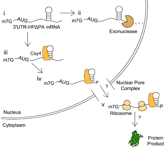
The latter observations might arise from the fact that following cleavage Csy4 remains bound to the cognate HP substrate (Haurwitz et al., 2010), which in turn could protect the 3' end of the transcript from degradation, despite removal of the poly(A) tail. Since translation is only slightly reduced, the Csy4/RNA complex must also be
transcribed cDNA using an oligo-dT20 primer. Regardless of the presence or absence of Csy4, 3'UTR-HP-Δp(A) reporter mRNA levels primed by oligo-dT20 appeared low
relative to 3'UTR-HP (Figure 5E, right column). This is consistent with the 3'UTR-HP-Δp(A) RNA lacking a poly(A) signal. Importantly, Csy4 co-expression does not alter the amount of poly-adenylated RNA detected. Furthermore, quantitation of GLuc activity revealed a 9-fold increase in signal with Csy4 co-expression relative to a control lacking Csy4 (Figure 5D). This corroborates the notion that Csy4 can potentially stabilize mRNA containing the HP substrate in the 3'UTR and support translation.
Csy4 binding and cleavage are necessary for regulation of transgene expression.
We next sought to determine whether Csy4 binding to the substrate HP alone was sufficient or if binding followed by enzymatic cleavage was essential for regulation of transgene expression. Specifically, we used two mutants of Csy4;
R115A/R119A, a binding deficient mutant that still retains catalytic activity, and Csy4-H29A, a catalytically inactive, but binding competent mutant (Haurwitz et al., 2010; Sternberg et al., 2012).
We first demonstrated Csy4 association with the target hairpin via RNA
immunoprecipitation (RNA-IP). Hemagglutainin (HA)-tagged Csy4-H29A was expressed with the ATG-HP-GFP reporter and Csy4-RNA complexes were isolated from cell
lysates using an -His6 antibody (Figure 6A). We were able to detect GFP RNA from
To evaluate the role of Csy4 binding and cleavage in regulating transgene expression, each reporter construct including the unmodified control, 5'UTR-HP, ATG-HP, 3'UTR-HP and the 3'UTR-HP-Δp(A) reporter (Figure 7A) was expressed in the presence or absence of native Csy4, Csy4-H29A or Csy4-R115A/R119A. Expression from unmodified GFP and GLuc constructs was unaffected by either mutant. The 5'UTR-HP GFP construct was also unaffected by co-expressing either the catalytically inactive or the binding deficient mutant (Figure 7B, 7D, 7E). Similar observations were made in case of the ATG-HP GFP reporter expressed with the catalytically inactive mutant, suggesting that steric hindrance from binding alone was insufficient to block translation initiation (Figure 7B, 7D). Further, the Csy4-R115A/R119A mutant also did not seem to substantially affect ATG-HP reporter expression (Figure 7B, 7E). Treatment with Csy4-H29A and Csy4-R115A/R119A did not dramatically affect expression of the corresponding GLuc reporters, supporting the previous data (Figure 7F). Taken
together, these data suggest that both binding and cleavage are essential for Csy4-mediated knockdown of transgene expression. In addition, neither mutant was able to rescue reporter signal in case of the 3'UTR-HP and the 3'UTR-HP-Δp(A) reporters (Figures 7B-F). These results support the notion that rescue of transgene expression from reporter constructs lacking a poly(A) tail requires both Csy4 binding to and
Selective processing by Csy4 is essential for rescue of 3'UTR-HP-Δp(A)
constructs.
To further confirm the role of Csy4-mediated recognition and cleavage in
rescuing 3'UTR-HP-Δp(A) reporters, we attempted to generate a similar cleaved 3' end product using a different endoribonuclease. Specifically, we engineered a novel reporter cassette (cGFP-HP-masc-Δp(A)), wherein a tRNA-like structure (mascRNA) was placed adjacent to the Csy4 HP (Figure 8A). This mascRNA motif is derived from the 3' end of a previously described MALAT1 RNA and is selectively cleaved by RNase P (Wilusz et al., 2008), releasing the capped 5' region of the transcript. In the endogenous MALAT1 RNA, the 3' end generated by RNase P processing folds into a triple helix structure that is capable of stabilizing the RNA and supporting translation in the absence of a poly(A) signal (Brown et al., 2012; Wilusz et al., 2012). The cGFP-HP-masc-Δp(A) construct was engineered in such a manner that RNase P would cleave at the same nucleotide as Csy4, at the base of the Csy4 HP stem, releasing the mascRNA motif, but generating the same RNA product as Csy4 would generate. In addition, we also constructed a cassette, wherein the Csy4 HP and the mascRNA motif were separated by 10 nucleotides to allow RNA processing at two different sites (Figure 8A, cGFP-cHP10-masc-Δp(A)). In this construct, the initial RNaseP product can be subsequently cleaved by Csy4. A construct containing the full MALAT1 3’ end, including the 3' end stabilizing triple helix element in place of the Csy4 hairpin was used as a control (Figure 8A, cGFP-mMALAT1-3').
observation suggests that generation of a similar 3' end using RNase P in conjunction with binding by the Csy4-H29A mutant can at least partially rescue expression as seen with wild type Csy4. It should be noted that Csy4-mediated cleavage of the hairpin results in a 3'-phosphate at the base of the HP stem, while RNase P processing generates a 3'-hydroxyl group at the same position (Wiedenheft et al., 2011; Wilusz et al., 2008). A specific role for the 3’-phosphate group, if any, on Csy4-mediated rescue of non-polyadenylated RNAs is the subject of further exploration. Nevertheless, when taken together, these results affirm that both Csy4 binding and 3' end processing at the base of the HP stem are essential for stabilizing mRNA.
Implications for studying and manipulating mRNA.
Our studies suggest that Csy4 is a robust tool for knockdown of transgene-derived mRNA. Potential applications for this versatile system include (i)
expression, the current studies might also help expand the application of CRISPR-based tools for understanding RNA processing.
Potential cellular processing events for 3'UTR constructs containing the Csy4 HP substrate and lacking a poly(A) signal (3'UTR-HP-Δp(A)) are shown (Figure 9). First, within the nucleus, poly(A)-deficient constructs are likely degraded by cellular
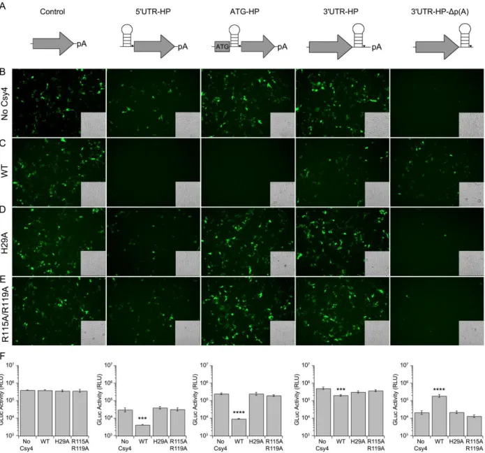
Figure 4: Csy4-mediated knockdown of 5' UTR-hairpin (HP) and ATG-HP
(B) Schematics of the unmodified control (left), 5'UTR-HP (middle), and ATG-HP (right) reporters. (C) PCR products of randomly primed RNA isolated from HEK293 cells transfected as indicated. (Left) unmodified control, (middle) 5'UTR-HP-GFP and (right) ATG-HP-GFP. (D) Fluorescence images of HEK293 cells expressing unmodified control (left), 5'UTR-HP (middle), and ATG-HP GFP (right) reporters in the absence (-) or
presence (+) of Csy4. Corresponding transmitted light images are shown as insets in each fluorescence image. (E) Quantitation of GLuc activity by luminometric analysis at 24 hours post-transfection for unmodified control (left), 5'UTR-HP (middle), and ATG-HP (right), GLuc reporters.Error bars indicate standard deviation of four replicates.
Figure 5: Effect of placing the HP in the 3’UTR on Csy4-mediated knockdown.(A)
Schematics of the 3'UTR-HP reporter (left) and 3'UTR-HP-Δp(A) reporter (right). (B)
GFP-3'UTR-HP reporter (left) or GFP-3'UTR-HP-Δp(A) reporter (right) and Csy4 as indicated. (C) Fluorescence images of HEK293 cells expressing GFP-3'UTR-HP (left) or GFP-3'UTR-HP-Δp(A) (right) in the absence (-) or presence (+) of Csy4. Corresponding transmitted light images are shown as insets in each fluorescence image. (D)
Figure 6: Cys4 interacts with the substrate hairpin in human cells. (A) Schematic representation of RNA immunoprecipitation (RNA-IP) detecting RNA associated with Csy4. An -His6 antibody is used to pull down His6-tagged Csy4-H29A and associated transcripts. (B) GFP-primed PCR products of cDNA generated from RNA purified from -His6 (Csy4-H29A) immunoprecipitates. Lysate from cells expressing ATG-HP-GFP
Figure 7:Csy4 binding and cleavage are necessary for regulation of transgene
expression.(A) Schematics of reporters (left to right) - unmodified control, 5'UTR-HP, ATG-HP, 3'UTR-HP, and 3'UTR-HP-Δp(A). Fluorescence images of HEK293 cells expressing each reporter in the absence of Csy4 (B), or presence of either native Csy4
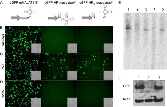
Figure 8: Selective processing by Csy4 is essential for rescue of 3'UTR-HP-Δp(A)
constructs. (A) Schematics of the unmodified mMALAT1-3' reporter (left), cGFP-HP-masc-Δp(A) (middle) and cGFP-HP10-masc-Δp(A) (right) reporters. Cleavage sites are indicated by black inverted triangles. Fluorescence images of HEK293 cells
expressing either mMALAT3' (left), HP-masc-Δp(A) (middle), or cGFP-HP10-masc-Δp(A) (right) in the absence of Csy4 (B), with Csy4 wild type (C), or Csy4-H29A (D). Corresponding transmitted light images are shown as insets in each
Figure 9: Potential events outlining Csy4-mediated processing of 3'UTR-HP-Δp(A)
CHAPTER 3: INDUCING CIRCULAR RNA FORMATION USING THE CRISPR ENDORIBONUCLEASE CSY4
3.1 Overview
Circular RNAs (circRNAs) are highly stable, covalently closed RNAs that are regulated in a spatiotemporal manner. With wide-ranging functions, these molecules have the potential to be incorporated into engineered systems with broad technological implications. Here we describe a switch for activating backsplicing of an engineered circRNA that relies on the CRISPR endoribonuclease, Csy4, as an activator of
3.2 Introduction
Circular RNAs (circRNAs) comprise an emerging large class of noncoding, covalently closed RNAs present in a wide variety of organisms ranging from archaea to humans (Danan et al., 2012; Wang et al., 2014). They are typically generated through the covalent joining of a downstream splicing donor site with an upstream splicing acceptor site through a process called direct backsplicing (Jeck et al., 2013; Salzman et al., 2012). Due to their circular nature, circRNAs exhibit marked stability relative to their corresponding linear isoforms (Enuka et al., 2015; Jeck et al., 2013; Lasda and Parker, 2014; Liang and Wilusz, 2014). The median half-life of circRNAs is at least 2.5X longer than their linear counterparts (Enuka et al., 2015). Furthermore, for some genes, the abundance of the circRNA exceeds that of the associated linear mRNA by a factor of 10 (Jeck et al., 2013; Salzman et al., 2012).
Despite their prevalence, the exact function of a large number of known
from stable, persistent RNA molecules. Accordingly, the development of molecular tools to exogenously control the circularization of a particular RNA of interest could prove useful.
In this regard, an interesting RNA targeting enzyme that has garnered recent interest from the synthetic biology community is Csy4 (Cas6f). Csy4 is an
endoribonuclease belonging to the Clustered Regularly Interspaced Short Palindromic Repeats (CRISPR) family. CRISPR systems utilize small RNAs (crRNAs) to guide CRISPR nucleases to nucleic acid, mediating sequence specific cleavage (Marraffini, 2015). Csy4 recognizes a 16 ribonucleotide hairpin which repeats throughout a
precursor CRISPR RNA, cleaving at the 3' base of the hairpin stem to generate crRNAs (Cady and O'Toole, 2011; Haurwitz et al., 2010). Importantly, Csy4 remains associated with the 5' cleavage product (Sternberg et al., 2012). These properties of Csy4 have been adapted recently to process operon-encoding RNAs for gene expression without interference from cis sequences in E. coli, B. subtilis, and S. cerevisiae (Qi et al., 2012). Further, Csy4 was employed for processing RNA encoding multiple guide-RNA (gRNA) to single gRNAs for Cas9-based applications (Nissim et al., 2014; Tsai et al., 2014). Csy4 was also used to isolate and identify proteins associated with a specific RNA of interest (Lee et al., 2013). We recently demonstrated that Csy4 can be exploited to modulate the stability and subsequent protein expression of specific RNAs of interest (Borchardt et al., 2015). In particular, we demonstrated that Csy4 can stabilize the cognate 5’ cleavage product to allow translation of a reporter protein.
system (circGFP-CD). This reporter may serve as a switch-based platform for the regulation of a variety of natural and engineered circRNA-mediated processes while also providing a new tool for shifting expression from one gene to another. Additionally, the Csy4-inducible circularization system has the potential for application in the study of factors involved in circRNA biogenesis.
3.3 Materials and Methods
Plasmids: The Csy4 gene was amplified from the pseudomonas aeruginosa UCBP-PA14 genome using primers 5’-ATC GTC TAG AAT GGA CCA CTA CCT CGA CAT TCG CTT GC-3’ and 5’-CGA TGC GGC CGC TCA GAA CCA GGG AAC GAA ACC TC CTT TGC-3’. The PCR product was cloned under the control of the CBA promoter in a plasmid backbone. Csy4-H29A was amplified from pHMGWA-Pa14Csy4H29A
(Addgene plasmid #41092) which was a gift from Dr. Jennifer Doudna (Haurwitz et al., 2010). Csy4-H29A was cloned under the control of the CBA reporter. CircGFP was a gift from Dr. Zefeng Wang (Wang and Wang, 2015). To construct circGFP-CD, a gBlock ® (IDT DNA Technologies) was synthesized consisting of the Csy4 targeted hairpin followed by the canonical intron 12 of IGF2BP1 and the P2A sequence in-frame with a dsRed coding sequence. This gblock was then cloned downstream of the split-GFP cassette such that the entire transcript is driven by the CMV promoter and terminated with a SV40 poly-A signal.
Cell culture: HEK293 cells were cultured in Dulbecco’s Modified Eagle’s Medium
penicillin/streptomycin, and 2.5 μg/mL amphotericin B (Sigma-Aldrich) and maintained at 37⁰C and 5% CO2.
Fluorescence microscopy and quantification of fluorescence: 5 X 10⁴ HEK293 cells were seeded overnight into 24 well plates and transfected with the indicated plasmids at equimolar quantities (500 ng). Cells were imaged at 72 hours post-transfection using an EVOS FL epifluorescence cell imaging system (AMC/Life Technologies) with the GFP light cube (excitation 470 nm, emission 510 nm), or RFP light cube (excitation 530, emission 590). Three images were analyzed per replicate using the FIJI image processing package to measure integrated density (Schindelin et al., 2012). Values for all nine images were averaged. Error bars indicate standard deviation of integrated density calculated from nine images from three biological replicates (three images per replicate). Statistical significance was calculated using a two-tailed Student’s t-test ([****] P≤.0001, [*] P≤.05).
RT-PCR: 3 X 10⁵ HEK293 cells were seeded overnight into wells of a 6-well plate and transfected with the indicated plasmids at equimolar quantities (totaling 2500 ng). RNA was harvested from these cells 48 hours post-transfection using the Total RNA
ACC C-3', 5'-GTT GTA CTC CAG CTT GTG CC-3') and glyceraldehyde 3-phosphate dehydrogenase (GAPDH) (5'-CCA CTC CTC CAC CTT TGA C-3', 5'-ACC CTG TTG CTG TAG CC-3'). PCR products were visualized on an agarose gel.
Western blot: HEK293 cells seeded overnight in 6 well plates at a density of 3 x 10⁵ cells per well were transfected with a total of 2 μg DNA as indicated. Lysates were recovered 48 hours post-transfection using 1X Passive Lysis Buffer (Promega) and diluted 1:10. Samples were heated to 100C before separation on a 10% Bis-Tris gel. Membranes were blocked overnight in 2% milk in TBST. After overnight incubation, membranes were blotted with primary antibody against either GFP (1:1000 Santa Cruz, SC9996) or Actin (1:2000, Abcam, Ab3280). Stabilized peroxidase-conjugated goat anti-mouse antibody was used as secondary antibody (1:20,000, Jackson
Immunologicals, 31430). Blots were developed using SuperSignal West Femto substrate (Thermo Scientific/Life Technologies).
RNaseR digestion: 2.2 X 10⁶ HEK293 cells were seeded overnight into 10 cm plates and transfected with the indicated plasmids at equimolar quantities (totaling 6
GTT GTG GC-3', 5'-CAA GCT GAC CCT GAA GTT CAT CTG CAC CAC C-3') or linear products (5'-CTT GGT CAC CTT CAG CTT GGC GGT CTG -3', 5'-GCT ACG TCC AGG GAT CCG GCG-3').
3.4 Results and Discussion
cleavage product by Csy4 could permit the cleaved split-GFP RNA to persist long enough for back-splicing to occur and support GFP expression (Figure 10B).
We first tested the circGFP-CD construct following plasmid transfection in mammalian cells and observed reporter expression via fluorescence microscopy. CircGFP, which is constitutively backspliced for GFP expression, is included as a control (Figure 11). When circGFP-CD is expressed alone, forward splicing appears to be favored as the cells strongly express dsRed but lack GFP fluorescence (Figure 11A). This expression pattern is consistent with predominant forward splicing and minimal backsplicing. It is important to note that the forward splice acceptor is identical to the backsplice acceptor in sequence. Therefore, the abundance of dsRed signal indicates that forward splicing is favored over backsplicing in this intron/exon configuration and host cell type. In contrast, when Csy4 is co-expressed with circGFP-CD, dsRed signal decreases while pronounced GFP signal appears, consistent with activation of circRNA biogenesis (Figure 11A). Fluorescence was quantified using FIJI image processing software (Schindelin et al., 2012) and the decrease in dsRed signal upon co-expression with Csy4 was measured at -6 fold. The corresponding increase in GFP signal was measured at +116 fold relative to Csy4 absent conditions (Figure 11B, 11C). The appearance of GFP signal concurrent with diminishing dsRed signal is consistent with Csy4 cleaving away the P2A-dsRed element and stabilizing the 5’ cleavage product. This allows back-splicing and subsequent GFP expression to occur.
hairpin, but is unable to cleave the RNA (Haurwitz et al., 2010). In the presence of H29A, GFP signal did not increase to the levels observed with native Csy4 (Figure 11A). This is consistent with Csy4 cleavage being required for predominant backsplicing to occur. However, when quantified, GFP signal did increase slightly by 1.8 fold (Figure 11C). Interestingly, H29A co-expression also induced a 2.4 fold increase in dsRed signal relative to Csy4 absent conditions (Figures 11A, 11B).
We then characterized the different RNA species generated from circGFP-CD using PCR and RNase R treatment. Two PCR primer sets were designed for the
detection of either circular or linear products. The primers for circRNA detection will only result in product when the GFP fragments have been spliced into the appropriate
orientation, for example, via RNA circularization. In the absence of Csy4, a faint band was detected using circular detection primers, indicating that back-splicing from
corroborate the requirement of Csy4 for circularization and consequently GFP expression in our reporter system.
We further confirmed circRNA production by RNase R digestion. RNase R is a 3'- to 5'- exonuclease to which circRNAs are inherently resistant due to their lack of a 3’ end (Suzuki et al., 2006). RNA derived from circGFP, which is constitutively back-spliced into circRNA, is included as a control and yields an RNase R resistant product (Figure 12C). When circGFP-CD RNA was expressed in the absence of Csy4, RNA was primarily detected using primers against linear template and was susceptible to RNase R (Figure 12C). This is consistent with predominant forward splicing in the absence of Csy4. Detection of RNase R susceptible linear product decreased when RNA from cells co-expressing Csy4 with circGFP-CD was analyzed (Figure 12C). Under these
circRNA synthesis. Importantly, in the absence of Csy4, circRNA levels remained low when expressed from the circGFP-CD cassette (Figure 13B). We next performed qRT-PCR with primers detecting forward splicing products (Figure 13A). Forward splicing products were expressed at high levels in case of the circGFP-CD without Csy4 (Figure 13A). Consistent with imaging and RNaseR studies, circGFP-CD co-transfected with Csy4 also expressed low levels of forward splice products. However, expression of these products remained consistently low relative to circGFP-CD transfected alone (Figure 13A). These results may indicate that a small proportion of RNA expressed from circGFP-CD is forward spliced before Csy4 cleaves away the forward splice acceptor.
In conclusion, we have designed a CRISPR-based, inducible system for circularization of RNAin mammalian cells. Csy4 acts as a reliable regulatory part for controlling the circGFP-CD switch, activating circRNA biogenesis, and protein
expression. Likewise, CircGFP-CD is an adaptable device for the expression of a variety of proteins or functional non-coding RNA sequences for use in genetic circuits. A wide variety of RNA-based tools have been engineered for manipulating gene
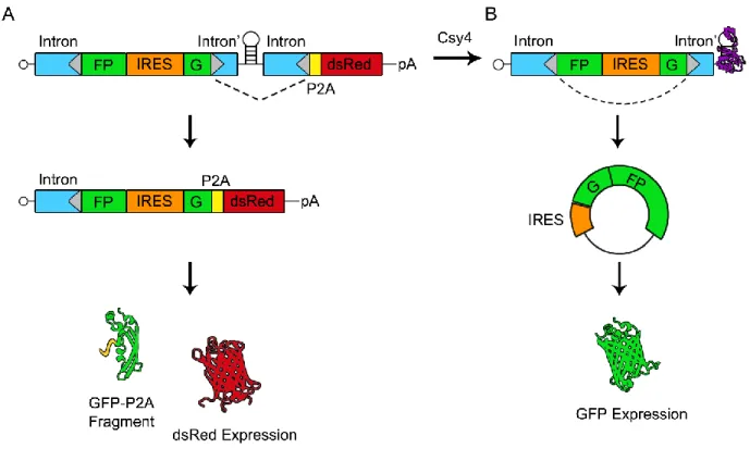
Figure 10: Schematic of circGFP-CD splicing patterns and reporter outputs. The mRNA expressed from circGFP-CD contains a split-GFP cassette flanked by
engineered introns (intron and intron’). This cassette is followed by a Csy4 targeted hairpin, another intron and a P2A-dsRed cassette. Splice sites are represented by grey triangles and the dotted line indicates the expected predominant splicing pattern. In the absence of Csy4 (A), forward splicing is favored and an IRES drives expression of dsRed. A non-fluorescing GFP fragment by-product is released via the P2A sequence. In the presence of Csy4 (B), Csy4 cleaves the RNA at the base of the hairpin stem, releasing the forward splice acceptor and P2A-dsRed cassette. Csy4 remains
Figure 11: Csy4 induces circGFP-CD reporter expression to switch from dsRed to
Figure 12: Detection of circGFP-CD expression products. Primers designed against either circular RNA or linear RNA products are depicted in each panel. (A) PCR
products obtained using cDNA template generated from randomly primed RNA that was isolated from HEK293 cells transfected as indicated. GAPDH PCR products were
products amplified using primers specific for linear or circular splicing patterns.
65
Figure 13: Accumulation of circRNA and linear splicing products over time. qRT-PCR of cDNA generated from RNA isolated at 8, 24, 48, 72, and 96 hours normalized to glyceraldehyde 3-phosphate dehydrogenase (GAPDH) and 0 hour transfection controls. Cells were transfected with either circGFP-CD (open circles, dotted line), circGFP-CD and Csy4 (closed circles, solid line), or with a circGFP control plasmid (open triangles, solid line). qRT-PCR was carried out using primers specific for either linear (A) or circular (B) splicing products as depicted above each graph. Error bars indicate standard deviation of three biological replicates. Statistical significance for the “circGFP-CD + Csy4” condition was
CHAPTER 4: IN VIVO DELIVERY OF TRANSLATABLE CIRCULAR RNA CASSETTES USING RECOMBINANT AAV VECTORS
4.1 Overview
Circular RNAs (circRNAs) are highly stable, persistent RNAs which serve a wide variety of cellular roles. With broad utility and potential for long-lasting effects due to their stability, circRNAs possess great promise as therapeutic agents. As currently understood, the natural functions of circRNAs are non-coding. However, circRNAs can be engineered for protein expression by the inclusion of an Internal Ribosome Entry Site (IRES) to drive translation. Recombinant Adeno-Associated Virus (rAAV) is commonly utilized in the field of gene therapy for the delivery of therapeutic cassettes. rAAV is an ideal candidate for transgene delivery due to well-defined tropism and minimal genome packaging requirements. Still, dose-related toxicity requires careful consideration when utilizing rAAV for therapeutic applications. Means to lower vector dosage while
and liver. CircRNA biogenesis was confirmed via RNase R digestion. These results provide the foundation for AAV-mediated delivery of circRNA cassettes for a variety of therapeutic applications.
4.2 Introduction
Circular RNAs (circRNAs) comprise a class of covalently closed, exceedingly stable non-coding RNAs (ncRNAs). Initially overlooked as splicing byproducts,
circRNAs are now recognized as highly abundant molecules with unique and complex expression patterns (Memczak et al., 2013; Salzman et al., 2012; Salzman et al., 2013) CircRNAs are found throughout the tree of life, with representation in organisms ranging from archaea, yeast, and amoeba, to more complex systems such as Caenorhabditis
elegans,Drosophila melanogaster, and humans (Danan et al., 2012; Memczak et al.,
2013; Salzman et al., 2013; Wang et al., 2014). The persistence of circRNAs throughout this wide range of organisms highlights the importance of these RNAs. It additionally supports further investigation into their biogenesis, function, and potential applications.
CircRNA biogenesis is thought to utilize a mechanism called backsplicing in order to join a downstream splicing donor to an upstream splicing acceptor. (Jeck et al., 2013). However, an alternate mechanism which employs a lariat intermediate has also been proposed (Zaphiropoulos, 1996).
their associated linear RNAs by a factor of 10 (Jeck et al., 2013; Lasda and Parker, 2014).
Though the abundance of circRNAs has been well established, the roles of most circRNAs remain undetermined. However, from the few circRNAs with
well-characterized functions, it is clear that circRNAs possess wide regulatory capacity. The circRNA Cdr1as (ciRS-7) acts as a miRNA sponge by interacting with miR-7 through 63 conserved miR-7 binding sites (Hansen et al., 2013; Memczak et al., 2013). Similarly, circSry also acts as a microRNA (miRNA) sponge by binding to miR-138 (Hansen et al., 2013). CircHIPK3 is an example of a circRNA capable of binding multiple different miRNAs and interacts with nine miRNAs over 18 binding sites (Zheng et al., 2016). CircMBL, on the other hand, is an example of a circRNA that binds protein. In an
autoregulatory fashion, circMBL interacts with the MBL (Muscleblind) protein (a product of the MBL linear RNA), through MBL binding sites. This in turn regulates circMBL formation, which relies on MBL protein (Ashwal-Fluss et al., 2014).