BASIC RESEARCH PAPER
Autophagy activation by novel inducers prevents BECN2-mediated drug tolerance
to cannabinoids
Kenta Kuramotoa,y,yy, Nan Wanga,b,y, Yuying Fana,c, Weiran Zhanga, Frank J. Schoenend, Kevin J. Frankowskie, Juan Maruganf, Yifa Zhouc, Sui Huanga, and Congcong Hea
aDepartment of Cell and Molecular Biology, Feinberg School of Medicine, Northwestern University, Chicago, IL, USA;bKey Laboratory of Industrial
Microbiology, Ministry of Education and Tianjin City, College of Biotechnology, Tianjin University of Science and Technology, Tianjin, China;cSchool of
Life Sciences, Northeast Normal University, Changchun, Jilin, China;dHiguchi Biosciences Center, University of Kansas, Lawrence, KS, USA;eUNC
Eshelman School of Pharmacy, University of North Carolina at Chapel Hill, Chapel Hill, NC, USA;fDivision of Pre-Clinical Innovation, National Center for
Advancing Translational Sciences, National Institutes of Health, Bethesda, MD, USA
ARTICLE HISTORY Received 17 December 2015 Revised 29 April 2016 Accepted 4 May 2016 ABSTRACT
Cannabinoids and related drugs generate profound behavioral effects (such as analgesic effects) through activating CNR1 (cannabinoid receptor 1 [brain]). However, repeated cannabinoid administration triggers lysosomal degradation of the receptor and rapid development of drug tolerance, limiting the medical use of marijuana in chronic diseases. The pathogenic mechanisms of cannabinoid tolerance are not fully understood, and little is known about its prevention. Here we show that a protein involved in macroautophagy/autophagy (a conserved lysosomal degradation pathway), BECN2 (Beclin 2), mediates cannabinoid tolerance by preventing CNR1 recycling and resensitization after prolonged agonist exposure, and deletion of Becn2 rescues CNR1 activity in mouse brain and conveys resistance to analgesic tolerance to chronic cannabinoids. To target BECN2 therapeutically, we established a competitive recruitment model of BECN2 and identified novel synthetic, natural or physiological stimuli of autophagy that sequester BECN2 from its binding with GPRASP1, a receptor protein for CNR1 degradation. Co-administration of these autophagy inducers effectively restores the level and signaling of brain CNR1 and protects mice from developing tolerance to repeated cannabinoid usage. Overall, our findings demonstrate the functional link among autophagy, receptor signaling and animal behavior regulated by psychoactive drugs, and develop a new strategy to prevent tolerance and improve medical efficacy of cannabinoids by modulating the BECN2 interactome and autophagy activity.
KEYWORDS analgesia; autophagy inducer; BECN2; behavior; cannabinoid; cannabinoid receptor 1; drug tolerance; intracellular trafficking
Introduction
Cannabinoids and related drugs, such as the marijuana-derived ingredients, generate profound behavioral effects (such as anal-gesic effects) that are therapeutic in many pathological condi-tions, including neurodegeneration, digestive disorders, spasticity, and chronic and cancer-related pain.1-4 However, long-term administration of cannabinoids for either medical or recreational purposes induces rapid development of tolerance (a demonstration of physical dependence), which is a limitation and concern of its medical use and may lead to addiction and withdrawal symptoms.5,6Clinical data showed that 9% of adult
cannabis users, and 17% of adolescent users, develop depen-dence and addiction after repeated dosage,6which is not trivial
given the widespread usage of illicit cannabinoids in many countries. Yet the pathogenic mechanisms of cannabinoid tol-erance are not fully understood, and little is known about its prevention methods. Consequently, only a very small number of cannabinoid therapeutics have been approved and used clin-ically in the market in limited regions; for example, the
cannabis medication for spasticity, Sativex, is prescribed as an oromucosal spray to ensure slow blood delivery and is adminis-tered at relatively low doses.
The effects of cannabinoids are mediated by binding and acti-vation of a G protein-coupled receptor (GPCR), CNR1/CB1
(cannabinoid receptor 1 [brain]), and chronic exposure to canna-binoids results in lysosomal trafficking and degradation of CNR1, which causes diminished cellular responses and requires higher doses to produce the same effects.7 Therefore, retaining the functionality of CNR1 may play an important role in the preven-tion of cannabinoid tolerance, especially in clinical applicapreven-tions. A recently identified autophagy gene, Becn2 (Beclin 2), which belongs to the Beclin (coiled-coil, myosin-like BCL2-interacting protein) family, may link CNR1-regulated cell signaling and animal behavior to the autophagy pathway.8 Autophagy is an essential catabolic process that breaks down damaged or unneces-sary structures in lysosomes, and the resulting metabolites are recycled and reused for new protein synthesis and energy produc-tion. Autophagy is intensely induced by physiological stimuli or
CONTACT Congcong He [email protected] 300 E Superior St, Tarry 8-767, Chicago, IL 60611, USA
yThese authors contributed equally to this work.
yyCurrent position: Department of Molecular Cancer Science, Yamagata University School of Medicine, Yamagata 990-9585, Japan.
Supplemental data for this article can be accessed on thepublisher’s website. © 2016 Taylor & Francis
stress, such as starvation9and physical exercise,10and malfunction of autophagy has been implicated in a variety of diseases, including neurodegeneration, cardiovascular diseases, cancer and metabolic disorders.11-13In addition to a role in autophagy, BECN2 is also essential for agonist-induced lysosomal degradation of a group of specific GPCRs, including CNR1, DRD2 (dopamine receptor D2) and OPRD1 (opioid receptor, delta 1).8In vitro biotin protection degradation data suggest that BECN2 mediates the degradation of these GPCRs by binding to GPRASP1/GASP1 (G protein-coupled receptor associated sorting protein 1),8 a receptor protein that
degrades GPCRs independently of ubiquitination and certain components of the canonical endosomal sorting complex required for transport (ESCRT) machinery.14-18
It is unclear whether BECN2 regulates the downstream events of these GPCRs in response to chronic agonist exposure, including receptor resensitization, signaling cascades and drug-responsive behaviors in vivo, which are important questions especially when many of the GPCRs in this specific group are targets of psychoactive drugs, such as CNR1. In addition, the function, genetic basis and molecular mechanism of autophagy in the regulation of drug tolerance and dependence after repeated usage remain mysterious. It is also unknown how the autophagy pathway crosstalks with drug-responsive GPCR sig-naling and behavioral regulation, and whether BECN2 plays a role in the process. Here we found that BECN2 is essential for the development of analgesic tolerance after chronic cannabi-noid exposure by preventing CNR1 recycling and resensitiza-tion in mice. We discovered that activaresensitiza-tion of autophagy restores CNR1 function and regulates cannabinoid analgesia through BECN2, and also identified novel synthetic and natural agents to prevent cannabinoid tolerance by modulating the BECN2 interactome and autophagy activity. Ourfindings dem-onstrated for the first time a new functional link among autophagy, drug responses and behaviors regulated by psycho-active substances, and opened up a new direction to potentially improve medical efficacy of cannabinoids in many circumstan-ces such as pain management.
Results
Becn2C/¡heterozygous knockout mice are resistant to analgesic tolerance induced by repeated cannabinoid dosage
We hypothesized that in response to prolonged cannabinoid exposure, BECN2 mediates the degradation and rapid deactiva-tion of CNR1. To test this hypothesis, we sought to use the Becn2C/¡heterozygous knockout (KO) mice, becausebecn2¡/¡ homozygotes rarely survive the embryonic development period. We treatedBecn2C/¡mice and wild-type (WT) littermates daily for 14 d with WIN55,212–2 (WIN), a synthetic cannabinoid drug and CNR1 agonist, and analyzed the levels of CNR1 in the brain. Compared to WT mice,Becn2C/¡mice maintained a significantly higher level of brain CNR1 after 14 d of chronic cannabinoid treatment (Fig. 1A), in addition to a trend of increased steady-state CNR1 levels under normal conditions (with DMSO treatment), which suggests that BECN2 loss-of-function increases the availability of CNR1 after repeated agonist dosage.
To study whether BECN2 functions in CNR1-regulated behavioral responsiveness following chronic cannabinoid treatment, we analyzed the anti-nociceptive (i.e., anti-pain) effect of WIN as a readout by analgesic tolerance tests (Fig. 1B), using WT mice and mice heterozygous forBecn2, or for Becn1, another Beclin family member that does not bind GPRASP1 or regulate GPCR degradation as a control. There were no genotype differences either in basal pain sensitivity to infrared heat-generated pain (agonist-free, d 0) (Fig. 1C, Video S1-S3), or in analgesic effects of acute WIN treatment prior to chronic WIN administration (d 1) (Fig. 1D, Video S4-S6). However, 14 d of repeated WIN dosage markedly decreased its analgesic effect in WT and Becn1C/¡ mice, but not in Becn2C/¡mice (d 14) (Fig. 1D, Video S7-S9), suggesting that Becn2C/¡mice are resistant to analgesic tolerance induced by prolonged cannabinoid exposure. Notably, body weight of mice of all genotypes remained constant during chronic WIN treatment (Fig. S1). Thus, these data demonstrate that BECN2 downregulates the level of brain CNR1 after prolonged WIN treatment, and BECN2 loss of function protects animals from developing analgesic tolerance to repeated exposure of canna-binoid drugs.
CNR1 is resensitized at the cell surface upon BECN2 loss
We next sought to investigate the cellular mechanism underlying the behavioral protection against tolerance upon loss of BECN2. Published data have shown that BECN2 is required for agonist-induced lysosomal transport of OPRD1, another GPRASP1-bound GPCR, which is relo-calized to the plasma membrane upon loss of BECN2.8 However, it is unknown whether in the absence of BECN2 these recycled receptors are functional, or whether BECN2 also plays a role in the trafficking, recycling and signaling of CNR1 after prolonged agonist exposure. Here we posed that BECN2 depletion stabilizes CNR1 levels by pro-moting its recycling and resensitization at the cell surface, and tested this hypothesis from 3 aspects. We first fol-lowed the intracellular trafficking of CNR1 in response to agonists, by examining its colocalization with endosome (EEA1) or lysosome (LAMP1) markers in the absence of BECN2. By pulse-labeling cell-surface CNR1 with anti-body, we found that compared to basal conditions (0 min), in a significantly higher number of cells treated with control siRNA, CNR1 was transported to endosomes after 30 min of WIN treatment and to lysosomes after 60 min, whereas in cells transfected with Becn2 siRNA,8 instead of being transported to lysosomes for degradation, internalized CNR1 was trapped in endosomal structures even after 60 min of WIN treatment (Fig. 2A, Fig. S2A). In addition, in Becn2 siRNA-treated cells the endocytosed CNR1 relocalized to the cell surface after 30 min of ago-nist removal and antagoago-nist treatment (Fig. 2B). Thus, these data demonstrate that loss of BECN2 leads to accu-mulation of CNR1 in the endosomes and its eventual recy-cling to the plasma membrane.
biochemical and cellular levels. CNR1 is coupled to the GNAI/ Gi/oprotein, and CNR1 activation inhibits the ADCY/adenylyl
cyclase activity and cyclic adenosine monophosphate (cAMP) production, while activating the mitogen-activated protein kinase (MAPK) pathways, including MAP2K1 (mitogen-acti-vated protein kinase 1)/MEK1-MAP2K2/MEK2 and MAPK8/ JNK1-MAPK9/JNK2 cascades.7,19Thus, we measured canna-binoid-induced cAMP suppression (Fig. 2C) and MAPK cas-cade phosphorylation (Fig. 2D) in cell lines and primary MEFs (mouse embryonic fibroblasts) following prolonged agonist treatment. We found that the intracellular cAMP level was reduced inBecn2siRNA-treated cells compared to control cells (Fig. 2C), which suggests an increased level of functional CNR1 at the cell surface and is an indication of CNR1 resensi-tization upon loss of BECN2. Furthermore, we detected a higher level of cannabinoid-induced phosphorylation of MAP2K1/2 and MAPK8/9 in Becn2 KO primary MEFs (Fig. 2D), as well as a trend of higher MAP2K1/2 and MAPK8/9 phosphorylation in the brain of Becn2C/¡ mice (Fig. S2B), than in WT MEFs or brain, suggesting higher CNR1 functionality after chronic agonist exposure in the absence of BECN2. Overall, these data suggest that loss of BECN2 leads to CNR1 resensitization.
Competitive recruitment model of BECN2 and identification of novel brain-penetrable autophagy inducers
Accordingly, an important translational question is how to reduce BECN2 activity to maintain CNR1 sensitivity and pre-vent drug tolerance induced by repeated cannabinoid exposure. To avoid the technical difficulty and risk of directly deleting Becn2 in vivo (such as by injecting shRNAs or CRISPR con-structs), which may disrupt BECN2-regulated autophagy, we developed and tested a novel and more convenient strategy to achieve the same goal via modulating the BECN2 interactome, which will not affect the essential autophagy function of BECN2 (Fig. 3A). As previously reported,8BECN2 is present in 2 distinct protein complex pools, the BECN2-class III phospha-tidylinositol 3-kinase complex for autophagy, and the BECN2-GPRASP1-GPCR complex in which BECN2-GPRASP1 binding is required for GPCR degradation. Thus, we hypothesized that BECN2 is a limiting factor that regulates the balance between the 2 lysosomal degradation pathways, and that competitive recruitment of BECN2 to the autophagy machinery by activat-ing autophagy prevents it from functionactivat-ing in CNR1 downre-gulation and thus restores CNR1 functionality and reverses Figure 1.Loss of BECN2 confers resistance to analgesic tolerance induced by chronic usage of cannabinoid drugs. (A) Brain samples fromBecn2C/¡heterozygous
knock-out (KO) and WT mice were collected after 14 d of prolonged treatment of vehicle or the synthetic cannabinoid drug WIN55,212–2 (WIN). CNR1 levels were analyzed by western blot of brain lysates (left) and quantified (right). Representative images of 3 mice in each group are shown. (B) Chronic cannabinoid treatment scheme, described in Materials and methods. Briefly, mice were injected with WIN for 14 d, and analgesic tolerance was measured without WIN treatment on day 0, and after 1-h WIN treatment on d 1 and d 14. (C) Baseline pain sensitivity of WT andBecn2C/¡ mice before chronic WIN treatment (day 0, agonist-free). N 16 mice/group. (D)Becn2C/¡mice show resistance to analgesic tolerance to chronic WIN treatment.Becn2C/¡,Becn1C/¡and WT mice were treated with daily WIN for 14 d, and the
anal-gesic effect of WIN was analyzed as in (B). Statistics are comparing the same genotype on d 1 and d 14. ND9–13 mice/group. Results represent mean§s.e.m.
Figure 2.BECN2 depletion triggers resensitization of endocytosed CNR1 at the plasma membrane. (A) Immunofluorescence imaging (upper) and quantification (lower) of the effects of nontargeting control (NC) orBecn2siRNA on the fate of endocytosed CNR1. HEK293 cells stably expressing FLAG-CNR1 were fed with anti-FLAG antibody, treated with the agonist WIN for 30 or 60 min and immunostained as in Materials and methods. Green, FLAG-CNR1; red, EEA1 or LAMP1. Percentages of cells with CNR1-EEA1 or CNR1-LAMP colocalization in>200 cells per experiment were quantified from 4 independent experiments. (B)Becn2knockdown promotes CNR1 recycling to the cell surface after prolonged agonist exposure, analyzed by immunofluorescence imaging of FLAG-CNR1. HEK293 FLAG-CNR1 cells were fed with anti-FLAG antibody, and treated with WIN for 30 or 180 min or with WIN for 140 min followed by agonist washout and antagonist (rimonabant) treatment for 30 min. Scale bar: 20mm. (C)Becn2 knockdown suppresses the ADCY/adenylyl cyclase activity downstream of CNR1 after prolonged agonist exposure. HEK293 FLAG-CNR1 cells transfected with the indicated siRNAs were treated either with vehicle for 120 min, or with WIN for 120 min to induce CNR1 internalization and degradation, followed by agonist washout and antago-nist (rimonabant) treatment for 30 min to trigger CNR1 recycling and another 20 min treatment of WIN to activate any CNR1 at the cell surface. Results represent mean§ s.e.m of 3 independent experiments. (D) BECN2 depletion increases cannabinoid-induced CNR1 signaling after prolonged exposure.Becn2C/C,Becn2C/¡orbecn2¡/¡
Figure 3.Identification of novel brain-penetrable autophagy-inducing compounds. (A) Competitive recruiting model of BECN2 by the 2 lysosomal degradation pathways: inducing autophagy shunts BECN2 away from the BECN2-GPRASP1 complex, and thus attenuates lysosomal degradation of CNR1 and maintains its responsiveness. Red arrows, pro-autophagy; black arrows, pro-CNR1 downregulation. PtdIns3K, phosphatidylinositol 3-kinase. (B) Chemical structure of ML246. (C and D) Representative images (C) and quantification (D) of GFP-LC3 puncta in HeLa cells stably expressing GFP-LC3 cultured for 3 h in normal or starvation medium, or treated with Rg2 or ML246 in normal medium for 3 h or 16 h, in the presence or absence of bafilomycin A1(BafA1). Results represent mean§s.e.m. Statistics are comparing the indicated
value with or without BafA1 to the one under the normal condition. Blue, DAPI; scale bar: 20mm. (E) Representative images (left) and quantification (right) of GFP-LC3 puncta in the brain of GFP-LC3 transgenic mice injected with vehicle or ML246. Results represent mean§ s.d. Scale bar: 25mm. N>100 cells or N4 mice.
drug tolerance. To test this hypothesis, we first examined whether autophagy activation by brain-penetrable inducers inhibits the BECN2-GPRASP1 interaction. From a small-mole-cule library screen20 and a phytochemical screen (Y. Fan, A. Rocchi, N. Wang, W. Zhang, R. Vassar, Y. Zhou and C. He, manuscript in preparation), we identified 2 novel brain-pene-trable autophagy-inducing compounds: ML246 (also named metarrestin based on its anti-metastasis effects in cancer,20 Fig. 3B), a synthetic small molecule derived from a high-throughput screen against a pan-cancer cellular marker; and Rg2,21 a 785 Da natural steroid glycoside compound isolated
fromPanax ginsengby analytical chemistry approaches. To determine autophagy activation by the 2 compounds, we analyzed several markers of autophagy induction, including formation of autophagosomes in cells and transgenic mice expressing GFP-tagged MAP1LC3/LC3 (microtubule-associ-ated protein 1 light chain 3; an autophagosome marker),9 deg-radation of SQSTM1/p62 (sequestosome 1; an autophagy receptor and substrate protein), and conversion of LC3 from the non-lipidated form (LC3-I) to the phagophore- and auto-phagosome-associated lipidated form (LC3-II). Both com-pounds markedly increased numbers of GFP-LC3 puncta (representing autophagosomes) and decreased levels of SQSTM1 in HeLa cells and in mouse brain, as potently as star-vation (Fig. 3C-E, Fig. S3A-C), although as previously
reported22we did not detect significant changes in LC3-II con-version in brain. In addition, when cotreated with the lyso-somal inhibitor bafilomycin A1, ML246 or Rg2 led to more
accumulation of GFP-LC3 puncta, LC3-II and SQSTM1 com-pared to normal conditions (Fig. 3C-D, Fig. S3D), suggesting an increased level of autophagic flux induced by either com-pound. Therefore, both agents are effective autophagy inducers in vitro and in vivo.
Activation of autophagy pharmacologically or physiologically attenuates BECN2-GPRASP1 binding, dependent on the early initiation step of autophagy
Based on our model, we subsequently examined the effect of the ML246 and Rg2 inducers on the BECN2-GPRASP1 interac-tion in vitro and in mouse brain. We found that autophagy induction either pharmacologically with ML246 or physiologi-cally with starvation medium for 3 h potently blocked co-immunoprecipitation of endogenous GPRASP1 by BECN2 in HEK293 cells (Fig. 4A). Importantly, early initiation of autophagy is required for the sequestration of BECN2 from GPRASP1 binding by autophagy inducers, as the BECN2-GPRASP1 complex remained intact during starvation or ML246 treatment after knockdown of any member in the upstream ATG13-RB1CC1/FIP200-ULK1 kinase complex
essential for autophagy induction (Fig. 4A).23-26Furthermore, injection of ML246 or Rg2 in vivo to WT mice, as well as 48-h starvation, dramatically decreased the binding between endoge-nous BECN2 and endogeendoge-nous GPRASP1 in mouse brain (Fig. 4B). Overall, these data suggest that both physiological and novel pharmacological autophagy inducers attenuate BECN2-GPRASP1 binding, which is dependent on the early initiating step of autophagy and supports our competitive recruitment model of BECN2. Thus, autophagy inducers serve as new candidates in the prevention of receptor downregulation and cannabinoid tolerance to prolonged exposure.
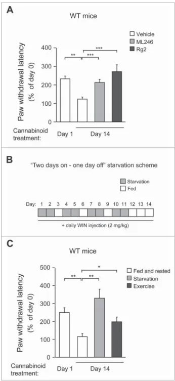
Synthetic, natural or physiological autophagy inducers prevent WT mice from analgesic tolerance after chronic cannabinoid usage
We then tested the efficacy of ML246 and Rg2 in the mainte-nance of cannabinoid analgesia in WT mice. We found that compared with vehicle injection, cotreatment of either ML246 or Rg2 with WIN potently rescued the pain-relieving effect of WIN in WT mice on d 14 to a d 1-like level (i.e., before repeated dosage) (Fig. 5A, Video S10-S12), suggesting that the autophagy-inducing compounds prevent analgesic tolerance induced by chronic WIN administration. The cotreatment regi-men did not alter body weight of the mice (Fig. S4A). To con-firm the findings with pharmacological inducers, we further asked whether autophagy induction by physiological methods also rescues cannabinoid tolerance. We adopted 2 methods that activate autophagy in mouse brain:22,27thefirst is daily volun-tary exercise by use of running wheels, which allows mice to run for approximately 1 km/night; and the second is a“2-day on/1-day off”periodic starvation protocol, in which mice were subjected to cycles of 48-h fasting followed by 24-h feeding that allowed them to return to normal weight in each cycle (Fig. 5B, Fig. S4B).
Although the increase in autophagosome formation was not significant in the brain (frontal cortex) of GFP-LC3 mice after a single bout of 48-h fasting as previously reported usingfl uo-rescence microscopy (Fig. S4C),9 we observed a cumulative
effect on autophagy induction in the same brain region after multiple rounds of alternating fasting and feeding, demon-strated by a significant induction of GFP-LC3 puncta in the frontal cortex after 4 cycles of“2-day on/1-day off”starvation (Fig. S4C). Although the exact mechanism of this additive effect is currently unclear, we propose that it may be due to a rela-tively stable glucose supply and low metabolism in the brain (compared to muscle and liver), leading to slow formation/ turnover of autophagosomes that can be detected after repeated induction. This hypothesis is supported by our observation that skeletal muscle, which has high metabolic activity, does not show much cumulative effect with regard to autophagy induc-tion by periodic starvainduc-tion cycles (Fig. S4C). At the end of chronic WIN treatment, similar to mice treated with autophagy-inducing compounds, mice undergoing daily run-ning or intermittent fasting showed significantly higher sensi-tivity to the analgesic effects of WIN (Fig. 5C, Video S13-S15), suggesting that physiological autophagy inducers, such as exer-cise and starvation, prevent analgesic tolerance as well. Alto-gether, these data suggest that both pharmacological and
physiological autophagy inducers prevent cannabinoid toler-ance to repeated dosage.
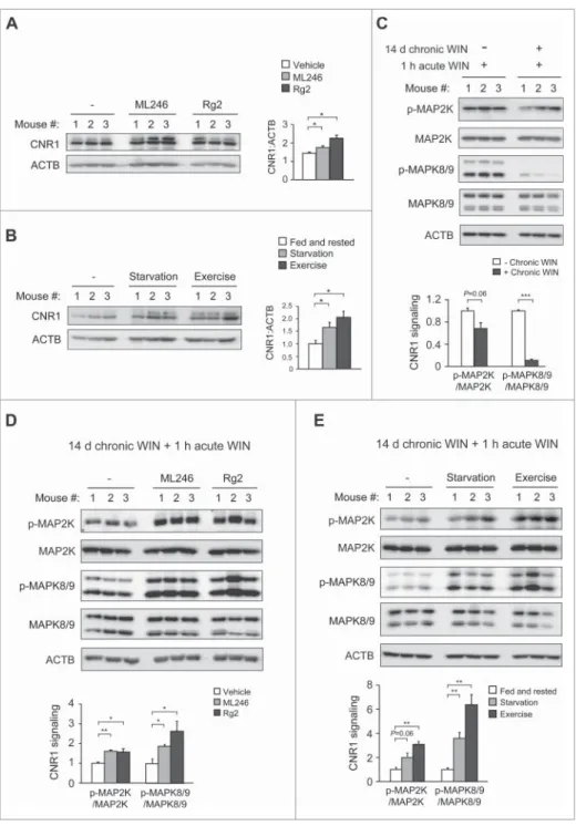
Autophagy induction preserves brain CNR1 level and activity in response to chronic cannabinoids
the level and functionality of CNR1 in mouse brain after co-administration of chronic cannabinoids and autophagy inducers. Consistent with behavioral sensitization to WIN, after chronic WIN treatment we detected higher levels of CNR1 in the brain of mice concurrently treated with ML246 or Rg2 (Fig. 6A), or fasted or exercised (Fig. 6B), which were compara-ble to the brain CNR1 level prior to chronic cannabinoid expo-sure (Fig. 1A). To determine whether the increased level of CNR1 represents CNR1 resensitization in the brain, we
autophagy pharmacologically or physiologically effectively improves CNR1 signaling after chronic cannabinoid exposure.
Discussion
Ourfindings characterized an autophagy gene as a novel regu-lator of drug-responsive behaviors, and linked autophagy for thefirst time to CNR1 sensitization and drug tolerance to can-nabinoids, a substance that has emerged as a major medical and social challenge in recent decades. We demonstrated that BECN2 is a new target in the prevention of tolerance to repeated cannabinoid dosage, and developed the concept of activating autophagy as an anti-tolerance therapeutic method. Furthermore, we identified novel autophagy-inducing com-pounds that achieve this goal by biochemically manipulating the BECN2-GPRASP1 protein complex, which may serve as candidate drug compounds to strengthen the pain-relieving effects of cannabinoids for chronic usage. Notably, we also found that periodic starvation and daily exercise are effective to disrupt the BECN2-GPRASP1 interaction and to prevent mice from cannabinoid tolerance after repeated usage, although we cannot rule out that in the cases of daily exercise and alternat-ing fastalternat-ing/feedalternat-ing, BECN2- or autophagy-independent mecha-nisms may also contribute to the anti-toleranceanti-tolerance effects or changes on CNR1 levels and signaling. To test this hypothesis, further studies using mutant mice deficient in early initiation of autophagy by starvation or exercise are required. Furthermore, to conclusively establish a causal relationship between BECN2-GPRASP1 binding and cannabinoid tolerance, it is necessary to directly disrupt the BECN2-GPRASP1 interac-tion or specifically perturb the BECN2 function in autophagy in vivo, by generation of knock-in mice containing loss-of-interaction mutations in BECN2 with either GPRASP1 or the autophagy machinery.
Cannabinoids can serve as promising therapeutics in many clinical occasions, such as alternative pain-relieving drugs for patients who develop tolerance to opioid medication. Yet canna-binoids’own potential of drug tolerance and dependence limits the medical use, which is sometimes a neglected issue due to the notion that cannabis may not cause as strong tolerance and dependence as some other drugs of abuse, such as opioids. Here ourfindings opened up many unanswered questions on analgesic tolerance to cannabinoids for further investigation. For example, drug tolerance is likely to develop at different kinetics to various CNR1 agonists of different efficacy;28 thus, in addition to WIN55,212–2, which is a highly potent CNR1 agonist, it will be essential to study whether the pharmacological or physiological autophagy inducers mediate the same effects on high doses of the natural cannabinoid THC (tetrahydrocannabinol) and other syn-thetic CNR1 agonists developed for therapeutic use. It is also important to analyze the role of BECN2 and autophagy in the reg-ulation of analgesic tolerance to cannabinoids in disease models of inflammatory or neuropathic pain,29-31as thermal pain gener-ated in the analgesic tolerance tests may not exactly mimic these types of clinical pain. Thus, future studies will involve surgical- or chemical-stimulation of inflammatory or neuropathic pain using the Becn2 heterozygous KO mouse model. Finally, given that BECN2 regulates the degradation of several additional drug-responsive GPCRs besides CNR1,8 such as DRD2 (dopamine
receptor D2) and OPRD1 (opioid receptor, delta 1), the current knowledge on cannabinoid tolerance may open up a new direc-tion for studying autophagy in tolerance or dependence to drugs of abuse targeting many other BECN2-regulated receptors.
Materials and methods
Mice
All mice were in the C57BL/6J background and housed on a 12-h lig12-ht/dark cycle. T12-he globalBecn2C/¡andBecn1C/¡ heterozy-gous knockout mice have been described previously.8,32All ani-mal protocols were approved by the Northwestern University Institutional Animal Care and Use Committee (IACUC).
Cell lines
HEK293 and HeLa cell lines were obtained from ATCC, and stable FLAG-CNR1 HEK293 cells were from J. Whistler (Uni-versity of California, San Francisco). Cells were cultured in Dulbecco’s modified Eagle’s medium (ThermoFisher Scientific, 11965) supplemented with 10% fetal bovine serum (Thermo-Fisher Scientific, 16140071).
Drug treatment
For in vivo use, WIN55,212–2 (Sigma-Aldrich, W102), ML246 (synthesized in-house) and Rg2 (purified in-house) were dis-solved in a solvent containing 5% NMP (Sigma-Aldrich, 494496), 20% PEG400 (EMD Millipore, PX1286B) and 75% of a 10% solution of HP-b-CD (Tokyo Chemical Industry, H0979) in water, and injected intraperitoneally at the dosage of 2 mg/kg, 10 mg/kg, and 20 mg/kg body weight, respectively. For cell culture use, WIN55,212–2 and ML246 were dissolved in 100% DMSO (Fisher Scientific, BP231) and Rg2 was dis-solved in 10% DMSO and used at the concentration of 1mM, 0.5 mM and 100 mM, respectively. Bafilomycin A1
(Sigma-Aldrich, B1793) was dissolved in 100% DMSO and used at 100 nM in cells.
Chronic WIN55,212–2 treatment and analgesic tolerance
test
Cyclic AMP measurement
Intracellular cAMP levels were analyzed in HEK293 cells stably expressing FLAG-CNR1 by the cAMP-Glo assay kit (Promega, V1681). Briefly, 40,000 cells/well were cultured in a 96-well plate in triplicate, and treated with either cAMP stabilizers 0.5 mM IBMX (Sigma-Aldrich, I5879) and 1 mM forskolin (Sigma-Aldrich, F6886) alone for 120 min, or with 1 mM WIN55,212–2 plus IBMX and forskolin for 120 min, followed by a phosphate-buffered saline (PBS; Sigma-Aldrich, D8537) wash and 200 nM rimonabant (Cayman Chemical, 9000484) treatment for 30 min, and then another 20 min incubation with 1 mM WIN55,212–2 in the presence of IBMX and forskolin. The cells were subsequently lysed and cAMP production was measured following the manufacturer’s instruction.
CNR1 trafficking and recycling by antibody pulse-chase assay
The antibody-feeding immunofluorescence internalization and recycling assays were performed essentially as previously described.33,34 Briefly, HEK293 cells stably expressing FLAG-CNR1 were grown on chamber slides, and incubated with 3.5mg/ml M1 anti-FLAG antibody (Sigma-Aldrich, F3040) for 30 min. The cells were either directlyfixed and immunostained as below as“0 min”control, or treated with the CNR1 agonist WIN55,212–2 (1mM) for 30 min or 60 min and then washed with calcium-free PBS (Sigma-Aldrich, D8537) to strip off the residual noninternalized M1 antibody (M1-FLAG binding is calcium-dependent). The cells were subsequently fixed in 2% paraformaldehyde (PFA; Sigma-Aldrich, P6148), permeabilized with 0.5% Triton X-100 (Bio-Rad, 161–0407), and then immu-nostained with anti-EEA1 (ThermoFisher Scientific, MA5– 14794) or LAMP1 (Cell Signaling Technology, 9091) anti-body for imaging. An Alexa Fluor 488-conjugated secondary antibody (ThermoFisher Scientific, A-21202) was used to detect FLAG and an Alexa Fluor 594-conjugated secondary antibody (ThermoFisher Scientific, A-11037) was used to detect EEA1 and LAMP1. For recycling experiments, after incubation with M1 anti-FLAG antibody, cells were either fixed directly as
“0 min” control, or treated with WIN55,212–2 (1 mM) for 30 min, 140 min or 180 min, and then washed with calcium-free PBS to strip off the non-internalized M1 antibody. To detect recycled CNR1 (“140 minC30 min washout”), cells were treated with the antagonist rimonabant (200 nM) for 30 min following the 140 min-agonist treatment and M1 antibody strip-off. The cells were fixed and permeablized as described above.
Co-immunoprecipitation (co-IP)
For co-IP in cell culture, cells were lysed in lysis buffer contain-ing 50 mM Tris-HCl, pH 7.4 (Sigma-Aldrich, 252859), 150 mM NaCl (Fisher Scientific, S271), 1 mM EDTA (Fisher Scientific, BP2482), 0.9% Triton X-100, Halt protease inhibitor cocktail (ThermoFisher Scientific, 1861279) and Halt phospha-tase inhibitor cocktail (ThermoFisher Scientific, 78427), and subjected to immunoprecipitation with anti-FLAG antibody (Sigma-Aldrich, F3165). Eluates were subjected to western blot
analyses with anti-FLAG-HRP (Sigma-Aldrich, A8592) and anti-GPRASP1 (Prestige Antibodies, HPA000161) antibodies. The Atg13, Rb1cc1 and Ulk1 siRNAs used for co-IP experi-ments were purchased from Cell Signaling Technology (M-020765-01-0005, M-021117-01-0005, and M-005049-00-0005, respectively). For co-IP in mouse brain, whole brain was dis-sected and homogenized in the above lysis buffer and subjected to immunoprecipitation with a polyclonal anti-mouse BECN2 antibody (custom made by GenScript Corporation). Eluates were separated and detected by anti-mouse BECN2 antibody and anti-GPRASP1 antibody using the ONE-HOUR Western Detection Kit (GenScript Corporation, L00204C) following the manufacturer’s instruction.
Autophagy analyses
HeLa cells stably expressing GFP-LC3 were cultured in normal medium, starvation (Earle’s balanced salt solution; Sigma-Aldrich, E7510) medium, or normal medium with the indicated drugs for 3 h or 16 h. The cells were fixed with 2% PFA for 20 min, and GFP-LC3 puncta were quantified by fluorescence microscopy.35 For assessment of autophagy in vivo, 8-wk-old male GFP-LC3 mice were subjected to 48-h fasting or 4 cycles of “2-day on/1-day off” fasting, or treated with the indicated drugs (5 mg/kg ML246 and 20 mg/kg Rg2) once daily for 3 d by intraperitoneal injection, and then anaesthetized and per-fused with 4% PFA. Brain samples werefixed in 4% PFA over-night, 15% sucrose (Sigma-Aldrich, S0389) for 4 h and 30% sucrose overnight before frozen sections were prepared. The number of GFP-LC3 puncta per unit area of tissue was quanti-fied byfluorescence microscopy.36Autophagy in vivo was also analyzed by western blot analysis of brain tissue extracts with antibodies against LC3 and SQSTM1 (see below for details).
Western blot analyses
Cell or mouse brain extracts were prepared in RIPA buffer (Sigma-Aldrich, R0278) containing Halt protease inhibitor cocktail and Halt phosphatase inhibitor cocktail, and subjected to western blot analysis with anti-human BECN2 (ProSci Inc., 7989), LC3 (Novus Biologicals, NB100-2220), anti-SQSTM1 (Abnova, H00008878-M01), anti-CNR1 (Cayman Chemical, 101500), anti-MAP2K1/2 (Cell Signaling Technol-ogy, 8727), anti-phosphorylated-MAP2K1/2 (Cell Signaling Technology, 9154), anti-MAPK8/9 (Cell Signaling Technology, 9258), anti-phosphorylated-MAPK8/9 (Cell Signaling Technol-ogy, 4668), anti-ATG13 (Cell Signaling TechnolTechnol-ogy, 13273), anti-RB1CC1 (Cell Signaling Technology, 12436), anti-ULK1 (Cell Signaling Technology, 8054) and anti-ACTB/b -actin-HRP (Santa Cruz Biotechnology, sc-47778 -actin-HRP) antibodies. Band intensity of western blot gel lanes was measured by ImageJ.
Statistical analyses
a brown coat color rather than black. No animals were excluded from the analyses.
Abbreviations
ATG13 autophagy- related 13 BECN1 Beclin 1, autophagy related cAMP cyclic adenosine monophosphate CNR1 cannabinoid receptor 1 (brain)
CRISPR clustered regularly-interspaced short palin-dromic repeats
GPCR G protein-coupled receptor
GPRASP1 G protein-coupled receptor associated sort-ing protein 1
KO knockout
MAP1LC3/LC3 microtubule-associated protein light chain 3 MAPK mitogen-activated protein kinase
MAP2K1/2 mitogen-activated protein kinase kinase 1/2 MEFs mouse embryonicfibroblasts
RB1CC1 RB1-inducible coiled-coil 1 SQSTM1 sequestosome 1
ULK1 unc-51 like kinase 1 WIN WIN55,212–2
WT wild-type
Disclosure of potential conflicts of interest
No potential conflicts of interest were disclosed.
Acknowledgments
We thank the Behavioral Phenotyping Core and the Mouse Histology and Phenotyping Laboratory at Northwestern University Feinberg School of Medicine for technical support and assistance, B. Levine (University of Texas Southwestern Medical Center) for providingBecn1C/¡ andBecn2C/¡mice,
N. Mizushima (University of Tokyo) for providing GFP-LC3 transgenic mice, and J. Whistler (University of California San Francisco) for providing HEK293/FLAG-CNR1 cells.
Funding
Synthesis of ML246 was supported by the University of Kansas Specialized Chemistry Center (Molecular Libraries Initiative grant U54HG005031 from National Institutes of Health, Jeffrey Aube PI). Y. Z. was supported by the grant from National Natural Science Foundation of China (No. 31470798) and the Doctoral Fund of Ministry of Education of China (No. 20120043130001). S. H. was supported by the grant from National Insti-tutes of Health (GM078555). K. K., N. W., Y. F., W. Z. and C. H. were sup-ported by the startup funds from Northwestern University and the grant from National Institutes of Health (DK094980). Y. F. was also supported by the National Natural Science Foundation of China (Grant No. 31300287). N. W. was also supported by the National Natural Science Foundation of China (Grant No. 31171303).
References
[1] Hill KP. Medical Marijuana for Treatment of Chronic Pain and Other Medical and Psychiatric Problems: A Clinical Review. Jama 2015; 313:2474-83; PMID:26103031; http://dx.doi.org/10.1001/jama.2015.6199 [2] Gerich ME, Isfort RW, Brimhall B, Siegel CA. Medical marijuana for digestive disorders: high time to prescribe? Am J Gastroenterol 2015; 110:208-14; PMID:25199471; http://dx.doi.org/10.1038/ajg.2014.245
[3] England TJ, Hind WH, Rasid NA, O’Sullivan SE. Cannabinoids in experimental stroke: a systematic review and meta-analysis. J Cere-bral Blood Flow Metab 2015; 35:348-58; PMID:25492113; http://dx. doi.org/10.1038/jcbfm.2014.218
[4] Whiting PF, Wolff RF, Deshpande S, Di Nisio M, Duffy S, Hernandez AV, Keurentjes JC, Lang S, Misso K, Ryder S, et al. Cannabinoids for Medical Use: A Systematic Review and Meta-analysis. Jama 2015; 313:2456-73; PMID:26103030; http://dx.doi.org/10.1001/jama.2015.6358 [5] Panlilio LV, Goldberg SR, Justinova Z. Cannabinoid abuse and addic-tion: Clinical and preclinical findings. Clin Pharmacol Therapeutics 2015; 97:616-27; PMID:25788435; http://dx.doi.org/10.1002/cpt.118 [6] Hall W, Degenhardt L. Adverse health effects of non-medical
canna-bis use. Lancet 2009; 374:1383-91; PMID:19837255; http://dx.doi. org/10.1016/S0140-6736(09)61037-0
[7] Fratta W, Fattore L. Molecular mechanisms of cannabinoid addic-tion. Curr Opin Neurobiol 2013; 23:487-92; PMID:23490548; http:// dx.doi.org/10.1016/j.conb.2013.02.002
[8] He C, Wei Y, Sun K, Li B, Dong X, Zou Z, Liu Y, Kinch LN, Khan S, Sinha S, et al. Beclin 2 functions in autophagy, degradation of g pro-tein-coupled receptors, and metabolism. Cell 2013; 154:1085-99; PMID:23954414; http://dx.doi.org/10.1016/j.cell.2013.07.035 [9] Mizushima N, Yamamoto A, Matsui M, Yoshimori T, Ohsumi Y. In
vivo analysis of autophagy in response to nutrient starvation using transgenic mice expressing a fluorescent autophagosome marker. Mol Biol Cell 2004; 15:1101-11; PMID:14699058; http://dx.doi.org/ 10.1091/mbc.E03-09-0704
[10] He C, Bassik MC, Moresi V, Sun K, Wei Y, Zou Z, An Z, Loh J, Fisher J, Sun Q, et al. Exercise-induced BCL2-regulated autophagy is required for muscle glucose homeostasis. Nature 2012; 481:511-5; PMID:22258505; http://dx.doi.org/10.1038/nature10758
[11] He C, Klionsky DJ. Regulation mechanisms and signaling pathways of autophagy. Annu Rev Genet 2009; 43:67-93; PMID:19653858; http://dx.doi.org/10.1146/annurev-genet-102808-114910
[12] Mizushima N, Komatsu M. Autophagy: renovation of cells and tis-sues. Cell 2011; 147:728-41; PMID:22078875; http://dx.doi.org/ 10.1016/j.cell.2011.10.026
[13] Mizushima N, Levine B. Autophagy in mammalian development and differentiation. Nat Cell Biol 2010; 12:823-30; PMID:20811354; http://dx.doi.org/10.1038/ncb0910-823
[14] Whistler JL, Enquist J, Marley A, Fong J, Gladher F, Tsuruda P, Mur-ray SR, Von Zastrow M. Modulation of postendocytic sorting of G protein-coupled receptors. Science 2002; 297:615-20; PMID:12142540; http://dx.doi.org/10.1126/science.1073308 [15] Martini L, Waldhoer M, Pusch M, Kharazia V, Fong J, Lee JH,
Freiss-muth C, Whistler JL. Ligand-induced down-regulation of the canna-binoid 1 receptor is mediated by the G-protein-coupled receptor-associated sorting protein GASP1. FASEB J 2007; 21:802-11; PMID:17197383; http://dx.doi.org/10.1096/fj.06-7132com
[16] Tappe-Theodor A, Agarwal N, Katona I, Rubino T, Martini L, Swiercz J, Mackie K, Monyer H, Parolaro D, Whistler J, et al. A molecular basis of analgesic tolerance to cannabinoids. J Neurosci 2007; 27:4165-77; PMID:17428994; http://dx.doi.org/10.1523/ JNEUROSCI.5648-06.2007
[17] Hanyaloglu AC, von Zastrow M. Regulation of GPCRs by endocytic membrane trafficking and its potential implications. Annu Rev Phar-macol Toxicol 2008; 48:537-68; PMID:18184106; http://dx.doi.org/ 10.1146/annurev.pharmtox.48.113006.094830
[18] Marchese A, Paing MM, Temple BR, Trejo J. G protein-coupled receptor sorting to endosomes and lysosomes. Annu Rev Pharmacol Toxicol 2008; 48:601-29; PMID:17995450; http://dx.doi.org/10.1146/ annurev.pharmtox.48.113006.094646
[19] Howlett AC, Blume LC, Dalton GD. CB(1) cannabinoid receptors and their associated proteins. Curr Medicinal Chem 2010; 17:1382-93; PMID:20166926; http://dx.doi.org/10.2174/ 092986710790980023
[21] Gui FJ, Yang XW, Li LY, Tian JM. Simultaneous enantiomer deter-mination of 20 (R)- and 20 (S)-ginsenoside-Rg2 in rat plasma after intravenous administration using HPLC method. J Chromatography B, Analytical Technologies Biomedical Life Sci 2007; 850:1-6; PMID:17113840; http://dx.doi.org/10.1016/j.jchromb.2006.11.008 [22] He C, Sumpter R, Jr, Levine B. Exercise induces autophagy in
periph-eral tissues and in the brain. Autophagy 2012; 8:1548-51; PMID:22892563; http://dx.doi.org/10.4161/auto.21327
[23] Hara T, Takamura A, Kishi C, Iemura S, Natsume T, Guan JL, Miz-ushima N. FIP200, a ULK-interacting protein, is required for auto-phagosome formation in mammalian cells. J Cell Biol 2008; 181:497-510; PMID:18443221; http://dx.doi.org/10.1083/jcb.200712064 [24] Hosokawa N, Hara T, Kaizuka T, Kishi C, Takamura A, Miura Y,
Iemura SI, Natsume T, Takehana K, Yamada N, et al. Nutrient-dependent mTORC1 association with the ULK1-Atg13-FIP200 com-plex required for autophagy. Mol Biol Cell 2009; 20(7):1981-91; PMID:19211835
[25] Jung CH, Jun CB, Ro SH, Kim YM, Otto NM, Cao J, Kundu M, Kim DH. ULK-Atg13-FIP200 complexes mediate mTOR signaling to the autoph-agy machinery. Mol Biol Cell 2009; 20(7):1992-2003; PMID:19225151 [26] Ganley IG, Lam du H, Wang J, Ding X, Chen S, Jiang X. ULK1.
ATG13.FIP200 complex mediates mTOR signaling and essential autophagy. J Biol Chem 2009; 284:12297-305
[27] Alirezaei M, Kemball CC, Flynn CT, Wood MR, Whitton JL, Kiosses WB. Short-term fasting induces profound neuronal autophagy. Autophagy 2010; 6:702-10; PMID:20534972; http://dx.doi.org/ 10.4161/auto.6.6.12376
[28] Flores-Otero J, Ahn KH, Delgado-Peraza F, Mackie K, Kendall DA, Yudowski GA. Ligand-specific endocytic dwell times control func-tional selectivity of the cannabinoid receptor 1. Nat Commun 2014; 5:4589; PMID:25081814; http://dx.doi.org/10.1038/ncomms5589
[29] Wang LX, Wang ZJ. Animal and cellular models of chronic pain. Adv Drug Deliv Rev 2003; 55:949-65; PMID:12935939; http://dx.doi. org/10.1016/S0169-409X(03)00098-X
[30] Boyce-Rustay JM, Honore P, Jarvis MF. Animal models of acute and chronic inflammatory and nociceptive pain. Methods Mol Biol 2010; 617:41-55; PMID:20336412; http://dx.doi.org/10.1007/978-1-60327-323-7_4
[31] Le Bars D, Gozariu M, Cadden SW. Animal models of nociception. Pharmacol Rev 2001; 53:597-652; PMID:11734620
[32] Qu X, Yu J, Bhagat G, Furuya N, Hibshoosh H, Troxel A, Rosen J, Eskeli-nen EL, Mizushima N, Ohsumi Y, et al. Promotion of tumorigenesis by heterozygous disruption of the beclin 1 autophagy gene. J Clin Invest 2003; 112:1809-20; PMID:14638851; http://dx.doi.org/10.1172/JCI20039 [33] Bartlett SE, Enquist J, Hopf FW, Lee JH, Gladher F, Kharazia V, Waldhoer M, Mailliard WS, Armstrong R, Bonci A, et al. Dopamine responsiveness is regulated by targeted sorting of D2 receptors. Proc Natl Acad Sci U S A 2005; 102:11521-6; PMID:16049099; http://dx. doi.org/10.1073/pnas.0502418102
[34] Finn AK, Whistler JL. Endocytosis of the mu opioid receptor reduces tolerance and a cellular hallmark of opiate withdrawal. Neuron 2001; 32:829-39; PMID:11738029; http://dx.doi.org/10.1016/S0896-6273 (01)00517-7
[35] Furuya N, Yu J, Byfield M, Pattingre S, Levine B. The evolutionarily conserved domain of Beclin 1 is required for Vps34 binding, autoph-agy and tumor suppressor function. Autophautoph-agy 2005; 1:46-52; PMID:16874027; http://dx.doi.org/10.4161/auto.1.1.1542