EXPLORING LOCAL NEURONAL CIRCUITRY WITH CONTROLLED IONTOPHORESIS
Anna Marie Belle
A dissertation submitted to the faculty of the University of North Carolina at Chapel Hill in partial fulfillment of the requirements for the degree of Doctor of Philosophy in the
Department of Chemistry
Chapel Hill 2013
Approved by:
Mark Wightman
James Jorgenson
Royce Murray
Regina Carelli
iii
ABSTRACT
Anna Marie Belle: EXPLORING LOCAL NEURONAL CIRCUITRY WITH CONTROLLED IONTOPHORESIS
(Under the direction of R. Mark Wightman)
Understanding how neurochemical messengers propagate neuronal signals and
ensure delivery of nutrients to fuel this process is the first step to proper treatment and
prevention of disorders involving neurological dysregulation. Here, the technique of
controlled iontophoresis is coupled to electrochemical detection of dopamine, serotonin, and
molecular oxygen (O2) and concurrent monitoring of cell firing to begin to probe the
mechanics of local neural circuits. Iontophoresis is a drug delivery technique where
application of current causes charged molecules to migrate through a glass capillary. In
controlled iontophoresis these capillaries are coupled to a carbon-fiber microelectrode and ejections are monitored electrochemically. The ability to control and modify iontophoretic
ejections in real-time makes controlled iontophoresis a significant advancement over
previous iterations of iontophoresis as explained and demonstrated herein. Use of this
technique first established that we can in fact selectively modulate electrically evoked
dopamine release in the striatum of anesthetized rats with local application of an
autoreceptor antagonist. Then, the technique was used to examine functional hyperemia,
the link between cerebral blood flow and neurochemical release, by monitoring local
changes in O2. Direct glutamate application, known to cause vasodilation, increased local
O2 concentration linking these O2 changes to blood flow. Additionally, with controlled
iontophoresis the relative concentrations of glutamate applied could be compared. This led
iv
oscillations that are attributed to calcium. Next, the role of serotonin in functional hyperemia
is examined in two regions with different serotonergic topography. Unlike glutamate,
serotonin application is shown have a much more complex role in functional hyperemia,
inducing increases, decreases, and no change in O2. Finally, in order to really understand
neuronal signaling, it must be given a context. This work concludes with collection of
concurrent electrochemical/electrophysiological data while controlled iontophoresis is used
to selectively modulate dopamine release and cell firing in freely-moving animals engaged in
behavioral tasks. Preliminary application of this technique has confirmed the existence of
subpopulations of medium spiny neurons in the nucleus accumbens and shown that each of
v
ACKNOWLEDGEMENTS
The work presented herein was directly and indirectly influenced and its author
nurtured by many people over the years. First, to my advisor, Mark Wightman, your
guidance, support, and enthusiasm for learning has shaped me into the scientist I am now
and for that I am truly grateful. You have provided a chemistry laboratory full of endless
opportunities limited only by my own creativity and need for sleep. To Dr. Natalie Rios Herr,
thank you for teaching me the ways of the Wightman Lab as a young first year and for your
own work on controlled iontophoresis, without which this dissertation would not be possible. To Dr. Catarina Owesson-White, thank you for confronting the seemingly impossible project
of iontophoresis in freely-moving animals with me and giving me a Zumba buddy. Our
efforts are presented in Chapters 5 and 6. To Kevin Wood, you were a fantastic
undergraduate researcher and thank you for collecting the serotonin data in Chapter 4. To
Preethi Gowrishankar, thank you so much for all your help over the past 3 years, without
your help there is no way I could have collected the data in Chapters 3 and 4. You
continually went above and beyond as an undergraduate student! Finally, to former and
current Wightmanites thank you so much for all of the encouragement, help, friendship and
fun both during and after work.
There are also several people who have fostered my love of science in general and
chemistry in particular over the years. To my extended family, thank you for all of the gifts
and holiday projects, it’s amazing the influence that Childcraft Encyclopedias and a crystal
vi
teacher, Marilyn Theisen. While there’s a chance that I would still love chemistry without her
instruction, I am not willing to risk it. Thanks also to Dr. Kevin Chambliss and Dr. Jason
Belden, for their support and mentorship during my undergraduate research. Finally, I will
be forever gratefully to Kevin Chambliss, not only for mentorship over the years but for
suggesting I look into ‘Chapel Hill’ for graduate school.
There have been a number of folks near and far who have helped me through this
experience as well. To Natalie, Kristi and Kristen, thanks for the free therapy and laughs
over the years. And, to Michael, my friend-for-life, thanks for making this whole experience
a bit more fun. Finally, thanks to my family for all of your support over the years. Justin
thanks for being my first collaborator and the best brother ever. My gratitude and love also
goes to my parents Joseph and Deborah Belle who are mentioned last simply because they
vii
TABLE OF CONTENTS
List of Tables ... xiii
List of Figures... xiiii
List of Abbreviations and Symbols ... xvv
Chapter 1. Monitoring and modulating dopamine release and unit activity in real-time ... 1
Introduction ... 1
Dopamine Neurotransmission ... 2
Evolution of a Waveform ... 5
Seeing The Brain Communicate: A combination of measurement procedures ... 8
Combining electrochemistry with electrophysiology ... 9
Controlled iontophoresis ...14
Conclusions ...17
Dissertation Overview ...18
References ...19
2. Iontophoresis for quantitative modulation of the D2 autoreceptor ...27
Introduction ...27
Drug receptor theory ...27
Drug delivery methods ...29
Iontophoresis ...31
viii
Materials & Methods ...39
Chemicals ...39
Construction of iontophoresis probes ...39
Electrochemistry ...42
Iontophoresis ejections ...42
Anesthetized rat surgery ...42
Calibration procedure ...43
Capillary electrophoresis ...43
In vivo protocol ...44
Construction of dose response curves ...45
Results & Discussion ...46
Determination of relative iontophoretic mobility ...46
Localization of iontophoretic ejections ...51
Effects of iontophoresed reagents on dopamine sensitivity ...53
Effects of large ejection currents on stimulated dopamine release in anesthetized animals...55
Effects of EOF marker on stimulated dopamine release ...58
Effect of systemic administration of haloperidol on the stimulated dopamine release ...61
Modulation of stimulated release of dopamine with quinelorane can be monitored over several hours for multiple ejections ...63
Dose response curves built using the neutral marker technique instead of pump current as an indicator of ejected drug concentration ...67
Construction of an intra-animal dose response curve ...70
Conclusions ...71
References ...74
3. Iontophoretic modulation of neuronal transmission and cerebral oxygen ...78
ix
Methods ...81
Chemicals ...81
Surgeries ...81
Electrochemical Data Acquisition and Presentation ...81
Iontophoresis Probe Preparation ...82
Experimental Protocol ...83
Flow Injection Apparatus ...84
Results & Discussion ...84
Increased medium spiny neuron activity with local glutamate application ...84
O2 changes with local glutamate application ...85
Conclusions ...91
References ...92
4. Serotonergic modulation of oxygen in two distinct brain regions ...96
Introduction ...96
Methods ...98
Chemicals ...98
Surgeries ...98
Slice preparation ...99
Electrochemical data acquisition and presentation ...99
Iontophoresis probe preparation ... 100
Experimental protocol... 101
Calibrations ... 102
Histology ... 102
Results ... 104
x
Effect of electrical stimulation parameters on O2 release in the SNr
and NAc. ... 104
Attenuation of stimulated O2 changes with methiothepin ... 106
Direct application of serotonin into the NAc via iontophoresis. ... 110
Discussion ... 112
Naturally occurring O2 fluctuations in the brain ... 115
Electrically induced O2 increases ... 115
Serotonin contributes to O2 changes in both the NAc and SNr ... 116
Variability in O2 with direct application of serotonin ... 117
Conclusions ... 118
References ... 119
5. Controlled iontophoresis coupled with fast-scan cyclic voltammetry/ electrophysiology in awake, freely-moving animals ... 122
Introduction ... 122
Results & Discussion ... 125
Voltammetry/single-unit/iontophoresis measurements ... 125
Electrode Modifications ... 127
Modulation of dopamine evoked release by autoreceptors ... 129
Modulation of cell firing with D1R and D2R antagonists ... 132
Modulation of cell firing with dopamine ... 138
Conclusions ... 141
Methods ... 143
Electrode modifications ... 143
Cannula and manipulator modifications ... 145
Combined electrochemistry/electrophysiology instrumentation ... 145
Surgeries ... 145
xi
Data Analysis ... 148
References ... 149
6. Role of medium spiny neurons in intracranial self-stimulation ... 154
Introduction ... 154
Methods ... 156
Electrode preparation ... 156
Combined electrochemistry/electrophysiology instrumentation ... 156
Surgeries ... 157
Training ... 157
Data acquisition ... 158
Data analysis ... 159
Results ... 159
Neuronal responses during behavior ... 159
Identification of MSNs by dopamine receptor subtype ... 161
Dopamine modulation of MSN firing patterns ... 163
Role of D2Rs in increased firing rate of MSNs to cue presentation... 163
Role of D1Rs in increased firing rate of MSNs in anticipation of lever press ... 163
Discussion ... 167
xii
LIST OF TABLES
Table Page
2.1. Rate of Iontophoretic Delivery Relative to Electroosmotic Mobility of AP ...50
2.2. Concentration of AP at the Carbon Fiber after Ejection Current is Turned Off. ...54 2.3. Effect of Drugs on Carbon Fiber Sensitivity for Dopamine ...56
5.1. Distribution of Prolonged and Immediate Responses of MSNs to Dopamine Antagonists ... 136
5.2. Medium Spiny Neuron Responses to Electrically Stimulated Dopamine and Locally Applied Dopamine ... 140
xiii
LIST OF FIGURES
Figure Page
1.1. Data output from combined fast-scan cyclic voltammetry/
electrophysiology experiments. ...12
2.1. Schematic of an iontophoresis probe coupled to a carbon-fiber microelectrode. ...30
2.2. The direct relationship between ejection current and amount of drug ejected with iontophoresis. ...34
2.3. The reproducibility of iontophoresis ejections from a single barrel ...35
2.4. Scanning electron microscopy image of a carbon-fiber iontophoresis probe. ...41
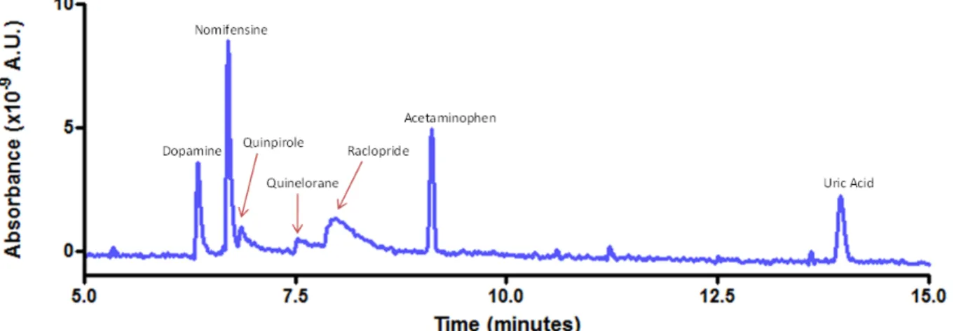
2.5. Representative electropherogram...48
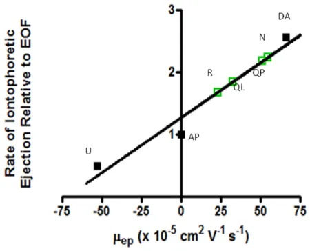
2.6. Relationship between iontophoretic and electrophoretic mobility. ...49
2.7. Spatial distribution of an iontophoretic ejection. ...52
2.8. Effect of current on stimulated dopamine release. ...57
2.9. Comparison of NaCl CVs in vitro and in vivo. ...59
2.10. Effect of AP on stimulated dopamine release. ...60
2.11. Temporal response (•) of extracellular DA concentration induced by electrical stimulation ...62
2.12. Stimulated dopamine release in an anesthetized animal before and after a localized ejection of solution containing both AP and quinelorane. ...64
2.13. Time course for changes in stimulated dopamine release with ejections of quinelorane. ...66
2.14. Linear dose-response curves ...68
2.15. Sigmoidal dose response curve for quinelorane in an anesthetized animal with quinelorane concentrations on a logarithmic scale. ...72
3.1. How cerebral blood flow fuels neuronal signaling. ...79
3.2. Glutamatergic modulation of MSN firing. ...86
xiv
3.4. Changes in the environment around the carbon-fiber electrode in vivo. ...89
4.1. Voltammetric response of dopamine (DA) and O2 in the NAc ... 103
4.2. Naturally occurring O2 changes. ... 105
4.3. Effect of electrical stimulation parameters on subsequent O2 release in the SNr and NAc. ... 107
4.4. Antagonism of dopamine receptors with raclopride and SCH 23390 and serotonin receptors with methiothepin. ... 109
4.5. Ejection of dopamine and serotonin into the NAc in an anesthetized animal. ... 111
4.6. Ejection of dopamine and serotonin into the NAc in a slice preparation. ... 113
4.7. O2 changes with local application of serotonin (5-HT) in the SNr. ... 114
5.1. Diagram of the electronics and outputs for combined fast-scan cyclic voltammetry/electrophysiology. ... 126
5.2. Modifications to probe construction and hardware for iontophoresis in freely moving animals. ... 128
5.3. Effect of dopamine transporter on exogenous dopamine diffusion. ... 131
5.4. Consistent modulation of electrically evoked dopamine release with raclopride antagonism of the pre-synaptic D2 receptor during ICSS in a behaving rat. ... 133
5.5. Immediate response of a single NAc MSN to iontophoretic application of dopamine receptor antagonists, SCH and raclopride, and dopamine in an awake animal. ... 134
5.6. Firing rates measured at another MSN indicating the prolonged analysis method. ... 139
6.1. Neuronal responses seen during VTO ICSS. ... 160
6.2. Firing rates for MSNs sorted by dopamine receptor subtype. ... 162
6.3. Pharmacological manipulation of cue cells during behavior. ... 165
xv
LIST OF ABBREVIATIONS AND SYMBOLS
* probability less than 0.05
** probability less than 0.01
*** probability less than 0.001
[O2]max maximal evoked O2 concentration
µA microamperes
µapp mobility due to applied current
µeo electroosmotic mobility
µep electrophoretic mobility
µM micromolar
µs microsecond
4-MC 4-methylcatechol
5-HT serotonin
aCSF artificial cerebral spinal fluid
Ag/AgCl silver/silverchloride
AP acetaminophen
AP anterior-posterior
Bmax drug concentration at maximal binding
BOLD blood-O2-level-dependent
Ca2+ calcium ion
CB1 cannabinoid receptor type 1
CBF cerebral blood flow
CE capillary electrophoresis
CMOS complementary metal–oxide–semiconductor
xvi
D1R D1 receptor
D2R D2 receptor
DA dopamine
DC direct current
DV dorsal-ventral
E applied electric field
ɛ permittivity
EC50 effective concentration for 50% of full effect
EGTA ethylene glycol tetraacetic acid
EOF electroosmotic flow
eVTO extended variable-time out
F Faraday's constant (96,485 C/mol) fMRI functional magnetic resonance imaging
FR fixed-ratio
FSCV fast-scan cyclic voltammetry
g gram
GABA gamma-aminobutyric acid
GPe external capsule of the globus pallidus
GPi internal capsule of the globus pallidus
H+ hydrogen ion
HDCV high definition cyclic voltammetry
Hz hertz
i applied current
i.p. intraperitoneal injection
xvii
K+ potassium ion
Kd drug concentration at 50% maximal binding
kg kilogram
kHz kilohertz
kV kilovolt
L distance between CE inlet and dectector Lt length of CE capillary
M molar
MFB medial forebrain bundle
mg milligram
Mg2+ magnesium ion
min minute
ML medial-lateral
mL milliliter
mm millimeter
ms millisecond
MSN medium spiny neuron
N nomifensine
n transport number
ƞ viscosity
nA nanoampere
Na+ sodium ion
NAc nucleus accumbens
NaCl sodium chloride
xviii NPE 2-(4-nitrophenoxy) ethanol
O2 molecular oxygen
PBS phosphate buffered saline
PEH peri-event histogram
PET Positron emission tomography
QL quinlorane
QP quinpirole
R raclopride
s second
SNr substantia nigra pars reticulata
T ejection time
tneutral time for neutral species to reach detector
tr time for analyte to reach detector
TRIS tris(hydroxymethyl)aminomethane
U uric acid
UNC University of North Carolina
UV-VIS ultraviolet-visible light
V applied voltage
V volt
ʋeo electroosmotic flow velocity
ʋep rate of electrophoretic migration
ʋobs observed linear velocity
VTA ventral tegmental area
VTO variable-time out
xix
ζ zeta-potential at glass capillary-solution interface
Chapter 1
Monitoring and modulating dopamine release and unit activity in real-time
INTRODUCTION
A vital step in creating effective treatments for addiction and learning disorders is to
understand the neuronal circuitry that drives voluntary reward-directed behaviors and how
drugs may hinder, help or hijack this system. Intracranial self stimulation (ICSS) is a well
established behavioral paradigm in which an animal learns to press a lever to stimulate a
region in their own brain and is considered a robust method to study learning and reward
(Olds and Milner, 1954). Use of this technique combined with selective lesions and pharmacological manipulation of selected brain regions has indicated that the nucleus
accumbens (NAc) is an important junction for the transmission of information during ICSS
(Saddoris et al., 2013).
The role of dopamine in circuits that control learning, goal-oriented behaviors, and
addiction has been of interest to neuroscientists for many decades. It is known that
dopaminergic neurons, many of which terminate in the NAc, change their firing patterns in
response to rewards and cues that predict reward (Mirenowicz and Schultz, 1994) and that
dopamine neurotransmission is vital for reinforcing a behavior (Di Chiara and Imperato,
1988). However, these discoveries were made using electrophysiology to monitor the firing
pattern of neurons or microdialysis to look at prolonged changes in extracellular dopamine
concentration, not dopamine concentration changes on the timescale of cell firing.
2
increased frequency (Grace and Bunney, 1984) so a technique that can distinguish changes
in dopamine concentration on this timescale is essential.
Fast-scan cyclic voltammetry (FSCV) can measure changes in local dopamine
concentrations up to 60 times per second (Kile et al., 2012), making it the perfect technique
to monitor dopamine release in freely moving animals (Garris et al., 1997; Rebec et al.,
1997, Robinson and Wightman, 2007). Thanks to technical advances over the past decade
FSCV can now be combined with electrophysiological recordings and iontophoresis,
allowing researchers to monitor and manipulate pre- and postsynaptic activity during
behavior. The technical considerations and resulting discoveries from the combination of
these techniques are discussed here.
DOPAMINE NEUROTRANSMISSION
Neurons are brain cells responsible for the integration and transmission of
information throughout the brain; they receive, process, and transmit information to and from
discrete populations of neurons within specific circuits in the brain. Classic
neurotransmission occurs when the activation of receptors on dendrites of a cell begins a
cascade of intracellular processes that often include the generation and propagation of an
action potential, a voltage difference across the neuronal membrane. The frequency of the
firing of action potentials of a cell is referred to as its unit activity, and the modulation of unit
activity is the way in which information is encoded. When an action potential propagates
along the neuron’s axon to its terminals, it triggers the release of neurotransmitter into the
extracellular space. There the neurotransmitters can bind to specific receptors on a
proximal neuron. The modulation of target neurons via neurotransmitter release is central to
3
Dopamine release in the NAc plays an extensive role in governing motivated
behaviors (Salamone and Correa, 2012) and we have shown that its release coincides with
learned associations for rewarding stimuli and drugs of abuse (Phillips et al., 2003;
Robinson and Wightman, 2007; Owesson-White et al., 2009; Beyene et al., 2010; Day et al.,
2010). There is a high density of dopamine cell bodies in the ventral tegmental area (VTA)
that send their axonal projections to many regions of the brain including the striatum. The
ventral striatum is divided into two subregions: the NAc core and NAc shell. In both NAc
subregions, dopamine terminals form synapses onto spines found on the dendrites of
medium spiny neurons (MSNs) (Yung et al., 1995). MSNs comprise 95% of the cell bodies
in the NAc and release GABA, an inhibitory neurotransmitter, upon firing (Chang and Kitai,
1985). Neurons with cell bodies located in regions other than the VTA, including other NAc
MSNs, also synapse onto these MSNs, making their activation a complex process known to
be essential for movement, learning, and motivation, and that is facilitated by dopamine
release in the region.
When dopamine is released from terminals it can then bind to any of the 5 types of dopamine receptors (D1-D5). The predominant dopamine receptors in the NAc, are the D1
receptor (D1R) and D2 receptor (D2R). Both of these receptors are G-protein linked, but
D1Rs activate G-proteins that stimulate adenylyl cyclase while D2Rs activate G-proteins that
inhibit adenylyl cyclase. D1Rs also have a lower binding affinity for dopamine than D2Rs.
This difference in binding affinities supports the idea that normal basal levels of dopamine in
the brain constantly ensure the activation of the majority of D2Rs, while sudden phasic
increases in dopamine release activate D1Rs (Dugast et al., 1997; Berke and Hyman,
2000).
D2Rs are found on both pre- and postsynaptic terminals. Pre-synaptic receptors are
commonly referred to as D2 autoreceptors (Roth, 1979) and are found to have inhibitory
4
modulation of dopamine release (Kita et al., 2007) making them an ideal target when
attempting to manipulate evoked dopamine release quickly. Additionally, there are
biochemical (Helmreich et al., 1982; Martin et al., 1982; Claustre et al., 1985),
electrophysiological (Skirboll et al., 1979), and behavioral (Bradbury et al., 1984) data
suggesting that dopamine agonists exhibit greater potency at D2 autoreceptors than at
postsynaptic D2Rs. While both pre- and postsynaptic D2 receptors exhibit similar
pharmacology (Elsworth and Roth, 1997), they differ in the G-proteins they use to inhibit
adenylyl cyclase (Montmayeur et al., 1993; Guiramand et al., 1995). Functionally, this
means more binding may be required at post-synaptic receptors to elicit comparable
adenylyl cylase inhibition. The net effect of dopamine release on MSNs is dependent on a
balance between the binding of D1Rs, postsynaptic D2Rs and D2 autoreceptors. Indeed, it
appears that the more we know about dopamine neurotransmission the more subtle and yet
complex its signaling capabilities seem.
While the different effects of D1R and D2R activation on MSNs has long been
recognized, it was recently discovered that D1Rs and D2Rs are in fact segregated onto two different MSN populations (Valjent et al., 2009; Gerfen and Surmeier, 2011). In the dorsal
striatum approximately half of MSNs contain D1Rs and project axons to the output nuclei of
the basal ganglia (substantia nigra and internal capsule of the globus pallidus). These D1
MSNs are termed the “direct pathway.” Other MSNs contain exclusively D2Rs and project
to the external capsule of the globus pallidus. These D2 MSNs are termed the “indirect
pathway” because they synapse with neurons that also project back to the output nuclei.
Although this circuitry was originally characterized in the dorsal striatum, it is also found in
5 EVOLUTION OF A WAVEFORM
A number of electrochemical techniques can be used to detect neurotransmitters in
vivo (Robinson et al., 2008) but cyclic voltammetry has the unique ability to provide a distinct current trace (cyclic voltammogram) for the oxidation and/or reduction of electroactive
compounds. This allows identification and quantification of compounds, a critical feature
that prevents incorrect conclusions about the role of a substance in a particular behavior
(Wightman and Robinson, 2002). FSCV has become the electrochemical technique of
choice for monitoring changes in neurotransmitter levels in the brain because it adds high
temporal resolution to the chemical selectivity and high sensitivity of cyclic voltammetry (Millar et al., 1985). The detection of monoamines with FSCV has been optimized over the
years to allow detection of low nanomolar concentrations of dopamine (Keithley et al.,
2011). However, the detection limit is not the only factor that must be considered when
deciding on a waveform. By varying the rate and range of potentials applied to the
carbon-fiber microelectrode the detection limit of a neuroactive species and the temporal response
of the electrode can be optimized.
The first studies in freely moving rats detected dopamine with a waveform that held
the carbon fiber at -0.4 V, increased linearly up to 1.0 V and back to -0.4 V in 9.3 ms once
every 100 ms. This potential waveform is less frequently used today for the detection of
neurotransmitters in freely moving animals because of its lack of sensitivity. However, many
important discoveries were made with its use because of its temporal resolution. These
include studies of rapid dopamine release in response to reward delivery and how the
magnitude of the dopamine changes as an animal learns a behavior (Garris et al., 1999;
Kilpatrick et al., 2000). This high speed recording of dopamine release during behavioral
tasks supported the hypothesis that dopamine release is required for learning a rewarding
6
The lack of sensitivity of the original waveform used for dopamine detection limited
FSCV to the measurement of “extracellular dopamine changes after electrical stimulation of
cell bodies in the substantia nigra compact, rather than spontaneous or gradual changes in
extracellular dopamine” (Budygin et al., 2001). Fortunately, extension of the original
waveform increased dopamine sensitivity 9-fold in vivo allowing detection of 5 nM, a level
seen for some naturally occurring changes in dopamine (Heien et al., 2003). The improved
detection limit was achieved by holding the electrode at -0.6 V and linearly increasing
voltage to 1.4 V and back to -0.6 V in 10 ms once every 100 ms. Increased sensitivity
resulted from two consequences of this extended waveform: adsorptive pre-concentration
of dopamine occurs when the electrode is held at a more negative potential between scans,
and the reactive carbon fiber surface is constantly cleaned as a consequence of
over-oxidation of its functional groups. This extended waveform decreased the temporal
response of the technique by 1.2 s, however, and decreased the selectivity of the electrode
for the oxidation and reduction of dopamine over other species. Thus this waveform is most
useful for experiments carried out in regions where only a single electroactive compound is present and when the detection limit of dopamine is of higher priority than the electrode’s
temporal response. This waveform allowed monitoring of naturally occurring increases in
dopamine release the NAc (Phillips et al., 2003) and allowed detection of basal dopamine
level changes over 90 s periods (Heien et al., 2005).
The waveform now used for dopamine detection in vivo combines the detection limit
of the extended waveform and the temporal response of the original waveform. The scan
starts at -0.4 V and linearly increases to 1.3 V at the same rate as the extended waveform,
taking 8.5 ms to complete (Heien et al., 2004). This is repeated every 100 ms. This
“dopamine waveform” allows detection of 8 nM DA without the sacrifice in temporal
7
where FSCV is used to monitor dopamine or norepinephrine and pH changes in the brain of
freely-moving animals.
With the development of methods to quantitatively measure the dynamics of several
analytes simultaneously (Heien et al., 2004) and to display thousands of cyclic
voltammograms simultaneously to qualitatively monitor the temporal dynamics of each
analyte (Michael et al., 1998), we were able to begin to ask and answer increasingly
complex questions about dopamine signaling dynamics in behavior. One of these questions
was how the dynamics of dopamine signaling affected the balance between cerebral blood
flow and metabolism in a region. A waveform was designed that allows for the reduction of
oxygen at the carbon fiber when biased to a negative potential (-1.4 V). By scanning up to
1.0 V we can also see the oxidation of dopamine and any changes in hydrogen ion
concentrations within the same scan, but the sensitivity of this waveform for monoamines is
greatly reduced due to the reduced magnitude of the positive scan and a holding potential of
0.0 V (Zimmerman and Wightman, 1991; Kennedy et al., 1992). Monitoring oxygen, pH,
and dopamine simultaneously has allowed the discoveries that dopamine seems to have little effect on the balance between cerebral blood flow (oxygen changes) and metabolism
(pH changes) in both anesthetized (Zimmerman and Wightman, 1991; Venton et al., 2003)
and awake animals (Cheer et al., 2006; Ariansen et al., 2012). The information about
metabolic dynamics during neurotransmission that this waveform allows us to monitor also
allowed us to be the first to show with the temporal and spatial resolution of our
microelectrodes that the endocannabinoid CB1 receptor mediates changes in the balance
between cerebral blood flow and metabolism in the brain of awake animals (Cheer et al.,
2006). This finding has implications for the development of new treatments for cerebral
vascular disorders and is an excellent example of the sorts of questions FSCV can answer
8
SEEING THE BRAIN COMMUNICATE: A COMBINATION OF MEASUREMENT PROCEDURES
Development of the FSCV method that is optimized for monitoring neuronal
processes was a task that analytical chemists with an interest in neuroscience could
confidently undertake. However, the design of small animal behavioral experiments to
probe the role of dopamine dynamics encompassed concepts far beyond those normally
addressed by chemists. We employed a number of unconventional stimuli such as the popping of bubble wrap outside of cages to monitor the startle response of rats (Robinson
and Wightman, 2004). We made the unanticipated discovery that the presence of female
rats induced subsecond dopamine increases in male rats. We also discovered that these
dopamine transients were unrelated to sexual differentiation (Rebec et al., 1997; Robinson
et al., 2001; Robinson et al., 2002). Nevertheless, the initial attempts at behavioral
experiments turned out to be excellent proof-of-concept for this new measurement
technique in freely moving rats.
In 2000, the Wightman lab partnered with the lab of Dr. Regina Carelli, a
psychologist with demonstrated proficiency in small animal behavior. Carelli was known for
using electrophysiology to monitor cell firing in behaviors involving reward-based learning
and drugs of abuse (Carelli and Deadwyler, 1997; Carelli, 2000; Carelli, 2002). Extracellular
electrophysiological recordings monitor voltage changes in the space around a cell that
result from potential changes arising from propagation of an action potential. Carelli and
coworkers had focused on the electrophysiology responses of dopaminergic neurons in rats
and thus it was relatively straight forward to reexamine the same behaviors while monitor
changes in dopamine release with FSCV (Carelli, 2002). The combined expertise brought
by psychologists and chemists working in parallel allowed significant breakthroughs in
9
indicating imminent lever availability from those that occur at the lever presentation 2 s later
(Stuber et al., 2005a; Day et al., 2007). In addition, the temporal resolution of FSCV
showed that the dopamine release occurred before the reward was presented once the
animal knew to expect lever presentation at a fixed interval (Cheer et al., 2007). This
directly supported earlier theories that hypothesized that dopamine is responsible for reward
prediction or its expectation instead of being a component of the hedonistic aspect (Schultz,
1998).
The early experiments used a fixed time between lever availability so it was unclear
whether dopamine was playing a role in the measurement of elapsed time or was signaling
that the lever was present. We now use a variable time-out period between lever
presentations to avoid teaching the animal that the timing of cue presentation is predictable
(Owesson-White et al., 2008). FSCV used in a variety of reward-based behavioral
paradigms established that in the NAc, dopamine release occurs during behavioral tasks
with the same rapid dynamics as changes in cell firing (Phillips et al., 2003; Roitman et al.,
2004; Stuber et al., 2005b; Stuber et al., 2005a; Owesson-White et al., 2008; Jones et al., 2010; Sugam et al., 2012).
Combining Electrochemistry with Electrophysiology
In a 2004 review, Carelli and Wightman reviewed the microcircuitry in the NAc
involved in drug addiction (Carelli and Wightman, 2004). The review contained a single
figure in which data from two separate experiments in two separate animals were overlaid to
show cell firing and dopamine changes both rapidly occurred at the administration of
cocaine. The modulation of cell firing in NAc MSNs during behavioral tasks appeared to be
dopamine-dependent (Yun et al., 2004) but the relationship between the two was (and still
is!) not fully understood. While the Wightman lab had previously ventured into simultaneous
10
(Ewing et al., 1983; Kuhr et al., 1987), the two techniques were carried out at separate
electrodes positioned 500 µm apart (Ewing et al., 1983) or in completely different brain
regions (Kuhr et al., 1987). The development of the electronics and methods to combine the
two techniques on a single electrode in a freely-moving animal was first accomplished by
the lab in 2005 (Cheer et al., 2005) and has been an ongoing area of research
(Owesson-White et al., 2009; Cacciapaglia et al., 2011; Takmakov et al., 2011; Belle et al., 2013).
The key to this combined technique is the ability to use a carbon-fiber microelectrode
both for electrochemistry and electrophysiology. This concept was first developed by Julian
Millar and coworkers in anesthetized animals (Armstrong-James and Millar, 1979, 1988;
Stamford et al., 1993). Several advantages arise from using the same electrode for both
measurements, including the ability to measure action potentials from the neurons that are
being influenced by the local neurotransmitter release sensed at the electrode (Su et al.,
1990), the minimization of tissue damage, and the decreased complexity of surgeries.
Rapidly alternating between these two techniques without glitches or artifacts requires
complex circuitry and modified experimental parameters to freely-moving systems (Garris et al., 1997), a project that required the collaboration of chemists, neurobiologists, and
electrical engineers (Cheer et al., 2005; Takmakov et al., 2011).
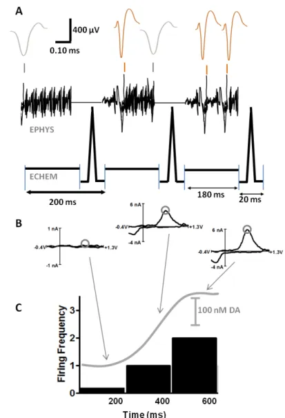
A switch is used to alternate the carbon fiber between a current amplifier for
electrochemical detections and a voltage follower for single-unit recording. The headstage
where this first stage of signal modification is completed is located on the rats’ head within a
few centimeters of the electrode, minimizing the noise in the amplified signal. Figure 1
shows the timing and output when switching between the two circuits. When the voltage
follower is connected, voltage changes occurring at the carbon fiber are recorded.
Alternatively when the current amplifier is connected to the carbon fiber, it is ramped through
a potential window and the resulting currents recorded. The original design of this
11
electrochemical voltages to leak over into the single-unit recordings (Cheer et al., 2005).
While these voltage changes looked distinct from unit activity, they often overwhelmed the
recordings making it difficult to elicit firing changes in the unit of interest. Two improvements
were made to help prevent this leakage.
The first modification was to the electrical circuit. There is now a new solid-state
relay chosen for its low leakage, low charge transfer, low and matched input capacitance,
low resistance (10 Ω), and fast (<1 µs) switching time (Takmakov et al., 2011). These
characteristics prevent excess charging currents during the switch and current leakage
between the two circuits. The second modification that helped prevent leakage of voltage
between the two systems was the timing of the voltammetric scan to the carbon fiber. The
unit-recording interval always has a 20 ms gap every 180 ms during which the 8.5 ms
voltammetric scan occurs. In initial applications of this technique, the potential scan
occurred at the very end of this 20 ms interval. This was done to maximize the adsorption of
dopamine to the electrode while it was held at a negative potential; however, this resulted in
current fluctuations when the electrode was switched over to electrophysiology. These fluctuations would manifest as large voltage changes in the single-unit data. To correct for
this, a potential of -0.4 V is applied for pre-concentration of dopamine and current
stabilization of the electrode for only the first 5 ms of the 20 ms interval. The electrode is
then ramped to 1.3 V and back to -0.4 V where it remains for ~5 ms before the mode is
switched. This timing is shown in Figure 1.1. The ability to look at these changes
concurrently and with respect to external stimuli or behavioral patterns allows us
to begin to directly tease apart the effects of dopamine neurotransmission by monitoring
dopamine release from terminals and the unit-activity of the cell with dopamine receptors in
the vicinity. The required modifications of the dopamine waveform result in some sacrifices
12
13
and the dopamine detection limit is only 62% of the traditional dopamine waveform (Cheer
et al., 2005).
This dual technique’s first discovery was to show that in the same location dopamine
release and changes in cell firing were both synchronized to lever pressing in trained
animals. MSNs that showed increases OR decreases at lever press (and electrical
stimulation), showed a simultaneous increase in dopamine (Cheer et al., 2005). This study
also showed that cell firing (but not dopamine release) changed with the administration of
GABAergic antagonists; supporting the idea that dopamine altered the probability of a cell
firing but was not the neurotransmitter directly responsible for NAc MSNs firing. Later work
demonstrated a positive correlation between the concentration of dopamine and magnitude
of cell firing change and observed that dopamine release was seen in all locations with
MSNs responsive to behavioral stimuli (Owesson-White et al., 2009). MSNs that were
unresponsive during the behavior were all in locations with no dopamine release during the
behavior (Owesson-White et al., 2009; Cacciapaglia et al., 2012). The difference between
correlation of dopamine release and cell firing in the two subregions of the NAc, the core and the shell, was also investigated. Dopamine in the core was closely timelocked to the
reinforced response of the lever press. In the shell, dopamine was released over a longer
duration and did not coincide as greatly with lever pressing (Owesson-White et al., 2009).
The same subregion specific dopamine dynamics were seen for natural rewards as well
(Cacciapaglia et al., 2012). These results demonstrate the heterogeneity of dopamine
release and suggest this release is positioned to selectively modulate specific MSNs.
After establishing the link between dopamine release and cell firing during ICSS
(Cheer et al., 2005; Cheer et al., 2007; Owesson-White et al., 2009), we investigated
whether this link was also seen with natural rewards (Cacciapaglia et al., 2011). Rats were
trained to press a lever for a sucrose pellet instead of a direct electrical stimulation. The
14
surge in dopamine concentration at the onset of the cue and (to a lesser extent) with lever
press, showed one of four alterations in MSN firing: inhibition at cue presentation, excitation
at cue presentation, inhibition at lever press, and excitation at lever press. To see if this
surge of dopamine was in fact responsible for the coincident changes in cell firing, NMDA
receptors were blocked in the VTA attenuating the burst firing of dopamine cells in the
region (Chergui et al., 1993). This decreased dopamine release in the NAc. MSNs that
showed an excitation to the cue onset or lever press became non-phasic, while cells that
were inhibited at either the cue or the lever press were unaffected by the diminished in
dopamine release. This showed dopamine’s ability to selectively modulate discrete
pathways within the NAc and suggested that this was a selective modulation of the direct
(D1 MSNs) or indirect (D2 MSNs) pathway.
Controlled Iontophoresis
Iontophoresis is a technique that uses current to induce the migration of a solution of
ions through a glass pipette. It was developed in the early 1950s by W.L. Natsuk, a student
of A.L. Hodgkin (Nastuk, 1953). While attempting to understand how ions contributed to the
actions of acetylcholine at the neuromuscular junction, he noticed that acetylcholine would
naturally leak out of a glass pipette pulled to a fine tip and that application of current to the
pipette solution ejected even more acetylcholine onto the junction (Hicks, 1984). From
there, iontophoresis increased in popularity and extensive studies on the technique were
carried out (Krnjevic et al., 1963a; Krnjevic et al., 1963b; Crawford and Curtis, 1964; Curtis
and Nastuk, 1964; Bradley and Candy, 1970; Bloom, 1974; Simmonds, 1974; Freedman et
al., 1975; Purves, 1977, 1979). The technique’s popularity for studying receptor dynamics in
vivo is due to the fact that drugs can be quickly, selectively, and locally delivered to the site of action with minimal disruption of tissue. Systemic drug administration is only useful for
15
where the drug can cross the blood-brain barrier, the drug affects the entire brain making it
difficult to study discrete brain region effects (Bloom, 1974). Additionally, systemic drug
administration can alter animal behavior, making it difficult to look at the drug effects in the
brain during behavior (Hernandez and Cheer, 2012). These problems are avoided by using
iontophoresis to study the pharmacology of the brain.
A drawback to iontophoresis was the inability to monitor or quantify the amount of
drug delivered. This made it impossible to differentiate a null response to drug application
from a clogged glass pipette. Additionally, too little drug delivered could result in a false
negative whereas excessive application could lead to nonspecific effects. Applied pump
currents are commonly used to compare ejections (Pierce and Rebec, 1995; Kiyatkin and
Rebec, 1996, 1999b), but the same pump current ejects different drug concentrations from
barrel to barrel (Herr et al., 2008). Modifications to the design of Millar and co-workers,
which coupled iontophoresis barrels to carbon-fiber microelectrodes, allow detection of
electroactive compounds ejected with iontophoresis at the neighboring electrode
(Armstrong-James et al., 1981).
Using these coupled iontophoresis probes, electroosmosis was found to contribute
significantly to the observed drug delivery (Herr et al., 2008). Electroosmosis is a
phenomenon caused by the attraction between the cations in solution and the ionizable
silanol groups on the glass capillary surface. When a positive current is applied to the
capillary, the cations along the wall migrate toward the anode, inducing a bulk movement of
solution, termed electroosmotic flow. The variability in iontophoretic ejections is associated
with variability of electroosmotic flow from barrel to barrel, while electrophoretic mobility
(ionic migration) for a given species is consistent. Using an electroactive neutral molecule
as an internal standard to monitor the variability in electroosmotic flow, and subsequently
the amount of drug delivered from different barrels, allows us to control for this variability.
16
electroinactive drugs by monitoring the coejection of an electroactive molecule from the
same barrel (Herr et al., 2008). When the relative mobilities of the coejected substances are
known, monitoring the concentration of the electroactive molecule with the carbon-fiber
electrode provides an indirect measure of the relative concentration of the coejected
nonelectroactive substance (Herr et al., 2008; Herr et al., 2010).
While FSCV had previously been coupled to iontophoresis to monitor modulation of
cell firing by electroactive compounds (Kiyatkin and Rebec, 1996, 1997; Rebec, 1998;
Kiyatkin and Rebec, 1999a, b; Kiyatkin et al., 2000; Kiyatkin and Rebec, 2000), it was not
until 2010 that the technique was used in vivo to alter release from dopamine terminals in
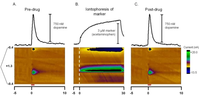
the region of drug application (Herr et al., 2010). These papers established that controlled
iontophoresis could be used to quickly (<60 s) modulate dopamine release by affecting
D2 autoreceptors and the dopamine transporter in anesthetized (Herr et al., 2010) and
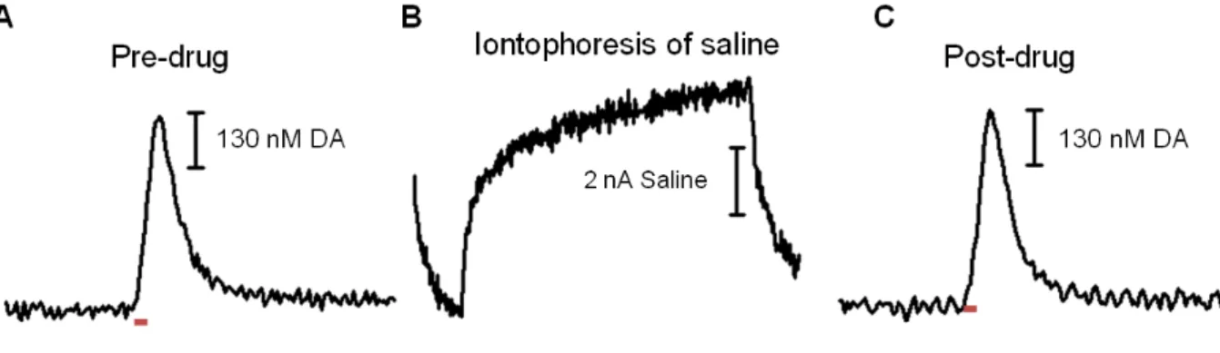
freely-moving animals (Belle et al., 2013). Monitoring iontophoresis ejections eliminates the
fear of ionic and electrical artifacts from the ejection current altering the neuronal
environment as well. With controlled iontophoresis coupled to the simultaneous
electrochemistry/electrophysiology technique we can watch for these problems during an
experiment. While changes to the neuronal environment are not seen from ejection of
saline or the electroactive marker molecule, if the ejection current is seen to affect the
electrode, electrical connections can be altered or a different barrel on the probe used to
prevent any data collection problems (Belle et al., 2013).
With the discovery of the discrete location of D1Rs and D2Rs on separate
populations of MSNs (Valjent et al., 2009; Gerfen and Surmeier, 2011) came a huge
breakthrough in how neuronal networks in the brain work together in reward, behavior and
addiction. In the NAc core, only 6% of MSNs have both D1Rs and D2Rs, while 53% of
MSN’s have D1Rs exclusively (D1 MSNs) and 41% of MSNs have D2Rs exclusively (D2
17
been investigated (Surmeier et al., 2007), direct electrophysiological evidence has been
lacking, partly because of the difficulties in differentiating D1 MSNs and D2 MSNs in vivo
(Venance and Glowinski, 2003).
Coupling controlled iontophoresis to the same microelectrode in a combined FSCV
and single-unit recording experiment allows for pharmacological identification of MSNs
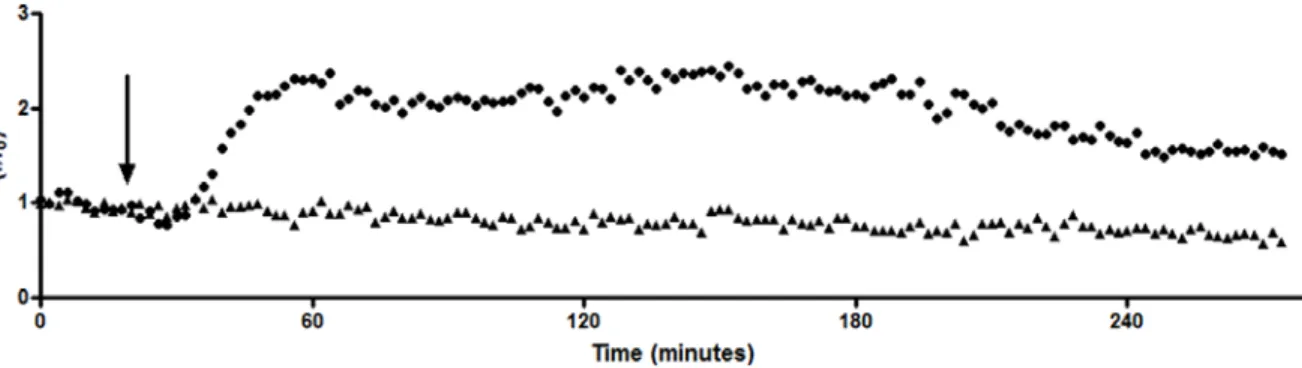
based on their responses to D1R and D2R antagonists (Belle et al., 2013). The firing rates
of NAc MSNs in awake animals were monitored before, during and after a 15 s iontophoretic
ejection of specific dopamine receptor antagonist. Changes in response to these
antagonists were seen both immediately and on a prolonged timescale (as an overall
change in the firing rate of a neuron after application). Looking at prolonged changes, 40%
of MSNs increased their firing rate after local application of a D2R antagonist, 46% of MSNs
exhibited a decreased firing rate after local application of the D1R antagonist, and only 11%
of MSNs responded to both antagonists. These results are in agreement with previously
reported distributions for dopamine receptor subtypes on MSNs (Valjent et al., 2009)
supporting the method as a way to discriminate between and selectively modulate D1 MSNs and D2 MSNs in vivo.
CONCLUSIONS
As our knowledge of the subtle and complex signaling required for the brain to encode
reward-directed behaviors increases, the ability to selectively modulate and monitor
dopamine release and MSN firing in freely-moving animals engaged in behavioral tasks
becomes even more essential. The development of a technique to monitor subsecond
dopamine fluctuations in freely-moving animals has allowed the study of naturally and
electrically evoked dopamine during reward-motivated tasks and provided the ability to
18
concurrent observation of cell firing patterns in vivo is essential for understanding
neurotransmission. The technique has shown that MSNs alter their firing during a
behavioral task, receive dopaminergic inputs during the behavior and that dopamine release
is required for excitations of MSNs during behavior. This combined technique is a passive
way to ‘listen in’ on neurotransmission and the ability to selectively and locally modulate this
neuronal conversation will allow an even greater understanding of the purpose of
dopaminergic pathways in learning and reward.
DISSERTATION OVERVIEW
While the final two chapters of this dissertation directly report the development and
first insights from the combined electrochemical/electrophysiological with iontophoretic
modulation discussed here, other applications of controlled iontophoresis in anesthetized
animals are first discussed in Chapters 2, 3 and 4. These Chapters lay the ground work for
further iontophoretic investigations in awake animals. Chapter 2 presents the utility and
methodology of controlled iontophoresis for pharmacological manipulation of
neurotransmission. Controlled iontophoresis is then used to probe the relationship between
cerebral metabolism (neurotransmission) and blood flow. First, in Chapter 3 this relationship
is examined for glutamate, a neurotransmitter with a known vasoactive role in the brain.
Then, in Chapter 4 methodology from Chapter 3 is used to investigate the role of serotonin
in regulation of cerebral blood flow. Finally, controlled iontophoresis is modified for use in
conscious animals (Chapter 5) to elicit the role of dopamine in ICSS (Chapter 6). These
projects all demonstrate the utility and improvements of controlled iontophoresis to look at
19 REFERENCES
Ariansen JL, Heien MLAV, Hermans A, Phillips PEM, Hernadi I, Bermudez M, Schultz W, Wightman RM (2012) Monitoring extracellular pH, oxygen, and dopamine during reward delivery in the striatum of primates. Front Behav Neurosci 6.
Armstrong-James M, Millar J (1979) Carbon fibre microelectrodes. J Neurosci Methods 1:279-287.
Armstrong-James M, Millar J (1988) High-speed cyclic voltammetry and unit recording with carbon fibre microelectrodes. In: Measurement Of Neurotransmitter Release in vivo (Marden C, ed), pp 209-224. New York: John Wiley and Sons Ltd.
Armstrong-James M, Fox K, Kruk ZL, Millar J (1981) Quantitative ionophoresis of
catecholamines using multibarrel carbon fibre microelectrodes. J Neurosci Methods 4:385-406.
Belle AM, Owesson-White C, Herr NR, Carelli RM, Wightman RM (2013) Controlled Iontophoresis Coupled with Fast-Scan Cyclic Voltammetry/Electrophysiology in Awake, Freely Moving Animals. ACS Chem Neurosci 5:761-71.
Berke JD, Hyman SE (2000) Addiction, dopamine, and the molecular mechanisms of memory. Neuron 25:515 -532.
Beyene M, Carelli RM, Wightman RM (2010) Cue-evoked dopamine release in the nucleus accumbens shell tracks reinforcer magnitude during intracranial self-stimulation. Neuroscience 169:1682-1688.
Bloom FE (1974) To spritz or not to spritz: The doubtful value of aimless iontophoresis. Life Sciences 14:1819-1834.
Bradbury AJ, Cannon JG, Costall B, Naylor RJ (1984) A comparison of dopamine agonist action to inhibit locomotor activity and to induce stereotyped behaviour in the mouse. Eur J Pharmacol 105:33-47.
Bradley PB, Candy JM (1970) Iontophoretic release of acetylcholine, noradrenaline, 5-hydroxytryptamine and D-lysergic acid diethylamide from micropipettes. Br J Pharmacol 40:194-201.
Budygin EA, Phillips PE, Robinson DL, Kennedy AP, Gainetdinov RR, Wightman RM (2001) Effect of acute ethanol on striatal dopamine neurotransmission in ambulatory rats. J Pharmacol Exp Ther 297:27-34.
Cacciapaglia F, Wightman RM, Carelli RM (2011) Rapid Dopamine Signaling Differentially Modulates Distinct Microcircuits within the Nucleus Accumbens during Sucrose-Directed Behavior. J Neurosci 31:13860-13869.
20
Carelli RM (2000) Activation of accumbens cell firing by stimuli associated with cocaine delivery during self-administration. Synapse 35:238-242.
Carelli RM (2002) Nucleus accumbens cell firing during goal-directed behaviors for cocaine vs. 'natural' reinforcement. Physiol Behav 76:379-387.
Carelli RM, Deadwyler SA (1997) Cellular mechanisms underlying reinforcement-related processing in the nucleus accumbens: electrophysiological studies in behaving animals. Pharmacol Biochem Behav 57:495-504.
Carelli RM, Wightman RM (2004) Functional microcircuitry in the accumbens underlying drug addiction: insights from real-time signaling during behavior. Curr Opin Neurobiol 14:763-768.
Chang HT, Kitai ST (1985) Projection neurons of the nucleus accumbens: an intracellular labeling study. Brain Res 347:112-116.
Cheer JF, Wassum KM, Wightman RM (2006) Cannabinoid modulation of electrically evoked pH and oxygen transients in the nucleus accumbens of awake rats. J Neurochem 97:1145-1154.
Cheer JF, Heien ML, Garris PA, Carelli RM, Wightman RM (2005) Simultaneous dopamine and single-unit recordings reveal accumbens GABAergic responses: implications for intracranial self-stimulation. Proc Natl Acad Sci USA 102:19150-19155.
Cheer JF, Aragona BJ, Heien ML, Seipel AT, Carelli RM, Wightman RM (2007) Coordinated accumbal dopamine release and neural activity drive goal-directed behavior. Neuron 54:237-244.
Chergui K, Charlety PJ, Akaoka H, Saunier CF, Brunet JL, Buda M, Svensson TH, Chouvet G (1993) Tonic activation of NMDA receptors causes spontaneous burst discharge of rat midbrain dopamine neurons in vivo. Eur J Neurosci 5:137-144.
Claustre Y, Fage D, Zivkovic B, Scatton B (1985) Relative selectivity of
6,7-dihydroxy-2-dimethylaminotetralin, N-n-propyl-3-(3-hydroxyphenyl)piperidine, N-n-propylnorapomorphine and pergolide as agonists at striatal dopamine
autoreceptors and postsynaptic dopamine receptors. J Pharmacol Exp Ther 232:519-525.
Crawford JM, Curtis DR (1964) The Excitation and Depression of Mammalian Cortical Neurones by Amino Acids. Br J Pharmacol Chemother 23:313-329.
Curtis DR, Nastuk WL (1964) Micro-electrophoresis. In: Physical Techniques in Biological Research, pp 144-190. New York: Academic Press.
Day JJ, Roitman MF, Wightman RM, Carelli RM (2007) Associative learning mediates dynamic shifts in dopamine signaling in the nucleus accumbens. Nat Neurosci 10:1020-1028.
21
Di Chiara G, Imperato A (1988) Drugs abused by humans preferentially increase synaptic dopamine concentrations in the mesolimbic system of freely moving rats. Proc Natl Acad Sci USA 85:5274-5278.
Dugast C, Brun P, Sotty F, Renaud B, Suaud-Chagny MF (1997) On the involvement of a tonic dopamine D2-autoinhibition in the regulation of pulse-to-pulse-evoked dopamine release in the rat striatum in vivo. Naunyn Schmiedebergs Arch Pharmacol 355:716-719.
Elsworth JD, Roth RH (1997) Dopamine Synthesis, Uptake, Metabolism, and Receptors: Relevance to Gene Therapy of Parkinson's Disease. Exp Neurol 144:4-9.
Ewing AG, Alloway KD, Curtis SD, Dayton MA, Wightman RM, Rebec GV (1983)
Simultaneous electrochemical and unit recording measurements: characterization of the effects of D-amphetamine and ascorbic acid on neostriatal neurons. Brain Res 261:101-108.
Freedman R, Hoffer BJ, Woodward DJ (1975) A quantitative microiontophoretic analysis of the responses of central neurones to noradrenaline: interactions with cobalt,
manganese, verapamil and dichloroisoprenaline. Br J Pharm 54:529-539.
Garris PA, Christensen JR, Rebec GV, Wightman RM (1997) Real-time measurement of electrically evoked extracellular dopamine in the striatum of freely moving rats. J Neurochem 68:152-161.
Garris PA, Kilpatrick M, Bunin MA, Michael D, Walker QD, Wightman RM (1999)
Dissociation of dopamine release in the nucleus accumbens from intracranial self-stimulation. Nature 398:67-69.
Gerfen CR, Surmeier DJ (2011) Modulation of striatal projection systems by dopamine. Annu Rev Neurosci 34:441-466.
Gratton A, Wise RA (1994) Drug- and behavior-associated changes in dopamine-related electrochemical signals during intravenous cocaine self- administration in rats. J Neurosci 14:4130-4146.
Grace AA, Bunney BS (1984) The control of firing patterns in nigral dopamine neurons: burst firing. J Neurosci 4:2877-2890.
Guiramand J, Montmayeur J-P, Ceraline J, Bhatia M, Borrelli E (1995) Alternative Splicing of the Dopamine D2 Receptor Directs Specificity of Coupling to G-proteins. J Biol Chem 270:7354-7358.
Heien ML, Johnson MA, Wightman RM (2004) Resolving neurotransmitters detected by fast-scan cyclic voltammetry. Anal Chem 76:5697-5704.
22
Heien ML, Khan AS, Ariansen JL, Cheer JF, Phillips PE, Wassum KM, Wightman RM (2005) Real-time measurement of dopamine fluctuations after cocaine in the brain of
behaving rats. Proc Natl Acad Sci USA 102:10023-10028.
Helmreich I, Reimann W, Hertting G, Starke K (1982) Are presynaptic dopamine
autoreceptors and postsynaptic dopamine receptors in the rabbit caudate nucleus pharmacologically different? Neuroscience 7:1559-1566.
Hernandez G, Cheer JF (2012) Effect of CB1 receptor blockade on food-reinforced
responding and associated nucleus accumbens neuronal activity in rats. J Neurosci 32:11467-11477.
Herr NR, Kile BM, Carelli RM, Wightman RM (2008) Electroosmotic flow and its contribution to iontophoretic delivery. Anal Chem 80:8635-8641.
Herr NR, Daniel KB, Belle AM, Carelli RM, Wightman RM (2010) Probing presynaptic regulation of extracellular dopamine with iontophoresis. ACS Chem Neurosci 1:627-638.
Hicks TP (1984) The history and development of microiontophoresis in experimental neurobiology. Prog Neurobiol 22:185-240.
Ikemoto S (2007) Dopamine reward circuitry: Two projection systems from the ventral midbrain to the nucleus accumbens-olfactory tubercle complex. Brain Res Rev 56:27-78.
Jones JL, Day JJ, Aragona BJ, Wheeler RA, Wightman RM, Carelli RM (2010) Basolateral amygdala modulates terminal dopamine release in the nucleus accumbens and conditioned responding. Biol Psychiatry 67:737-744.
Keithley RB, Takmakov P, Bucher ES, Belle AM, Owesson-White CA, Park J, Wightman RM (2011) Higher sensitivity dopamine measurements with faster-scan cyclic
voltammetry. Anal Chem 83:3563-3571.
Kennedy RT, Jones SR, Wightman RM (1992) Simultaneous measurement of oxygen and dopamine: coupling of oxygen consumption and neurotransmission. Neuroscience 47:603-612.
Kile BM, Walsh PL, McElligott ZA, Bucher ES, Guillot TS, Salahpour A, Caron MG, Wightman RM (2012) Optimizing the Temporal Resolution of Fast-Scan Cyclic Voltammetry. ACS Chem Neurosci 3:285-292.
Kilpatrick MR, Rooney MB, Michael DJ, Wightman RM (2000) Extracellular dopamine dynamics in rat caudate-putamen during experimenter-delivered and intracranial self-stimulation. Neuroscience 96:697-706.
23
Kiyatkin EA, Rebec GV (1996) Dopaminergic modulation of glutamate-induced excitations of neurons in the neostriatum and nucleus accumbens of awake, unrestrained rats. J Neurophysiol 75:142-153.
Kiyatkin EA, Rebec GV (1997) Iontophoresis of amphetamine in the neostriatum and nucleus accumbens of awake, unrestrained rats. Brain Res 771:14-24.
Kiyatkin EA, Rebec GV (1999a) Striatal neuronal activity and responsiveness to dopamine and glutamate after selective blockade of D1 and D2 dopamine receptors in freely moving rats. J Neurosci 19:3594-3609.
Kiyatkin EA, Rebec GV (1999b) Modulation of striatal neuronal activity by glutamate and GABA: iontophoresis in awake, unrestrained rats. Brain Res 822:88-106.
Kiyatkin EA, Rebec GV (2000) Dopamine-independent action of cocaine on striatal and accumbal neurons. Eur J Neurosci 12:1789-1800.
Kiyatkin EA, Kiyatkin DE, Rebec GV (2000) Phasic inhibition of dopamine uptake in nucleus accumbens induced by intravenous cocaine in freely behaving rats. Neuroscience 98:729-741.
Krnjevic K, Laverty R, Sharman DF (1963a) Iontophoretic release of adrenaline,
noradrenaline and 5-hydroxytryptamine from micropipettes. Br J Pharmacol Chem 20:491-496.
Krnjevic K, Mitchell JF, Szerb JC (1963b) Determination of iontophoretic release of acetylcholine from micropipettes. J Physiol 165:421-436.
Kuhr WG, Wightman RM, Rebec GV (1987) Dopaminergic neurons: simultaneous
measurements of dopamine release and single-unit activity during stimulation of the medial forebrain bundle. Brain Res 418:122-128.
Martin GE, Williams M, Haubrich DR (1982) A pharmacological comparison of 6,7-dihydroxy-2-dimethylaminotetralin (TL-99) and
N-n-propyl-3-(3-hydroxyphenyl)piperidine with (3-PPP) selected dopamine agonists. J Pharmacol Exp Ther 223:298-304.
Michael D, Travis ER, Wightman RM (1998) Color images for fast-scan CV measurements in biological systems. Anal Chem 70:586A-592A.
Millar J, Stamford JA, Kruk ZL, Wightman RM (1985) Electrochemical, pharmacological and electrophysiological evidence of rapid dopamine release and removal in the rat caudate nucleus following electrical stimulation of the median forebrain bundle. Eur J Pharmacol 109:341-348.
Mirenowicz J, Schultz W (1994) Importance of unpredictability for reward responses in primate dopamine neurons. J Neurophysiol 72:1024-1027.
24
Nastuk WL (1953) Membrane potential changes at a single muscle endplate produced by transitory application of acetylcholine with an electrically controlled microjet. Fed Proc 102.
Olds J, Milner P (1954) Positive reinforcement produced by electrical stimulation of septal area and other regions of rat brain. J Comp Physiol Psychol 47:419-427.
Owesson-White CA, Cheer JF, Beyene M, Carelli RM, Wightman RM (2008) Dynamic changes in accumbens dopamine correlate with learning during intracranial self-stimulation. Proc Natl Acad Sci USA 105:11957-11962.
Owesson-White CA, Ariansen J, Stuber GD, Cleaveland NA, Cheer JF, Wightman RM, Carelli RM (2009) Neural encoding of cocaine-seeking behavior is coincident with phasic dopamine release in the accumbens core and shell. Eur J Neurosci
30:1117-1127.
Phillips PE, Stuber GD, Heien ML, Wightman RM, Carelli RM (2003) Subsecond dopamine release promotes cocaine seeking. Nature 422:614-618.
Pierce RC, Rebec GV (1995) Iontophoresis in the neostriatum of awake, unrestrained rats: differential effects of dopamine, glutamate and ascorbate on motor- and nonmotor-related neurons. Neuroscience 67:313-324.
Purves RD (1977) The release of drugs from iontophoretic pipettes. J Theor Biol 66:789-798.
Purves RD (1979) The physics of iontophoretic pipettes. J Neurosci Methods 1:165-178. Rebec GV (1998) Real-time assessments of dopamine function during behavior: single-unit
recording, iontophoresis, and fast-scan cyclic voltammetry in awake, unrestrained rats. Alcohol Clin Exp Res 22:32-40.
Rebec GV, Christensen JR, Guerra C, Bardo MT (1997) Regional and temporal differences in real-time dopamine efflux in the nucleus accumbens during free-choice novelty. Brain Res 776:61-67.
Robinson DL, Wightman RM (2004) Nomifensine amplifies subsecond dopamine signals in the ventral striatum of freely-moving rats. J Neurochem 90:894-903.
Robinson DL, Wightman RM (2007) Rapid dopamine release in freely moving rats. In: Electrochemical Methods for Neuroscience (Michael AC, Borland LM, eds), pp 17-36. Boca Raton, FL: CRC Press.
Robinson DL, Heien ML, Wightman RM (2002) Frequency of dopamine concentration transients increases in dorsal and ventral striatum of male rats during introduction of conspecifics. J Neurosci 22:10477-10486.
25
Robinson DL, Phillips PE, Budygin EA, Trafton BJ, Garris PA, Wightman RM (2001) Sub-second changes in accumbal dopamine during sexual behavior in male rats. Neuroreport 12:2549-2552.
Roitman MF, Stuber GD, Phillips PE, Wightman RM, Carelli RM (2004) Dopamine operates as a subsecond modulator of food seeking. J Neurosci 24:1265-1271.
Roth RH (1979) Dopamine autoreceptors: pharmacology, function and comparison with post-synaptic dopamine receptors. Commun Psychopharmacol 3:429-445.
Saddoris MP, Sugam JA, Cacciapaglia F, Carelli RM (2013) Rapid dopamine dynamics in the accumbens core and shell: learning and action. Front Biosci (Elite Ed) 5:273-288. Salamone JD, Correa M (2012) The mysterious motivational functions of mesolimbic
dopamine. Neuron 76:470-485.
Schultz W (1998) Predictive reward signal of dopamine neurons. J Neurophysiol 80:1-27. Simmonds MA (1974) Quantitative evaluation of responses to microiontophoretically applied
drugs. Neuropharmacol 13:401-406.
Skirboll LR, Grace AA, Bunney BS (1979) Dopamine auto- and postsynaptic receptors. Electrophysiological evidence for differential sensitivity to dopamine agonists. Science 206:80-82.
Stamford JA, Palij P, Davidson C, Jorm CM, Millar J (1993) Simultaneous "real-time" electrochemical and electrophysiological recording in brain slices with a single carbon-fibre microelectrode. J Neurosci Methods 50:279-290.
Stuber GD, Wightman RM, Carelli RM (2005a) Extinction of cocaine self-administration reveals functionally and temporally distinct dopaminergic signals in the nucleus accumbens. Neuron 46:661-669.
Stuber GD, Roitman MF, Phillips PE, Carelli RM, Wightman RM (2005b) Rapid dopamine signaling in the nucleus accumbens during contingent and noncontingent cocaine administration. Neuropsychopharmacol 30:853-863.
Su MT, Dunwiddie TV, Gerhardt GA (1990) Combined electrochemical and
electrophysiological studies of monoamine overflow in rat hippocampal slices. Brain Res 518:149-158.
Sugam JA, Day JJ, Wightman RM, Carelli RM (2012) Phasic Nucleus Accumbens Dopamine Encodes Risk-Based Decision-Making Behavior. Biol Psychiatry 71:199-205.
26
Takmakov P, McKinney CJ, Carelli RM, Wightman RM (2011) Instrumentation for fast-scan cyclic voltammetry combined with electrophysiology for behavioral experiments in freely moving animals. Rev Sci Instrum 82:074301-4306.
Valjent E, Bertran-Gonzalez J, Herve D, Fisone G, Girault JA (2009) Looking BAC at striatal signaling: cell-specific analysis in new transgenic mice. Trends Neurosci 32:538-547. Venance L, Glowinski J (2003) Heterogeneity of spike frequency adaptation among medium
spiny neurones from the rat striatum. Neuroscience 122:77-92.
Venton BJ, Michael DJ, Wightman RM (2003) Correlation of local changes in extracellular oxygen and pH that accompany dopaminergic terminal activity in the rat caudate-putamen. J Neurochem 84:373-381.
Wightman RM, Robinson DL (2002) Transient changes in mesolimbic dopamine and their association with 'reward'. J Neurochem 82:721-735.
Yun IA, Wakabayashi KT, Fields HL, Nicola SM (2004) The ventral tegmental area is required for the behavioral and nucleus accumbens neuronal firing responses to incentive cues. J Neurosci 24:2923-2933.
Yung KK, Bolam JP, Smith AD, Hersch SM, Ciliax BJ, Levey AI (1995) Immunocytochemical localization of D1 and D2 dopamine receptors in the basal ganglia of the rat: light and electron microscopy. Neuroscience 65:709-730.