HARNESSING INTER-INDIVIDUAL VARIABILITY TO
IDENTIFY MOLECULAR MECHANISMS SHAPING AIRWAY EPITHELIAL CELL TRANSCRIPTIONAL RESPONSES TO OZONE EXPOSURE
Emma Ciel Bowers
A dissertation submitted to the faculty at the University of North Carolina at Chapel Hill in partial fulfillment of the requirements for the degree of Doctor of Philosophy in the Curriculum
of Toxicology.
Chapel Hill 2017
Approved by
iii ABSTRACT
Emma Ciel Bowers: Harnessing Inter-Individual Variability to Identify the Molecular Mechanisms Shaping Airway Epithelial Cell Transcriptional Responses to Ozone Exposure
(Under the direction of David Diaz-Sanchez)
Millions of people are exposed to levels of the ambient air pollutant ozone that are known to produce pulmonary inflammation; however, inflammatory responses exhibit extensive inter-individual variability. Moreover, during multi-day exposures acute inflammatory responses are attenuated, resulting in a phenomenon known as “ozone adaptation.” The mechanisms
governing these phenomena are not understood, but their identification is essential in
understanding the health impacts of air pollutant exposure. The ozone-mediated induction of pro-inflammatory genes is a key step in the release of cytokines and chemokines in the airway. Thus, differences in the regulation of transcription may be a potential source of ozone
iv
thus I investigated whether these kinases also controlled gene induction inter-individual variability. I found that phBECs with higher inductions of IL-8 are distinguished by elevated activation of ERK1/2, but not p38, following ozone exposure. Upon repeated ozone exposure, ozone-responsive gene expression was suppressed and was paralleled by decreases in ERK1/2 activation, suggesting that this may be an important adaptive mechanism. In collaboration with other scientists, I also found that epigenetic modifications at pro-inflammatory gene promoters were strongly associated with ozone-associated gene expression, suggesting that the epigenome is critical part of epithelial cell response ‘programming.’ In summary, this work identifies novel molecular mechanisms that dictate responsiveness to ozone exposure. This information can be used to refine definitions of susceptible populations and better predict health outcomes
v
I dedicate this work to my parents,
vi
ACKNOWLEDGMENTS
This work would not have been possible without the exceptional guidance and
mentorship of David Diaz-Sanchez and Shaun McCullough. David’s experience and knowledge of the field were essential in helping me choose my battles and strategically plan my research. I would often arrive in David’s office gushing ideas about experiments, but would leave with one resonating word: “FOCUS.” David’s insight and criticism were essential in preparation for
committee meetings, presentations, publications, etc. David considers himself to be a harsh critic, but I tend to disagree. I think it’s because when someone is ripping your work apart in an English accent, it distracts you from having your feelings hurt. Importantly, whenever I felt overwhelmed, downtrodden, or panicked, David’s ability to put life (and science) into
perspective always made me feel better.
I became Shaun McCullough’s ‘ward’ when he was transitioning between a post-doctoral
position and a principle investigator. Initially Shaun’s role was to teach me laboratory
techniques and chromatin biology; however, his role thereafter became much more of a friend and mentor. Shaun’s attention to detail, technical knowledge, enthusiasm for science, and
emphasis on professionalism were essential in my development as a scientist. Many of my accomplishments have been the result of his incessant goading and encouragement. I will never forget the hours we spent side-by-side on the computer honing my scientific writing skills. Doctoral students should be so lucky to have somebody like that in their corner.
vii
Doan and David were great friends, but also great technicians. Together we harvested cells, optimized ChIP protocols, performed clinical studies, and ran a lot of qPCR (I mean a lot). Doan even forgave me when I messed up our three-day protocol when I got too distracted by juicy gossip (it was the third day). Lisa Dailey, also part of our team, was essential in curating
primary cells and growing phBEC cultures. Given the hundreds (likely thousands) of Transwells that I have used in my research and the months of care they required, I wouldn’t have
accomplished nearly as much if I had to manage it all on my own. Hannah Smith, a newer addition to our team, is a promising undergraduate from the UNC School of public health. Hannah performed several of the analyses included in this dissertation, which speaks to her impressive capabilities as a young researcher.
I would be remiss if I didn’t thank the rest of the EPA family who helped me throughout my graduate work. Scott Meade and the rest of the TRC Engineering team built and maintain the exposure chambers that were critical to my research. I am also grateful to the nurses Tracey, Mary Anne, and Julie for their assistance, sense of humor, and for allowing me to be a co-investigator in clinical studies. I would also like to acknowledge the heroic efforts of Ilona Jaspers and Julie Cannefax. These ladies have gone out of their way to help me many times, especially with the STAR fellowship that I had to turn down, and I can’t imagine what the program would be like without them (seriously, it would be scary).
I would like to think my mother and father for being great parents, being unapologetically eccentric, and thinking the world of me (somebody’s got to). I would also like to think my
viii
I would also thank the funding sources that have made my studies possible. My research was funded by the Toxicology T32 training grant T32-ES007126 and the Center for
ix PREFACE
Although I was the lead in this dissertation, much of this work was done in collaboration with other scientists. I would like to take this opportunity to acknowledge these individuals and describe their role in this research. Thought the dissertation, David Diaz-Sanchez and Shaun McCullough helped me design and plan experiments, hence they are the senior and second authors, respectively, on my publications. Other individuals also greatly contributed to this research. Lisa Dailey, our primary cell technician, was instrumental in curating phBEC donor libraries and preparing cell cultures. David Morgan and Doan On, both technicians in the McCullough lab, were helpful throughout my dissertation research. Doan, David, and Lisa assisted with sample collection during large harvests. David also helped process some of the RNA extractions and PCR reactions from Chapters 1 and 2. Hannah Smith, a bright
undergraduate in our lab, was able to use my banked samples to perform several of her own experiments, which include the baseline transcript analysis described in Figure 2-9, the oxidative stress PCR panel featured in Figure 3-5, and the GSTM1 analysis in Figure 5-5.
Large portions of Chapters 2 and 3 have been or are currently in preparation for publication submission:
Bowers EC, McCullough SD, Morgan DS, Dailey LA, Diaz-Sanchez D. (Under review 2017) Ozone-mediated IL-8 responsiveness is an intrinsic property of airway epithelial cells and is determined by the activation of the MAP kinase ERK1/2.
x
Chapter 4 represents unpublished data that is the result of a collaborative effort between myself and the aforementioned individuals. Doan On, David Morgan, and Shaun McCullough banked the material that was used in this chapter. Under the supervision of Shaun McCullough, I designed the ChIP experiments and performed the data analysis. To increase the throughput of our sample processing, Doan On and I performed the ChIP reactions in tandem. In order to generate this data our lab spent a significant amount of time deconstructing and optimizing our ChIP protocol. Our efforts resulted in a streamlined, efficient ChIP protocol that we were then invited to publish, which can be found at:
McCullough, S.D., On, D.M., Bowers, E.C. Using Chromatin Immunoprecipitation in Toxicology: A Step-by-Step Guide to Increasing Efficiency, Reducing Variability, and Expanding Applications. Current Protocols in Toxicology, 3-14.
Several of the figures and discussion points in Chapters 1 and 4 were previously published in a review that I coauthored with Shaun McCullough:
Bowers, E. C., & McCullough, S. D. (2017). Linking the Epigenome with Exposure Effects and Susceptibility: The Epigenetic Seed and Soil Model. Toxicological Sciences, 155 (2): 302-314. Used with permission.
xi
TABLE OF CONTENTS
LIST OF TABLES ... xiv
LIST OF FIGURES ... xv
LIST OF ABBREVIATIONS ... xviii
CHAPTER 1: INTRODUCTION ... 1
Ozone: a major public health issue ... 1
Ozone formation and pollution trends ... 2
Health effects of ozone inhalation ... 4
Ozone adaptation ... 7
Inter- and intra-individual variability in the ozone inflammatory response ... 9
IL-8 is a hallmark of the ozone inflammatory response ... 11
Molecular events linking ozone exposure to pro-inflammatory gene expression ... 13
The epigenome: a potential source inter-individual variability in gene induction ... 13
Model System: Primary human bronchial epithelial cells (phBECs) ... 17
Scope of Dissertation ... 19
REFERENCES ... 23
CHAPTER 2: OZONE-MEDIATED IL-8 RESPONSIVENESS IS AN INTRINSIC PROPERTY OF AIRWAY EPITHELIAL CELLS AND IS DETERMINED BY THE ACTIVATION OF THE MAP KINASE ERK1/2 ... 30
Introduction ... 30
Materials and Methods ... 33
xii
Discussion ... 54
Conclusions ... 61
Supplementary Figures ... 62
REFERENCES ... 64
CHAPTER 3: USING PRIMARY EPITHELIAL CELLS TO MODEL OZONE ADAPTATION: INVESTIGATING THE ROLE OF ANTIOXIDANT CAPACITY AND MAP KINASE SIGNALING ... 67
Introduction ... 67
Materials and Methods ... 70
Results ... 76
Discussion ... 88
Conclusions ... 94
REFERENCES ... 95
CHAPTER 4: OZONE-ASSOCIATED HISTONE MODIFICATION CHANGES AT BIVALENT GENE PROMOTERS ARE ASSOCIATED WITH THE MAGNITUDE OF GENE INDUCTION ... 99
Introduction ... 99
Materials and Methods ... 105
Results ... 108
Discussion ... 115
Conclusions ... 119
REFERENCES ... 120
CHAPTER 5: ADDITIONAL EXPERIMENTS ... 122
Cytotoxicity Assessment of Multiple Ozone Exposures ... 122
Comparisonof in vitro IL-8 induction and the in vivo inflammatory response ... 125
xiii
The Influence of phBEC Donor Characteristics on IL-8 Induction ... 135
REFERENCES ... 140
CHAPTER 6: CONCLUSIONS, PERSPECTIVES, AND FUTURE DIRECTIONS ... 141
Epithelial cell programming: genetic and epigenetic considerations ... 141
Additional characterization of the adaptive mechanism ... 144
Epigenetic influences: association vs. causation ... 146
Considerations in modeling the ozone pro-inflammatory response ... 148
Conclusions: The MAPK-epigenome axis ... 150
REFERENCES ... 152
xiv
LIST OF TABLES
Table 2-1. Characteristics of human subjects who underwent in vivo ozone exposure ... 38
Table 2-2. Characteristics of phBEC donors. ... 39
Table 3-1. Donor characteristics for phBECs depicted in Figure 3-2. ... 76
Table 3-2. Oxidative stress and antioxidant genes with differing expression between single (1X) versus repeated (4X) ozone exposure ... 83
Table 5-1. Comparison of in vitro phBEC IL-8 responses to in vivo acute inflammatory responses. ... 126
Table A1-1. Antibodies used for Western blotting and ChIP. ... 154
Table A1-2. ChIP buffer formulations. ... 154
Table A1-3. Primer and probe sequences used for detection of ChIP DNA. ... 155
xv
LIST OF FIGURES
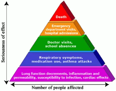
Figure 1-1. Health effects and repercussions of ozone exposure. ... 2
Figure 1-2. Number of days reaching unhealthy ozone levels in major U.S cities. ... 4
Figure 1-3. Reaction of ozone with organic molecules in the airway lining fluid and cell membranes. ... 5
Figure 1-4. Inter-individual variability in the ozone inflammatory response. ... 9
Figure 1-5. Ozone responses within individuals are highly reproducible. ... 10
Figure 1-6. Inter-individual variability in airway IL-8 levels following ozone exposure. ... 12
Figure 1-7. Histone modifications and DNA methylation: The epigenetic code that determines chromatin state. ... 15
Figure 1-8. Correlations between specific baseline chromatin modification levels and O3-induced gene expression. ... 17
Figure 1-9. Mucociliary differentiation of phBECs at US EPA using technique described by Ross et al., (2007). ... 19
Figure 1-10. Diagram of dissertation approach. ... 21
Figure 2-1. Inter-individual variability in epithelial cell IL-8 induction from both controlled human ozone exposure studies (in vivo) and primary cell cultures exposure to ozone (in vitro). ... 40
Figure 2-2. Donor-specificity of phBEC IL-8 induction following in vitro ozone exposure. ... 41
Figure 2-3. The influence of ERK1/2 and p38 inhibition on ozone associated IL-8 induction in high responders. ... 43
Figure 2-4. Representative Western blot showing the effects of SCH772984 and LY2228820 on ERK1/2 and p38 phosphorylation. ... 44
Figure 2-5. The activation of ERK1/2 and its associated kinases in high and low-responding cultures. ... 46
Figure 2-6. The activation of p38 and MKK4 in high and low-responding cultures... 48
Figure 2-7. Comparison of p65 activation in high and low-responding cultures. ... 49
xvi
Figure 2-9. Comparison of baseline with induced values of IL-8 expression and MAPK phosphorylation. ... 51 Figure 2-10. Induction and MAPK inhibition in other ozone-responsive genes. ... 53 Figure 2-11. Paradigm describing how differences in MAPK signaling
lead to inter-individual variability in phBEC ozone-mediated IL-8 induction. ... 56 Figure 2-12. In vitro ozone IL-8 induction in cells collected from single
bronchoscopy and exposed to ozone at two different times. ... 62 Figure 2-13. The influence of ERK1/2 and p38 inhibition on ozone
associated IL-8 induction in low responders. ... 63 Figure 3-1. Collection, culture, and exposure of primary human bronchial epithelial cells. ... 71 Figure 3-2. Ozone-responsive gene induction following single and repeated ozone exposure. .. 77 Figure 3-3. Relationship between single ozone exposure induction
(1XO3) and the magnitude of the adaptive effect. ... 79
Figure 3-4. TEER after single and repeated ozone exposures. ... 80 Figure 3-5. Comparing oxidative stress and antioxidant gene expression
in single and repeated ozone exposures. ... 82 Figure 3-6. Antioxidant potential of apical cell secretions measured before Day 4 exposure. ... 84 Figure 3-7. MAPK pathway activation after single and repeated ozone exposures. ... 87 Figure 4-1. Bivalent gene promoters regulate expression based on the
balance of activating and repressive histone modifications. ... 101 Figure 4-2. ENCODE ChIP-seq data showing selected epigenetic
features of the promoter regions of ozone-responsive genes. ... 102 Figure 4-3. Exposure design: comparing ozone-associated changes in
the chromatin landscape with peak gene induction. ... 104 Figure 4-4. Inductions of ozone-responsive genes during ozone exposure
and 1 hour post exposure. ... 109 Figure 4-5. The abundance of total histone H3 at candidate gene promoters... 110 Figure 4-6. The magnitude of ozone-responsive gene expression is related
xvii
Figure 4-7. Relationships between peak gene induction and
post-exposure chromatin modifications. ... 112
Figure 4-8. Changes in K27 methylation from 1H to 2H O3 are associated with the magnitude of IL-8 induction. ... 113
Figure 4-9. Summary of relationships between post-exposure chromatin modifications and gene expression. ... 114
Figure 5-1. Cytotoxicity assessment of repeated ozone exposures using trypan blue exclusion assay. ... 123
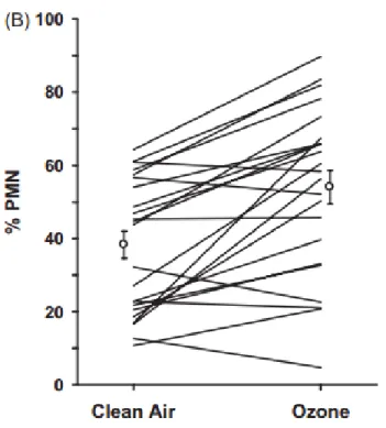
Figure 5-2. Relationship between in vitro IL-8 induction and PMN infiltration. ... 127
Figure 5-3. Number of exposure days required for ozone responsive genes to exhibit suppression. ... 131
Figure 5-4. Do ‘adapted’ cells regain responsiveness? ... 132
Figure 5-5. Ozone inductions in GSTM1 wild type and GSMT1 null phBEC donors. ... 137
Figure 5-6. PhBEC IL-8 induction in different donor age groups. ... 137
Figure 5-7. PhBEC IL-8 induction in male and female donors. ... 138
xviii
LIST OF ABBREVIATIONS
BALF Bronchoalveolar lavage fluid CGRP Calcitonin gene-related peptide
ChIP-qPCR Chromatin Immunoprecipitation- quantitative polymerase chain reaction EGFR Epidermal Growth Factor Receptor
ERK1/2 Extracellular Related Kinase 1/2
FA Filtered Air
GSTM1 Glutathione S-transferase Mu 1
GSTP1 Glutathione S-Transferase Pi 1
H3K27me3 Histone H3 lysine 27 trimethyl H3K4me3 Histone H3 lysine 4 trimethyl H4Ac Pan-acetylated histone H4
HMOX-1 Heme oxygenase 1
IL-6 Interleukin 6
IL-8 Interleukin 8
LPS Lipopolysaccharide
MAPK Mitogen Associated Protein Kinase
MEK MAPK/ERK Kinase
NAAQS National Ambient Air Quality Standards NOx Nitrogen oxides
NQO1 NAD(P)H Quinone Dehydrogenase 1
O3 Ozone
xix
PTGS2/COX-2 Prostaglandin-endoperoxide synthase 2 ROS Reactive oxygen species
RSK Ribosomal S6 Kinase
TEER Transepithelial Electrical Resistance TNF Tumor necrosis factor
1
CHAPTER 1: INTRODUCTION Ozone: a major public health issue
2
Figure 1-1. Health effects and repercussions of ozone exposure. This material was generated by the U.S. EPA and is not copyrighted. Available at https://www.epa.gov/ozone-pollution-and-your-patients-health/health-effects-ozone-general-population.
Ozone formation and pollution trends
Ozone is a ubiquitous oxidant pollutant that is often used to study the inflammatory response caused by air pollutant exposure. Unlike many other ambient air pollutants, ozone is not directly emitted, and is instead formed during secondary reactions between sunlight, oxygen, and other air pollutants such as volatile organic compounds (VOCs) and nitrogen oxides (NOx). Because sunlight and heat catalyze ozone formation, ozone concentrations often parallel sunlight intensity, peaking in the afternoon and dissipating in the evening. Ozone formation also exhibits seasonal variation, where high concentrations may occur more frequently during warm, sunny seasons such as summer and early fall. In addition to the previously described factors, other components such as topography, humidity, and wind patterns can play an important role in determining ambient ozone levels (US EPA 2013).
3
4
Figure 1-2. Number of days reaching unhealthy ozone levels in major U.S cities. Figure indicates the number of days exceeding the ozone NAAQS standard in major U.S. cities. This figure was published by the US EPA (2016) and is not copyrighted. Available at
https://www3.epa.gov/airnow/airaware/trends.html.
Health effects of ozone inhalation
While ozone can interact with all surfaces of the airway, the tissues receiving the highest doses in humans are thought to be the terminal bronchioles and the centriacinar region, which is located between the tracheobronchial and gas exchange regions (Plopper et al., 1998). Ozone interacts with extracellular lining fluid as well as the phospholipids comprising cell membranes, forming secondary oxidation products such as aldehydes, H2O2, and lipid ozonation products
5
Figure 1-3. Reaction of ozone with organic molecules in the airway lining fluid and cell membranes. Ozone is thought to react with double bonds in polyunsaturated fatty acids
(PUFA), producing lipid oxidation products, aldehydes, and hydrogen peroxide. These oxidation products mediate many of the health effects associated with ozone exposure. This image
previously appeared in US EPA 2013 Integrated Science Assessment for Ozone and Related Photochemical Oxidants. This material is not copyrighted and can be found at
https://cfpub.epa.gov/ncea/isa/recordisplay.cfm?deid=247492.
These ozonation products and pro-inflammatory eicosanoids are responsible for many of the adverse health effects associated with ozone exposure. Ozonation products activate transient receptor potential ankyrin 1 (TRPA1) receptors on vagal pulmonary C-fibers innervating the lungs, causing the release of various neurotransmitters including substance P, neurokinin A, and calcitonin gene-related peptide (CGRP) (US EPA 2013). The release of these neurotransmitters sensitizes the airway, leading to painful and truncated inspiration and restrictive decrements in lung function. Ozone-mediated lung function decrements are mostly restrictive in nature; however, the release of some neurotransmitters such as tachykinins and substance P may also cause bronchoconstriction and create obstructive lung function decrements (Verhein et al., 2011). Vagal nerve stimulation is also responsible for several autonomic changes in the cardiopulmonary system, including changes in breathing rate and altered cardiac
6
In addition to lung function decrements ozone exposure also causes pulmonary inflammation, predominantly characterized by airway neutrophilia. The ozone inflammatory response is mediated by the release of pro-inflammatory mediators into the airway. These include pro-inflammatory eicosanoids as well as a variety of cytokines and chemokines, such as IL-8, IL-6, IL-1, and TNFα. The pathways and receptors that are associated with the release of these pro-inflammatory mediators are still being elucidated, and are discussed in subsequent sections. Cytokines and markers of vascular inflammation are also elevated in the blood
following ozone exposure (Devlin et al., 2012, Thompson et al., 2010). Although the source of these circulating cytokines is unclear, this may be an important finding in determining whether there is a relationship between ozone exposure and cardiovascular mortality.
Airway neutrophil influx typically peaks six hours after exposure, and then declines 18-24 hours post exposure (Schelegle et al., 1991; US EPA 2013). Infiltrating neutrophils are thought to play an important role in the removal of necrotic debris from the airway (Hu et al., 1982); however, they may also cause further epithelial injury (Balmes et al., 1996; Vesely et al., 1999) via the release of bactericidal ROS. In addition to neutrophils, ozone exposure is also associated with the chemotaxis of other leukocytes into the airway, including lymphocytes, macrophages, and mast cells, which comprise a secondary component of the ozone inflammatory response (US EPA 2013). While changes in lung function are reversible, repeated episodes of inflammation may lead to tissue injury, permanent lung damage, airway remodeling, and metaplasia (Harkema et al., 1999). Given these findings, individuals who are more susceptible to ozone-associated inflammation may be at a greater risk of long-term airway damage.
7
as they occur within different time frames and don’t necessarily occur in the same individuals
(Balmes et al., 1996; Blomberg et al., 1999). Regardless of their origin, their combined effect has a substantial impact on public health. Increases in ambient ozone levels are correlated
hospitalizations and emergency department visits due to respiratory complications (Burnett et al., 1997; Moore et al., 2008), and are also associated with increased mortality (Jerrett et al., 2009).
The health effects of ozone exposure must also be considered in the context of realistic exposure scenarios, which often involve multiple pollutants and can result in additive or synergistic effects. Previous ozone exposure sensitizes individuals to the effects of subsequent allergen exposure (Jorres et al., 1996; Schelegle et al., 2003), and has an additive effect with a variety of other air pollutants such as diesel exhaust, particulate matter, and NO2 (Ehrlich et al.,
1977; Kafoury and Kelley 2005; Madden et al., 2014).
Ozone adaptation
Predicting the health effects of ozone exposure is complicated by the fact that outcomes are highly dependent on exposure history. While a single exposure is associated with lung function decrements and pulmonary inflammation, these effects are greatly reduced or even abolished during repeated exposures, an effect termed “ozone adaptation.” Ozone adaptation can be divided into two distinct components: lung function adaptation and inflammatory adaptation.
8
studies monitoring the effects of repeated daily ozone exposures found that lung function
decrements occur following one to two exposures, but in subsequent exposures these declines are abated or even disappear (Folinsbee et al., 1980, 1994). Lung function adaptation lasts
approximately one week, but its persistence varies between individuals (Linn et al., 1982). Inflammatory ozone adaptation has been observed in both animal and human clinical ozone exposure studies. In such studies the acute inflammatory response typically peaks after one to two exposure days, but after four days of repeated exposures the level of airway
cytokines, such as IL-8 and IL-6, are reduced as are airway leukocytes (Christian et al., 1998; Devlin et al., 1996; Jörres et al., 2000; Schelegle et al., 2003; Van der Wal et al., 1994). Unlike lung function adaptation, the persistence of inflammatory adaptation is more complex. Some inflammatory markers return to baseline levels within a week of exposure, while other markers remain altered for over 20 days (Devlin et al., 1996).
9
‘culling’ effect, the role of adaptation in ozone-associated mortality needs to be further investigated.
Inter- and intra-individual variability in the ozone inflammatory response
Human clinical ozone exposure studies have demonstrated that ozone exposure results in an average increase in neutrophil (PMN) influx into the lungs; however, there is substantial inter-individual variability in this response (Figure 1-4). Some inter-individuals exhibit no change in neutrophil infiltration, or even a reduction, while others have substantial increases.
Figure 1-4. Inter-individual variability in the ozone inflammatory response. Twenty-four human subjects were exposed to 0.06 ppm ozone or clean air for six hours. Leukocyte
10
Care Med 183:1215-1221. The American Journal of Respiratory and Critical Care Medicine is an official journal of the American Thoracic Society.
Risk factors such as genotype, age, and disease state (Alexis et al., 2009; Scannell et al., 1996; Vancza et al., 2009), appear to be associated with increased inflammatory responses; however, they have poor predictive value and the mechanisms by which they exert their effects are unknown. Moreover, inflammatory responses in healthy individuals who don’t exhibit any risk factors can exceed those in traditional risk groups (Holz et al., 1999). While ozone responses are extremely variable between individuals, responses within an individual are highly
reproducible (Figure 1-5), which suggests that the ozone inflammatory response adheres to a set of biological rules that remain to be discovered.
11
American Thoracic Society. Copyright © 2017 American Thoracic Society. Holz O, Jorres RA, Timm P, Mucke M, Richter K, Koschyk S, et al., 1999. Ozone-induced airway inflammatory changes differ between individuals and are reproducible. Am J Respir Crit Care 159:776-784. The American Journal of Respiratory and Critical Care Medicine is an official journal of the American Thoracic Society.
IL-8 is a hallmark of the ozone inflammatory response
Inflammatory responses require the coordinate activity of many different chemical messengers and cell types. A critical mediator in this process is IL-8, a potent neutrophil
12
Figure 1-6. Inter-individual variability in airway IL-8 levels following ozone exposure. Twelve healthy humans were exposed to 0.2 ppm ozone and filtered air. Bronchoalveolar lavage fluid was collected six hours post exposure and IL-8 concentrations were assessed. Reproduced with permission of the European Respiratory Society ©. European Respiratory Journal Jun 1998, 11 (6) 1294-1300.
Although multiple cell types produce IL-8, airway epithelial cells may be the most important source of this chemokine during ozone exposure (Devlin et al., 1994; Jaspers et al 1997; Chang et al., 1998). The induction of the IL-8 gene in response to ozone exposure is a key
step in the release of IL-8 protein and may be an important source of inter-individual variability. While previous studies in epithelial cell lines have attributed ozone-mediated IL-8 induction to NFκB, in physiologically-relevant primary human bronchial epithelial cells, the activation of the mitogen activated protein kinase (MAPK) pathway appears to be the most important
(McCullough et al., 2014). The activation of two MAPKs, extracellular related kinase (ERK) 1/2 and p38, appear to be particularly important in the ozone-mediated induction of
13
Molecular events linking ozone exposure to pro-inflammatory gene expression There are several hypotheses regarding how ozone exposure leads to pro-inflammatory gene expression via the MAPK pathway. Ozone interacts with surface liquids and phospholipid membranes leading to the production of reactive oxygen species (ROS) such as hydroxyl
radicals, aldehydes, and hydrogen peroxide (Pryor et al., 1995). These ROS are thought to play a role in the activation of membrane proteins, specifically the epidermal growth factor receptor (EGFR; McCullough et al., 2014; Wu et al., 2015), which signals downstream to ERK1/2. ROS such as H2O2 are known to directly modify EGFR and increase its kinase activity (Paulsen et al.,
2012). Others have hypothesized that ozone-associated ROS may change lipid membrane raft formation (Park et al., 2009), thereby encouraging EGFR receptor dimerization and
autophosphorylation. ROS are also known to oxidize specific residues within phosphatases, thereby inactivating them and removing the ‘brake’ from MAPK pathway activation (Bonini et al., 2014; Tal et al., 2006; Yan et al., 2016). Finally, ozone exposure is also thought to increase
the production of heat shock proteins, which directly activate ERK1/2 (Bauer et al., 2011). Less
is known about the molecular events that lead to ozone-mediated p38 activation, but it is possible that the ROS-mediated deactivation of phosphatases is involved. Moreover, the activation of toll-like-receptor 4 (TLR4) may also play an important role (Bauer et al., 2011; Williams et al., 2007).
14
important regulator of gene induction and a potential source of inter-individual variability. Unlike the DNA sequence, the epigenome is malleable- shaped from birth by environmental factors such as diet, stress, and chemical exposures, among others(Bowers and McCullough 2017; Bredfeldt et al., 2010; Dolinoy et al., 2010; Fiel and Fraga 2012). This duality - the ability to shape expression and be shaped by external forces- grants the epigenome predictive and/or explanatory power that other fixed predictors (i.e., genotype, sex, age) may not possess.
15
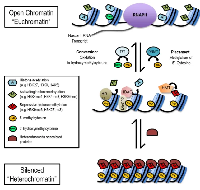
Figure 1-7. Histone modifications and DNA methylation: The epigenetic code that determines chromatin state.1 Histone modifications and DNA methylation function
cooperatively to regulate chromatin structure, accessibility to transcription factors, and gene expression. DNA methylation is the addition of a methyl group by a DNA methyltransferase (DNMT) to the cytosine residue of CpG dinucleotides in DNA. Methylation of DNA in gene regulatory regions (promoters and enhancers) often results in transcriptional repression; however, the oxidation of 5-methylcytosine to 5-hydroxymethylcytosine by the ten-eleven translocation (TET) family of methylcytosine dioxygenases is associated with the activation of gene expression. The genome is packaged on a protein scaffolding composed of histone proteins arranged into repeating units known as nucleosomes. The unstructured tails of these histones extend outside of the core nucleosome and are subject to numerous modifications such as acetylation, methylation, phosphorylation, ubiquitination, et cetera. These modifications can be activating (e.g. H3K4me3 and acetylation) or repressive/silencing (e.g. H3K9me3 and
1 This figure and caption previously appeared in Bowers, E. C., & McCullough, S. D. (2017). Linking the
16
H3K27me3). Activating histone acetylation and methylation, modifications made by histone acetyltransferases (HATs) and histone methyltransferases (HMTs), facilitate chromatin accessibility (euchromatin), recruitment of the transcriptional machinery, including RNA polymerase II (RNAPII), and initiation/elongation of transcription. DNA methylation and repressive histone modifications function cooperatively, through proteins such as methyl-CpG binding protein 2 (MeCP2), histone deacetylases (HDACs), histone demethylases (HDMs), and repressive HMTs, in the recruitment of transcriptional co-repressors and the formation of repressed and inactive (heterochromatin) epigenetic states.
Chromatin modifications have been particularly implicated in controlling pulmonary inflammation (Adcock et al., 2007; Barnes et al., 2005; Saccani and Natoli 2012) and they are
highly predictive of gene expression(Heintzman et al., 2007; Karlić et al., 2010; Wang et al., 2008). Our lab recently found that patterns of chromatin modifications in unexposed phBECs were associated with ozone-associated gene induction, suggesting that pre-exposure chromatin states may be an important source of inter-individual variability (Figure 1-8). While
pre-exposure chromatin modifications may be influential, epigenetic changes occurring as a result of ozone exposure may be just as important, as the induction of some genes may be dependent on the removal or placement of certain epigenetic modifications. Thus, profiling
17
Figure 1-8. Correlations between specific baseline chromatin modification levels and O3
-induced gene expression.2 (A) Induction of the pro-inflammatory genes COX-2, IL-8, and IL-6
and the oxidative stress gene HMOX-1 were measured in pHBECs following a two hour
exposure to 0.5 ppm O3. Baseline levels of H3K4me3, H3K27me2/3, and 5-hmC were compared
to the peak induction of HMOX-1 (B) and COX-2 (C). Fold induction is shown as O3/air and was
normalized to corresponding fold change in the housekeeping gene ACTB according to the Pffafl method. R2 and p-values were determined by simple linear regression.
Model System: Primary human bronchial epithelial cells (phBECs)
The majority of studies examining inflammatory response inter-individual variability and inflammatory adaptation have been human clinical exposure studies. While highly relevant,
18
clinical studies are inefficient and have a limited capacity for mechanistic investigation. While mechanisms are more easily studied in animal models, there is less genetic complexity to model inter-individual variability and the question of applicability to human biology is ever present. Alternatively, in vitro research using cell lines has a limited capacity to study inter-individual variability, as each cell line represents only one genotype. Moreover, most epithelial cell lines do not exhibit contact inhibition, and therefore become over-confluent during multiple days of exposures. Many airway epithelial cell lines also require submersion in media, which
complicates exposing cell surfaces to ozone gas.
19
Figure 1-9. Mucociliary differentiation of phBECs at US EPA using technique described by Ross et al., (2007). ALI cultures were fixed at Day 0 (A–C), Day 6 (D–F), Day 14 (G–I), Day 21 (J–L), and Day 29 (M–O). Sections were stained with H&E, Alcian blue/PAS reagent, or immunostained to label acetylated α-tubulin (red) and MUC5AC (green) with DAPI nuclear staining (blue). J, M: Ciliated cells=black arrows; basal cells=black arrowheads. K, N: Cells with PAS and Alcian blue staining, indicating the presence of mucins, are indicated with green
arrows. I, L, O: Acetylated α-tubulin staining indicates ciliated cells (yellow arrows) and mucin staining indicates secretory cells (white arrowheads). Reprinted with permission of the
American Thoracic Society. Copyright © 2017 American Thoracic Society. Ross et al., 2007. Am J Respir Cell Mol Biol Vol 37. pp 169–185. The American Journal of Respiratory Cell and Molecular Biology is an official journal of the American Thoracic Society.
Scope of Dissertation
Two long-observed but poorly characterized phenomena may hold the key to
20
ozone exposure, which may prevent excessive inflammation and permanent lung damage. These phenomena have been observed in human clinical studies and exposed populations for nearly fifty years, but is unclear if these responses are a feature of the airway environment specific to each individual or they are mediated on a cellular level. If we can identify the mechanisms that dictate inflammatory response inter-individual variability and adaptation, we may be able use this information to both engineer protective interventions and also refine predictions of susceptible populations.
The inflammatory response to ozone exposure exhibits extensive inter-individual variability, but responses within an individual are highly reproducible. This indicates that the inflammatory ozone response obeys biological rules that have yet to be discovered. Epithelial cells line the airway and are the first to encounter inhaled pollutants. These cells act as sentinels by sensing pollutants and/or tissue damage and releasing pro-inflammatory mediators such as IL-8. IL-8, a potent neutrophil chemokine, is an important driver of the ozone-associated neutrophil response. A key step in the release of IL-8 from epithelial cells is the ozone-mediated
transcription of the IL-8 gene. By understanding the factors dictating ozone-mediated IL-8 transcription in epithelial cells, we may be able to understand a critical component of ozone inflammatory response ‘programming.’
Using the phBEC model system, the goal of my dissertation is to understand the
21
to acute ozone exposure (Chapter 2) and may be an important mechanism underlying adaptive responses during repeated ozone exposure (Chapter 3). While ozone-associated MAPK activation is an important component of pro-inflammatory signaling, focusing on cellular signaling alone does not take into account the role of the epigenome at pro-inflammatory gene promoters. Thus, I also tested the hypothesis that changes in promoter epigenetic modifications may explain inter-individual variability in ozone-mediated IL-8 induction (Chapter 4).
Figure 1-10. Diagram of dissertation approach. Chapters 2 and 3 address the contributions of cellular signaling in response inter-individual variability and ozone adaptation. While examining MAPK signaling accounts for one of the main pro-inflammatory signals entering the nucleus, it does not take into consideration the role epigenome, which is explored in Chapter 4.
The findings from my dissertation provide compelling evidence that phBEC IL-8
22
cultures had elevated activation of ERK1/2. Moreover, when phBECs were subjected to repeated ozone exposure, many cultures exhibited suppression of ozone-responsive gene
expression, recapitulating the in vivo adaptive response. Upon further investigation, I discovered that antioxidant capacity is not increased during repeated ozone exposure as was previously hypothesized. Instead, phBECs that exhibited suppressed IL-8 induction showed reductions in ERK activation, suggesting this is an important mechanism driving the adaptive response. While MAPK signaling is an important component of the ozone response, epigenetic changes at pro-inflammatory gene promoters may also shape response inter-individual variability. I observed that the magnitude of pro-inflammatory gene induction is associated with changes in activating and repressive chromatin modifications.
23
REFERENCES
Abdul-Wahab SA, Bakheit CS, Al-Alawi SM. 2005. Principal component and multiple regression analysis in modelling of ground-level ozone and factors affecting its concentrations. Environmental Modelling & Software 20:1263-1271.
Adcock IM, Tsaprouni L, Bhavsar P, Ito K. 2007. Epigenetic regulation of airway inflammation. Current opinion in immunology 19:694-700.
Alexis NE, Zhou H, Lay JC, Harris B, Hernandez ML, Lu T-S, et al. 2009. The glutathione-s-transferase mu 1 null genotype modulates ozone-induced airway inflammation in human subjects. Journal of Allergy and Clinical Immunology 124:1222-1228. e1225.
Balmes JR, Chen LL, Scannell C, Tager I, Christian D, Hearne PQ, et al. 1996. Ozone-induced decrements in fev1 and fvc do not correlate with measures of inflammation. American journal of respiratory and critical care medicine 153:904-909.
Barnes P, Adcock I, Ito K. 2005. Histone acetylation and deacetylation: Importance in inflammatory lung diseases. European Respiratory Journal 25:552-563.
Bauer AK, Rondini EA, Hummel KA, Degraff LM, Walker C, Jedlicka AE, et al. 2011.
Identification of candidate genes downstream of TLR4 signaling after ozone exposure in mice: A role for heat-shock protein 70. Environmental health perspectives 119:1091. Blomberg A, Mudway I, Nordenhäll C, Hedenström H, Kelly F, Frew A, et al. 1999. Ozone‐ induced lung function decrements do not correlate with early airway inflammatory or antioxidant responses. European respiratory journal 13:1418-1428.
Bonini MG, Consolaro MEL, Hart PC, Mao M, de Abreu ALP, Master AM. 2014. Redox control of enzymatic functions: The electronics of life's circuitry. IUBMB Life 66:167-181. Bowers EC, McCullough SD. 2017. Linking the epigenome with exposure effects and
susceptibility: The epigenetic seed and soil model. Toxicological Sciences 155:302-314. Bredfeldt TG, Greathouse KL, Safe SH, Hung M-C, Bedford MT, Walker CL. 2010.
Xenoestrogen-induced regulation of ezh2 and histone methylation via estrogen receptor signaling to pi3k/akt. Molecular endocrinology 24:993-1006.
Burnett RT, Brook JR, Yung WT, Dales RE, Krewski D. 1997. Association between ozone and hospitalization for respiratory diseases in 16 Canadian cities. Environmental Research 72:24-31.
Chang MM-J, Wu R, Plopper CG, Hyde DM. 1998. IL-8 is one of the major chemokines
24
Christian DL, Chen LL, Scannell CH, Ferrando RE, Welch BS, Balmes JR. 1998. Ozone-induced inflammation is attenuated with multiday exposure. American journal of respiratory and critical care medicine 158:532-537.
Devlin R, Mckinnon KP, Noah T, Becker S, Koren H. 1994. Ozone-induced release of cytokines and fibronectin by alveolar macrophages and airway epithelial cells. American Journal of Physiology-Lung Cellular and Molecular Physiology 266:L612-L619.
Devlin RB, McDonnell WF, Becker S, Madden MC, McGee MP, Perez R, et al. 1996. Time-dependent changes of inflammatory mediators in the lungs of humans exposed to 0.4 ppm ozone for 2 hr: A comparison of mediators found in bronchoalveolar lavage fluid 1 and 18 hr after exposure. Toxicol Appl Pharmacol 138:176-185.
Devlin RB, Duncan KE, Jardim M, Schmitt MT, Rappold AG, Diaz-Sanchez D. 2012.
Controlled exposure of healthy young volunteers to ozone causes cardiovascular effects. Circulation 126:104-111.
Dolinoy DC, Weinhouse C, Jones TR, Rozek LS, Jirtle RL. 2010. Variable histone modifications at the Avy metastable epiallele. Epigenetics 5:637-644.
Ebi KL, McGregor G. 2008. Climate change, tropospheric ozone and particulate matter, and health impacts. Environmental health perspectives 116:1449.
Ehrlich R, Findlay JC, Fenters JD, Gardner DE. 1977. Health effects of short-term inhalation of nitrogen dioxide and ozone mixtures. Environmental Research 14:223-231.
Feil R, Fraga MF. 2012. Epigenetics and the environment: Emerging patterns and implications. Nature Reviews Genetics 13:97-109.
Folinsbee L, Bedi J, Horvath S. 1980. Respiratory responses in humans repeatedly exposed to low concentrations of ozone. American Review of Respiratory Disease 121:431-439. Folinsbee LJ, Horstman DH, Kehrl HR, Harder S, Abdul-Salaam S, Ives PJ. 1994. Respiratory
responses to repeated prolonged exposure to 0.12 ppm ozone. American journal of respiratory and critical care medicine 149:98-105.
Farraj AK, Hazari MS, Winsett DW, Kulukulualani A, Carll AP, Naykal-Coates N, et al. 2012. Overt and latent cardiac effects of ozone inhalation in rats: Evidence for autonomic modulation and increased myocardial vulnerability. Environmental health perspectives 120:348.
Fry RC, Rager JE, Zhou H, Zou B, Brickey JW, Ting J, et al. 2012. Individuals with increased inflammatory response to ozone demonstrate muted signaling of immune cell trafficking pathways. Respiratory research 13:1.
Hackney JD, Linn WS, Buckley RD, Hislop HJ. 1976. Studies in adaption to ambient oxidant air pollution: Effects of ozone exposure in Los Angeles residents vs. new arrivals.
25
Hackney JD, Linn WS, Karuza SK, Buckley RD, Law DC, Bates DV, et al. 1977. Effects of ozone exposure in Canadians and southern Californians: Evidence for adaptation? Archives of Environmental Health: An International Journal 32:110-116.
Harkema JR, Hotchkiss JA, Barr EB, Bennett CB, Gallup M, Lee JK, et al. 1999. Long-lasting effects of chronic ozone exposure on rat nasal epithelium. American Journal of
Respiratory Cell and Molecular Biology 20:517-529.
Heintzman ND, Stuart RK, Hon G, Fu Y, Ching CW, Hawkins RD, et al. 2007. Distinct and predictive chromatin signatures of transcriptional promoters and enhancers in the human genome. Nature genetics 39:311-318.
Holz O, Jorres RA, Timm P, Mucke M, Richter K, Koschyk S, et al. 1999. Ozone-induced airway inflammatory changes differ between individuals and are reproducible. American journal of respiratory and critical care medicine 159:776-784.
Hu PC, Miller FJ, Daniels MJ, Hatch GE, Graham JA, Gardner DE, et al. 1982. Protein accumulation in lung lavage fluid following ozone exposure. Environmental Research 29:377-388.
Jaspers I, Flescher E, Chen L. 1997. Ozone-induced IL-8 expression and transcription factor binding in respiratory epithelial cells. American Journal of Physiology-Lung Cellular and Molecular Physiology 272:L504-L511.
Jerrett M, Burnett RT, Pope CAI, Ito K, Thurston G, Krewski D, et al. 2009. Long-term ozone exposure and mortality. New England Journal of Medicine 360:1085-1095.
Jorres RA, Holz O, Zachgo W, Timm P, Koschyk S, Muller B, et al. 2000. The effect of repeated ozone exposures on inflammatory markers in bronchoalveolar lavage fluid and mucosal biopsies. American journal of respiratory and critical care medicine 161:1855-1861. Jörres R, Nowak D, Magnussen H. 1996. The effect of ozone exposure on allergen
responsiveness in subjects with asthma or rhinitis. American journal of respiratory and critical care medicine 153:56-64.
Kafoury R, Kelley J. 2005. Ozone enhances diesel exhaust particles (dep)-induced interleukin-8 (IL-8) gene expression in human airway epithelial cells through activation of nuclear factors- κB (NF-κB) and IL-6 (NF-IL6). International Journal of Environmental Research and Public Health 2:403.
Kafoury RM, Pryor WA, Squadrito GL, Salgo MG, Zou X, Friedman M. 1998. Lipid ozonation products activate phospholipases a2, c, and d. Toxicology and Applied Pharmacology 150:338-349.
26
Kim CS, Alexis NE, Rappold AG, Kehrl H, Hazucha MJ, Lay JC, et al. 2011. Lung function and inflammatory responses in healthy young adults exposed to 0.06 ppm ozone for 6.6 hours. American journal of respiratory and critical care medicine 183:1215-1221. Kodavanti UP, Hatch GE, Starcher B, Giri SN, Winsett D, Costa DL. 1995. Ozone-induced
pulmonary functional, pathological, and biochemical changes in normal and vitamin c-deficient guinea pigs. Toxicological Sciences 24:154-164.
Krishna M, Madden J, Teran L, Biscione G, Lau L, Withers N, et al. 1998. Effects of 0.2 ppm ozone on biomarkers of inflammation in bronchoalveolar lavage fluid and bronchial mucosa of healthy subjects. European Respiratory Journal 11:1294-1300.
Linn WS, Medway DA, Anzar UT, Valencia LM, Spier CE, Tsao FS-D, et al. 1982. Persistence of adaptation to ozone in volunteers exposed repeatedly for six weeks. American Review of Respiratory Disease 125:491-495.
Madden MC, Stevens T, Case M, Schmitt M, Diaz-Sanchez D, Bassett M, et al. 2014. Diesel exhaust modulates ozone-induced lung function decrements in healthy human volunteers. Particle and fibre toxicology 11:37.
McCullough SD, Duncan KE, Swanton SM, Dailey LA, Diaz-Sanchez D, Devlin RB. 2014. Ozone induces a proinflammatory response in primary human bronchial epithelial cells through mitogen-activated protein kinase activation without nuclear factor-κB activation. American Journal of Respiratory Cell and Molecular Biology 51:426-435.
McCullough SD, Bowers EC, On DM, Morgan DS, Dailey LA, Hines RN, et al. 2016. Baseline chromatin modification levels may predict interindividual variability in ozone-induced gene expression. Toxicological Sciences 150:216-224.
Moore K, Neugebauer R, Lurmann F, Hall J, Brajer V, Alcorn S, et al. 2008. Ambient ozone concentrations cause increased hospitalizations for asthma in children: An 18-year study in southern California. Environmental Health Perspectives 116:1063.
Nambu Z, Yokoyama E. 1983. Antioxidant system and ozone tolerance. Environmental research 32:111-117.
Park SJ, Kim HY, Kim H, Park SM, Joe E-h, Jou I, et al. 2009. Oxidative stress induces lipid-raft-mediated activation of Src homology 2 domain-containing protein-tyrosine phosphatase 2 in astrocytes. Free Radical Biology and Medicine 46:1694-1702. Paulsen CE, Truong TH, Garcia FJ, Homann A, Gupta V, Leonard SE, et al. 2012.
Peroxide-dependent sulfenylation of the EGFR catalytic site enhances kinase activity. Nat Chem Biol 8:57-64.
27
Pryor WA, Squadrito GL, Friedman M. 1995. A new mechanism for the toxicity of ozone. Toxicology Letters 82:287-293.
Rahman I, Clerch L, Massaro D. 1991. Rat lung antioxidant enzyme induction by ozone. The American journal of physiology 260:L412-418.
Ross AJ, Dailey LA, Brighton LE, Devlin RB. 2007. Transcriptional profiling of mucociliary differentiation in human airway epithelial cells. American journal of respiratory cell and molecular biology 37:169-185.
Saccani S, Natoli G. 2002. Dynamic changes in histone H3 lys 9 methylation occurring at tightly regulated inducible inflammatory genes. Genes & development 16:2219-2224.
Scannell C, Chen L, Aris RM, Tager I, Christian D, Ferrando R, et al. 1996. Greater ozone-induced inflammatory responses in subjects with asthma. American journal of respiratory and critical care medicine 154:24-29.
Schelegle ES, Miller LA, Gershwin LJ, Fanucchi MV, Van Winkle LS, Gerriets JE, et al. 2003. Repeated episodes of ozone inhalation amplifies the effects of allergen sensitization and inhalation on airway immune and structural development in rhesus monkeys. Toxicology and Applied Pharmacology 191:74-85.
Tal T, Graves LM, Silbajoris R, Bromberg PA, Wu W, Samet J. 2006. Inhibition of protein tyrosine phosphatase activity mediates epidermal growth factor receptor signaling in human airway epithelial cells exposed to Zn2+. Toxicology and applied pharmacology 214:16-23.
Tepper JS, Costa DL, Lehmann JR, Weber MF, Hatch GE. 1989. Unattenuated structural and biochemical alterations in the rat lung during functional adaptation to ozone. American Journal of Respiratory and Critical Care Medicine 140:493-501.
Thompson AM, Zanobetti A, Silverman F, Schwartz J, Coull B, Urch B, et al. 2010. Baseline repeated measures from controlled human exposure studies: Associations between ambient air pollution exposure and the systemic inflammatory biomarkers IL-6 and fibrinogen. Environ Health Perspect 118:120-124.
U.S. EPA. 2011. The Benefits and Costs of the Clean Air Act from 1990 to 2020.
https://www.epa.gov/sites/production/files/2015-07/documents/fullreport_rev_a.pdf. Accessed 5-17-17.
U.S. EPA. 2013. Final Report: Integrated Science Assessment of Ozone and Related Photochemical Oxidants. U.S. Environmental Protection Agency, Washington, DC, EPA/600/R-10/076F, 2013.
U.S. EPA. 2016. Ozone Trends. https://www.epa.gov/air-trends/ozone-trends. Accessed 5-16-17. U.S. EPA. 2017. Air Quality - National Summary.
28
Van der Wal W, Van Bree L, Marra M, Rombout P. 1994. Attenuation of acute lung injury by ozone inhalation—the effect of low level pre-exposure. Toxicology letters 72:291-298. Vancza EM, Galdanes K, Gunnison A, Hatch G, Gordon T. 2009. Age, strain, and gender as
factors for increased sensitivity of the mouse lung to inhaled ozone. Toxicological sciences 107:535-543.
Verhein KC, Hazari MS, Moulton BC, Jacoby IW, Jacoby DB, Fryer AD. 2011. Three days after a single exposure to ozone, the mechanism of airway hyperreactivity is dependent on substance p and nerve growth factor. American Journal of Physiology - Lung Cellular and Molecular Physiology 300:L176-L184.
Vesely KR, Schelegle ES, Stovall MY, Harkema JR, Green JF, Hyde DM. 1999. Breathing pattern response and epithelial labeling in ozone-induced airway injury in neutrophil-depleted rats. American Journal of Respiratory Cell and Molecular Biology 20:699-709. Wang Z, Zang C, Rosenfeld JA, Schones DE, Barski A, Cuddapah S, et al. 2008. Combinatorial
patterns of histone acetylations and methylations in the human genome. Nat Genet 40:897-903.
Wiester M, Tepper J, Winsett D, Crissman K, Richards J, Costa D. 1996. Adaptation to ozone in rats and its association with ascorbic acid in the lung. Toxicological Sciences 31:56-64. Wiester MJ, Winsett DW, Richards JH, Jackson MC, Crissman KM, Costa DL. 2000. Ozone
adaptation in mice and its association with ascorbic acid in the lung. Inhalation toxicology 12:577-590.
Williams AS, Leung S-Y, Nath P, Khorasani NM, Bhavsar P, Issa R, et al. 2007. Role of TLR2, TLR4, and MYD88 in murine ozone-induced airway hyperresponsiveness and
neutrophilia. Journal of applied physiology 103:1189-1195.
Williams AS, Issa R, Durham A, Leung S-Y, Kapoun A, Medicherla S, et al. 2008. Role of p38 mitogen-activated protein kinase in ozone-induced airway hyperresponsiveness and inflammation. European Journal of Pharmacology 600:117-122.
World Health Organization (WHO). 2014. 7 million premature deaths annually linked to air pollution. http://www.who.int/mediacentre/news/releases/2014/air-pollution/en/. Accessed 5-17-17.
Wu W, Wages PA, Devlin RB, Diaz-Sanchez D, Peden DB, Samet JM. 2015. Src-mediated EGF receptor activation regulates ozone-induced interleukin 8 expression in human bronchial epithelial cells. Environmental health perspectives 123:231.
29
30
CHAPTER 2: OZONE-MEDIATED IL-8 RESPONSIVENESS IS AN INTRINSIC PROPERTY OF AIRWAY EPITHELIAL CELLS AND IS DETERMINED BY THE
ACTIVATION OF THE MAP KINASE ERK1/2 3
Introduction
Millions of individuals are exposed to levels of the ambient air pollutant ozone that are known to produce pulmonary inflammation; however, inflammatory responses exhibit extensive inter-individual variability, making it difficult to anticipate health effects of exposed individuals. The variability of ozone associated inflammation is not reliably explained by biological factors, nor can it be predicted by traditional risk groups; for example, the magnitude of inflammatory response in healthy individuals may be greater than that observed in asthmatics (Holtz et al.,
1999; US EPA 2013). While ozone inflammatory responses are poorly understood and exhibit a high degree of inter-individual variability, they also exhibit low intra-individual variability, as inflammatory responses are reproducible if the same individual is exposed to ozone at different times (Holz et al., 1999 and 2005). This indicates that there are biological factors dictating the ozone inflammatory response that have yet to be defined, and suggests that their discovery could facilitate the reliable identification of individuals who may be particularly susceptible to ozone exposure.
31
IL-8 is an important pro-inflammatory chemokine that is a hallmark of environmentally-induced airway inflammation (Aris et al., 1993; Sundeep et al., 2000). Multiple studies have shown that ozone exposure results in the release of IL-8 protein into the airways and that these levels exhibit extensive variation between individuals (Fry et al., 2012; Krisha et al., 1998). Given the importance of IL-8 in airway inflammation, we sought to further understand the molecular mechanisms driving IL-8 response variability, as these may lead to the discovery of novel ozone susceptibility factors.
We examined in vitro IL-8 response in parallel with the activation of cellular signaling pathways that are known to be important in mediating the ozone pro-inflammatory response. We modeled IL-8 expression in epithelial cells as they are one of the first cells to respond to inhaled pollutants and are a major source of IL-8 following ozone exposure (Devlin et al., 1994; Jaspers et al 1997; Chang et al., 1998). While IL-8-associated gene transcription has previously been shown to occur through the NFκB pathway in cell lines, our lab demonstrated that in primary human bronchial epithelial cells (phBECs), the mitogen-activated protein kinases (MAPKs) are central signaling mediators (McCullough et al., 2014). These kinases are activated by
phosphorylation at specific residues and, in turn, phosphorylate other downstream effectors, which can include kinases, transcription factors, stress-associated proteins, etc. Within the MAPK pathway, the activity of extracellular-signal related kinases (ERK) 1 and 2, as well as p38 have an essential role in ozone pro-inflammatory signaling, as inhibiting these signaling
32
To determine if IL-8 transcriptional variability occurred in vivo, we first assessed IL-8 transcription in epithelial cells collected from human subjects following controlled ozone exposure. We then investigated the role of MAPKs in modulating IL-8 transcriptional response using differentiated phBECs cultured at air-liquid interface (ALI). Here we report that both in vivo and in vitro, ozone exposure results in inter-individual variability in epithelial cell IL-8
transcription. PhBEC IL-8 transcriptional responses from individual donors were consistent across repeated collections, suggesting that the magnitude of IL-8 induction is an intrinsic
property of phBECs and may be donor-specific. By extension, this epithelial cell ‘programming’ could partially explain the reproducibility of ozone responses observed in vivo (Holz et al., 1999 and 2005). When we examined ozone-associated pro-inflammatory signaling pathways in phBECs, we found that MAPK activity differed between cultures with high and low IL-8
33
Materials and Methods
In vivo epithelial cell ozone responses: Clinical ozone exposures and epithelial cell collection
Nine subjects participated in a randomized single-blind crossover study as described in Devlin et al., (2012). The median age was 24 years (range 21-32 years) and all were male (1 bi-racial, 7 Caucasian, 1 Hispanic). Briefly, young healthy volunteers gave a detailed medical history and underwent a physical examination. All subjects were lifetime nonsmokers. Each subject was exposed twice for 2 hours: once to filtered air (FA) and once to 0.3 ppm ozone. This ozone dose is frequently used in human exposure studies and is equivalent to an exposure at the 2005 National Ambient Air Quality Standard (NAAQS) of 75 ppb over an eight-hour period, which is slightly higher than the current NAAQS of 70 ppb/8h. Exposures were separated by at least 2 weeks. During each 2-hour exposure, subjects alternated between 15 minutes of rest and 15 minutes of exercise on a cycle ergometer and exposure exercise levels were adjusted to obtain a target minute ventilation of 25 L·min-1·m-2 body surface area. One hour after the exposure concluded, epithelial cells were collected from the bronchi of subjects via bronchial brushing, mixed with Trizol (Life Technologies, Carlsbad, CA), and the extracts were stored at -80 °C until they were ready for processing. The informed consent and collection protocol were approved by the UNC School of Medicine Committee on the Protection of the Rights of Human Subjects and by the US EPA.
In vitro epithelial cell ozone responses: Primary cell collection, plating, and ozone exposure
34
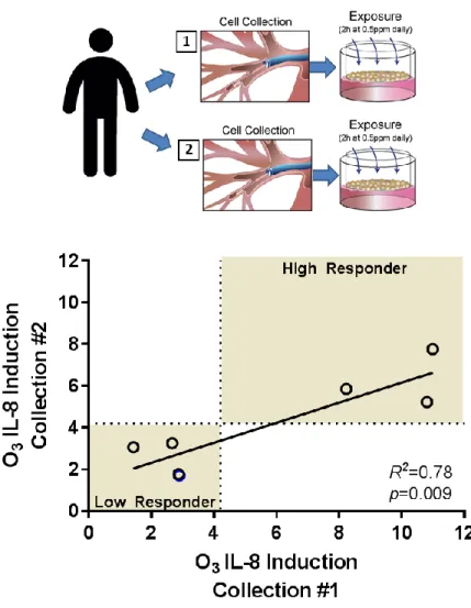
protocol were approved by the UNC School of Medicine Committee on the Protection of the Rights of Human Subjects and by the US EPA. To determine if phBECs from the same donor had consistent responses to ozone over time we compared IL-8 inductions between cells from the same donor that were collected during two different bronchoscopies, which were a minimum of three weeks apart. Cells were cultured and differentiated as described by Ross et al., (2007). Briefly, cells were expanded for three passages and then plated on 24 mm Transwell inserts with 0.4 µm pores (Corning Life Sciences, Tewksbury, MA). Three inserts per treatment per donor were used. Once confluent, cells were submerged for 48 hours with 500 nM retinoic acid. Afterward the apical layer of medium was removed creating an air-liquid interface (ALI). Cells were maintained for 24 days at ALI and supplemented with 100 nM retinoic acid to promote differentiation into a pseudostratified columnar epithelium. Prior to each ozone exposure, the basolateral medium was replaced and the apical surface was washed with 500 µL Dulbecco’s PBS (Life Technologies, Carlsbad, CA) to remove cellular debris and secretions. To investigate the role of MAPK activity in ozone-associated IL-8 induction, the ERK1/2 and p38 kinase inhibitors SCH772984 and LY2228820 (Cayman Chemical, Ann Arbor, MI), respectively, were used. LY2228800 and SCH772984 both inhibit these kinases by competitively binding the ATP binding domain, but SCH772984 also induces a conformation change that prevents the
35
cells designated for MAPK analysis were harvested as described below. Two hours following exposure, cells designated for gene expression analysis were harvested in RNA lysis buffer (Life Technologies) and stored at -80° C until they were ready for processing. As a positive control for NFκB pathway activation, primary cells were stimulated with 10 ng/mL TNFα for 20
minutes.
RT-qPCR
We used IL-8 transcription as our primary read-out, as protein levels may be influenced by further regulatory processes such as degradation. For in vivo samples, RNA was extracted from lysed samples using RNeasy kit (Qiagen, Valencia, CA), while for in vitro samples, an RNA Mini Kit (Life Technologies) was used. RNA was quantified using a Nanodrop ND1000. For in vivo and in vitro samples 100 ng and one (1) μg, respectively, was used to synthesize cDNA using iScript Reverse Transcription Kits (Bio-Rad, Hercules, CA). Gene expression was assessed in technical triplicates using TaqMan RT-qPCR primers and probes (Supplementary Methods) and the CFX96 qPCR system (Bio-Rad). Target gene expression was normalized to β-actin (ACTB) and then expressed as a fold changes between filtered air and ozone exposure treatments using the Pfaffl method (Pfaffl 2001).
It has been shown that in individuals who have elevated IL-8 protein in the airway after ozone exposure also exhibit increased IL-8 protein prior to exposure (Fry et al., 2010); thus, we wanted to determine if there was a relationship between baseline IL-8 transcription and ozone-induced transcription. To answer this question, we performed absolute quantification of the IL-8
pUC57-36
based plasmids (synthesized by Genewiz, Inc.) contained the IL-8 cDNA sequence. The resulting values were normalized to the absolute quantity of ACTB transcript in the same PCR reaction.
MAPK Pathway Analysis
We compared ozone-associated MAPK activation between donors that had high versus low IL-8 inductions by examining phosphorylation at specific residues. Cellular extracts were prepared in RIPA buffer (50mM Tris, pH 8.0; 150mM NaCl; 1% Triton X-100; 400μM EDTA; 10% glycerol; 0.1% SDS; 0.1% deoxycholate) with 1X protease (cOmplete EDTA-free, Roche, Indianapolis, IN) and 1X phosphatase (PhosSTOP, Roche) inhibitors. Cellular debris was then centrifuged and aliquots RIPA extract were removed for protein quantification via BCA assay (ThermoFisher, Waltham, MA). The remaining supernatant was supplemented with Laemmli buffer to a final concentration of 1X (60mM Tris, pH 6.8; 200 mM DTT; 10% glycerol; 2% SDS; 0.05% bromophenol blue) incubated at 95°C for five minutes, aliquoted, and stored at -80°C. For each sample, equal amounts of protein were loaded into SDS-PAGE gels,
37 Statistical Analysis
All statistical analyses were conducted using GraphPad Prism 6.07 (GraphPad Software, La Jolla, California, USA). For all analyses a p-value of less than 0.05 was considered
statistically significant. Simple linear regression was used to relate IL-8 responses between repeated bronchoscopies. To determine if inhibitor treatments significantly reduced IL-8 induction from the ozone-vehicle (O3-V) control, a two-way ANOVA with Dunnett’s Multiple
38 Results
Inter-individual variability in ozone induced epithelial derived IL-8 response
Epithelial cells collected from human subjects exposed to ozone demonstrated variable
IL-8 expression following exposure (Table 2-1; Figure 2-1A). When normalized to matched filtered air control exposures, themean IL-8 induction (± SD) was 1.30 ± 0.69. In phBEC cultures exposed to ozone in vitro (Table 2-2; Figure 2-1 B), the mean IL-8 induction was 4.18 ± 2.29. Although the magnitude of IL-8 inductions differed in vitro and in vivo, the coefficients of variability were nearly identical between model systems, 53% and 55%, respectively, indicating that the data were similarly distributed. For downstream analysis using the phBEC in vitro
system, we used the arithmetic mean value of 4.2 fold change from FA to separate donor cultures into “high responders” and “low responders” depending on whether they fell above or below this
value.
In vivo exposure subjects
Subject Sex Age Ethnicity Mean
IL-8
1 M 23 White 0.52
2 M 26 White 2.55
3 M 32 White 1.23
4 M 24 Hispanic 1.27
5 M 24 White 0.4
6 M 21 White 1.19
7 M 23 White 1.86
8 M 22 Bi-racial 0.88
9 M 26 White 1.81
39 phBEC donors
Donor Sex Age Ethnicity
Mean 1XO3
IL-8
1 F 35 Black 5.8
2 M 32 White 7.9
3 M 39 White 2.8
4 F 29 White 6.5
5 M 26 White 2.7
6 F 18 Black 5.0
7 M 26 White 9.2
8 M 20 Black 2.4
9 M 34 White 2.9
10 M 21 Asian 1.9
11 M 27 White 3.1
12 M 33 Black 2.4
13 M 39 White 5.7
14 F 35 White 4.5
15 M 28 White 1.4
16 F 21 Black 2.6
40
Figure 2-1. Inter-individual variability in epithelial cell IL-8 induction from both controlled human ozone exposure studies (in vivo) and primary cell cultures exposure to ozone (in vitro). (A) IL-8 induction in bronchial epithelial cells collected from ozone-exposed human subjects. Subjects were exposed to 0.3 ppm ozone for 2h and after a one-hour acclimation period subjects underwent bronchoscopy to collect epithelial cells via bronchial brushing. IL-8
expression was first normalized to β-actin and then expressed as a fold change from matched filtered air (control) exposures from the same subjects. Mean ± SD shown, n=9 subjects. (B) IL-8 expression in phBECs cultured at ALI exposed to ozone (0.5 ppm/2h). Each data point
represents a phBEC culture collected from a different human donor. Expression was normalized to β-actin and expressed as fold change from filtered air exposures. Mean ± SD shown, n=16 donors. To further investigate the mechanisms underlying ozone responsiveness using phBECs, we classified cultures as being “high” or “low” responders based on whether IL-8 inductions fell above or below the group mean (4.2 fold change).
Donor-specificity in the IL-8 response
PhBECs collected from the same donor over time exhibited donor-specificity in ozone associated IL-8 induction (Figure 2-2). Data were available for seven phBEC donors who had two epithelial cell collections. The time between bronchoscopies ranged from 49-453 days. Once again, we used the mean described in Figure 2-1B, 4.2-fold change from FA, to