ASSESSMENT AND RE-‐TREATMENT OF RESILON OBTURATION SYSTEM
PART 1: RESILON: ASSESSMENT OF DEGRADED FILLING MATERIAL IN NON-‐HEALED CASES
PART 2: RESILON: A CASE SERIES OF RETREATMENTS OF NON-‐HEALED CASES
Lesleigh Alexandra Payne
A thesis submitted to the faculty of the University of North Carolina at Chapel Hill in partial fulfillment of the requirements for the degree of Master of Science in the Endodontics
Department in the School of Dentistry.
Chapel Hill
2018
Approved by:
©2018 Lesleigh A. Payne ALL RIGHTS RESERVED
ABSTRACT
Lesleigh Alexandra Payne: Assessment and Re-‐Treatment of Resilon Obturation System; Part 1: Resilon: Assessment of Degraded Filling Material in Non-‐ Healed Cases; Part 2: Resilon: A
case series of Retreatments of Non-‐Healed Cases (Under the direction of Peter Z. Tawil)
Resilon obturation system was discontinued following its introduction as an alternative to traditional gutta percha and sealer. In-‐vitro models support anecdotal reports of degraded Resilon filling material. This may represent a significant health concern for the patient. The aim of this study was to determine the proportion of
Resilon degradation in non-‐healed endodontic cases and to present a case series describing retreatments of non-‐healed Resilon obturated teeth. Patients previously treated with Resilon (R) or Gutta-‐Percha (GP) that had a non-‐healed canal that needed a retreatment were enrolled. The proportion of degraded filling material was statistically significantly different between R and GP treated groups (p=.0003) with 78% of R canals being degraded compared to 0% of GP canals. The difference in the proportionality of degradation between the two materials was marginally significant (p=.054) when dichotomized time to follow-‐ up and presence of an orifice barrier were controlled for in the multivariate analysis.
ACKNOWLEDGEMENTS
I wish to acknowledge the following people who made this research project possible:
Dr. Peter Z. Tawil for your mentorship, friendship, and dedication.
Dr. Ashraf F. Fouad for your leadership, passion, and expertise.
Dr. Ceib Phillips for your guidance and for helping with the statistical analysis.
Dr. Krista A. Strange for your support throughout this study.
TABLE OF CONTENTS
LIST OF FIGURES ... vii
LIST OF TABLES ... viii
LIST OF ABBREVIATIONS ... ix
Thesis Introduction ... 1
References ... 5
MANUSCRIPT 1: RESILON: ASSESSMENT OF DEGRADED FILLING MATERIAL IN NON-‐HEALED CASES ... 8
Introduction ... 8
Materials and Methods ... 9
Statistical Analysis ... 14
Results ... 14
Discussion ... 18
Conclusions ... 21
References ... 22
MANUSCRIPT 2: RESILON: A CASE SERIES OF RETREATMENTS OF NON-‐HEALED CASES ... 24
Introduction ... 24
Case Reports ... 25
Case 2 ... 30
Case 3 ... 32
Discussion ... 34
References ... 37
Thesis Summary ... 38
References ... 41
Appendix A: Patient Consent Form ... 42
Appendix B: HIPPAA Authorization ... 46
Appendix C: IRB Approval Letter ... 48
LIST OF FIGURES
Figure 1: Degraded Resilon filling material. Note the passive
insertion of the 15K file ………..…….……13
Figure 2: Intact GP filling material. Note the resistance to
insertion of the 15K file resulting in bending ………13
Figure 3: Radiographs and images from Case 1 (A) Pre-‐operative radiograph (B) Post-‐ operative radiograph of retreatment (C) Image of Resilon in distal canals (D) Resilon debris
removed with a D3 rotary instrument……..………..29
Figure 4: Radiographs and images from Case 2 (A) Pre-‐operative radiograph (B) Post-‐operative radiograph of retreatment (C) Image of Resilon upon access with size 15K file being inserted passively to working length (D) Resilon debris
on a size 15K file……….………...31
Figure 5: Radiographs and images from Case 3 (A) Pre-‐operative radiograph (B) Post-‐ operative radiograph of retreatment (C) Image of degraded Resilon in canals upon access through intraorifice barriers (D) Image of degraded Resilon in canals upon access through intraorifice barriers (E) D3 Protaper Retreatment Rotary file with Resilon debris (F) 4x4 gauze
after wiping Resilon debris off of a file ………..33
LIST OF TABLES
Table 1: Patient demographics in the bivariate analysis where material (Resilon or gutta percha) was the
explanatory variable……….15
Table 2: Clinical characteristics in the bivariate analysis where
material (Resilon or gutta percha) was the explanatory variable………....16
Table 3: Wilcoxon Scores (Rank Sums) for variable age and time
since previous treatment classified by variable material……….………....16
Table 4: Degradation in the bivariate analysis where material
(Resilon or gutta percha) was the explanatory variable………...……….…16
Table 5: Patient demographics in the bivariate analysis where
degradation was the explanatory variable……….……17
Table 6: Clinical characteristics in the bivariate analysis where
degradation was the explanatory variable……….………17
Table 7: Wilcoxon Scores (Rank Sums) for variable age and time
since previous treatment classified by variable degraded……….….…..17
Table 8: Likelihood Ratio Statistics for Type 3 Analysis………...….18
LIST OF ABBREVIATIONS
AAE American Association of Endodontists ASA American Society of Anesthesiologists DMD Doctor of Dental Medicine
ET. AL And Others GP Gutta Percha
HIPPA Health Insurance Portability and Accountability Act IRB Institutional Review Board
OB Orifice Barrier
PA Periapical Radiograph PCL Polycaprolactone R Resilon
THESIS INTRODUCTION
Endodontic success relies on the ability to eliminate bacteria from the root canal system and to create a fluid tight seal within the canal space (1). Endodontic therapy may be initiated for a variety of reasons including trauma, carious pulpal exposure, mechanical pulpal exposure, and restorative needs, however the goal of sealing and disinfecting the root canal system remains. Following chemomechanical preparation, adequate obturation is required to prevent future reinfection. Obturation of root canals traditionally involves the use of gutta percha in combination with a sealing cement (2). Gutta percha was first used by Bowman as a root canal filling material in 1867 and is still the most widely used obturation core in endodontics (3). Despite its popularity, the material has several
complicate the formation of high-‐ strength bonds within the canal, which may result in decreased retention and increased marginal leakage (7,8).
Resilon is a thermoplastic synthetic polymer based root canal filling material that was introduced as a replacement for gutta percha in 2004 (9). It is composed of a parent polymer, a biodegradable aliphatic polyester called polycaprolactone, bioactive glass, methacrylate resin, barium sulphate, and bismuth oxychloride (10). Resilon handles
similarly to gutta percha, and can be dissolved by solvents during retreatment (10). It is used in conjunction with Epiphany, a methacrylate-‐ based sealer, and a self-‐ etching primer (9). These products, when used in combination, were designed to form a monoblock with the primed dentin, resulting in an alleged superior obturation resistant to bacterial leakage (11) and increased fracture resistance of the root canal treated tooth (12).
Although initial studies showed Resilon having improved characteristics over traditional gutta percha (11,13), several in vitro studies have since reported otherwise. The idea of a monoblock has been challenged due to the unpredictability of the
Epiphany/dentin bond, which was shown to result in significantly more gap formation at the dentin-‐ sealer interface than AH Plus/ gutta percha fillings (14). In a rat subcutaneous implantation model, Resilon showed inferior long-‐term biocompatibility when compared to a gutta percha control (15). Its believed that Resilon’s cytotoxicity may be due to its
biodegradability (16). The Resilon polymeric matrix consists of 25-‐40% polycaprolactone (PCL) and 3-‐10% dimethacrylates (17–19). PCL accounts for Resilon’s thermoplasticity and is susceptible to biodegradation through the cleavage of ester bonds (16). Its inclusion in the Resilon formulation
has caused concern. In a series of in vitro studies, Tay et. al
as alkaline hydrolases and biotic factors including endodontically relevant bacteria and fungi (16,20,21). When compression molded into disks, both Resilon and PCL exhibited significant surface pitting and erosion compared to gutta percha following incubation in dental sludge for 4 months (16). Emulsified Resilon was shown to degrade in the presence of cholesterol esterase, a component of salivary hydrolases and an inflammatory–cell derived enzyme (20).
The significance of Resilon’s susceptibility to biodegradation as demonstrated in the before mentioned studies, is its potential impact on outcome of Resilon obturated root canals. A recent study comparing long-‐term clinical outcomes found that Resilon had 5.7 times greater odds of failure compared with gutta percha (22). Of the Resilon obturated teeth, 56% were classified as successful compared to 88% of gutta percha obturated teeth (22). The biodegradation of Resilon as demonstrated in in vitro models may present a potential explanation for this higher clinical failure rate. Should a non-‐healed Resilon filled tooth require a retreatment, significant concern also exists over the increased possibility of experiencing a flare-‐up due to the potentially degraded status of the Resilon.
While most patients return to normal function and experience a significant relief of pain following endodontic therapy, some patients may experience an acute exacerbation of severe pain and/or swelling requiring an unscheduled visit and active treatment known as a flare-‐up (23). Though this occurrence is rare, it is still a significant concern for both the patient and the provider. Several factors have been recognized as predisposing patients to flare-‐ups. Walton and Fouad found positive correlations between flare-‐ups and pre-‐
radiolucencies (25), the number of visits, tooth type, allergies, and patient demographics including sex and age are all factors shown to be associated with endodontic flare-‐ups (26). Retreatments have also been associated with a higher prevalence of inter-‐appointment emergencies (26–28). Azim et al. found that previously treated teeth resulted in more than four times the rate of flare-‐ups compared to vital teeth (28). A study looking at
retreatments of teeth with resorcinol-‐formaldehyde fillings noted a higher than normal incidence of flare-‐ups (29). These findings may be attributed to acute periapical
inflammation caused by microbial factors including apical extrusion of infected debri, changes in endodontic microbiota or in environmental conditions due to incomplete chemo-‐mechanical preparation, increase of the oxidation-‐ reduction potential, or due to changes in local adaptation syndrome, mechanical irritation from overinstrumentation, extrusion of irrigating solutions, or extrusion of cytotoxic filling materials (30-‐32). While the Barborka et. al study identified a higher clinical failure rate of Resilon, there remains a gap in knowledge as to the cause of Resilon failures (22). The in vitro studies mentioned previously provide hypotheses including the susceptibility of Resilon and its components to degradation, however there is no previous clinical proof of this degradation. The aim of this study is to identify the proportion of degradation of Resilon filling material in non-‐healed retreatment cases compared to gutta percha. We also present a case series of Resilon retreatments illustrative of the clinical presentation of the material, the process of retreatment, and the occurrence of flare-‐ups. These findings will be
significant in providing information to guide the practitioner in future treatment planning of Resilon filled teeth, such as the decision between retreatment versus periapical
REFERENCES
1. Dow P, Ingle J. Isotope Determination of Root Canal Failure. Oral Surgery, Oral Med Oral Pathol Oral Pathol. 1955;8(10):1100–4.
2. Prakash R, Gopikrishna V, Kandaswamy D. Gutta-‐percha – an untold story. Endodontology. 2005;17(2):32–6.
3. Anthony L, Grossman L. A brief history of root canal therapy in the United States. J Am Dent Assoc. 1945;32:43–50.
4. Mcmahon T, Zijl PCM Van, Gilad AA. Ability of new obturation materials to improve the seal of the root canal system-‐ a review. 2015;27(3):320–31.
5. Madison S, Wilcox LR. An Evaluation of Coronal Microleakage in Endodontically Treated Teeth . Part 1. Time Periods. J Endod. 1988;14(9):455–8.
6. Schwartz RS. Adhesive Dentistry and Endodontics. Part 2: Bonding in the Root Canal System-‐The Promise and the Problems: A Review. J Endod. 2006;32(12):1125–34.
7. Tay F, Loushine R, Lambrechts P, Weller R, Pashley D. Geometric Factors Affecting Dentin Bonding in Root Canals: A Theoretical Modeling Approach. J Endod.
2005;31(8):584–9.
8. Bouillaguet S, Troesch S, Wataha JC, Krejci I, Meyer JM, Pashley DH. Microtensile bond strength between adhesive cements and root canal dentin. Dent Mater. 2003;19:199–205.
9. Tay FR, Pashley DH. Monoblocks in Root Canals: A Hypothetical or a Tangible Goal. J Endod. 2007;33(4):391–8.
10. Shanahan DJ, Duncan HF. Root canal filling using Resilon: A review. Br Dent J. 2011;211(2):81–8.
11. Shipper G, Orstavik D, Teixeira F, Trope M. An Evaluation of Microbial Leakage in Roots Filled with a Thermoplastic Synthetic Polymer-‐Based Root Canal Filling Material (Resilon). J Endod. 2004;30(5):342–7.
12. Teixeira FB, Teixeira ECN, Thompson JY, Trope M. Fracture resistance of roots endodontically treated with a new resin filling material. J Am Dent Assoc. 2004;135(5):646–52.
14. De-‐Deus G, Reis C, Di Giorgi K, Brandão MC, Audi C, Fidel RAS. Interfacial adaptation of the Epiphany self-‐adhesive sealer to root dentin. Oral Surgery, Oral Med Oral Pathol Oral Radiol Endodontology. 2011;111(3):381–6.
15. Cardoso M, Marques RF, Lopes MF, Cabrita A SJ. In vivo biocompatibility of Resilon compared with gutta-‐percha in a pre-‐clinical model. Dent Res J. 2013;10(5):652–8.
16. Tay F, Pashley DH, Loushine R, Kuttler S, Garcia-‐Godoy F, King N, et al. Susceptibility of a polycaprolactone-‐based root canal filling material to degradation. Evidence of biodegredation from a simulated field test. Am J Dent. 2007;20(6):265–369.
17. Jia W, Alpert B. Root canal filling material. United States Patent Application 20030113686, US Patent & Trademark Office. 2003.
18. JIa W, Trope M, Alpert B. Dental filling material. United States Patent Application 20050069836, US Patent & Trademark Office. 2005.
19. Jia W. Dental filling material. United States Patent Application 20050066854, US Patent & Trademark Office. 2005.
20. Hiraishi N, Sadek F, King N, Ferrari M, Pashley D, Tay F. Susceptability of a
polycaprolactone-‐ based root canal filling material to degradation using an agar-‐well diffusion assay. Am J Dent. 2008;21(2):119–23.
21. Tay F, Pashley D, Williams M, Raina R, Loushine R. Susceptibility of of a polycaprolactone-‐ based root canal filling material to degradation. I. Alkaline hydrolysis. J Endod. 2005;31(8):593–8.
22. Barborka BJ, Woodmansey KF, Glickman GN, Schneiderman E, He J. Long-‐term Clinical Outcome of Teeth Obturated with Resilon. J Endod. 2017;43(4):556–60.
23. Walton R, Fouad A. Endodontic interappointment flare-‐Ups: A prospective study of incidence and related factors. J Endod. 1992;18(4):172–7.
24. Chávez De Paz Villanueva LE. Fusobacterium nucleatum in endodontic flare-‐ups. Oral Surg Oral Med Oral Pathol Oral Radiol Endod. 2002;93(2):179–83.
25. Iqbal M, Kurtz E, Kohli M. Incidence and factors related to flare-‐ups in a graduate endodontic programme. Int Endod J. 2009;42(2):99–104.
26. Torabinejad M, Kettering JD, McGraw JC, Cummings RR, Dwyer TG, Tobias TS. Factors associated with endodontic interappointment emergencies of teeth with necrotic pulps. J Endod. 1988;14(5):261–6.
27. Imura N, Zuolo M. Factors associated with endodontic flare-‐ups: a prospective study. Int Endod J. 1995;28:261–5.
28. Azim AA, Azim KA, Abbott P V. Prevalence of inter-‐appointment endodontic flare-‐ups and host-‐related factors. Clin Oral Investig. 2017;21(3):889–94.
29. Gound TG, Marx D, Schwandt NA. Incidence of flare-‐ups and evaluation of quality after retreatment of resorcinol-‐formaldehyde resin (“Russian Red cement”) endodontic therapy. J Endod. 2003;29(10):624–6.
30. Siqueira J. Microbial causes of endodontic flare-‐ups. Int Endod J. 2003;36:453–63.
31. Seltzer S, Naidorf IJ. Flare-‐ups in endodontics: I. Etiological factors. J Endod. 2004;30(7):476–81.
32. Sipavičiūtė E, Manelienė R. Pain and flare-‐up after endodontic treatment procedures. Balt Dent Maxillofac J. 2014;16(1):25–30.
MANUSCRIPT 1: RESILON: ASSESSMENT OF DEGRADED FILLING MATERIAL IN NON-‐ HEALED CASES
Introduction
Resilon is a thermoplastic synthetic polymer based root canal filling material that was introduced as a replacement for traditionally used gutta percha in 2004 (1). It is
composed of a parent polymer, a biodegradable aliphatic polyester called polycaprolactone, bioactive glass, methacrylate resin, barium sulphate, and bismuth oxychloride (2). Resilon handles similarly to gutta percha, and can be dissolved by solvents during retreatment (2). It is used in conjunction with Epiphany, a methacrylate-‐ based sealer, and a self-‐ etching primer (1). These products, when used in combination, were designed to form an alleged monoblock with the primed dentin, resulting in a superior obturation resistant to bacterial leakage (3) and increased fracture resistance of the root canal treated tooth (4).
Although initial studies showed Resilon having improved characteristics over traditional gutta percha (3,5), several in vitro studies have since reported otherwise. The idea of a monoblock has been challenged due to the unpredictability of the
Epiphany/dentin bond, which was shown by De-‐Deus to result in significantly more gap formation at the dentin-‐ sealer interface than AH Plus/ gutta percha fillings (6). In a rat subcutaneous implantation model, Resilon showed inferior long-‐ term biocompatibility when compared to a gutta percha control (7). Its believed that Resilon’s cytotoxicity may be due to its biodegradability (8). The Resilon polymeric matrix consists of 25-‐ 40%
thermoplasticity and is susceptible to biodegradation through the cleavage of ester bonds (8). Its inclusion in the Resilon formulation
has caused concern. In a series of in vitro
studies, Tay et. al demonstrated the biodegradation of Resilon and PCL in the presence of abiotic factors such as alkaline hydrolases and biotic factors including endodontically relevant bacteria and fungi (8,12,13). When compression molded into disks, both Resilon and PCL exhibited significant surface pitting and erosion compared to gutta percha following incubation in dental sludge for 4 months (8). Emulsified Resilon was shown to degrade in the presence of cholesterol esterase, a component of salivary hydrolases and an inflammatory–cell derived enzyme (12).
The significance of Resilon’s susceptibility to biodegradation as demonstrated in the before mentioned studies, is its potential impact on outcome of Resilon obturated root canals. A recent study comparing long-‐term clinical outcomes found that Resilon had 5.7 times greater odds of failure compared with gutta percha (14). While a higher clinical failure rate has been identified, there remains a gap in knowledge as to the cause of Resilon failures (20). The in vitro studies mentioned previously provide hypotheses including the susceptibility of Resilon and its components to degradation, however there is no previous clinical proof of this degradation. The aim of this study is to identify the proportion of degradation of Resilon filling material in non-‐ healed retreatment cases compared to gutta percha.
Materials and Methods
of North Carolina at Chapel Hill (#16-‐1069). Patients who were 18 years or older, ASA classification I or II, whose primary treatment included use of Resilon with Epiphany sealer or gutta percha and AH-‐Plus sealer as the obturation material, and whose treatment was completed in either the predoctoral or graduate endodontic practices at University of North Carolina School of Dentistry (UNC-‐SOD) during the time period of 08/2004 -‐
08/2013 were identified through a search of the electronic patient record. Patient’s whose dental records did not objectively indicate which material was used for obturation
(Resilon or gutta percha) or whose dental records did not contain an adequate post-‐ operative radiograph of the primary root canal treatment were excluded.
Five hundred eighty patients were randomly selected from a pool of 7,376 possible patients identified in the electronic patient record search. A telephone call was made to the 580 randomly selected patients at the phone numbers registered in the database. If there was no answer, a scripted voice message was left and the secondary number on file was then called. Thirty eight patients agreed to come into the endodontic clinic for a follow-up examination. Of the 38 patients, 11 were determined to have a non-‐healed root canal requiring
retreatment based on the follow-‐up examination described below.
Follow-‐ up Examination
A head and neck exam and intra oral evaluation were performed. Endodontic diagnostic tests were completed including percussion and palpation testing, tooth slooth response, and probing depth measurements. A periapical radiograph (PA) was taken at an angle similar to the original post-‐ operative radiograph. A tooth was considered non-‐ healed if the patient presented with signs or symptoms of infection or if there was a periapical lesion of the same or larger size than in the original post-‐operative radiograph.
Retreatment
Patients identified as requiring retreatment were assigned to one of two endodontic residents by front desk personnel at UNC-‐SOD. Patients signed the standardized UNC-‐SOD consent form for endodontic treatment as well as consent to participate in the study and HIPAA Authorization. Patients were given the opportunity to ask the investigator questions concerning treatment. The retreatment began with local anesthesia based on the individual needs of the subject. The investigator isolated the tooth to be retreated with a single punch rubber dam. The rubber dam was disinfected before and after access with 4% NaOCl. After adequate anesthesia and isolation, the investigator removed any carious tooth structure or compromised restorations. The tooth was then accessed to expose the filling material. Upon access of the root canal system, the investigator classified the Resilon or gutta percha filling material in each canal as either degraded or intact. Classification was based on the following criteria:
bending of the size 15 K file) (Fig.1).
- Intact: Presence of solid dense filling material that provides resistance to size 15 K file insertion resulting in the file bending (Fig. 2).
The following clinical observations were recorded during treatment for each tooth: - Type of restoration (full coverage or not)
- Presence of caries
- Presence of a missed canal - Apical stop
- Tooth discoloration
- Presence of an orifice barrier
After documentation of the findings, the investigator continued with the retreatment, which was completed over two-‐visits with inter-‐appointment calcium hydroxide.
Figure 1: Degraded Resilon filling material. Note the passive insertion of the 15K file
Figure 2: Intact GP filling material. Note the resistance to insertion of the 15K file resulting
in bending
Statistical Analysis
A power analysis indicated that a Fisher’s exact test with a 0.05 two-‐sided
significance level would have greater than 80% power to detect a difference in proportions of 0.70 when the sample size in each group was 10.
Fisher’s exact test was used to compare the demographics and clinical
characteristics of the two materials (Resilon or gutta percha) and to assess the effect of material and demographics and clinical characteristics on the presence of degradation. Logistic Regression using likelihood ratio tests in PROC GENMOD (SAS ® Vers 9.3) was used to assess the effect of three variables (obturation material, presence of an orifice barrier, and time since previous treatment dichotomized at the median time of all subjects (TP_N)) on the presence of degradation. The level of significance was established at p <.05.
Results
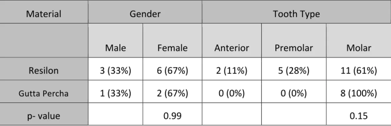
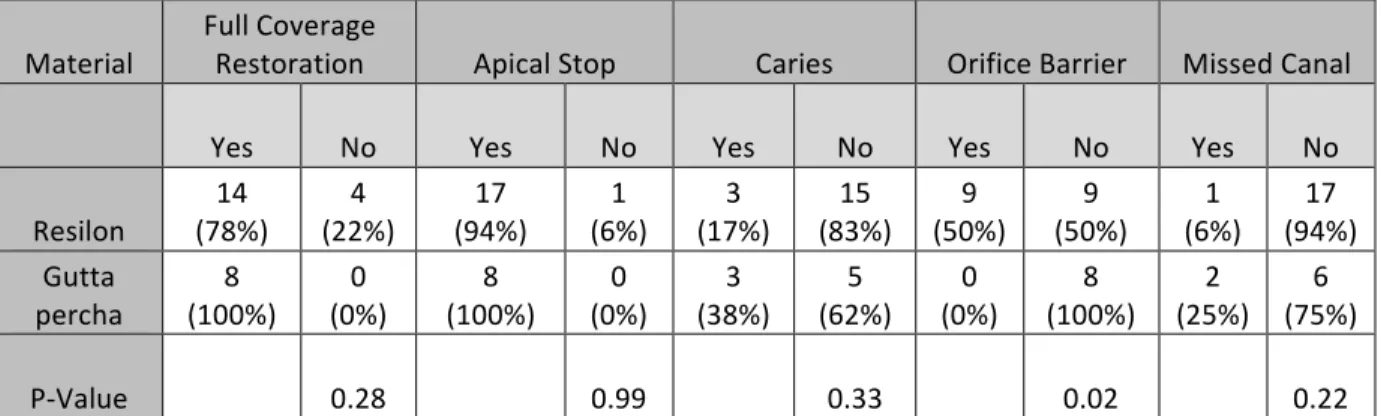
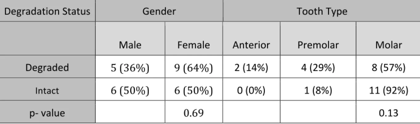
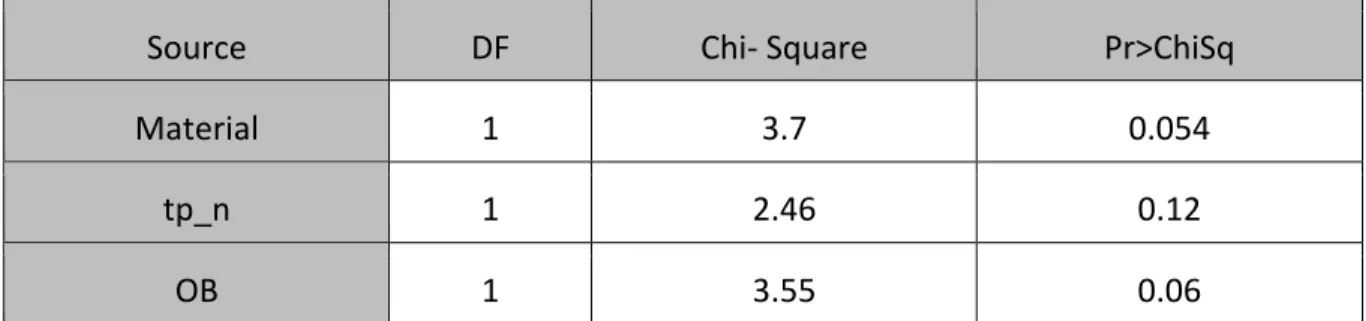
percha in the bivariate analysis (Table 4). Of the Resilon obturated canals, 78% were degraded compared to 0% of the gutta percha canals. The bivariate analysis indicated a statistically significant difference in the periapical diagnosis between Resilon and gutta percha obturated canals at the primary treatment appointment with a higher proportion of chronic apical abscesses and asymptomatic apical peridodontitis among Resilon canals (p<.05). However, the data was too sparse to include in the logistic regression. In the bivariate analysis where degradation was the outcome, presence of an orifice barrier (Table 6) was the only variable that was statistically significantly different between degraded and intact canals (p<.05) (Table 5-‐7). The multivariate analysis indicated that neither presence of an orifice barrier nor dichotomized time to follow-‐up were associated with degradation (Table 8). The difference in the proportionality of degradation between the two materials was marginally significant (p=.054) when the dichotomized time to follow up and presence of an orifice barrier were controlled for (Table 8).
Material Gender Tooth Type
Male Female Anterior Premolar Molar
Resilon 3 (33%) 6 (67%) 2 (11%) 5 (28%) 11 (61%)
Gutta Percha 1 (33%) 2 (67%) 0 (0%) 0 (0%) 8 (100%)
p-‐ value 0.99 0.15
Table 1: Patient demographics in the bivariate analysis where material (Resilon or gutta percha) was the explanatory variable
Material Full Coverage Restoration Apical Stop Caries Orifice Barrier Missed Canal
Yes No Yes No Yes No Yes No Yes No
Resilon (78%) 14 (22%) 4 (94%) 17 (6%) 1 (17%) 3 (83%) 15 (50%) 9 (50%) 9 (6%) 1 (94%) 17
Gutta
percha (100%) 8 (0%) 0 (100%) 8 (0%) 0 (38%) 3 (62%) 5 (0%) 0 (100%) 8 (25%) 2 (75%) 6
P-‐Value
0.28 0.99 0.33 0.02 0.22
Table 2: Clinical characteristics in the bivariate analysis where material (Resilon or gutta percha) was the explanatory variable
Material Variable Lower Quartile Median Upper Quartile
Resilon Age TP 48 96 64 99 142 69
Gutta Percha Age TP 51 50 68 84 68.5 99
P-‐value Age TP
0.58 0.04
Table 3: Wilcoxon Scores (Rank Sums) for variable age and time since previous treatment classified by variable material
Material Degraded Intact
Resilon 14 (78%) 4 (22%)
Gutta Percha 0 (0%) 8 (100%)
P-‐Value 0.0003
Table 4: Degradation in the bivariate analysis where material (Resilon or gutta percha) was the explanatory variable
Degradation Status Gender Tooth Type
Male Female Anterior Premolar Molar
Degraded 5 (36%) 9 (64%) 2 (14%) 4 (29%) 8 (57%)
Intact 6 (50%) 6 (50%) 0 (0%) 1 (8%) 11 (92%)
p-‐ value 0.69 0.13
Table 5: Patient demographics in the bivariate analysis where degradation was the explanatory variable
Degradation Status
Full Coverage
Restoration Apical Stop Caries Orifice Barrier Missed Canal
Yes No Yes No Yes No Yes No Yes No
Degraded
12 (86%)
2 (14%)
13 (93%)
1 (7%)
1 (7%)
13 (93%)
8 (57%)
6 (43%)
1 (7%)
13 (93%)
Intact (83%) 10 (17%) 2 (100%) 12 (0%) 0 (42%) 5 (58%) 7 (8%) 1 (92%) 11 (7%) 2 (83%) 10
P-‐Value
0.99
0.99
0.07
.015
0.58
Table 6: Clinical characteristics in the bivariate analysis where degradation was the explanatory variable
Degradation
Status Variable Lower Quartile Median Upper Quartile
Degraded Age TP 48 96 63 96 142 69
Intact Age TP 54 54 67 84 68.5 108
P-‐value Age TP
0.30 0.13
Table 7: Wilcoxon Scores (Rank Sums) for variable age and time since previous treatment classified by variable degraded
Source DF Chi-‐ Square Pr>ChiSq
Material 1 3.7 0.054
tp_n 1 2.46 0.12
OB 1 3.55 0.06
Table 8: Likelihood Ratio Statistics for Type 3 Analysis
Discussion
The Resilon/ Epiphany obturation system serves as an example as to the difficulties surrounding bonded obturation materials. There remains little published evidence on intracanal adhesion and the advantages/ disadvantages over traditionally used gutta percha. In vitro studies have demonstrated complications with intracanal bonding including stresses due to polymerization shrinkage resulting from a high cavity
configuration factor (C-‐factor). These shrinkage stresses complicate the formation of high-‐ strength bonds within the canal resulting in decreased retention and increased marginal leakage (15, 16). While initial in vitro studies showed promising results including
improved fracture resistance and less bacterial leakage of Resilon obturated root canals (3,4), later studies showed less favorable results including degradation of Resilon and its components as well as diminishing biocompatibility over time (6,7). While Resilon is no longer in use clinically, its effects resonate in the patients who have retained Resilon obturated root canal treated teeth.
polycaprolactone (PCL) in the Resilon formulation. PCL is a biodegradable polyester responsible for Resilon’s thermoplasticity (8). This resorbable polymer has traditionally been used as a component in drug-‐delivery devices due to its low melting point,
biocompatibility, and susceptibility to biodegradation by enzymes and microorganisms (17). Hydrolases released by bacteria, yeast and fungi can degrade PCL through the cleavage of ester bonds (18). In a series of in vitro studies, Tay et. al demonstrated the biodegradation of Resilon and PCL in the presence of abiotic factors such as alkaline hydrolases and biotic factors including endodontically relevant bacteria and fungi (8,12,18). Following exposure to dental sludge consisting of saliva, sputum, coagulated blood, plaque and calculus, enamel, dentin and shavings from restorative procedures, compression molded disks of Resilon and PCL both exhibited significant surface
degradation compared to gutta percha (8). Emulsified Resilon was shown to degrade in the presence of cholesterol esterase, a component of salivary hydrolases and an inflammatory – cell derived enzyme (12). Penetration of the coronal and apical seals could result in exposure of the Resilon filling material to this enzyme (12). Gutta percha and its main constituent poly(trans-1,4-isoprene) in comparison, have not been associated with
biodegradation by microbial enzymes (19).
The significance of Resilon’s susceptibility to biodegradation as demonstrated in this study is its potential impact on outcome of Resilon obturated root canals. A study comparing Resilon and gutta percha obturated root canals with follow-‐ up times ranging from 2-‐25 months indicated statistically indistinguishable differences in clinical outcome
difference in outcome. Of the Resilon obturated teeth, 56% were classified as successful compared to 88% of gutta percha obturated teeth (14). With comparable protocol and success criteria, it is likely that the significant factor between these two studies is the time to follow-up. Should the inferior clinical outcome of Resilon be related to its biodegradation, time may prove to be a significant variable in the process. Due to the small N number in our study, we were unable to determine the full effect of time on degradation status, thus further studies are needed to determine the significance of this factor.
Within the limitations of this study, it was determined that the presence of an orifice barrier was not protective against degradation. There may be several explanations for these findings. Orifice barriers have been shown to result in coronal leakage of varying degrees based on material and thickness (21, 22). An alternative explanation is that the source of enzymatic degradation is apical. Resilon has been shown to exhibit degradation in the presence of
cholesterol esterase, a component of salivary hydrolases and an inflammatory– cell derived enzyme (12). Monocyte-derived macrophages that secrete these enzymes have been shown to
be present within periapical granulomas associated with endodontically infected teeth (23). While our study demonstrates a higher proportion of degradation of Resilon obturated non-healed root canals compared to gutta percha obturated canals, further investigation should focus on the analysis of the degraded material to identify its chemical and microbiological content. This may provide us with better understanding as to the cause of biodegradation and its potential impact on the patient’s health. It is also important to detect factors associated with non-healed degraded cases such as periapical status, quality of coronal seal, and radiographic
some concern exists. Resilon should serve as a reminder to stress the need for clinical outcome studies in the implementation of new products.
Conclusions
Within the limitations of this clinical observational study, the results indicate that Resilon with Epiphany sealer has a higher proportion of degradation when compared to gutta percha and sealer after adjusting for presence of an orifice barrier and dichotomized at median time since previous treatment. Further studies are needed to assess the effect of time on degradation and to analyze the degraded material to identify its chemical and
microbiological content.
REFERENCES
1. Tay FR, Pashley DH. Monoblocks in Root Canals: A Hypothetical or a Tangible Goal. J Endod. 2007;33(4):391–8.
2. Shanahan DJ, Duncan HF. Root canal filling using Resilon: A review. Br Dent J. 2011;211(2):81–8.
3. Shipper G, Orstavik D, Teixeira F, Trope M. An Evaluation of Microbial Leakage in Roots Filled with a Thermoplastic Synthetic Polymer-‐Based Root Canal Filling Material (Resilon). J Endod. 2004;30(5):342–7.
4. Teixeira FB, Teixeira ECN, Thompson JY, Trope M. Fracture resistance of roots endodontically treated with a new resin filling material. J Am Dent Assoc. 2004;135(5):646–52.
5. Hammad M, Qualtrough A, Silikas N. Effect of New Obturating Materials on Vertical Root Fracture Resistance of Endodontically Treated Teeth. J Endod. 2007;33(6):732– 6.
6. De-‐Deus G, Reis C, Di Giorgi K, Brandão MC, Audi C, Fidel RAS. Interfacial adaptation of the Epiphany self-‐adhesive sealer to root dentin. Oral Surgery, Oral Med Oral Pathol Oral Radiol Endodontology. 2011;111(3):381–6.
7. Cardoso M, Marques RF, Lopes MF, Cabrita A SJ. In vivo biocompatibility of Resilon compared with gutta-‐percha in a pre-‐clinical model. Dent Res J. 2013;10(5):652–8.
8. Tay F, Pashley DH, Loushine R, Kuttler S, Garcia-‐Godoy F, King N, et al. Susceptibility of a polycaprolactone-‐based root canal filling material to degradation. Evidence of biodegredation from a simulated field test. Am J Dent. 2007;20(6):265–369.
9. Jia W, Alpert B. Root canal filling material. United States Patent Application 20030113686, US Patent & Trademark Office. 2003.
10. JIa W, Trope M, Alpert B. Dental filling material. United States Patent Application 20050069836, US Patent & Trademark Office. 2005.
11. Jia W. Dental filling material. United States Patent Application 20050066854, US Patent & Trademark Office. 2005.
12. Hiraishi N, Sadek F, King N, Ferrari M, Pashley D, Tay F. Susceptability of a
polycaprolactone-‐ based root canal filling material to degradation using an agar-‐well diffusion assay. Am J Dent. 2008;21(2):119–23.
13. Tay F, Pashley D, Williams M, Raina R, Loushine R. Susceptibility of of a polycaprolactone-‐ based root canal filling material to degradation. I. Alkaline hydrolysis. J Endod. 2005;31(8):593–8.
14. Barborka BJ, Woodmansey KF, Glickman GN, Schneiderman E, He J. Long-‐term Clinical Outcome of Teeth Obturated with Resilon. J Endod. 2017;43(4):556–60.
15. Tay F, Loushine R, Lambrechts P, Weller R, Pashley D. Geometric Factors Affecting Dentin Bonding in Root Canals: A Theoretical Modeling Approach. J Endod.
2005;31(8):584–9.
16. Bouillaguet S, Troesch S, Wataha JC, Krejci I, Meyer JM, Pashley DH. Microtensile bond strength between adhesive cements and root canal dentin. Dent Mater. 2003;19:199–205.
17. Woodruff MA, Hutmacher DW. The return of a forgotten polymer -‐ Polycaprolactone in the 21st century. Prog Polym Sci. 2010;35(10):1217–56.
18. Lefèvre C, Tidjani A, Vander Wauven C, David C. The interaction mechanism between microorganisms and substrate in the biodegradation of polycaprolactone. J Appl Polym Sci. 2002;83(6):1334–40.
19. Rose K, Steinbu A. Minireviews. Biodegradation of Natural Rubber and Related Compounds : Recent Insights into a Hardly Understood Catabolic Capability of Microorganisms. Appl Environ Microbiol. 2005;71(6):2803–12.
20. Cotton TP, Schindler WG, Schwartz SA, Watson WR, Hargreaves KM. A Retrospective Study Comparing Clinical Outcomes after Obturation with Resilon/Epiphany or Gutta-‐Percha/Kerr Sealer. J Endod. 2008;34(7):789–97.
21. Ghulman MA, Gomaa M. Effect of intra-‐orifice depth on sealing ability of four
materials in the orifices of root-‐filled teeth: An ex-‐vivo study. Int J Dent. 2012;2012.
22. Yavari H, Samiei M, Eskandarinezhad M, Shahi S, Aghazadeh M, Pasvey Y. An in vitro comparison of coronal microleakage of three orifice barriers filling materials. Iran Endod J. 2012;7(3):156–60.
23. Stern MH, Dreizen S, Mackler BF, Selbst AG, Levy BM. Quantitative analysis of cellular composition of human periapical granuloma. J Endod 1981;7:117–22.
MANUSCRIPT 2: RESILON: A CASE SERIES OF RETREATMENTS OF NON-‐HEALED CASES
Introduction
Resilon is a thermoplastic synthetic polymer based root canal filling material that was introduced as a replacement for traditionally used gutta percha in 2004 (1). When used in combination with Epiphany sealer and a self-‐etching primer, Resilon was designed to form an alleged monoblock with dentin, aiming for a superior obturation resistant to bacterial leakage (2) and increased fracture resistance of the root canal treated tooth (3). Although initial studies showed Resilon having improved characteristics over gutta percha (2-‐4), several investigators began publishing in vitro studies demonstrating the biodegradation of Resilon in the presence of abiotic factors such as alkaline hydrolases and biotic factors including endodontically relevant bacteria and fungi (5-‐7). This is likely attributed to the inclusion of polycaprolactone, a biodegradable aliphatic polyester, in the Resilon formulation (5). PCL accounts for Resilon’s thermoplasticity and is susceptible to biodegradation through the cleavage of ester bonds (5). Degradation of Resilon may be a contributing factor to its higher clinical failure rate reported in a recent study comparing long-‐term clinical outcomes of Resilon and gutta percha obturated root canals (8).
relief of pain following endodontic therapy, some patients may experience an acute exacerbation of severe pain and/or swelling requiring an unscheduled visit and active treatment known as a flare-‐up (9). Retreatments in particular have been associated with a higher prevalence of inter-‐appointment emergencies (10–12). A study looking at
retreatments of teeth with resorcinol-‐formaldehyde fillings noted a significantly higher than normal incidence of flare-‐ups (13). Due to the potential biodegradation of Resilon within the root canal, Resilon retreatments may present with comparable flare-‐up incidence to paste retreatments.
Throughout the literature, there is still little information to guide clinicians on the retreatment of Resilon cases including clinical presentation, challenges with removal, and flare-‐ups. This series aims to contribute to the knowledge of the clinical approaches of Resilon retreatments by describing three clinical cases.
Case Reports
filling material, the same instrumentation and irrigation protocol was used to remove the existing Resilon and clean/shape the canals. This protocol is described below:
A 15K file was used to obtain an apical glide path and determine working length (WL) using an ApexID apex locator. Protaper Universal retreatment rotary instruments were used in the coronal and middle third of the canals. Hedstrom files were used to remove Resilon from the apical third of the canals. Irrigation involved the use of 4% NaOCl with activation using an EndoActivator. No solvents were used in removal of the Resilon. Vortex Blue files were used sequentially to enlarge and shape the canals with
recapitulation with a size 10K file and 4% NaOCl irrigation between files. A final rinse was performed using 4% NaOCl and 17% EDTA. The canals were dried with paper points and calcium hydroxide was placed to WL with a Lentulo spiral. A piece of sponge was placed over the orifices followed by Cavit and FUJI 9 to seal the access.
At the conclusion of the first visit, the patient was provided with the following post-‐ operative instructions:
- It is normal for the tooth and the area around the tooth to be sore for the first 24-‐48 hours
- This soreness should not be more than an over-‐the-‐counter analgesic can control ex: ibuprofen 600mg
Contact me if:
- The pain is severe and cannot be controlled by over-‐the-‐counter analgesics - The pain is persisting or increasing
A flare-‐up was defined as the patient experiencing an acute exacerbation of severe pain and/or swelling requiring an unscheduled visit and active treatment. If a patient
experienced a flare-‐up, it was recorded.
At the next scheduled appointment, the root canal was obturated with gutta percha and AH Plus sealer using the warm vertical technique and the retreatment was completed. Described in detail below is the clinical presentation of Resilon including the pattern of degradation, as well as descriptions of its removal and technical challenges, and the occurrence of flare-‐ups.
Case 1
A 49-‐year-‐old Hispanic female with medical history including lower back pain for which she takes ibuprofen 600mg and a thyroidectomy in 2015 for which she takes Levothyroxine 100 mcg/day presented to UNC-‐SOD. Tooth #19 was previously root canal treated 5 years prior. Clinically #19 presented with intact porcelain fused to metal crown and composite core build-‐up. The diagnosis was as follows: previously treated,
symptomatic apical periodontitis.
Upon access, four canals were located, two mesial and two distal canals which shared a common orifice. The round bur made a drop into the canals upon penetration of the composite and a potent smell was immediately evident. The Resilon in all four canals was similar in consistency and coloration, presenting as soft and degraded with
predominately gray coloration with light pink material intermixed (Fig. 3C). A 15 K file was inserted into the canals and was able to be immediately inserted to working length
Retreatment Rotary instruments to auger Resilon from the coronal and middle thirds, the material wrapped around the files in ribbons and travelled coronally where it could be removed from the canals in segments (Fig. 3D). The rotary was circumferentially scraped along the walls of the canals and was able to remove the bulk of the material, however a gray film was smeared around the canal walls and could not be removed with
instrumentation. A Hedstrom was scraped along the walls and in the apical third, to remove the remaining material, but the film along the walls remained. The texture and density of the Resilon was consistent throughout the entire length of the canals. The smeared layer of Resilon was not loosened from the canal walls until the sonic activator was used in each canal with NaOCl. During activation, the irrigant turned gray and small debris could be seen floating. After a rinse with NaOCl and EDTA, the majority of the walls were residue free and clean. Some extrusion of the material was noted radiographically. Following the initial retreatment visit, the patient did not experience a flare-‐up.
Figure 3: Radiographs and images from Case 1 (A) Pre-‐operative radiograph (B) Post-‐ operative radiograph of retreatment (C) Image of Resilon in distal canals
Case 2
A 43-‐year-‐old Caucasian female with non-‐contributory medical history presented to UNC-‐SOD. Tooth #8 received primary root canal therapy 12 years prior. The patient
presented with a lingual IRM with clinically open margins that was placed at the time of treatment. The crown was discolored with a gray hue compared to the adjacent teeth. The diagnosis was as follows: previously treated, symptomatic apical periodontitis.
Figure 4: Radiographs and images from Case 2 (A) Pre-‐operative radiograph (B) Post-‐operative radiograph of retreatment (C) Image of Resilon upon access
Case 3:
A 20-‐year-‐old Caucasian female with a penicillin allergy and no other contributory medical history presented to UNC-‐SOD.Tooth #3 was previously root canal treated 7 years prior. Clinically #3 presented with an acrylic provisional crown and FUJI build-‐up. The diagnosis was as follows: previously treated, symptomatic apical periodontitis.
Upon access, intact PermaFlo Purple composite orifice barriers were found covering four canals. Upon penetration of the orifice barriers, a potent smell was again noted that was significant enough for the patient to inquire about. The Resilon was discolored in all four canals, predominately dark gray (Fig 5C&D), The material was degraded in the mesial and palatal canals, however it was slightly more dense in the distal canal as determined by the 15 K file requiring some apical pressure to penetrate. As the file approached the middle third of the canal, the material became softer and an apical glide path was obtained
Figure 5: Radiographs and images from Case 3 (A) Pre-‐operative radiograph (B) Post-‐ operative radiograph of retreatment (C) Image of degraded Resilon
in canals upon access through intraorifice barriers (D) Image of degraded Resilon in canals upon access through intraorifice barriers
(E) D3 Protaper Retreatment Rotary file with Resilon debris (F) 4x4 gauze after wiping Resilon debris off of a file

![cis Ethyl 5 amino 1 [4 (4 chlorophenyl) 5,5 dimethyl 2 oxo 1,3,2 dioxaphosphinan 1 yl] 3 (6 chloro 3 pyridylmethylamino) 1H pyrazole 4 carboxylate](data:image/gif;base64,R0lGODlhAQABAIAAAP///wAAACH5BAEAAAAALAAAAAABAAEAAAICRAEAOw==)