CHARACTERIZATION AND MODULATION OF AUTOREACTIVE CD4+ T CELLS IN TYPE 1 DIABETES
Li Li
A dissertation submitted to the faculty of the University of North Carolina at Chapel Hill in partial fulfillment of the requirements for the degree of PhD in the Department of
Microbiology and Immunology
Chapel Hill 2007
Approved by
Advisor: Roland M. Tisch, PhD
Reader: Stephen H. Clarke, PhD
Reader: Edward J. Collins, PhD
Reader: Jeffrey A. Frelinger, PhD
© 2007
Li Li
ABSTRACT
Characterization and Modulation of Autoreactive CD4+ T cells in Type 1 Diabetes
Type 1 diabetes (T1D) is an autoimmune disease mediated by pathogenic β cell-specific T
cells. The soluble (s) IAg7- immunoglobulin (Ig) dimers covalently linked to GAD65 peptides or the mimetic BDC2.5 epitope (mBDC) were utilized in two studies. The first use was to
enhance the efficacy of peptide treatment. NOD female mice with established β cell
autoimmunity received a short course of sIAg7-Ig dimers intravenously (i.v.). NOD mice treated with sIAg7-mBDC continued to develop diabetes. In marked contrast, the majority of NOD mice treated with sIAg7-Ig complexed with the GAD65-specific peptides p217 or p286 remained diabetes-free. Protection correlated with an increased frequency of IL-10 secreting
immunoregulatory CD4+ T cells that delayed diabetes in a co-adoptive transfer model. These results demonstrate that treatment with a short-course of sIAg7-GAD65 peptide dimers is an effective approach to suppress T1D.
Secondly, the relative role for BDC2.5 clonotypic CD4+ T cells in the progression of the diabetogenic response was tracked and temporal analyzed using sIAg7-mBDC multimers. The frequency and/or number of T cells binding sIAg7-mBDC multimers (g7-mBDC+) increase
with in peripheral blood lymphocytes (PBL) and the islets at the onset of β cell autoimmunity
in NOD female mice. In contrast, a reduced frequency and number of g7-mBDC+ T cells was
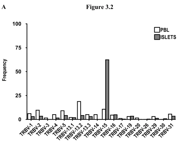
gene complementary determinant region 3 (CDR3) sequences revealed that BDC2.5
clonotypic CD4+ T cells in the islets but not PBL selectively expressed TRBV15. These data demonstrate that g7-mBDC+ T cells are an early indicator of the development of destructive insulitis, and that both clonotypic expansion and preferential usage of TCR characterize islet
ACKNOWLEDGEMENTS
I wish to give my warmest thanks to Dr. Roland Tisch. He introduced me to this field and
generously gave me advice and suggestions during these years. I would like to express my
gratitude to him for spending valuable time inspiring me and conducting my dissertation
research. Likewise, I am grateful to Dr. Bo Wang for providing me with thought-provoking
discussions and advice. I am also grateful to my dissertation committee members, Dr. Jeff
Frelinger, Dr. Ed Collins, Dr. Glenn Matsushima, and Dr. Steve Clark. Thank you for
generously giving me valuable time and advice during the last few years.
Finally, I would like to specially dedicate this work to my wife, Hong Yu and my son,
Evan. Thank you for supporting me both intellectually and emotionally during these years. I
could not have done this without your immense love. Thank you Evan for bringing felicity to
my life. As well as my parents, Shiyan Li and Yaqin Zhao, your endless love for me and
continuous encouragement throughout my life are deeply appreciated. Without the generous
help of my sister, Yueying Li and brother, Zongrui Li, this investigation would not have been
TABLE OF CONTENTS
Page
LIST OF TABLES……….vii
LIST OF FIGURES………..…..ix
LIST OF ABBREVIATIONS………...x
CHAPTER 1. INTRODUCTION……….……..…1
1.1 Type 1 Diabetes……….………2
1.2 The NOD Mouse……….………….……..4
1.3 Properties of IAg7……….……….……….5
1.4 T1D is a T cell-mediated Autoimmune Disease………..……..6
1.5 Multiple β cell Autoantigens are Targeted in T1D………9
1.6 Multiple Defects Account for the Breakdown of Self-tolerance to β cells……….11
1.6.1 Thymic Selection………..12
1.6.2 Immunoregulation……….13
1.7 Immunotherapy of T1D………..……….18
1.7.1 Antigen-independent Immunotherapies………...…….18
1.7.2 Antigen-dependent Immunotherapies………...……20
2. SUPPRESSION OF AUTOIMMUNE DIABETES BY TREATMENT
WITH PEPTIDE-MHC CLASS II DIMERS………36
2.1 Abstract……….…37
2.2 Introduction………..38
2.3 Results………..………41
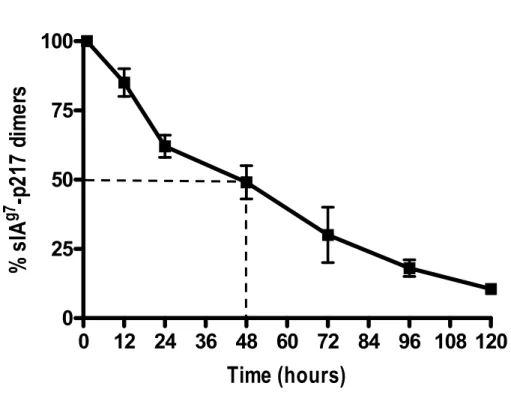
The production and characterization of sIAg7-Ig dimers……….………...41
sIAg7-p217 and sIAg7-p286 but not sIAg7-mBDC dimers prevent diabetes in NOD mice………....…...43
Protection mediated by sIAg7-p217 and sIAg7-p286 dimers correlates with the induction of peptide-specific immunoregulatory Tr1 cells……….…..…44
2.4 Discussion………..….…..46
2.5 Materials and Methods……….………50
2.6 References………...…….61
3. SELECTIVE EXPANSION OF β CELL-SPECIFIC T CELL RECEPTORS IN AUTOIMMUNE DIABETES……….66
3.1 Abstract………..………..67
3.2 Introduction………..………68
3.3 Results………..………71
Detection of mBDC-specific CD4+ T cells in PBL and the islets………...………71
The TCR Vβ repertoire of mBDC2.5-specific CD4+ T cells is skewed in the Islets……….………72
3.4 Discussion………..……..74
3.5 Materials and Methods………..………...77
3.6 References………..………..93
4.1 sIAg7-Ig dimer administration: A potent strategy of
peptide vaccination………..………....96
4.2 Characterization of autoreactive T cells in islet infiltrates…………..………97
4.3 References………..100
5. APPENDICES………..101
Appendix I………101
LIST OF TABLES
Page Table 3.1 CDR3β analysis of g7-mBDC+ CD4+ T cells in PBL and islets
from Mouse 1………83
Table 3.2 CDR3β analysis of g7-mBDC+ CD4+ T cells in PBL and islets
from Mouse 2………84
Table 3.3 CDR3β analysis of g7-mBDC+ CD4+ T cells in PBL and islets
from Mouse 3………85
Table 3.4 CDR3β analysis of g7-mBDC+ CD4+ T cells in PBL and islets
from Mouse 4………86
Table 3.5 CDR3β analysis of g7-mBDC+ CD4+ T cells in PBL and islets
from Mouse 5………87
Table 3.6 CDR3β analysis of g7-mBDC+ CD4+ T cells in PBL and islets
from Mouse 6………88
Table 3.7 CDR3β analysis of g7-mBDC+ CD4+ T cells in PBL and islets
from Mouse 7………89
Table 3.8 CDR3β analysis of g7-mBDC+ CD4+ T cells in PBL and islets
from Mouse 8………90
Table 3.9 CDR3β analysis of g7-mBDC+ CD4+ T cells in PBL and islets
from Mouse 9………91
Table 3.10 CDR3β analysis of g7-mBDC+ CD4+ T cells in PBL and islets
LIST OF FIGURES
Page
Figure 2.1 Characterization of sIAg7-Ig dimers……….…53
Figure 2.2 T cell binding by sIAg7-Ig multimers is peptide specific……….54 Figure 2.3 sIAg7-Ig dimers induce T cell proliferation in a peptide-specific manner…....55 Figure 2.4 In vivo detection of sIAg7-Ig dimers………...……..56 Figure 2.5 sIAg7-p217 and sIAg7-p286 but not sIAg7-mBDC dimers
prevent diabetes in NOD female mice……….………57
Figure 2.6 Treatment of sIAg7-p217 or sIAg7-p286 dimers
blocks the progression of insulitis……….…..……58
Figure 2.7 Protection mediated by sIAg7-p217 and sIAg7-p286 dimers correlates
with the induction of peptide-specific immunoregulatory Tr1 cells…………59
Figure 2.8 sIAg7-p217 dimer treatment induces immuoregulatory T cells………60 Figure 3.1 g7-mBDC+ CD4+ T cells are increased in PBL and the islets
of NOD female mice………79
Figure 3.2 The Vβ repertoire of most islet infiltrating g7-mBDC+ CD4+ T cells
LIST OF ABBREVIATIONS AND SYMBOLS
AC apoptotic cell
Ag antigen
APC antigen presenting cell
CCR chemokine receptor
CDR3 complementarity determining region 3
ConA concavilin A
CTE cortical thymic epithelial cell
CTL cytotoxic T lymphocyte
CTLA-4 cytotoxic T lymphocyte antigen
DC dendritic cell
DN double-negative
DP double-positive
ELISA enzyme-linked immunosorbent assay
ELISPOT enzyme-linked immuospot asay
FACS fluorescent activated cell sorting
FITC fluorescein isothiocyanate
FoxP3 Forkhead box P3
GAD65 glutamic acid decarboxylase 65
GITR glucocorticoid-induced tumor necrosis factor receptor family-related gene
HA hemagglutinin
HEL hen egg lysozyme
HRP horseradish peroxidase
IA-2 insulinoma-associated protein 2
Idd insulin-dependent genes
IDDM Insulin dependent diabetes mellitus
IFN interferon
Ig immunoglobulin
IGRP islet-specific glucose-6-phosphatasecatalytic subunit–related protein
IL interleukine
InsB insulin B
i.v. intravenous
MAb monoclonal antibody
MHC major histocompatibility complex
MTE medullary thymic epithelial cell
MΦ macrophage
NK natural killer
NOD non-obese diabetic
Ova ovalbumin
PBL peripheral blood lymphocyte
PE phyco-erythrin
PerCP peridinin-chlorophyll protein
PLN pancreatic lymph node
RIP rat insulin promoter
SAV streptavidin
mBDC mimetic BDC
scid severe-combined immunodeficient
SP single-positive
T1D type 1 diabetes
TCR t cell receptor
TGF transforming growth factor
Tg tansgenic
TH1 type 1 T helper
TH2 type 2 T helper
TNF tumor necrosis factor
TRBV T cell receptor beta chain variable gene
Treg regulatory T cell
α alpha
β beta
γ gamma
CHAPTER 1
1.1 Type 1 Diabetes
Type 1 diabetes (T1D) or insulin dependent diabetes mellitus (IDDM) is a T cell-mediated
autoimmune disease characterized by the destruction of the insulin producing β cells of the
islets of Langerhans (Bach 1994; Tisch and McDevitt 1996). T1D affects mostly children and
young adults, but can occur at any age. Currently, 0.3% of the population in the United States
is affected by T1D with 30,000 new clinical cases diagnosed each year and importantly, the
incidence of T1D is increasing in developed countries. Diabetic individuals suffer from
long-term complications including blindness, kidney failure and premature vascular disease
leading to a reduced life expectancy by an average of 15 years. Although diabetes can be
controlled by daily insulin injections, there is no effective therapy to prevent or “cure” the
disease in humans. Accordingly, there is a pressing need to understand the pathogenic
mechanisms of β cell autoimmunity in order to design effective and rational
immunotherapies for the prevention and treatment of T1D.
T1D is a multi-factorial disease with both environmental and genetic factors contributing to
the development and progression of β cell autoimmunity. Epidemiological studies have
found that environmental factors such as diet, toxins, and viral and bacterial infections are
associated with T1D (Hyoty and Taylor 2002). The most important evidence for
environmental factors having a role in T1D comes from observations that 1) migrants from
countries with low incidence rates are more susceptible to T1D in countries with high
incidence rates, and 2) genetically identical twins are only 36% concordant in disease
development. However, whether environmental insults play a role in the initiation and/or
provide evidence that viral infections may trigger β cell autoimmunity through for instance,
molecularmimicry between viral and β cell proteins (Ohashi, Oehen et al. 1991; Faideau,
Larger et al. 2005) or by direct β cell injury (Oldstone, Southern et al. 1984).
T1D is polygenic and more than 20 chromosal loci have been found to be associated with
disease susceptibility in humans and the nonobese diabetic (NOD) mouse, a spontaneous
model of T1D (see below) (Davies, Kawaguchi et al. 1994; Todd 1995; Concannon,
Gogolin-Ewens et al. 1998). The majority of the susceptibility genes within theseloci have yet to be
identified with the exceptionof the major histocompatibility complex (MHC) class II genes,
the 5' flanking region of the insulingene, and the CTLA4 gene (Rotwein, Chyn et al. 1981;
Winter, Beppu et al. 1987; Owerbach and Gabbay 1993; Kristiansen, Larsen et al. 2000). The
strongest genetic association with T1D susceptibility and resistance maps to the MHC class
II region in both humans (IDDM1) and NOD mice (idd1). The HLA-DRB1*0301,
HLADRB1*0401, HLA-DQB1*0302, and HLADQA1*0301 alleles confer high-risk
susceptibility in humans, whereas other alleles such as DRB1*0403,
HLA-DQB1*0602, and HLADQA1*0102 confer resistance to T1D (Wicker, Todd et al. 1995;
Undlien, Lie et al. 2001). Currently, it is believed that MHC class II susceptibility alleles are
necessary but not sufficient for the development of diabetes, and that the combined effects of
other susceptibility genes influence the progression of β cell autoimmunity in the context of
the appropriate environmental insult (Redondo, Rewers et al. 1999).
1.2 The NOD Mouse
NOD mice are considered to be the leading animal model for T1D. β cell autoimmunity
spontaneously develops in NOD mice and several aspects of the diabetogenic response
closely reflect the human disease. For instance, both NOD mice and humans share a number
of T1D genetic susceptible loci, and both autoimmune responses are affected by
environmental factors. In addition, multiple immune effector cells, such as CD4+ and CD8+ T cells, B cells, macrophages (MΦ), dendritic cells (DCs), and natural killer (NK) cells are
involved in the respective disease processes (Miller, Appel et al. 1988; O'Reilly, Hutchings et
al. 1991; Cooke, Phillips et al. 2001). Finally, several β cell autoantigens, such GAD and
insulin, are targeted in both diabetic patients and NOD mice (Kent, Chen et al. 2005).
Islet infiltration or insulitis is initially detected in NOD mice at 3-4 weeks of age. The first
step in insulitis referred to peri-insulitis, is characterized by pancreatic infiltrates surrounding
an islet. Peri-insulitis then progresses to intra-insulitis in which cells invade the islets. MΦs
and DCs are the first cells to traffick to the islets (Jansen, Homo-Delarche et al. 1994) and
appear to play essential albeit ill-defined roles in the disease process (Jun, Santamaria et al.
1999; Yoon, Yoon et al. 1999). Islet infiltrating antigen-presenting cells (APC) may mediate
initial β cell injury through the secretion of proinflammatory cytokines such as interferon-γ
(IFNγ), tumor necrosis factor-α (TNFα) and interleukin (IL)-1 and reactive oxygen species,
in addition to processing and presenting β cell autoantigens to promote the subsequent
recruitment of T cells (Chervonsky, Wang et al. 1997; Kagi, Odermatt et al. 1997;
Suarez-Pinzon, Mabley et al. 2001; Ogasawara, Hamerman et al. 2003). Insulitis continues over a
lesion undergoes a qualitative change and β cells are efficiently destroyed (Mathis, Vence et
al. 2001). The events that promote this destructive phase of insulitis are poorly understood
and may involve changes in the composition of DC subsets residing in the islets (Ludewig,
Odermatt et al. 1998; Summers, Behme et al. 2003), affinity/avidity maturation of pathogenic
T effectors (Amrani, Verdaguer et al. 2000), and a reduced frequency of immunoregulatory T
cells (see below) (Gregori, Giarratana et al. 2003; Pop, Wong et al. 2005). Once 90% of β
cell mass is lost, hyperglycemic blood levels are achieved and overt diabetes are established.
Typically, diabetes is initially detected at 13-15 weeks of age in NOD mice. The frequency
of diabetes differs markedly between NOD male and female mice. By 30 weeks of age 30%
and 80% of NOD males and females develop diabetes, respectively. This sex bias in diabetes
development appears due to the ill-defined effects of testosterone and estrogen on the
immune system (Fox 1992; Bao, Yang et al. 2002).
1.3 Properties of IAg7
The IAg7 MHC class II molecule expressed by NOD mice plays a key role in the initiation and progression of T1D . The association of IAg7 with T1D is believed to be due to the
molecule’s unique structure. The α chain of IAg7 is identical to that of the IAdα chain.
However, the sequence of the IAg7β chain is unique compared to all other mouse IAβ alleles
(Acha-Orbea and McDevitt 1987). Specifically, histidine and serine residues are found at
positions 56 and 57 in the IAg7β chain, respectively, whereas all other IAβ chain alleles
contain proline and aspartic acid residues, respectively. Noteworthy is that the human T1D
susceptible DQ8β chain is also characterized by a non-aspartic residue at position 57 (Tisch
A direct role for IAg7 in T1D has been confirmed by introduction of transgenes encoding
various IA alleles, such as IAk (Akα/Akβ) or IAd (Adβ) that prevent or reduce the frequency of diabetes in Tg NOD mice (Nishimoto, Kikutani et al. 1987; Lund, O'Reilly et al. 1990).
Furthermore, substitution of the histidine and/or serine residues at β56 and β57 with a proline
and aspartic acid, respectively, significantly reduces the frequency of insulitis and prevents
diabetes in the corresponding lines of Tg NOD mice (Lund, O'Reilly et al. 1990;
Quartey-Papafio, Lund et al. 1995). Consequently, it has been proposed that the amino acid residues
at β56 and β57 influence the peptide binding properties of IAg7 (Carrasco-Marin, Shimizu et
al. 1996; Latek, Suri et al. 2000). Nevertheless, the precise mechanism(s) by which the β56
and β57 amino acids govern peptide binding, and in turn the diabetogenic capacity of IAg7 remains ill defined. Studies have suggested that IAg7 binds to peptides weakly, and/or the
surface half-life of IAg7-peptide complexes are relatively short-lived (Carrasco-Marin, Shimizu et al. 1996). Either of these properties would be predicted to impact the repertoire
and reactivity of β cell-specific T cells.
1.4 T1D is a T cell-mediated Autoimmune Disease.
As alluded to above, T cells are the primary mediators of β cell destruction (Wicker, Miller
et al. 1986; Bach 1994). B cells have also been implicated in T1D, and appear to primarily
serve as APC (Baekkeskov, Nielsen et al. 1982; Baekkeskov, Aanstoot et al. 1990; Kaufman,
Clare-Salzler et al. 1993). The first evidence indicating that T1D is a T cell-mediated
autoimmune disease came from histological examination of pancreases from diabetic patients
mass (Gepts 1965). Subsequent work in prediabetic and diabetic NOD mice has also
demonstrated T cell infiltrates in the islets. A direct role for T cells in β cell destruction was
initially shown by depleting T cells via administration of anti-CD3 antibody, and preventing
diabetes in NOD mice (Hayward and Shreiber 1989; Chatenoud, Thervet et al. 1992;
Hayward and Shriber 1992; Chatenoud, Thervet et al. 1994). Furthermore, NOD mice
deficient of T cells develop neither insulitis nor diabetes, and diabetes can be adoptively
transferred in immunodeficient NOD.scid mice by T cells from diabetic NOD donors. A
number of Tg mouse lines have been established expressing T cell receptors (TCR) specific
for native or neo β cell autoantigens that further substantiate the critical role for T cells in the
initiation and progression of β cell autoimmunity (Ohashi, Oehen et al. 1991; Oldstone,
Nerenberg et al. 1991; Pankewycz, Strom et al. 1991; Katz, Wang et al. 1993)
Both CD4+ and CD8+ T cells are required to mediate efficient β cell destruction based on
adoptive transfer studies using NOD.scid or irradiated NOD mice as recipients (Bendelac,
Carnaud et al. 1987; Miller, Appel et al. 1988). For example, diabetes is transferred to
NOD.scid recipients by a mixture of naïve CD4+ and CD8+ T cells prepared from the spleens of diabetic NOD mice, but not by either T cell subset alone (Bendelac, Carnaud et al. 1987;
Miller, Appel et al. 1988). These pathogenic β cell-specific CD4+ and CD8+ T cells typically
exhibit a type 1 phenotype characterized by the production of IFNγ and TNFα (Healey,
Ozegbe et al. 1995; Katz, Benoist et al. 1995; Liblau, Singer et al. 1995).
the early and late stages of β cell autoimmunity. Various groups have shown that activated
CD4+ T cells alone transfer diabetes to irradiated NOD or NOD.scid recipients (Katz, Wang et al. 1993). Furthermore, depletion of CD4+ T cells in NOD mice via anti-CD4 antibody treatment at early and late preclinical stages of T1D prevents diabetes (Shizuru,
Taylor-Edwards et al. 1988). Moreover, NOD mice lacking expression of the MHC class II
transcriptional regulator CIITA and which have few CD4+ T cells, remain diabetes-free although insulitis is detected (Wong, Visintin et al. 1998). The latter observation is important
since NOD mice depleted of CD8+ T cells by anti-CD8 antibody treatment prior to the
initiation of β cell autoimmunity, remain diabetes-free but also fail to develop insulitis
(Wang, Gonzalez et al. 1996). Prevention of diabetes, however, is only detected when young
but not older NOD mice (>5 weeks of age) are treated with anti-CD8 antibody (Wang,
Gonzalez et al. 1996). Together, these findings suggest that CD8+ T cells play an important
role in the initiation of β cell autoimmunity. Further support for this notion comes from work
with NOD mice lacking expression of the β2 microglobulin (β2m) (NOD. β2mnull) gene and
which have significantly reduced numbers of CD8+ T cells. NOD. β2mnull mice exhibit no insulitis and consequently fail to develop diabetes (Serreze, Leiter et al. 1994; Wicker, Leiter
et al. 1994). Based on these observations a model has been proposed in which CD8+ T cells are required to promote early β cell injury, and CD4+ T cells amplify the response and drive
autoimmunity through the destructive phase of the disease process (Wang, Gonzalez et al.
1996). However, this model is too simplistic. For instance, reports by the Santamaria group
demonstrate that detection of increasing numbers of CD8+ T cells specific for islet-specific glucose-6-phosphatasecatalytic subunit–related protein (IGRP)206-214 in the peripheral blood
Kelly-Smith et al. 2003). This result would argue that (at least) IGRP-specific CD8+ T cells are also involved in the late stages of β cell destruction. Therefore, β cell antigen specificity and
affinity/avidity of the clonotypic TCR may be additional parameters determining the relative
role of CD4+ and CD8+ T cells in the disease process.
1.5 Multiple β cell Autoantigens are Targeted in T1D
Given the overall importance of T cells in the pathogenesis of T1D, a large body of work
has been devoted to identifying the β cell antigens that drive the autoimmune T cell
responses in NOD mice and diabetic patients. A number of β cell autoantigens are targeted
throughout the diabetogenic response, but only a few have been identified. The autoantigens
targeted in T1D can be distinguished by tissue distribution, namely: 1) β-cell-specific
antigens such as insulin, insulin derivatives, and IGRP; 2) neuroendocrine antigens such as
carboxypeptidase H, insulinoma-associated antigen (IA-2), GAD, and carboxypeptidase E;
and 3) those that are expressed ubiquitously such as heat shock protein 60 (HSP60)
(Anderson and Bluestone 2005). Among these autoantigens, insulin, GAD and IGRP appear
to have significant roles in disease initiation and progression. BDC2.5 clonotypic CD4+ T cells also appear to have an important role in the diabetogenic response of NOD mice, which
will be discussed in Chapter 3 (Candeias, Katz et al. 1991; Katz, Wang et al. 1993).
Insulin is abundantly expressed by β cells and processed from a precursor molecule,
preproinsulin. Mice, unlike humans, express two isoforms of insulin, insulin 1 and 2. In mice,
proinsulin 1 is expressed mainly in the pancreas, whereas proinsulin 2 is expressed in both
inducing self-tolerance to insulin. Compelling evidence suggests that insulin is a major target
of pathogenic T cells in NOD mice (French, Allison et al. 1997; Eisenbarth, Moriyama et al.
2002). For instance, CD4+ T cells isolated from islet infiltrates of young NOD mice display high reactivity to insulin, with more than 90% recognizing the insulin B chain derived
peptide 9 to 23 (InsB9−23) (Wegmann, Norbury-Glaser et al. 1994). Furthermore,
InsB9-23-specific CD4+ T cell lines or clones induce diabetes upon transfer into young NOD or NOD.scid mice (Wegmann, Norbury-Glaser et al. 1994; Daniel, Gill et al. 1995).
Insulin-specific CD8+ T cells that are H2Kd-restricted and recognize an InsB15-23 epitope have also been identified in islet infiltrates. Recently, Nakayama et al. demonstrated that insulitis and
diabetes are prevented in Tg NOD mice expressing a transgene encoding proinsulin 2 in
which the IAg7 and H2Kd epitopes had been mutated (Nakayama, Abiru et al. 2005). These results argue that insulin is a key causative self-antigen of T1D in NOD mice.
Two isoforms of GAD exist, namely GAD65 and GAD67, which catalyze the production
of the neurotransmitter γ amino butyric acid in the central nerve system (Erlander,
Tillakaratne et al. 1991; Martin and Rimvall 1993). Both isoforms of GAD are expressed in β
cells, the thymus and brain (Faulkner-Jones, Cram et al. 1993; Kim, Richter et al. 1993). The
role of GAD in the pancreas is unclear, and may be involved in regulating the response of β
cells to glucose. GAD65 is an important autoantigen implicated in the pathogenesis of T1D.
The earliest autoantibodies found in pre-diabetic patients are GAD65-specific (Verge,
Gianani et al. 1996) and presentation of these autoantibodies indicate a strong likelihood for
the development of diabetes (Baekkeskov, Aanstoot et al. 1990). In NOD mice, the earliest
T cell reactivity (Kaufman, Clare-Salzler et al. 1993; Tisch, Yang et al. 1993). Furthermore,
immunization with plasmid DNA (pDNA) encoding GAD65 exacerbates the onset of
diabetes in NOD mice, further suggesting a pathogenic role in T1D (Tisch, Wang et al. 2001).
IGRP-specific CD8+ T cells are detected in the earliest insulitic lesions, and up to 30% of islet infiltrating CD8+ T cells in adult NOD female mice are specific for IGRP206-214 or the
corresponding mimetic peptide NRP-V7 (Anderson, Park et al. 1999). Interestingly, the
majority of IGRP-specific CD8+ T cells are characterized by a recurrent amino acid sequence
motif in the complementarity determining region 3 (CDR3) of the TCR α chain, with a
prevalence of Vα17 joined to the Jα42 segment (Santamaria, Utsugi et al. 1995). Several
lines of evidence suggest that IGRP-specific CD8+ T cells play a critical role in the
diabetogenic response of NOD mice. First, the frequency and TCR avidity of IGRP-specific
CD8+ T cells increase in the islet infiltrates during disease progression (Amrani, Verdaguer et al. 2000; Lieberman, Evans et al. 2003). Second, diabetes onset is accelerated in NOD mice
that express a transgenic IGRP-specific TCR (Verdaguer, Yoon et al. 1996). Finally,
depletion of IGRP-specific CD8+ T cells via treatment with the mimetic peptide NRP-A7 protects NOD mice from diabetes (Han, Serra et al. 2005).
1.6 Multiple Defects Account for the Breakdown of Self-Tolerance to β cells.
Induction of self–tolerance is essential for regulating the development, activation and
expansion of autoreactive lymphocytes. A number of mechanisms exist to establish tolerance
to self antigens, including central deletion, peripheral clonal anergy and/or deletion and
Studies in NOD mice suggest that the development and expansion of pathogenic β
cell-specific T cells are largely due to defective thymic selection, and aberrant peripheral
immunoregulation (Thomas-Vaslin, Damotte et al. 1997; Kishimoto and Sprent 2001;
Anderson and Bluestone 2005).
1.6.1 Thymic Selection.
Positive selection of thymocytes occurs in the cortex of the thymus. Double-positive (DP)
CD4+CD8+ thymocytes bearing newly rearranged TCR interact with self-peptide/MHC complexes presented by cortical thymic epithelial cells (CTE). This interaction can lead to
transduction of a survival signal, thereby ensuring that the TCR expressed by thymocytes is
“restricted” to self-MHC (Liu 2006). On the other hand, DP thymocytes expressing TCR that
fail to recognize a self-peptide/MHC complex die by neglect (Nossal 1994). DP thymocytes
that are positively selected traffick to the corticomedullary junction and medulla to undergo
negative selection. The majority of DP thymocytes with autoreactive potential are eliminated
by negative selection (Anderson, Partington et al. 1998). DP thymocytes are clonally deleted
which express TCR that bind with high affinity/avidity to peptide/MHC complexes on the
surface of medullary thymic epithelial (MTE) cells and/or thymic DC (Shortman, Vremec et
al. 1998). The efficiency of MTE to mediate negative selection is partly regulated by the
transcription factor Aire, which drives expression of a large number of self-antigens normally
found in peripheral tissues (Kyewski, Derbinski et al. 2002; Anderson, Venanzi et al. 2005).
cells in NOD mice. For instance, Ridgway et al. have proposed that the relatively short
half-life of peptide/IAg7 complexes reduces the “stimulatory” capacity of CTE, therefore resulting in positive selection skewed towards thymocytes expressing TCR with increased
affinity/avidity (Ridgway, Fasso et al. 1999). In the periphery, these T cells would be
expected to have an increased autoreactive potential. Poor peptide binding by and/or a
decreased half-life of peptide/IAg7 complexes would also be expected to diminish the efficacy of thymic negative selection, and result in increased development of autoreactive T
cells (Carrasco-Marin, Shimizu et al. 1996). Recent work by the Mathis and Benoist group
has demonstrated that DP thymocytes from NOD mice exhibit a decreased sensitivity to the
apoptotic-inducing events promoted by negative selection (Anderson, Venanzi et al. 2005),
which would further promote the development of T cells with a pathogenic potential.
Interestingly, CTE of NOD mice have been reported to be inefficient in inducing the
development of natural CD4+CD25+ regulatory T cells (Treg) (Thomas-Vaslin et al., 1997). CD4+CD25+ Treg play an important role in preventing the expansion and/or differentiation of autoreactive T cells in the periphery (Sakaguchi 2000) (see below). Therefore, defective
thymic selection in NOD mice may both enhance the development of autoreactive T cells,
and limit the production of natural CD4+CD25+ Treg.
1.6.2 Immunoregulation
Negative selection of thymocytes is not absolute and mature T cells with autoreactive
potential reside in the periphery. Nevertheless, activation, expansion and/or effector cell
differentiation of these autoreactive T cells are normally held “in check” through several
2004; Gonzalez-Rey, Chorny et al. 2007). The most dominant mechanism by which
self-tolerance within the T cell compartment is maintained, however, is through the function of
immunoregulatory T cells. Until recently immunoregulation of T cells was considered in
terms of a functional balance between CD4+ T helper (Th)1 and Th2 cells (Abbas, Murphy et al. 1996; Coffman and Reiner 1999). Th1 cells are generally associated with
pro-inflammatory/cell-mediated responses, and are characterized by the secretion of IFNγ (Scott
1993). As mentioned above, pathogenic β cell-specific CD4+ T effectors exhibit a Th1
phenotype (Katz, Benoist et al. 1995; Haskins and Wegmann 1996). Th2 cells typically
mediate humoral immunity, are characterized by the production of IL-4, and in T1D exhibit
an immunoregulatory function (Stevens, Bossie et al. 1988; Kopf, Le Gros et al. 1993). IFNγ
and IL-4 have reciprocal down-regulatory effects on the differentiation of naïve Th cells into
Th1 or Th2 cells. For example, IFNγ aids in the differentiation of Th1 cells, and blocks Th2
cell development (Mosmann, Cherwinski et al. 1986). In contrast, IL-4 promotes and inhibits
the differentiation of Th2 and Th1 cells, respectively (Abbas, Murphy et al. 1996).
The current view of immunoregulation and self-tolerance is considerably more complex.
Several subsets of immunoregulatory T cells with distinct phenotypes and mechanisms of
action have been identified (Ramsdell 2003). These subsets include: i) Th3 cells, which
primarily secrete IL-4 and transforming growth factor-β (TGFβ) and are induced via
mechanisms of oral tolerance (Chen, Kuchroo et al. 1994), ii) Tr1 cells, which secrete high
levels of IL-10 (Groux, O'Garra et al. 1997), and iii) natural and adaptive CD4+CD25+ Treg which are defined by the expression of the transcription factor Forkhead box P3 (FoxP3) and
Nomura et al. 2003). More recently, CD8+ T cells exhibiting immunoregulatory function have also been identified (Chang, Ciubotariu et al. 2002; Hu, Ikizawa et al. 2004). For the
purpose of this thesis and in view of their potent immunoregulatory function, the following
discussion will focus primarily on Tr1 cells and CD4+CD25+ Treg. Discussion of other subsets of immunoregulatory T cells can be obtained in the following reviews (Bluestone and
Boehmer 2006; Weaver, Harrington et al. 2006).
Similar to Th1 and Th2 cells, Tr1 cells differentiate from naïve CD4+ T precursors. IL-10 is critical for Tr1 cell differentiation. Tr1 cells are characterized by the secretion of high levels
of IL-10, low amounts of IL-5, IFNγ, and no IL-2 and IL-4 production (Groux, O'Garra et al.
1997). The surface phenotype of Tr1 cells is similar to that of naïve T cells with regard to
expression levels of CD40L, CD69, CD28, cytotoxic T-lymphocyte antigen-4 (CTLA-4)
(Bacchetta, Sartirana et al. 2002). The observation that Tr1 cells express CCR5 and T1-ST2,
which are surface markers expressed preferentially by Th1 and Th2 cells, respectively,
suggests that Tr1 cells are a phenotypically distinct subset of CD4+ T cells (McGuirk, McCann et al. 2002). Functional studies have shown that upon antigen stimulation, Tr1 cells
promote bystander suppression mediated by the local release of IL-10. IL-10 has effects on
both APC and T cells (Groux, O'Garra et al. 1997). IL-10 blocks the effector function of
APC by inhibiting upregulation of costimulatory molecules and pro-inflammatory cytokine
secretion, and directly inhibits IL-2 and TNFα production by CD4+ T cells (Conti, Kempuraj
et al. 2003). Numerous studies have demonstrated that Tr1 cells prevent the development of
type 1-mediated autoimmune and inflammatory bowel diseases (Roncarolo, Bacchetta et al.
Natural FoxP3-expressing CD4+CD25+ Treg are considered to be the most potent subset of immunoregulatory T cells, and as such play a pivotal role in establishing and maintaining
self-tolerance (Hori, Nomura et al. 2003). Indeed, mice lacking natural CD4+CD25+ Treg develop a highly aggressive, systemic form of autoimmunity (Chatila, Blaeser et al. 2000;
Bennett, Christie et al. 2001; Brunkow, Jeffery et al. 2001). Unlike Th2 or Tr1 cells which
differentiate into immunoregulatory T effector cells upon antigen stimulation in the periphery,
the suppressor function of natural CD4+CD25+ Treg is established in the thymus upon recognition of self-peptide/MHC complexes. Expression of FoxP3 is essential for the
differentiation of CD4+CD25+ Treg. For instance, retroviral transduction of a FoxP3 transgene into naïve CD4+CD25- T cells is sufficient to induce differentiation of bona fide CD4+CD25+ Treg (Hori, Nomura et al. 2003). Furthermore, humans and mice lacking FoxP3-expression fail to develop natural CD4+CD25+ Treg (Khattri, Cox et al. 2003; Fontenot, Rasmussen et al. 2005; Sakaguchi 2005). Natural CD4+CD25+ Treg exhibit an anergic-like phenotype in vitro but proliferate extensively in vivo (Bluestone and Boehmer
2006), and constitutively express CTLA-4 and the glucocorticoid-inducedTNF receptor
(GITR) among other surface molecules. Natural CD4+CD25+ Treg have been shown to mediate suppression in vitro by a cell-cell contact mechanism. However, the mechanism by
which natural CD4+CD25+ Treg elicit suppression in vivo is a matter of debate, and may also involve the production of TGFβ. This controversy may partly be explained by the presence
of “adaptive” FoxP3-expressing CD4+CD25+ Treg, which differentiate in the periphery from
naïve CD4+ T precursors upon antigen stimulation and in the presence of TGFβ (Chen, Jin et
function of adaptive versus natural CD4+CD25+ Treg may nevertheless differ. Natural CD4+CD25+ Treg can directly inhibit CD4+ and CD8+ T cells regardless of the activation, proliferative and effector status of the T cells (Sakaguchi 2000; Shevach 2002).
Immunoregulation of T cells by natural CD4+CD25+ Treg can also be achieved indirectly by the latter’s modulatory effects on DC. For example, DC co-cultured with natural CD4+CD25+ Treg produce indoleamine 2,3-dioxygenase (IDO) which catalyzes the degradation of
tryptophans and in turn promotes T cell apoptosis (Fallarino, Grohmann et al. 2003).
The breakdown of T cell immunoregulation is believed to be a key factor in driving β cell
autoimmunity in NOD mice and diabetic individuals. In NOD female mice, the frequency of
β cell-specific Th2 and Tr1 cells is reduced relative to NOD males, which develop diabetes
less frequently (Haskins and Wegmann 1996). Notably, work by Peakman and colleagues has
shown that in individuals that are at high risk of developing diabetes, the frequency of
proinsulin- and IA-2-specific type 1 T effectors is increased, and the percentage of Tr1 cells
markedly decreased compared to HLA-matched healthy control subjects (Peakman, Stevens
et al. 1999). Furthermore, the frequency and function of natural FoxP3-expressing
CD4+CD25+ Treg progressively decline with age in NOD female but not male mice (Pop, Wong et al. 2005). In diabetic subjects, FoxP3-expressing CD4+CD25+ Treg exhibit a reduced suppressor activity in vitro (Gregori, Giarratana et al. 2003). Finally, adoptive
transfer of Th2 cells, Tr1 cells or CD4+CD25+ Treg into NOD female mice can effectively
prevent the development of diabetes, providing additional evidence that β cell autoimmunity
progresses due to insufficient T cell immunoregulation (Healey, Ozegbe et al. 1995; Chen,
1.7 Immunotherapy of T1D
A number of different strategies of immunotherapy have been tested experimentally and in
the clinic to prevent and/or suppress β cell autoimmunity. These immunotherapies can be
generally categorized as antigen-independent versus antigen-dependent strategies.
Antigen-independent strategies typically have the benefit of targeting large numbers of autoreactive T
cells that are found at late preclinical T1D or once diabetes has been established. However, a
major drawback of this approach is that both autoreactive and nonautoreactive T cells are
affected, therefore raising the possibility that a treated subject may be immunocompromised.
“Vaccinating” with a β cell autoantigen provides the means to selectively target disease
relevant T cells, leaving the nonautoimmune component of the immune system unaffected.
However, the efficacy of antigen-specific immunotherapies tends wane at late stages of
disease progression due to the high numbers of established pathogenic T effectors, and the
relatively low frequency of immunoregulatory T cells. In general, the most effective
antigen-independent or –dependent immunotherapies at late stages of β cell autoimmunity promote
the development of immunoregulatory T cells. As alluded to above, immunoregulatory T
cells when found at a sufficient frequency are highly effective at inhibiting the differentiation
of type 1 T effectors, in addition to suppressing the activity of established pathogenic T cells
depending on the subset of immunoregulatory T cell.
1.7.1 Antigen-independent Immunotherapies.
One of the first approaches tested in the clinic to target T cells and treat T1D was the
1983; Stiller, Dupre et al. 1984). Remission of diabetes was observed but the treatment had to
be discontinued due to adverse effects, and unfortunately both T cells and diabetes
reappeared. This study provided proof of principle that targeting T cells in the clinic can be
effective for the treatment of T1D, and that establishing self-tolerance (e.g.
immunoregulation) is essential for suppressing β cell autoimmunity long-term. A variety of
approaches have since been investigated attempting to modulate the activity of β cell-specific
T cells. Continuous administration of anti-inflammatory cytokines such as IL-4, IL-10, IL-13
and TGFβ to young NOD mice prevents diabetes (Cameron, Arreaza et al. 1997; Nitta,
Tashiro et al. 1998; Piccirillo, Chang et al. 1998; Zaccone, Phillips et al. 1999). However, the
efficacy of cytokine immunotherapy is significantly diminished when initiated after β cell
autoimmunity is well established. Noteworthy, is that treatment with a short course of IL-10
and rapamycin protects islet grafts in diabetic NOD recipients via induction of Tr1 cells
(Battaglia, Stabilini et al. 2006). T cell-depleting antibodies specific for CD4, CD8, and
CD25 have been used in NOD mice and Tg models of T1D to successfully prevent or induce
remission of diabetes (Wang, Hao et al. 1987; Shizuru, Taylor-Edwards et al. 1988;
Lenschow, Ho et al. 1995; Wang, Gonzalez et al. 1996; Balasa, Krahl et al. 1997; Kuttler,
Rosing et al. 1999). Despite their efficacy, the depleting action of these antibodies establishes
a state of immunosuppression, and protection is only maintained by continuous treatment
with the antibodies. Nevertheless, an approach based on low dose administration of an
anti-CD3 (Fab’)2 antibody has proven to be highly effective at inducing remission in recent onset
diabetic NOD mice. At effective doses, T cell depletion is minimal and long-term protection
is mediated by adaptive FoxP3-expressing CD4+CD25+ Treg that express TGFβ (Chatenoud,
protects β cell mass in recent onset diabetics (Herold, Bluestone et al. 1992; Chatenoud 2005;
Herold, Gitelman et al. 2005; Schwartz 2005; Li, Davis et al. 2006). The protective effect,
however, is only transient and there are concerns regarding adverse cytokine release due to T
cell activation, and recurrent viral infections due to transient depletion of T cells (Xu, Wang
et al. 2005; Chatenoud 2006).
1.7.2 Antigen-dependent Immunotherapies.
Treatment with self-antigen can affect autoreactive T cells in two mutually nonexclusive
ways. First, T cells may undergo clonal anergy and/or deletion, which is typically seen when
soluble antigen is administered at high doses (Liblau, Pearson et al. 1994; Liblau, Tisch et al.
1996). This approach is effective if there is a single or dominant autoantigen as in the case of
myasthenia gravis (Drachman, Okumura et al. 1996; Barchan, Souroujon et al. 1999).
However, inducing anergy/deletion in a select set of clonotypes is only marginally effective
when multiple autoantigens are targeted, as seen in the late stages of T1D (Sohnlein, Muller
et al. 2000; Tian, Gregori et al. 2001). The second possible outcome of self-antigen
vaccination is the induction/expansion of immunoregulatory T cells. This outcome is
appealing since once established, immunoregulatory T cells traffick to the site of
inflammation and suppress the differentiation and/or activity of pathogenic T effectors
independent of antigen-specificity. Indeed, administration of whole GAD65 or a pool of
GAD65-specific peptides (e.g. p217-236, p290-309) induces GAD65-specific
immunoregulatory CD4+ T cells in 12 week-old NOD female mice, which traffick to the
islets and draining pancreatic lymph nodes (PLN) to suppress β cell autoimmunity and
extracellular milieu established by GAD65-specific immunoregulatory T cells also promotes
the development of additional immunoregulatory T cells with distinct β cell-specificities to
amplify the protective effect (Kaufman, Clare-Salzler et al. 1993; Tisch, Yang et al. 1993;
Elliott, Qin et al. 1994).
There are at least two key issues that need to be addressed when developing an
antigen-based approach to induce β cell-specific immunoregulatory T cells. First, the identity of the β
cell antigen used for vaccination is critical. Preventing the initiation of the diabetogenic
response in young NOD mice has been readily achieved by treatment with several β cell
autoantigens such as insulin, GAD65, and HSP60 (Kaufman, Clare-Salzler et al. 1993; Tisch,
Yang et al. 1993; Daniel and Wegmann 1996; Cohen 1997; Elias, Meilin et al. 1997).
However, in older NOD mice in which β cell autoimmunity is ongoing, only administration
of intact GAD65 or derived peptides has consistently induced a sufficient frequency of
immunoregulatory T cells to prevent diabetes (Kaufman, Clare-Salzler et al. 1993; Tian,
Clare-Salzler et al. 1996; Tisch, Liblau et al. 1998; Tisch, Wang et al. 1999). The efficacy of
GAD65 treatment may partly be explained by a significant number of naïve GAD65-specific
T precursors present at the late stages of T1D, which in turn can differentiate into
immunoregulatory T effectors. The second key issue is how immunoregulatory versus
pathogenic T effectors can be selectively induced/expanded by self-antigen vaccination. The
use of adjuvants such as complete Freund’s adjuvant or alum have proven to be relatively
effective in inducing antigen-specific Th2 and Tr1-like cells (Sadelain, Qin et al. 1990; Qin,
Sadelain et al. 1993). Furthermore, co-administration of antigen and anti-inflammatory
subsets of immunoregulatory T cells (Tisch, Wang et al. 2001; Weaver, Liu et al. 2001; Pop,
Wong et al. 2007). Properties intrinsic to a given approach of self-antigen vaccination may
also skew towards the induction/expansion of immunoregulatory T cells. One such example
is the use of soluble MHC class II molecules containing a covalently linked peptide (Casares,
Bona et al. 1997; Casares, Zong et al. 1999; Appel, Seth et al. 2001; Zuo, Cullen et al. 2002).
These fusion molecules consist of the extracellular domains of an MHC class II molecule that
is supported by an immunoglobulin (Ig) scaffold. Remission of diabetes has been reported in
a TCR Tg mouse model following treatment with a peptide-MHC class II-Ig fusion molecule
(Casares, Hurtado et al. 2002; Masteller, Warner et al. 2003). Notably, protection
corresponds with clonal anergy/deletion of pathogenic T effectors, and induction of IL-10
1.8 References
Abbas, A. K., K. M. Murphy, et al. (1996). "Functional diversity of helper T lymphocytes." Nature 383(6603): 787-93.
Acha-Orbea, H. and H. O. McDevitt (1987). "The first external domain of the nonobese diabetic mouse class II I-A beta chain is unique." Proc Natl Acad Sci U S A 84(8): 2435-9.
Amrani, A., J. Verdaguer, et al. (2000). "Progression of autoimmune diabetes driven by avidity maturation of a T-cell population." Nature 406(6797): 739-42.
Anderson, B., B. J. Park, et al. (1999). "Prevalent CD8(+) T cell response against one peptide/MHC complex in autoimmune diabetes." Proc Natl Acad Sci U S A 96(16): 9311-6.
Anderson, G., K. M. Partington, et al. (1998). "Differential effects of peptide diversity and stromal cell type in positive and negative selection in the thymus." J Immunol
161(12): 6599-603.
Anderson, M. S. and J. A. Bluestone (2005). "The NOD mouse: a model of immune dysregulation." Annu Rev Immunol 23: 447-85.
Anderson, M. S., E. S. Venanzi, et al. (2005). "The cellular mechanism of Aire control of T cell tolerance." Immunity 23(2): 227-39.
Appel, H., N. P. Seth, et al. (2001). "Anergy induction by dimeric TCR ligands." J Immunol
166(8): 5279-85.
Bacchetta, R., C. Sartirana, et al. (2002). "Growth and expansion of human T regulatory type 1 cells are independent from TCR activation but require exogenous cytokines." Eur J Immunol 32(8): 2237-45.
Bach, J. F. (1994). "Insulin-dependent diabetes mellitus as an autoimmune disease." Endocr Rev 15(4): 516-42.
Baekkeskov, S., H. J. Aanstoot, et al. (1990). "Identification of the 64K autoantigen in insulin-dependent diabetes as the GABA-synthesizing enzyme glutamic acid decarboxylase." Nature 347(6289): 151-6.
Baekkeskov, S., J. H. Nielsen, et al. (1982). "Autoantibodies in newly diagnosed diabetic children immunoprecipitate human pancreatic islet cell proteins." Nature 298(5870): 167-9.
Bao, M., Y. Yang, et al. (2002). "Molecular mechanisms for gender differences in
susceptibility to T cell-mediated autoimmune diabetes in nonobese diabetic mice." J Immunol 168(10): 5369-75.
Barchan, D., M. C. Souroujon, et al. (1999). "Antigen-specific modulation of experimental myasthenia gravis: nasal tolerization with recombinant fragments of the human acetylcholine receptor alpha-subunit." Proc Natl Acad Sci U S A 96(14): 8086-91. Battaglia, M., A. Stabilini, et al. (2006). "Rapamycin and interleukin-10 treatment induces T
regulatory type 1 cells that mediate antigen-specific transplantation tolerance." Diabetes 55(1): 40-9.
Bendelac, A., C. Carnaud, et al. (1987). "Syngeneic transfer of autoimmune diabetes from diabetic NOD mice to healthy neonates. Requirement for both L3T4+ and Lyt-2+ T cells." J Exp Med 166(4): 823-32.
Bennett, C. L., J. Christie, et al. (2001). "The immune dysregulation, polyendocrinopathy, enteropathy, X-linked syndrome (IPEX) is caused by mutations of FOXP3." Nat Genet 27(1): 20-1.
Bluestone, J. A. and H. Boehmer (2006). "Regulatory T cells." Semin Immunol 18(2): 77. Brunkow, M. E., E. W. Jeffery, et al. (2001). "Disruption of a new forkhead/winged-helix
protein, scurfin, results in the fatal lymphoproliferative disorder of the scurfy mouse." Nat Genet 27(1): 68-73.
Cameron, M. J., G. A. Arreaza, et al. (1997). "IL-4 prevents insulitis and insulin-dependent diabetes mellitus in nonobese diabetic mice by potentiation of regulatory T helper-2 cell function." J Immunol 159(10): 4686-92.
Candeias, S., J. Katz, et al. (1991). "Islet-specific T-cell clones from nonobese diabetic mice express heterogeneous T-cell receptors." Proc Natl Acad Sci U S A 88(14): 6167-70. Carrasco-Marin, E., J. Shimizu, et al. (1996). "The class II MHC I-Ag7 molecules from
non-obese diabetic mice are poor peptide binders." J Immunol 156(2): 450-8.
Casares, S., C. A. Bona, et al. (1997). "Engineering and characterization of a murine MHC class II-immunoglobulin chimera expressing an immunodominant CD4 T viral epitope." Protein Eng 10(11): 1295-301.
Casares, S., A. Hurtado, et al. (2002). "Down-regulation of diabetogenic CD4+ T cells by a soluble dimeric peptide-MHC class II chimera." Nat Immunol 3(4): 383-91.
Chang, C. C., R. Ciubotariu, et al. (2002). "Tolerization of dendritic cells by T(S) cells: the crucial role of inhibitory receptors ILT3 and ILT4." Nat Immunol 3(3): 237-43. Chatenoud, L. (2005). "CD3-specific antibodies restore self-tolerance: mechanisms and
clinical applications." Curr Opin Immunol 17(6): 632-7.
Chatenoud, L. (2006). "[Anti-CD3 monoclonal antibodies: a new step towards therapy in new-onset type 1 diabetes]." Med Sci (Paris) 22(1): 5-6.
Chatenoud, L., E. Thervet, et al. (1992). "[Remission of established disease in diabetic NOD mice induced by anti-CD3 monoclonal antibody]." C R Acad Sci III 315(6): 225-8. Chatenoud, L., E. Thervet, et al. (1994). "Anti-CD3 antibody induces long-term remission of
overt autoimmunity in nonobese diabetic mice." Proc Natl Acad Sci U S A 91(1): 123-7.
Chatila, T. A., F. Blaeser, et al. (2000). "JM2, encoding a fork head-related protein, is mutated in X-linked autoimmunity-allergic disregulation syndrome." J Clin Invest
106(12): R75-81.
Chen, C., W. H. Lee, et al. (2003). "Induction of autoantigen-specific Th2 and Tr1 regulatory T cells and modulation of autoimmune diabetes." J Immunol 171(2): 733-44.
Chen, W., W. Jin, et al. (2003). "Conversion of peripheral CD4+CD25- naive T cells to CD4+CD25+ regulatory T cells by TGF-beta induction of transcription factor Foxp3." J Exp Med 198(12): 1875-86.
Chen, Y., V. K. Kuchroo, et al. (1994). "Regulatory T cell clones induced by oral tolerance: suppression of autoimmune encephalomyelitis." Science 265(5176): 1237-40. Chervonsky, A. V., Y. Wang, et al. (1997). "The role of Fas in autoimmune diabetes." Cell
89(1): 17-24.
Coffman, R. L. and S. L. Reiner (1999). "Instruction, selection, or tampering with the odds?" Science 284(5418): 1283, 1285.
Cohen, I. R. (1997). "The Th1/Th2 dichotomy, hsp60 autoimmunity, and type I diabetes." Clin Immunol Immunopathol 84(2): 103-6.
Concannon, P., K. J. Gogolin-Ewens, et al. (1998). "A second-generation screen of the human genome for susceptibility to insulin-dependent diabetes mellitus." Nat Genet
19(3): 292-6.
Conti, P., D. Kempuraj, et al. (2003). "IL-10 subfamily members: IL-19, IL-20, IL-22, IL-24 and IL-26." Immunol Lett 88(3): 171-4.
Daniel, D., R. G. Gill, et al. (1995). "Epitope specificity, cytokine production profile and diabetogenic activity of insulin-specific T cell clones isolated from NOD mice." Eur J Immunol 25(4): 1056-62.
Daniel, D. and D. R. Wegmann (1996). "Protection of nonobese diabetic mice from diabetes by intranasal or subcutaneous administration of insulin peptide B-(9-23)." Proc Natl Acad Sci U S A 93(2): 956-60.
Davies, J. L., Y. Kawaguchi, et al. (1994). "A genome-wide search for human type 1 diabetes susceptibility genes." Nature 371(6493): 130-6.
Drachman, D. B., S. Okumura, et al. (1996). "Oral tolerance in myasthenia gravis." Ann N Y Acad Sci 778: 258-72.
Eisenbarth, G. S., H. Moriyama, et al. (2002). "Insulin autoimmunity:
prediction/precipitation/prevention type 1A diabetes." Autoimmun Rev 1(3): 139-45. Elias, D., A. Meilin, et al. (1997). "Hsp60 peptide therapy of NOD mouse diabetes induces a Th2 cytokine burst and downregulates autoimmunity to various beta-cell antigens." Diabetes 46(5): 758-64.
Elliott, J. F., H. Y. Qin, et al. (1994). "Immunization with the larger isoform of mouse glutamic acid decarboxylase (GAD67) prevents autoimmune diabetes in NOD mice." Diabetes 43(12): 1494-9.
Erlander, M. G., N. J. Tillakaratne, et al. (1991). "Two genes encode distinct glutamate decarboxylases." Neuron 7(1): 91-100.
Faideau, B., E. Larger, et al. (2005). "Role of beta-cells in type 1 diabetes pathogenesis." Diabetes 54 Suppl 2: S87-96.
Fallarino, F., U. Grohmann, et al. (2003). "Modulation of tryptophan catabolism by regulatory T cells." Nat Immunol 4(12): 1206-12.
Fantini, M. C., C. Becker, et al. (2004). "Cutting edge: TGF-beta induces a regulatory phenotype in CD4+CD25- T cells through Foxp3 induction and down-regulation of Smad7." J Immunol 172(9): 5149-53.
Faulkner-Jones, B. E., D. S. Cram, et al. (1993). "Localization and quantitation of expression of two glutamate decarboxylase genes in pancreatic beta-cells and other peripheral tissues of mouse and rat." Endocrinology 133(6): 2962-72.
Fontenot, J. D., J. P. Rasmussen, et al. (2005). "Regulatory T cell lineage specification by the forkhead transcription factor foxp3." Immunity 22(3): 329-41.
French, M. B., J. Allison, et al. (1997). "Transgenic expression of mouse proinsulin II prevents diabetes in nonobese diabetic mice." Diabetes 46(1): 34-9.
Gepts, W. (1965). "Pathologic anatomy of the pancreas in juvenile diabetes mellitus." Diabetes 14(10): 619-33.
Gonzalez-Rey, E., A. Chorny, et al. (2007). "Regulation of immune tolerance by anti-inflammatory neuropeptides." Nat Rev Immunol 7(1): 52-63.
Gorelik, L. and R. A. Flavell (2000). "Abrogation of TGFbeta signaling in T cells leads to spontaneous T cell differentiation and autoimmune disease." Immunity 12(2): 171-81. Green, E. A., L. Gorelik, et al. (2003). "CD4+CD25+ T regulatory cells control anti-islet
CD8+ T cells through TGF-beta-TGF-beta receptor interactions in type 1 diabetes." Proc Natl Acad Sci U S A 100(19): 10878-83.
Gregori, S., N. Giarratana, et al. (2003). "Dynamics of pathogenic and suppressor T cells in autoimmune diabetes development." J Immunol 171(8): 4040-7.
Groux, H., A. O'Garra, et al. (1997). "A CD4+ T-cell subset inhibits antigen-specific T-cell responses and prevents colitis." Nature 389(6652): 737-42.
Han, B., P. Serra, et al. (2005). "Prevention of diabetes by manipulation of anti-IGRP autoimmunity: high efficiency of a low-affinity peptide." Nat Med 11(6): 645-52. Haskins, K. and D. Wegmann (1996). "Diabetogenic T-cell clones." Diabetes 45(10):
1299-305.
Hayward, A. R. and M. Shreiber (1989). "Neonatal injection of CD3 antibody into nonobese diabetic mice reduces the incidence of insulitis and diabetes." J Immunol 143(5): 1555-9.
Hayward, A. R. and M. Shriber (1992). "Reduced incidence of insulitis in NOD mice
following anti-CD3 injection: requirement for neonatal injection." J Autoimmun 5(1): 59-67.
Healey, D., P. Ozegbe, et al. (1995). "In vivo activity and in vitro specificity of CD4+ Th1 and Th2 cells derived from the spleens of diabetic NOD mice." J Clin Invest 95(6): 2979-85.
Herold, K. C., J. A. Bluestone, et al. (1992). "Prevention of autoimmune diabetes with nonactivating anti-CD3 monoclonal antibody." Diabetes 41(3): 385-91.
Herold, K. C., S. E. Gitelman, et al. (2005). "A single course of anti-CD3 monoclonal antibody hOKT3gamma1(Ala-Ala) results in improvement in C-peptide responses and clinical parameters for at least 2 years after onset of type 1 diabetes." Diabetes
Hori, S., T. Nomura, et al. (2003). "Control of regulatory T cell development by the transcription factor Foxp3." Science 299(5609): 1057-61.
Hu, D., K. Ikizawa, et al. (2004). "Analysis of regulatory CD8 T cells in Qa-1-deficient mice." Nat Immunol 5(5): 516-23.
Hyoty, H. and K. W. Taylor (2002). "The role of viruses in human diabetes." Diabetologia
45(10): 1353-61.
Jansen, A., F. Homo-Delarche, et al. (1994). "Immunohistochemical characterization of monocytes-macrophages and dendritic cells involved in the initiation of the insulitis and beta-cell destruction in NOD mice." Diabetes 43(5): 667-75.
Jun, H. S., P. Santamaria, et al. (1999). "Absolute requirement of macrophages for the development and activation of beta-cell cytotoxic CD8+ T-cells in T-cell receptor transgenic NOD mice." Diabetes 48(1): 34-42.
Jun, H. S. and J. W. Yoon (2001). "The role of viruses in type I diabetes: two distinct cellular and molecular pathogenic mechanisms of virus-induced diabetes in animals."
Diabetologia 44(3): 271-85.
Kagi, D., B. Odermatt, et al. (1997). "Reduced incidence and delayed onset of diabetes in perforin-deficient nonobese diabetic mice." J Exp Med 186(7): 989-97.
Katz, J. D., C. Benoist, et al. (1995). "T helper cell subsets in insulin-dependent diabetes." Science 268(5214): 1185-8.
Katz, J. D., B. Wang, et al. (1993). "Following a diabetogenic T cell from genesis through pathogenesis." Cell 74(6): 1089-100.
Kaufman, D. L., M. Clare-Salzler, et al. (1993). "Spontaneous loss of T-cell tolerance to glutamic acid decarboxylase in murine insulin-dependent diabetes." Nature 366(6450): 69-72.
Kent, S. C., Y. Chen, et al. (2005). "Expanded T cells from pancreatic lymph nodes of type 1 diabetic subjects recognize an insulin epitope." Nature 435(7039): 224-8.
Khattri, R., T. Cox, et al. (2003). "An essential role for Scurfin in CD4+CD25+ T regulatory cells." Nat Immunol 4(4): 337-42.
Kim, J., W. Richter, et al. (1993). "Differential expression of GAD65 and GAD67 in human, rat, and mouse pancreatic islets." Diabetes 42(12): 1799-808.
Kishimoto, H. and J. Sprent (2001). "A defect in central tolerance in NOD mice." Nat Immunol 2(11): 1025-31.
Krishnamurthy, B., N. L. Dudek, et al. (2006). "Responses against islet antigens in NOD mice are prevented by tolerance to proinsulin but not IGRP." J Clin Invest 116(12): 3258-65.
Kristiansen, O. P., Z. M. Larsen, et al. (2000). "CTLA-4 in autoimmune diseases--a general susceptibility gene to autoimmunity?" Genes Immun 1(3): 170-84.
Kuttler, B., K. Rosing, et al. (1999). "Prevention of autoimmune but not allogeneic destruction of grafted islets by different therapeutic strategies." J Mol Med 77(1): 226-9.
Kyewski, B., J. Derbinski, et al. (2002). "Promiscuous gene expression and central T-cell tolerance: more than meets the eye." Trends Immunol 23(7): 364-71.
Latek, R. R., A. Suri, et al. (2000). "Structural basis of peptide binding and presentation by the type I diabetes-associated MHC class II molecule of NOD mice." Immunity 12(6): 699-710.
Lenschow, D. J., S. C. Ho, et al. (1995). "Differential effects of anti-B7-1 and anti-B7-2 monoclonal antibody treatment on the development of diabetes in the nonobese diabetic mouse." J Exp Med 181(3): 1145-55.
Li, J., J. Davis, et al. (2006). "Modulation of antigen-specific T cell response by a non-mitogenic anti-CD3 antibody." Int Immunopharmacol 6(6): 880-91.
Liblau, R. S., C. I. Pearson, et al. (1994). "High-dose soluble antigen: peripheral T-cell proliferation or apoptosis." Immunol Rev 142: 193-208.
Liblau, R. S., S. M. Singer, et al. (1995). "Th1 and Th2 CD4+ T cells in the pathogenesis of organ-specific autoimmune diseases." Immunol Today 16(1): 34-8.
Liblau, R. S., R. Tisch, et al. (1996). "Intravenous injection of soluble antigen induces thymic and peripheral T-cells apoptosis." Proc Natl Acad Sci U S A 93(7): 3031-6.
Lieberman, S. M., A. M. Evans, et al. (2003). "Identification of the beta cell antigen targeted by a prevalent population of pathogenic CD8+ T cells in autoimmune diabetes." Proc Natl Acad Sci U S A 100(14): 8384-8.
Liu, Y. J. (2006). "A unified theory of central tolerance in the thymus." Trends Immunol
27(5): 215-21.
Ludewig, B., B. Odermatt, et al. (1998). "Dendritic cells induce autoimmune diabetes and maintain disease via de novo formation of local lymphoid tissue." J Exp Med 188(8): 1493-501.
Martin, D. L. and K. Rimvall (1993). "Regulation of gamma-aminobutyric acid synthesis in the brain." J Neurochem 60(2): 395-407.
Masteller, E. L., M. R. Warner, et al. (2003). "Peptide-MHC class II dimers as therapeutics to modulate antigen-specific T cell responses in autoimmune diabetes." J Immunol
171(10): 5587-95.
Mathis, D., L. Vence, et al. (2001). "beta-Cell death during progression to diabetes." Nature
414(6865): 792-8.
McGuirk, P., C. McCann, et al. (2002). "Pathogen-specific T regulatory 1 cells induced in the respiratory tract by a bacterial molecule that stimulates interleukin 10 production by dendritic cells: a novel strategy for evasion of protective T helper type 1 responses by Bordetella pertussis." J Exp Med 195(2): 221-31.
Miller, B. J., M. C. Appel, et al. (1988). "Both the Lyt-2+ and L3T4+ T cell subsets are required for the transfer of diabetes in nonobese diabetic mice." J Immunol 140(1): 52-8.
Mosmann, T. R., H. Cherwinski, et al. (1986). "Two types of murine helper T cell clone. I. Definition according to profiles of lymphokine activities and secreted proteins." J Immunol 136(7): 2348-57.
Nakayama, M., N. Abiru, et al. (2005). "Prime role for an insulin epitope in the development of type 1 diabetes in NOD mice." Nature 435(7039): 220-3.
Nishimoto, H., H. Kikutani, et al. (1987). "Prevention of autoimmune insulitis by expression of I-E molecules in NOD mice." Nature 328(6129): 432-4.
Nitta, Y., F. Tashiro, et al. (1998). "Systemic delivery of interleukin 10 by intramuscular injection of expression plasmid DNA prevents autoimmune diabetes in nonobese diabetic mice." Hum Gene Ther 9(12): 1701-7.
Nossal, G. J. (1994). "Negative selection of lymphocytes." Cell 76(2): 229-39.
O'Garra, A., P. L. Vieira, et al. (2004). "IL-10-producing and naturally occurring CD4+ Tregs: limiting collateral damage." J Clin Invest 114(10): 1372-8.
Ogasawara, K., J. A. Hamerman, et al. (2003). "Impairment of NK cell function by NKG2D modulation in NOD mice." Immunity 18(1): 41-51.
Ohashi, P. S., S. Oehen, et al. (1991). "Ablation of "tolerance" and induction of diabetes by virus infection in viral antigen transgenic mice." Cell 65(2): 305-17.
Oldstone, M. B., P. Southern, et al. (1984). "Virus persists in beta cells of islets of Langerhans and is associated with chemical manifestations of diabetes." Science
224(4656): 1440-3.
O'Reilly, L. A., P. R. Hutchings, et al. (1991). "Characterization of pancreatic islet cell infiltrates in NOD mice: effect of cell transfer and transgene expression." Eur J Immunol 21(5): 1171-80.
Owerbach, D. and K. H. Gabbay (1993). "Localization of a type I diabetes susceptibility locus to the variable tandem repeat region flanking the insulin gene." Diabetes 42(12): 1708-14.
Pankewycz, O., T. B. Strom, et al. (1991). "Islet-infiltrating T cell clones from non-obese diabetic mice that promote or prevent accelerated onset diabetes." Eur J Immunol
21(4): 873-9.
Peakman, M., E. J. Stevens, et al. (1999). "Naturally processed and presented epitopes of the islet cell autoantigen IA-2 eluted from HLA-DR4." J Clin Invest 104(10): 1449-57. Piccirillo, C. A., Y. Chang, et al. (1998). "TGF-beta1 somatic gene therapy prevents
autoimmune disease in nonobese diabetic mice." J Immunol 161(8): 3950-6. Pop, S. M., C. P. Wong, et al. (2005). "Single cell analysis shows decreasing FoxP3 and
TGFbeta1 coexpressing CD4+CD25+ regulatory T cells during autoimmune diabetes." J Exp Med 201(8): 1333-46.
Pop, S. M., C. P. Wong, et al. (2007). "The type and frequency of immunoregulatory CD4+ T-cells govern the efficacy of antigen-specific immunotherapy in nonobese diabetic mice." Diabetes 56(5): 1395-402.
Qin, H. Y., M. W. Sadelain, et al. (1993). "Complete Freund's adjuvant-induced T cells prevent the development and adoptive transfer of diabetes in nonobese diabetic mice." J Immunol 150(5): 2072-80.
Quartey-Papafio, R., T. Lund, et al. (1995). "Aspartate at position 57 of nonobese diabetic I-Ag7 beta-chain diminishes the spontaneous incidence of insulin-dependent diabetes mellitus." J Immunol 154(10): 5567-75.
Ramsdell, F. (2003). "Foxp3 and natural regulatory T cells: key to a cell lineage?" Immunity
19(2): 165-8.
Redondo, M. J., M. Rewers, et al. (1999). "Genetic determination of islet cell autoimmunity in monozygotic twin, dizygotic twin, and non-twin siblings of patients with type 1 diabetes: prospective twin study." Bmj 318(7185): 698-702.