REGULATION OF HIV-1 SPLICING
Ann Emery
A dissertation submitted to the faculty at the University of North Carolina at Chapel Hill in partial fulfillment of the requirements for the degree of Doctor of Philosophy in the Curriculum in Genetics and Molecular
Biology.
Chapel Hill 2017
Approved by: Ronald Swanstrom Corbin Jones Piotr Mieczkowski Nathaniel Moorman Jan Prins
ABSTRACT
Ann Emery: Regulation of HIV-1 Splicing (Under the direction of Ronald Swanstrom)
The Human Immunodeficiency Virus Type 1 (HIV-1) has a single primary transcript – full-length genomic RNA. Left unspliced, it serves as either genomic RNA or as mRNA for the viral reverse
transcriptase, protease, integrase, and structural proteins. The mRNAs for all other viral proteins require splicing of the full-length transcript. HIV-1 undergoes a complex program of splicing and suppression of splicing to make more than 50 transcript types. Since these complex splicing patterns are essential for viral replication, splicing disruption could be a point of vulnerability given a detailed understanding of the steps involved.
In order to assess the regulation of splicing, the products of splicing have to be quantifiable. This dissertation describes two Primer ID-tagged deep sequencing assays developed to quantify HIV-1 splicing in the context of viral infection/transfection and in the context of a full-length viral genome. The depth of sequencing allows quantification of even rare splicing events.
PREFACE
Much of the work described here was done in collaboration with others. Chapter 2 describes the splicing assay that is the basis for my thesis work. I am the lead author and did the experiments and wrote the data analysis code. Two members of the Swanstrom lab, Elizabeth Pollom and Shuntai Zhou, provided background work and help with deep sequencing respectively. This paper has been published with the following citation:
Emery A, Zhou S, Pollom E, Swanstrom R. Characterizing HIV-1 Splicing by Using Next-Generation Sequencing. Journal of virology. 2017;91(6). Epub 2017/01/13. doi:
10.1128/jvi.02515-16. PubMed PMID: 28077653; PubMed Central PMCID: PMCPMC5331825. Chapter 3 was done in collaboration with members of Paul Bieniasz’s lab at The Rockefeller University: Matthew Takata, Steven J. Soll, and Daniel Blanco-Melo. This study involved large mutations across the HIV-1 genome and my part in this work was to identify splicing changes in the mutants and revertants. This involved adapting the splicing assay protocol and data analysis program to each mutant. I also looked for cryptic splicing and this required development of another program that identifies splice sites from sequencing data. The manuscript was written by Matthew Takata and Paul Bieniasz. I assisted with editing. This paper is currently under revision at Plos Pathogens.
Chapter 5 is a bioinformatics analysis of a subset of mutations and their effects on splicing, and is my own work.
Chapter 6 is a collaboration with the Tolbert (Case Western) and Telesnitsky (University of Michigan) labs. Jeff Levengood and John Collins designed and constructed the mutations to the splicing regulatory elements. I transfected the constructs and grew the virus, and then prepared the samples for deep sequencing. I developed the random reverse primer assay to use with these samples, and also adapted the splicing assay and splice site finder program to process the data.
The paper in the appendix has been previously published with the following citation: Miller CM, Akiyama H, Agosto LM, Emery A, Ettinger CR, Swanstrom RI, Henderson AJ, Gummuluru S. 2017. Virion-Associated Vpr Alleviates a Postintegration Block to HIV-1 Infection of Dendritic Cells. J Virol 91.
My contribution was the analysis of splicing in cells infected with wild type or mutant Vpr viruses, with the results shown in Figure 8.5 E-G.
TABLE OF CONTENTS
LIST OF FIGURES ... xiii
LIST OF TABLES ... xvi
LIST OF ABBREVIATIONS... xvii
CHAPTER 1: INTRODUCTION ... 1
Human Immunodeficiency Virus ... 1
Splicing ... 3
Alternative Splicing ... 11
Splicing in HIV-1 ... 15
Thesis Overview ... 21
REFERENCES ... 23
CHAPTER 2: CHARACTERIZING HIV-1 SPLICING USING NEXT GENERATION SEQUENCING ... 33
Overview ... 33
Importance ... 33
Introduction ... 34
Materials and Methods ... 36
Viruses ... 36
Cells... 37
RNA extraction, cDNA synthesis, and Illumina MiSeq library preparation ... 38
Sequencing ... 41
Splicing analysis ... 41
Results ... 42
Description of a new splicing assay ... 42
Differential effects of temperature on splicing ... 45
Changes in secondary structure can affect splicing ... 47
Combined effects of temperature and structure ... 48
Splicing in transfected cells ... 49
Comparison of splicing in transmitted/founder viruses ... 50
Detection of cryptic/rare splicing events... 52
Splicing in subtype C ... 55
Splicing quantification in in SIVmac239 ... 57
Discussion ... 60
REFERENCES ... 67
CHAPTER 3: GLOBAL SYNONYMOUS MUTAGENESIS IDENTIFIES CIS-ACTING RNA ELEMENTS THAT REGULATE HIV-1 SPLICING AND REPLICATION ... 72
Overview ... 72
Introduction ... 73
Results ... 76
Design and synthesis of synonymously mutated HIV-1 strains ... 76
Protein expression and replication properties of synonymously mutated HIV-1 mutants ... 76
Assays for splicing perturbations in replication-defective HIV-1 mutants ... 79
Activation of cryptic splice sites in mutants A and B (Group 2a) ... 81
Overuse of canonical splice acceptor sites in mutants I, J and K (Group 2b) ... 81
Viruses with blocks of synonymous mutations with three different phenotypes that map to distinct regions of the HIV-1 genome ... 82
Mapping elements responsible for splicing perturbations in Group 2 viruses... 84
Redundant HIV-1 RNA sequences mediate suppression of cryptic splice sites that are activated in Group 2a mutant viruses ... 85
Multiple novel cis-acting RNA elements suppress HIV-1 splicing at A1 and A2 ... 88
A novel cis-acting RNA element inhibits HIV-1 splicing at A3 ... 93
Material and Methods ... 100
Cell lines, viruses and infections ... 100
Construction of mutant proviral plasmids ... 100
PCR quantification of unspliced HIV-1 RNA ... 100
Antibodies and western blotting ... 101
Analysis of HIV-1 splicing with fluorescent primer PCR ... 101
Analysis of HIV-1 splicing using Primer ID-based deep sequencing ... 102
REFERENCES ... 103
CHAPTER 4: CLIP-SEQ REVEALS A KEY ROLE FOR hnRNP H1 IN REGULATION OF HIV-1 ALTERNATIVE mRNA SPLICING ... 106
Overview ... 106
Introduction ... 107
Results ... 109
Generation of 3xHA-tagged hnRNP variants ... 109
Binding sites of hnRNPs on viral RNAs ... 111
HnRNPs bind primarily to intronic sequences on cellular RNAs ... 113
Direct effect of hnRNP RNAi on HIV-1 splicing ... 115
hnRNP H1 binds to purine-rich sequences on viral RNAs ... 117
Calorimetric and NMR titrations validate hnRNP H1 RNA binding preferences ... 119
Effect of mutations within the hnRNP H1 binding sites ... 120
Discussion ... 123
Materials and Methods ... 125
Cell culture, transfection and infections ... 125
Antibodies ... 126
Plasmids ... 126
CLIP-seq experiments ... 128
Protein expression and purification ... 130
Isothermal titration calorimetry ... 130
NMR experiments ... 130
REFERENCES ... 132
CHAPTER 5: USING APOBEC3G AS A MUTAGEN TO PROBE HIV-1 SPLICING SIGNALS ... 136
Background ... 136
Sequencing and Bioinformatics Methods... 139
Results ... 139
Mutations are not equally represented in the two HIV-1 spliced transcript size classes ... 139
G-to-A and A-to-G mutations are skewed in size class distribution ... 142
Mutations between A5 and D4 affect A5 usage ... 143
Discussion ... 144
REFERENCES ... 149
CHAPTER 6: A SURVEY OF KNOWN HIV-1 SPLICE REGULATORY ELEMENTS (SRE): A RE-EVALUATION ... 153
Background ... 153
Materials and Methods ... 154
Cells and constructs ... 154
Sequencing and bioinformatics ... 154
The Random Reverse Primer Assay (RRPA) ... 155
Results ... 157
Mutations to ESEVif, ESEM1, and ESEM2 alter splicing to acceptor A1 ... 157
Mutation of ESSV activates acceptor A2 usage and also activates D1 to cause oversplicing ... 159
Mutation of ESS2 and ESS2p synergistically activate A3 and D1 to cause oversplicing ... 161
Mutations to the ESS3 stem-loop cause a small increase in splicing from D4
to A7 ... 165
Discussion ... 167
REFERENCES ... 169
CHAPTER 7: CONCLUSIONS AND FUTURE DIRECTIONS ... 171
REFERENCES ... 179
APPENDIX: VIRION ASSOCIATED VPR ALLEVIATES A POST-INTEGRATION BLOCK TO HIV-1 INFECTION OF DENDRITIC CELLS ... 181
Overview ... 181
Importance ... 181
Introduction ... 182
Results ... 183
Vpr-deficient viruses display a replication defect in DCs ... 183
Defects in Vpr infection are independent of viral glycoprotein expression ... 186
Infection with Vpr-deficient HIV-1 does not induce type 1 IFN ... 188
Infection with ∆Vpr viruses results in decreased infection in a single round of replication and is rescued by virion-associated Vpr ... 190
Proviral LTR-mediated transcriptional activity is attenuated in Vpr-deficient virus infection in DCs... 191
Mutations in the C-terminal end of Vpr or those that disrupt binding to CRL4DCAF1 ubiquitin ligase attenuate viral replication in DCs ... 193
Discussion ... 197
Materials and Methods ... 200
Plasmids ... 200
Cells and viruses ... 200
Drug treatments ... 201
Quantitative western blotting ... 201
Quantitative RT-PCR... 201
Quantification of viral integration ... 202
LIST OF FIGURES
Figure 1.1. Splicing Signal Sequences. ... 4
Figure 1.2. Spliceosome E Complex. ... 5
Figure 1.3. Spliceosome A Complex. ... 5
Figure 1.4. Spliceosome B Complex. ... 6
Figure 1.5. Spliceosome B* Complex ... 7
Figure 1.6. Spliceosome C Complex. ... 7
Figure 1.7. Post-Splicing Products. ... 8
Figure 1.8. Exon Recognition and Inclusion. ... 12
Figure 1.9. Intron Retention. ... 12
Figure 1.10. Undefined Exon Skipped. ... 12
Figure 1.11. Alternative Acceptor Usage. ... 13
Figure 1.12. Inclusion or Skipping of an Alternative Exon. ... 14
Figure 1.13. Splicing in HIV-1. ... 15
Figure 1.14. Major and Minor HIV-1 Splice Sites. ... 17
Figure 1.15. HIV-1 from and Exon/Intron Perspective. ... 18
Figure 1.16. Known HIV-1 SREs. ... 18
Figure 2.1. HIV-1 Splice Patterns and Primer Locations. ... 35
Figure 2.2 Quantification of HIV-1 Splicing Patterns. ... 44
Figure 2.3. Temperature Dependent Splicing Regulation. ... 46
Figure 2.4. Effect of SLSA1 Structural Mutations on Splicing. ... 48
Figure 2.5. Combined Effects of Temperature and SLSA1 Mutation. ... 49
Figure 2.6. Splicing in Transmitted/Founder Viruses. ... 51
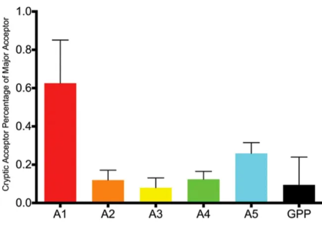
Figure 2.7. Cryptic Acceptor Usage as a Percentage of Major Acceptor Usage in Transmitted/Founder Viruses and NL4-3. ... 53
Figure 2.8. Quantification of Splicing in Subtype C Strain pZM247Fv2. ... 56
Figure 2.9. SIVmac239 Splice Patterns and Primer Locations. ... 57
Figure 3.1. Design and analysis of panel of synonymously mutated HIV-1 viruses. ... 75
Figure 3.2. Spreading replication properties of mutant viruses. ... 78
Figure 3.3. Analysis of HIV-1 splicing in WT and synonymously mutated HIV-1. ... 80
Figure 3.4. Phenotypes of synonymously mutated HIV-1 viruses. ... 83
Figure 3.5. Activation of cryptic splice sites by synonymous mutations in Gag. ... 87
Figure 3.6. Activation of canonical splice acceptor sites (A1 and A2) by synonymous mutations in mutant I. ... 90
Figure 3.7. Activation of canonical splice acceptor site A2 by synonymous mutations in mutant J... 92
Figure 3.8. Activation of canonical splice acceptor site A3 by synonymous mutations in mutant K. ... 94
Figure 3.9. Summary of splicing control in HIV-1. ... 97
Figure 4.1. Generation of cell lines stably expressing HA-tagged hnRNPs and summary of CLIP-seq. ... 110
Figure 4.2. CLIP-seq of hnRNPs reveal their binding sites on viral RNAs. ... 112
Figure 4.3. Classification of hnRNP-bound cellular RNAs and the binding motifs identified within them. ... 114
Figure 4.4. RNAi of hnRNP H1 decreases Gag expression and alters viral splicing in cells. ... 116
Figure 4.5. HnRNP H1 binds to purine-rich sequences on the viral genome. ... 118
Figure 4.6. HnRNP H1 exhibits specificity for G-rich sequences in vitro. ... 119
Figure 4.7. Mutation of hnRNP H1 binding sites alters HIV-1 replication and alternative splicing. ... 122
Figure 5.1. Target sequence between A5 and D4. ... 138
Figure 5.2. Mutation rates for WT or SLSA1 (low vif) in the 1.8 kb or 4 kb size classes. ... 140
Figure 5.3. G-to-A mutations as a proportion of total mutations. ... 141
Figure 5.4. Skew of G-to-A mutations. ... 142
Figure 5.5. Skew of A-to-G mutations. ... 143
Figure 5.6. Mutations affecting the use of acceptor A5. ... 144
Figure 6.1. Relative positions of known HIV-1 SREs. ... 154
Figure 6.3. Splicing regulatory elements mutated. ... 157
Figure 6.4. Usage of acceptor A1 in mutated SREs between A1 and D2. ... 158
Figure 6.5. Acceptor usage in ESSV mutant relative to WT. ... 160
Figure 6.6. Percent of unspliced transcripts in the WT and the ESSV mutant. ... 161
Figure 6.7. Acceptor usage for mutations to ESS2 and ESS2p. ... 162
Figure 6.8. Effects of ESS2 and ESS2p mutation on percentage of unspliced transcripts. ... 163
Figure 6.9. Two transcript types make up the majority of transcripts in the ESS2/ESS2p/ESSV combined mutations. ... 164
Figure 6.10. ISS suppresses cryptic splicing near A7. ... 165
Figure 6.11. The ESS3 stem-loop structure located downstream of splice acceptor A7. ... 166
Figure 6.12. Ratio of 1.8 kb transcripts to 4 kb transcripts in each ESS3 mutant and its internal NL4-3 control. ... 167
Figure 7.1. A Proposed Model for hnRNP Binding. ... 177
Figure 8.1. Infection with Vpr-deficient HIV-1 results in attenuated virus replication in MDDCs and MDDC-T co-cultures. ... 185
Figure 8.2. Vpr does not regulate Env expression in infected MDDCs or incorporation of Env into MDDC-derived virions. ... 187
Figure 8.3. Vpr-deficiency does not result in enhanced type I IFN production in productively infected MDDCs. ... 189
Figure 8.4. Infection of MDDCs with Vpr-deficient viruses results in block to HIV-1 replication in single round infection analysis. ... 191
Figure 8.5. Viral transcription is attenuated in ΔVpr virus infected MDDCs. ... 192
LIST OF TABLES
LIST OF ABBREVIATIONS 4-SU 4-Thiouridine
A Adenine
A3G APOBEC 3G family
ABI Applied Biosystems AX Splice Acceptor Number X
ADAR Adenosine Deaminase Acting on RNA AIDS Acquired Immunodeficiency Syndrome APC Allophycocyanin
APOBEC Apolipoprotein B mRNA Editing Enzyme, Catalytic Polypeptide-Like
Arg Arginine
ASF/SF2 Serine Arginine Rich Splicing Factor 1 ATM Ataxia-Telangiectasia Mutated Kinase ATP Adenosine Triphosphate
AZT Azidothymidine
BST-2 Bone Marrow Stromal Cell Antigen 2 or Tetherin
C Cytosine
CA HIV-1 Capsid Protein
CCR5 C-C Motif Chemokine Receptor 5 CD4 Cluster of Differentiation 4 Receptor
CD11c Integrin, AlphaX (complement component 3 receptor 4 subunit) cDNA Complementary DNA
cERMIT (Conserved) Evidence-Ranked Motif Identification Tool Algorithm CLIP-seq Crosslinking Immunoprecipitation and Sequencing
CRL4DCAF1 Cul4A/DCAF/DDB1 E3 Ubiquitin Ligase Complex Crm1 Chromosomal Maintenance 1 or Exportin 1 C-terminal Carboxyl Terminal
CXCR4 CXC Motif Chemokine Receptor
D Splice Donor
DC Dendritic Cells
DCAF DDB1 and CUL4-Associated Factors
DC-SIGN Dendritic Cell-Specific Intercellular Adhesion Molecule-3-Grabbing Non-Integrin Receptor DDB1 DNA Damage-Binding Protein 1
DDR DNA Damage Response
DGCR8 DiGeorge Syndrome Critical Region 8 DMEM Dulbecco’s Modified Eagle Medium DMSO Dimethyl Sulfoxide
DNA Deoxyribonucleic Acid
dNTP Deoxynucleotide Triphosphates E3 Ubiquitin Ligase
EDTA Ethylenediaminetetraacetic Acid ELISA Enzyme-Linked Immunosorbent Assay Env HIV-1 Envelope Protein
ER Endoplasmic Reticulum ESE Exonic Splicing Enhancer ESS Exonic Splicing Silencer
FACS Fluorescence Activated Cell Sorting FSC Forward Scatter
F/T Founder/Transmitted
G Guanine
G2/M Cell Cycle Interphase and Mitosis Gag HIV-1 Group Specific Antigen
GFP Green Fluorescent Protein
Gly Glycine
GPP Gag-Pro-Pol polyprotein
HA Hemagglutinin
HD Phosphohydrolase Protein Family HIS6 Histadine Tag
HIV-1 Human Immunodeficiency Virus Type 1 HLTF Helicase-like Transcription Factor
hnRNP Heterogeneous Nuclear Ribonucleoproteins
HSQC Heteronuclear Single Quantum Coherence Spectroscopy IFN Interferon
Ig Immunoglobulin
IL-2 Interleukin-2
IN HIV-1 Integrase Protein IRF Interferon Regulatory Factor ISE Intronic Splicing Enhancer ISS Intronic Splicing Silencer ITC Isothermal Titration Calorimetry
Kb Kilobase
LTR Long Terminal Repeat MA HIV-1 Matrix Protein
MDDC Monocyte-Derived Dendritic Cells MDM Monocyte-Derived Macrophages MHC Major Histocompatibility Complex
MHz Megahertz
Nef Viral Negative Regulatory Factor
NF-B Nuclear Factor Kappa-light-chain-enhancer of Activated B Cells NGS Next Generation Sequencing
NIAID National Institute of Allergy and Infectious Diseases NIH National Institute of Health
Ni-IDA Nickle-iminodiacetic Acid NMD Nonsense Mediated Decay NMR Nuclear Magnetic Resonance
NT No Treatment
ORF Open Reading Frame
p24 24kDa HIV-1 Capsid Protein PAGE Polyacrylamide Gel Electrophoresis PBS Phosphate Buffered Saline
PCR Polymerase Chain Reaction PEI Polyethylenimine
PFA Paraformaldehyde PHA Phytohaemagglutinin P
Pol HIV-1 Reverse Transcriptase Polymerase PPT Polypyrimidine Tracts
PR HIV-1 Protease
P-TEFb Positive Transcription Elongation Factor Q-PCR Quantitative Polymerase Chain Reaction qRRM Quasi-RNA Recognition Motif
Rev Regulator of Expression of Virion Proteins RGG boxes Arginine-Glycine-Glycine repeats
RPMI Roswell Park Memorial Institute Medium RRE Rev Response Element
RRPA Random Reverse Primer Assay RT Reverse Transcriptase
RT-PCR Reverse Transcriptase Polymerase Chain Reaction SAM Sterile Alpha Motif
SAMHD1 SAM and HD Domain Containing Deoxynucleoside Triphosphate Triphosphohydrolase 1 SDS Sodium Dodecyl Sulfate
SELEX Systematic Evolution of Ligands by Exponential Enrichment SERINC Serine Incorporator
SC35 Serine Arginine Rich Splicing Factor 35 SEM Standard Error of the Mean
SHAPE Selective 2’-Hydroxyl Acylation Analyzed by Primer Extension SIV Simian Immunodeficiency Virus
SLSA1 Stem-Loop Containing Splice Acceptor A1 SLX1, SLX4 Structure Specific Endonuclease Subunits SLX4com Structure-Specific Endonuclease Complex SMN Survival Motor Neuron Gene
snRNP Small Nuclear Ribonucleoproteins SR Proteins Serine Arginine Protein Family SRA Sequence Read Archive Database SRE Splicing Regulatory Element
SRp40 Serine and Arginine Rich Splicing Factor 5
SSC Side Scatter
SX Small Exon
T Thymine
TBE Tris Borate EDTA Buffer TCEP Tris(2-Carboxyethyl)Phosphine
TET Ten-Eleven Translocation Methylcytosine Dioxygenase T/F Transmitted/Founder
TLR4 Toll Like Receptor 4
U2AF65 Splicing Factor 65 kDa Subunit UNG2 Uracil DNA Glycosylase Vif Viral Infectivity Factor Vpr Viral Protein R Vpu Viral Protein U Vpx Viral Protein X
VSV-G Vesicular Stomatitis Virus
CHAPTER 1: INTRODUCTION Human Immunodeficiency Virus
The human immunodeficiency virus is the cause of the current AIDS pandemic. There are two virus types, HIV-1 and HIV-2, both resulting from multiple cross-species transmission events of simian immunodeficiency viruses (SIV) (1). HIV-1 is the primary and most virulent of the two types and the one studied in this thesis.
HIV-1 is a retrovirus and like all retroviruses, it has a positive single-stranded RNA genome. Two copies of this genome are packaged into a viral protein capsid and surrounded by a membrane derived from the host cell. HIV-1 targets CD4+ cells and the initial step of HIV-1 infection is recognition of the host cell CD4 receptor by a trimer of the viral envelope (Env) surface antigen. After binding the CD4 receptor Env becomes competent to bind a co-receptor, either CCR5 or CXCR4, and undergoes a conformational change that fuses the viral membrane with the host cell membrane and releases the viral contents into the cell (2). After entry the viral capsid largely disassembles, allowing the viral reverse transcriptase to make a double-stranded DNA copy from the genomic RNA template. The viral integrase protein (IN) forms a complex with the ends of the linear viral DNA and inserts it into the host cell genome. High levels of transcription of viral RNA begin after integration, leading to high levels of viral protein production.
The viral proteins have multiple known functions and ongoing studies continue to discover and characterize additional functions (6). Protein function is linked to the regulation of its expression and alternative splicing is a major feature of gene regulation for HIV-1.
Env and Vpu have a common transcript. Although the Vpu open reading frame is upstream of Env, leaky or discontinuous ribosomal scanning may bypass the Vpu start codon and continue to the Env start codon (7). Vpu stands for “viral protein U” (8) and it functions to downregulate CD4 from the cell surface and degrade it in the endoplasmic reticulum (ER). This may facilitate viral particle production by preventing Env from binding to CD4 in the ER, directing it instead to virus particles (9). Vpu also functions to downregulate BST-2 (also known as tetherin), a cellular membrane-associated protein that stalls release of viral particles. In the absence of Vpu, tetherin inhibits viral particle release by tethering the budding virus to the host cell membrane (10).
Viral infectivity factor (Vif) counteracts the cellular restriction factor APOBEC3G (A3G) and 3F and is indispensable for viral replication in cells expressing these proteins. A3G and A3F proteins are packaged in HIV-1 virions and cause mutations during reverse transcription. Vif targets A3G/F for proteosomal degradation (6). In the absence of Vif, the viral genome becomes hypermutated (11, 12).
Viral Protein R (Vpr) has several known functions. It induces cell cycle arrest in the G2 stage (13). Vpr interacts with the ubiquitin ligase complex to target cellular proteins for degradation and is involved in the nuclear import of the pre-integration complex (14). Vpr-defective virus also has a transcription defect (15).
The HIV-1 Tat protein is required for efficient elongation of viral transcripts. It recruits the
transcription factor P-TEFb from a cellular storage complex and brings it to the TAR loop, a region of viral RNA secondary structure. This Tat/P-TEFb/TAR complex interacts with the elongating RNA polymerase to greatly increase processivity (4). Viral transcripts abort prematurely without sufficient Tat (16) and it is thought that this may be associated with viral latency (17).
response element (RRE) and Crm1 and in this way transcripts with retained introns are exported to the cytoplasm (19-21).
Negative factor (Nef) modulates the immune response of the infected cell to accommodate viral replication by downregulating CD4 and MHC Class I from the cell surface (22, 23). Nef also suppresses the apoptotic response and counteracts the anti-viral effect of SERINCs, a family of host proteins (24) that are included in viral particles and block entry into the cell (25).
Viral assembly takes place at the cellular membrane. Multiple domains of Gag target it to the cell membrane, bind two copies of the viral genome, and make numerous protein-protein interactions. These proteins are incorporated into the viral capsid and include the viral reverse transcriptase, Vif, and
integrase. Trimers of envelope proteins are embedded in the cell membrane so as to be on the outer surface of the virion as it buds from the cell (3).
Splicing
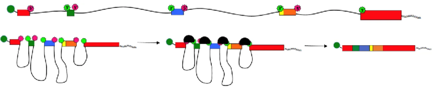
Most eukaryotic genes contain short coding sequences (exons) interspersed with long non-coding sequences (introns). In 1977 reports of splicing in adenovirus were published (26, 27) and soon splicing of discontinuous genes was recognized as a common feature across eukaryotic species (28). After a pre-mRNA is transcribed it must be completely processed before it can be exported to the cytoplasm and translated. This processing includes adding a poly A sequence at the 3’ end, attaching a 5’ cap, and splicing to excise the introns and join the exons.
The terms exon and intron can be confusing so it helps to know where these names originated. Some parts of the pre-mRNA were expressed in protein so they were called exons. Exons were flanked by intervening sequences, hence called introns. Exons are typically 100-200 bases in length. The
average length of a vertebrate exon is 137 bases with less than 1% of exons longer than 400 bases (29). Introns are longer – generally 1000-2000 bases although some are much longer. The 5’ end of an intron is called the donor and the 3’ end is the acceptor. Splicing cuts out the sequence between the donor and acceptor.
sequences (both recognized by U2). The 5’ and 3’ consensus sequences are located at each end of the intron (Figure 1.1). The branch point and polypyrimidine tract (PPT) sequences are about 20 bases long and begin about 25 to 30 bases upstream of the 3’ acceptor site. Almost without exception intronic sequences begin with GU and end with AG and the branch point is an A. HIV-1 is an exception and has some non-adenosine branch points (30, 31). The terminal exons obviously need a different set of recognition signals. For the 5’ terminal exon, the 7-methyl-guanosine cap may be essential for exon recognition, as the cap and its binding proteins are required for splicing in experimental systems (32). The 3’ terminal exon may be recognized by polyadenylation. Splicing and polyadenylation are coupled and splicing of an upstream intron upregulates polyadenylation but mutation of the final 3’ splice site interferes with the polyadenylation process (33).
Figure 1.1. Splicing Signal Sequences.
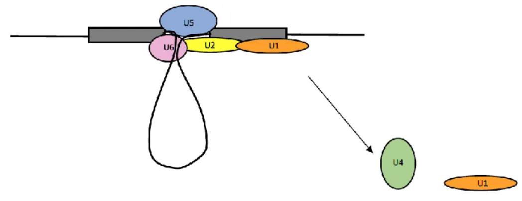
Splicing requires two transesterification reactions catalyzed by the spliceosome, a huge cellular splicing machine (34). This two-step process is catalyzed by a set of five ribonucleoproteins called snRNPs (35), numbered U1, U2, U4, U5, and U6. These snRNPs and a large number of associated proteins combine to create the spliceosome. A proteomic analysis was done to identify spliceosomal proteins and 145 distinct proteins were identified (36), although the actual number may be even larger. A collective review of splicing factor analyses lists almost 300 proteins, making the spliceosome the biggest macromolecular machine in the cell (37, 38).
complex (Figure 1.2) (35, 39, 40) and are often depicted as U1 and U2 binding to opposite ends of an intron. In most eukaryotic genes, however, U1 and U2 actually pair up on opposite ends of an exon. This is called exon definition (29).
Figure 1.2. Spliceosome E Complex.
In the next step, spliceosome proteins assist U1 and U2 to interact across the intron to form the A complex (Figure 1.3). The transition from exon definition to intron definition is not as yet understood. This U1-U2 interaction puts the bulged-out branch point A into proximity with the 5’ donor site (35, 39, 40).
Figure 1.3. Spliceosome A Complex.
Figure 1.4. Spliceosome B Complex.
Figure 1.5. Spliceosome B* Complex
The resulting partially spliced products form complex C (Figure 1.6). Another set of
rearrangements leads to catalysis of the second splicing nucleophilic attack –the 3’hydroxyl at the free 5’ donor splice site attacks the phosphate at the end of the intron (the 3’ splice site), displacing the lariat and connecting the exons (35, 39, 40).
Figure 1.6. Spliceosome C Complex.
Figure 1.7. Post-Splicing Products.
Splicing requires ATP. The multiple remodeling steps involve a collection of core splicing factors, many of them ATP-dependent RNA helicases (37). The stages of spliceosome assembly are reversible
in vitro, and this may give the splicing machinery the ability to form, release, and reform on optimal or
correct sites to increase splicing fidelity (45, 46).
The spliceosome does not processively travel down the pre-mRNA, but appears to be formed de novo for each intron excision, meaning that a transcript with multiple introns will require a new
spliceosome to assemble for each splicing event (47). The spliceosome can self-organize in vitro and does so in the precise series of steps described above. These steps are well documented and agreed on (37, 39, 40, 48, 49).
Recent studies describe the structural conformations of the spliceosome intermediate and catalytic states (50-57). These structural studies have used in vitro one intron systems that block the splicing reaction at a specific point. The results are instructive but may not fully represent the situation in vivo where cellular transcripts have multiple introns and also undergo additional pre-mRNA processing.
Although the spliceosome is generally thought of as the “splicing machine,” work by the Sperling laboratory has found that in nuclei isolated in physiological conditions, spliceosomes are assembled into a tetrameric form called the supraspliceosome (58-61). A supraspliceosome consists of four individual spliceosomal subunits multimerized around a single pre-mRNA. Whereas studies involving individual spliceosomes indicate that a spliceosome forms on each intron/exon, catalyzes splicing, and then
movement/rearrangement of the transcript through the supraspliceosome structure - that mechanism has not yet been described. Although studies of individual spliceosomes detail a complex temporal
association of the five splicing snRNPs, all five are continually associated with the supraspliceosome. Additional factors involved in pre-mRNA processing have been found in the supraspliceosome structure. hnRNP and SR proteins control alternative splicing and are both found in the
supraspliceosome. Additionally, Drosha and its cofactor DGCR8, components of pre-miRNA processing, have also been found in the supraspliceosome, suggesting that miRNA processing and splicing may be connected. mRNAs must be capped at the 5’ end and polyadenylated at the 3’ end, and components required for end processing have also been found in supraspliceosomes (63). ADAR editing is another component of pre-mRNA processing. ADAR proteins make A to I edits in double stranded RNA. Supraspliceosomes were found to contain both ADAR1 and ADAR2 (64). The collective presence of all of these pre-mRNA processing elements suggests that the supraspliceosome is a cellular machine designed to carry out multiple pre-mRNA processing steps in a common location (65). Much work has been done to characterize the structure of the supraspliceosome (40). The resulting structures suggest that a single supraspliceosome is associated with a single pre-mRNA and fragmenting the transcript results in dissolution of the supraspliceosome into four separate spliceosomes, which can be
reassembled by adding an intact transcript. This hints that the pre-mRNA itself is an integral part of the supraspliceosome structure. Each spliceosome directs one exon to a central cavity of the
supraspliceosome, bringing the exons into close proximity with the introns looped out around the exterior of the structure. Each spliceosome subunit can splice out the intron looped out around it, thus the supraspliceosome could splice out four introns at a time, though it is not known if splices occur in a 5’ to 3’ order within the supraspliceosome. It has been hypothesized that an exon that got included in the external intron loop would be skipped (61), one possible pathway for alternative splicing.
Splicing happens with high accuracy but the information in the core splicing signals is not
proteins. This helps identify valid splice sites and enhance or repress splicing. These SREs can occur in introns, where they are called intronic splicing suppressors (ISS) or intronic splicing enhancers (ISE). More often they are found in exons, called exonic splicing silencers (ESS) or exonic splicing enhancers (ESE) (68).
The two classes of cellular proteins that act as trans-factors are the SR proteins and the hnRNP proteins. SR proteins are a family of cellular proteins named for their serine-arginine rich motifs (RS motifs) (69). Although SR proteins are a related family of proteins, the heterogeneous nuclear
ribonucleoproteins (hnRNP) are a heterogeneous group of RNA binding proteins that associate with pre-mRNAs in the nucleus, as the name implies. In general, SR proteins activate splicing and hnRNP proteins suppress splicing, but their activity is context dependent (70-72). This context-dependent behavior of SREs initially seems random but in fact follows some highly predictable, if complex, patterns and rules (73).
It’s known that SR and hnRNP proteins bind to splicing regulatory elements (74) and available evidence suggests that this binding recruits other proteins, either to construct a spliceosome or to make a large enough protein concatenation to block splice sites. This idea is further supported by experiments that tether an hnRNP or SR protein near a splice site, producing the effect of an ESS or an ESE respectively (75-78). Efforts have been made to discover the binding motifs of hnRNP and SR proteins using SELEX (to identify optimal binding sequences (Tian, 1995 #196)) and computational approaches (79, 80) to predict SREs, including an HIV-1 specific screening algorithm (81). These approaches have met with variable success. The redundancy of the proteins and the degeneracy of binding motifs make it difficult to say for certain which protein or proteins control a particular splicing event.
ESEs are important for exon recognition and constitutive exons and all exons have an ESE (84, 85). An ESE and an ESS in the same exon can competitively bind to SR or hnRNP proteins respectively to enhance or repress spliceosome formation across the exon (86).
There is evidence that cellular SREs are associated with RNA secondary structures, in particular the loops of stem-loop structures (89, 90), and that perturbation or stabilization of structural features may alter splicing by masking or unmasking splicing sites or regulatory elements (91). Secondary structures control some HIV-1 splicing events (92), as will be demonstrated in Chapter 2.
As with the core splicing signals, splicing motifs are highly overrepresented relative to valid usage. Splicing regulatory elements are short sequences and thus any given element will be present in far more locations than actual SREs exist. Presumably all these candidate sites are not recognized by trans-acting cellular factors, just as all pseudo-splice sites are not recognized. Splicing factors can recognize multiple sequence motifs, and a given sequence motif can be recognized by redundant splicing factors. Any one exon can be controlled by multiple SREs. The possibilities that feed into splicing decisions are numerous and complex, and many co-operating/competing factors play into the final splicing outcome (35). This suggests that some complex combinatorial code exists to selectively identify genuine splice sites distinct from the background noise, a code that as yet remains uncracked (68, 85, 87, 88).
Alternative Splicing
Alternative splicing is a mechanism in which exons can be used in different arrangements to give specific activities and functions to the resulting proteins (93, 94). There are various types of alternative splicing seen in cellular transcripts. The following figures illustrate the three types of alternative splicing also seen in HIV-1: intron retention, exon skipping, and multiple acceptor sites (68, 95).
Figure 1.8. Exon Recognition and Inclusion.
It is interesting that in cellular transcripts, all the introns are spliced out – intron retention is a rare type of alternative splicing (Figure 1.9). In HIV-1, introns are retained and splicing is so heavily
suppressed that the majority of HIV-1 transcripts remain completely unspliced. The mechanism for intron retention is not known.
Figure 1.9. Intron Retention.
Constitutive exons are present in every transcript but there are also alternate exons that are sometimes skipped. If the exon does not get defined, as shown in Figure 1.10, then it is considered part of the intron and is left out.
Figure 1.10. Undefined Exon Skipped.
Figure 1.11. Alternative Acceptor Usage.
More than one-third of alternate splicing involves alternate exons, and more than one-fourth involves the use of alternate donor or acceptor splice sites (95). Other types of alternative splicing include multiple 5’ donor sites, mutually exclusive exons, and alternative leader or terminal sequences (68).
Figure 1.12. Inclusion or Skipping of an Alternative Exon.
Pseudo/cryptic splice sites typically have silencer elements, particularly those near a constitutive splice site. This may help ensure the usage of correct splice sites, and this type of silencer is seen in HIV-1 (76, 95, 97).
It is not always clear what function, if any, results from many alternatively spliced cellular transcripts. One interesting idea is that some transcripts may include exons that bind and sequester protein factors, making them unavailable (98). Most alternatively spliced transcripts are present at low levels. One evolutionary hypothesis is that these low levels give the cell a chance to try out a new protein isoform while maintaining adequate production of the previous isoform (83). Only about 10% of alternate splicing products have a clearly predictable functional effect but many would be predicted to alter protein surfaces and thereby alter interaction partners (95).
Some functional roles for alternative splicing have been discovered in development and immune response. One well-studied area of alternative splicing is its role in neuronal development (99). The mechanisms are not yet known, but the splicing differences are specific to cell types. Alternative splicing has also been documented in activated T-cells, the primary host cells for HIV-1 (100, 101). HIV-1 infection of macrophages was shown to change the balance of SR and hnRNP proteins over time (102).
decay (NMD) (103). It’s been predicted that up to one-third of alternative splicing events introduce premature stop codons that subject them to NMD (104).
Changes to splicing can have pathological effects. A well-known example is mutation of the SMN (survival motor neuron) gene. A point mutation damages an ESE, causing an exon to be skipped. The shorter protein is rapidly degraded and this loss of function is the cause of spinal muscular atrophy (105). Interestingly, spinal muscular atrophy is one of the first diseases to be successfully treating by controlling a specific splicing event with an antisense oligonucleotide (106). Perhaps as many as half of genetic diseases are mutations that cause aberrant alternative splicing (95).
Splicing in HIV-1
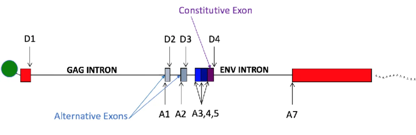
Figure 1.13is a simplified illustration of splicing in HIV-1. There are four donor sites and six acceptor sites. This figure is not drawn to scale – splice sites A1 through A5 actually occupy a 1 kilobase sequence in the middle of the genome. Only one mRNA type is transcribed – a full length genomic RNA, represented by the black bar. If this RNA is not spliced it serves as either a gag/pro/pol mRNA, or as genomic RNA (5, 107).
If HIV-1 mRNA splices there are two basic splicing events. The first event is when D1 is used to splice to one of the six downstream acceptor sites. The spliced transcript generally codes for the first open reading frame after the splice, so a splice from D1 to A1 creates an mRNA for Vif, a splice to A2 for Vpr, and Tat for A3. The transcript type for splicing to A4, and A5 is determined by the second splicing event.
The second major splicing event is from D4 to A7. This splice removes the env intron and the RRE, and joins together the two exons of tat and rev. All spliced transcripts first splice from D1 to an acceptor A1 through A5, and having done so, they may or may not splice out the D4 to A7 sequence. Spliced transcripts are divided into two size classes, depending on whether or not the D4-A7 splice happens. Retaining the env intron makes a larger class of transcripts collectively known as the 4 kb class or incompletely spliced transcripts, while transcripts that splice from D4 to A7 are called the 1.8 kb class or completely spliced transcripts. As discussed above, it is not usual for cellular transcripts to remain unspliced or to retain introns. HIV-1 unspliced or incompletely spliced transcripts keep all introns or the env intron respectively, and this prevents their nuclear export, so they need the RRE and Rev to connect to the Crm1 export pathway (19).
There are two additional donor sites, D2 and D3. Use of D2 or D3 creates small non-coding exons. The red bars in Figure 1.13 illustrate the constitutive exons in HIV-1 transcript types. The grey bars represent the small alternative exons created by use of D2 and/or D3. Either, both, or none can be included in a transcript. These small exons greatly increase the number of possible splice variants (5, 107). For each acceptor there are four possible types of transcripts with variable inclusion of the small exons, but they are not made in equal amounts. In general, direct splices, with no small exons, make up 75-85 % of transcripts. The exception is nef. The direct splice from D1 to A7 makes a nef transcript but this is an uncommon splicing event. The majority of nef transcripts splice to A5 (with or without including the two small upstream exons) and thus the region between A5 and D4 becomes a 3rd small exon.
already been observed for D4 and env transcripts (described below). One study claimed the first small exon stimulated protein production while the second small exon reduced it, with the improbable claim that this effect was activated after transcription but before splicing (when the small exons come into existence) (109). This notion was refuted in a later study that claimed the small exons do not affect transcript stability or protein production (108). Small exon inclusion does not change any known coding sequences or create a new ORF, yet the small exons are well conserved across HIV and SIV.
It is of interest that certain splicing events have not been seen. One such event is the inclusion of the small exons in nef transcripts that splice to A7. A second more interesting omission is transcripts that fail to splice at D1 but later splice from D4 to A7. This transcript type has never yet been reported in otherwise fairly comprehensive splicing analyses (5, 107), and I have not found it in quantities above what might be caused by PCR artifacts. This and other data presented later suggest that there are
mechanisms to suppress certain types of HIV-1 splicing early and completely.
Splicing of cellular genes is an accurate process but splicing in HIV-1 is much less precise. Splicing is primarily from the major donors to acceptors but there are a number of lesser used sites, most often in close proximity to major sites as illustrated in Figure 1.14. This diagram shows some of the splice sites commonly found across different HIV-1 strains but each strain also has its individual minor splice sites, sometimes leading to idiosyncratic splicing and protein products (110-112) and even a novel transcript size class (107).
Figure 1.14. Major and Minor HIV-1 Splice Sites.
constitutive but it uses alternate acceptors, so some control elements regulating acceptor usage would be predicted.
Figure 1.15. HIV-1 from and Exon/Intron Perspective.
HIV-1 donor sites D1 and D4 are efficient, D2 and D3 less so (108). HIV-1 acceptor sites are described as “weak” compared to the cellular splicing consensus sequence; nevertheless, they must still be suppressed to achieve balanced splicing (113-115). It is certain that all known HIV-1 SREs are not yet discovered and characterized but Figure 1.16 shows those that have been described to date.
Figure 1.16. Known HIV-1 SREs.
There are numerous SREs in and immediately downstream of the first small exon from A1 to D2. This exon is only 50 bases long, and such short exons are particularly likely to be skipped, possibly due to steric hindrance between the two portions of the spliceosome that must form on each end. Such tiny exons may need additional enhancers to be included (29). Three ESEs have been characterized between A1 and D2: ESEVif (122), and M1 and M2- repeats of the same enhancer sequence (123). A GGGG sequence just downstream of D2 suppresses D2 use (122). Additional elements have been found downstream of D2 that suppress splicing from a cryptic donor site (97, 124). The strength of splice site D2 also controls splicing, and mutations that strengthen or weaken its ability to bind U1 snRNP increase or decrease recognition of the exon and thereby splicing to A1 (108, 125). Attempts have been made to identify the SR and hnRNP binding proteins that bind HIV-1 SREs and confer splicing control. ESEvif was found to bind SRp75, M1 and M2 bind ASF/SF2, but the binding protein for GGGG is not yet known.
The second small exon between A2 and D3 has an enhancer, ESEVpr (126-128) and a silencer ESSV. ESSV binds the hnRNP A/B proteins and prevents binding of U2AF65 to the viral PPT, preventing U2AF65 from stabilizing the binding of U2 and initiating spliceosome formation (126, 129). ESEVpr may bind the SR proteins Tra2 alpha/beta to support binding of U1 to D3 (128). Another poly-G run called G13-2 just downstream of D3 suppresses D3 and thereby splicing to A2. G13-2 binds hnRNP F/H and hnRNP A2/B1 (130), which is typical of G runs (131). As with D2 and the first small exon, mutations that enhance or reduce D3 complementarity to U1 snRNP increase or reduce splicing to A2 and inclusion of the second small exon. Mutations to ESSV or G13-2, or upregulation of D3 cause activation of splicing from D1 to A2 such that the number of unspliced transcripts is dramatically reduced. This situation is known as oversplicing and has a severe fitness cost. Interestingly, ESSV is not well conserved across HIV groups (127). Although splicing branch points are almost invariably A residues, the branch point for A2 is a G. This unusual branch point does not appear to make A2 a less effective acceptor (31).
by D4 and yet little is known about the interaction of these two sites on each other. Like the silencer ESSV that represses use of acceptor A2, ESS2 is also not well conserved across HIV groups (138).
A4 has 3 or 4 subsites (depending on the strain) named A4a, A4b, A4c, and A4d and A5 has two subsites, A5 and A5b. All the A4 and A5 sites lie within a 45 base sequence and are alternative
acceptors in the constitutive exon defined by D4 (see Figure 1.15). A4a, A4b, A5 and A5b are within a 26 base sequence. They share some common regulatory sequences. Their polypyrimidine tracts overlap and are short and interspersed with purines. There are eight branch points in the A4/5 region and they may be used by more than one acceptor. In the NL4-3 strain, the splice site A4b is also a branch point used by A5 (138, 139). An enhancer named ESE GAR (GAR for guanine/adenine rich) is located just downstream of D4 and binds to SF2/ASF and/or SRp40 to activate splicing to A4 and A5 sites. The SR protein used may influence the acceptor site that gets defined (140, 141). D4 is the donor involved in the second splicing event – removal or retention of the env intron. While binding of U1 does not always lead to splicing at D4, binding of U1 is necessary for the stability of the partially spliced env transcripts (140, 142), similar to U1 binding in the gag-pro-pol gene region that confers stability to gag transcripts, also mediated by SF2/ASF (143). E42 is the sequence between ESE GAR and D4. It is required for D4 exon definition but the specific sequence elements are not yet defined. In the partially spliced 4 kb transcript class, transcripts that splice to either an A4 or A5 acceptor make a vpu/env transcript. All of the A4 and A5 transcripts support Env protein production, but only transcripts that splice to A5 are efficiently translated into Vpu. The presence or absence of the small exons has no differential effect (144).
Although D4 is a strong donor, A7 is a weak acceptor and therefore it is A7 efficiency that determines removal or retention of the env intron (30, 113). Optimizing the A7 splicing signal sequence activates D4-A7 splicing even if D4 is weakened (123). The region surrounding A7 is highly structured and contains three SREs that control splicing to A7. The ISS, an intronic splicing silencer, is just
bind to the specific ESS3a/b and ISS sequences (147, 149) and cooperatively multimerize to prevent binding of SF2/ASF to ESE3 (74, 145, 146).
Thesis Overview
Research is ongoing to correctly identify and characterize HIV-1 SREs, regions of regulatory RNA secondary structure, and the trans-acting SR and hnRNP factors that control splicing. This research requires methods to quantify splicing and splicing changes. Previous work has used artificial sub-genomic constructs to measure splicing. These sub-sub-genomic constructs are transfected or used in cellular extracts and analyzed using agarose gels or Northern blots. As splicing and the effects of SREs are context and structure dependent (68), these artificial constructs, while useful in discovering SREs, may not accurately report their effects on splicing in the context of viral infection.
Chapter 2 describes a new deep sequencing assay I developed to quantify splicing in HIV-1. It combines Primer ID technology with Illumina paired-end deep sequencing to tally the number of all the different splice types in each of the two HIV-1 transcript size classes. This assay was used to measure splicing changes caused by changes in temperature and mutation of a secondary structure. Splicing was quantified in a panel of founder and transmitted viruses. The assay was adapted to measure splicing in SIVmac239. An additional set of programs was written to scan for cryptic splicing and trans-splicing was found. These experiments demonstrate the utility of this splicing assay.
The project in Chapter 3 was done in collaboration with the Bieniasz lab at The Rockefeller University. Blocks of synonymous mutations were made across the HIV-1 genome, some of which had serious replication defects. My part in the collaboration was to characterize and quantify changes in splicing and to identify new donor and acceptor sites in the original mutations and compensating revertant mutants. Previously unknown splicing control elements were discovered.
Chapter 4 is a collaboration with the Kutluay lab at Washington University and the Tolbert lab at Case Western University. This paper presents a study of binding proteins and their effects on HIV-1 splicing, with a particular emphasis on the role of hnRNP H1. My contribution quantified the effects of cellular splicing factor knock downs and the effect of mutations to hnRNP H1 binding sites.
Chapter 6 was done in collaboration with the Tolbert lab (Case Western University) and the Telesnitsky lab (University of Michigan). This chapter describes a new random reverse primer and deep sequencing assay used to distinguish fully spliced, partially splice, and unspliced transcripts. Mutations to a subset of known HIV-1 SREs were analyzed for splicing.
REFERENCES
1. Sharp PM, Hahn BH. 2011. Origins of HIV and the AIDS Pandemic. Cold Spring Harb Perspect Med 1.
2. Wilen CB, Tilton JC, Doms RW. 2012. HIV: cell binding and entry. Cold Spring Harb Perspect Med 2.
3. Sundquist WI, Krausslich HG. 2012. HIV-1 assembly, budding, and maturation. Cold Spring Harb Perspect Med 2:a006924.
4. Karn J, Stoltzfus CM. 2012. Transcriptional and posttranscriptional regulation of HIV-1 gene expression. Cold Spring Harb Perspect Med 2:a006916.
5. Purcell DF, Martin MA. 1993. Alternative splicing of human immunodeficiency virus type 1 mRNA modulates viral protein expression, replication, and infectivity. J Virol 67:6365-6378.
6. Malim MH, Bieniasz PD. 2012. HIV Restriction Factors and Mechanisms of Evasion. Cold Spring Harb Perspect Med 2:a006940.
7. Schwartz S, Felber BK, Fenyo EM, Pavlakis GN. 1990. Env and Vpu proteins of human immunodeficiency virus type 1 are produced from multiple bicistronic mRNAs. J Virol 64:5448-5456.
8. Cohen EA, Terwilliger EF, Sodroski JG, Haseltine WA. 1988. Identification of a protein encoded by the vpu gene of HIV-1. Nature 334:532-534.
9. Estrabaud E, Le Rouzic E, Lopez-Verges S, Morel M, Belaidouni N, Benarous R, Transy C, Berlioz-Torrent C, Margottin-Goguet F. 2007. Regulated degradation of the HIV-1 Vpu protein through a betaTrCP-independent pathway limits the release of viral particles. PLoS Pathog 3:e104.
10. Neil SJ, Zang T, Bieniasz PD. 2008. Tetherin inhibits retrovirus release and is antagonized by HIV-1 Vpu. Nature 451:425-430.
11. Sheehy AM, Gaddis NC, Choi JD, Malim MH. 2002. Isolation of a human gene that inhibits HIV-1 infection and is suppressed by the viral Vif protein. Nature 418:646-650.
12. Lecossier D, Bouchonnet F, Clavel F, Hance AJ. 2003. Hypermutation of HIV-1 DNA in the absence of the Vif protein. Science 300:1112.
13. He J, Choe S, Walker R, Di Marzio P, Morgan DO, Landau NR. 1995. Human immunodeficiency virus type 1 viral protein R (Vpr) arrests cells in the G2 phase of the cell cycle by inhibiting p34cdc2 activity. J Virol 69:6705-6711.
14. Vodicka MA, Koepp DM, Silver PA, Emerman M. 1998. HIV-1 Vpr interacts with the nuclear transport pathway to promote macrophage infection. Genes Dev 12:175-185.
15. Miller CM, Akiyama H, Agosto LM, Emery A, Ettinger CR, Swanstrom RI, Henderson AJ, Gummuluru S. 2017. Virion-Associated Vpr Alleviates a Postintegration Block to HIV-1 Infection of Dendritic Cells. J Virol 91.
17. Lassen KG, Bailey JR, Siliciano RF. 2004. Analysis of human immunodeficiency virus type 1 transcriptional elongation in resting CD4+ T cells in vivo. J Virol 78:9105-9114.
18. Jacob AG, Smith CW. 2017. Intron retention as a component of regulated gene expression programs. Hum Genet doi:10.1007/s00439-017-1791-x.
19. Malim MH, McCarn DF, Tiley LS, Cullen BR. 1991. Mutational definition of the human immunodeficiency virus type 1 Rev activation domain. J Virol 65:4248-4254.
20. Hope TJ. 1999. The ins and outs of HIV Rev. Arch Biochem Biophys 365:186-191.
21. Hammarskjold MH, Rekosh D. 2011. A long-awaited structure is rev-ealed. Viruses 3:484-492. 22. Harris M. 1999. HIV: a new role for Nef in the spread of HIV. Curr Biol 9:R459-461.
23. Azad AA. 2000. Could Nef and Vpr proteins contribute to disease progression by promoting depletion of bystander cells and prolonged survival of HIV-infected cells? Biochem Biophys Res Commun 267:677-685.
24. Usami Y, Wu Y, Gottlinger HG. 2015. SERINC3 and SERINC5 restrict HIV-1 infectivity and are counteracted by Nef. Nature 526:218-223.
25. Trautz B, Wiedemann H, Luchtenborg C, Pierini V, Kranich J, Glass B, Krausslich HG, Brocker T, Pizzato M, Ruggieri A, Brugger B, Fackler OT. 2017. The host-cell restriction factor SERINC5 restricts HIV-1 infectivity without altering the lipid composition and organization of viral particles. J Biol Chem 292:13702-13713.
26. Chow LT, Gelinas RE, Broker TR, Roberts RJ. 1977. An amazing sequence arrangement at the 5' ends of adenovirus 2 messenger RNA. Cell 12:1-8.
27. Berget SM, Moore C, Sharp PA. 1977. Spliced segments at the 5' terminus of adenovirus 2 late mRNA. Proc Natl Acad Sci U S A 74:3171-3175.
28. Flint SJ. 1979. Spliced viral messenger RNA. Am Sci 67:300-311.
29. Berget SM. 1995. Exon recognition in vertebrate splicing. J Biol Chem 270:2411-2414. 30. Dyhr-Mikkelsen H, Kjems J. 1995. Inefficient spliceosome assembly and abnormal branch site
selection in splicing of an HIV-1 transcript in vitro. J Biol Chem 270:24060-24066.
31. Damier L, Domenjoud L, Branlant C. 1997. The D1-A2 and D2-A2 pairs of splice sites from human immunodeficiency virus type 1 are highly efficient in vitro, in spite of an unusual branch site. Biochem Biophys Res Commun 237:182-187.
32. Izaurralde E, Lewis J, McGuigan C, Jankowska M, Darzynkiewicz E, Mattaj IW. 1994. A nuclear cap binding protein complex involved in pre-mRNA splicing. Cell 78:657-668.
33. Niwa M, Rose SD, Berget SM. 1990. In vitro polyadenylation is stimulated by the presence of an upstream intron. Genes Dev 4:1552-1559.
34. Brody E, Abelson J. 1985. The "spliceosome": yeast pre-messenger RNA associates with a 40S complex in a splicing-dependent reaction. Science 228:963-967.
36. Zhou Z, Licklider LJ, Gygi SP, Reed R. 2002. Comprehensive proteomic analysis of the human spliceosome. Nature 419:182-185.
37. Jurica MS, Moore MJ. 2003. Pre-mRNA splicing: awash in a sea of proteins. Mol Cell 12:5-14. 38. Nilsen TW. 2003. The spliceosome: the most complex macromolecular machine in the cell?
Bioessays 25:1147-1149.
39. Rino J, Carmo-Fonseca M. 2009. The spliceosome: a self-organized macromolecular machine in the nucleus? Trends Cell Biol 19:375-384.
40. Sperling R. 2017. The nuts and bolts of the endogenous spliceosome. Wiley Interdiscip Rev RNA 8.
41. Madhani HD, Guthrie C. 1994. Dynamic RNA-RNA interactions in the spliceosome. Annu Rev Genet 28:1-26.
42. Makarov EM, Makarova OV, Urlaub H, Gentzel M, Will CL, Wilm M, Luhrmann R. 2002. Small nuclear ribonucleoprotein remodeling during catalytic activation of the spliceosome. Science 298:2205-2208.
43. Valadkhan S, Mohammadi A, Jaladat Y, Geisler S. 2009. Protein-free small nuclear RNAs catalyze a two-step splicing reaction. Proc Natl Acad Sci U S A 106:11901-11906.
44. Fica SM, Tuttle N, Novak T, Li NS, Lu J, Koodathingal P, Dai Q, Staley JP, Piccirilli JA. 2013. RNA catalyses nuclear pre-mRNA splicing. Nature 503:229-234.
45. Hoskins AA, Friedman LJ, Gallagher SS, Crawford DJ, Anderson EG, Wombacher R, Ramirez N, Cornish VW, Gelles J, Moore MJ. 2011. Ordered and dynamic assembly of single spliceosomes. Science 331:1289-1295.
46. Yang F, Wang XY, Zhang ZM, Pu J, Fan YJ, Zhou J, Query CC, Xu YZ. 2013. Splicing proofreading at 5' splice sites by ATPase Prp28p. Nucleic Acids Res 41:4660-4670.
47. Staley JP, Guthrie C. 1998. Mechanical devices of the spliceosome: motors, clocks, springs, and things. Cell 92:315-326.
48. Smith DJ, Query CC, Konarska MM. 2008. "Nought may endure but mutability": spliceosome dynamics and the regulation of splicing. Mol Cell 30:657-666.
49. Valadkhan S. 2007. The spliceosome: caught in a web of shifting interactions. Curr Opin Struct Biol 17:310-315.
50. Galej WP, Wilkinson ME, Fica SM, Oubridge C, Newman AJ, Nagai K. 2016. Cryo-EM structure of the spliceosome immediately after branching. Nature 537:197-201.
51. Kosmyna B, Query CC. 2016. Structural biology: Catalytic spliceosome captured. Nature 537:175-176.
52. Wan R, Yan C, Bai R, Huang G, Shi Y. 2016. Structure of a yeast catalytic step I spliceosome at 3.4 A resolution. Science 353:895-904.
54. Yan C, Wan R, Bai R, Huang G, Shi Y. 2016. Structure of a yeast activated spliceosome at 3.5 A resolution. Science 353:904-911.
55. Bertram K, Agafonov DE, Liu WT, Dybkov O, Will CL, Hartmuth K, Urlaub H, Kastner B, Stark H, Luhrmann R. 2017. Cryo-EM structure of a human spliceosome activated for step 2 of splicing. Nature 542:318-323.
56. Fica SM, Oubridge C, Galej WP, Wilkinson ME, Bai XC, Newman AJ, Nagai K. 2017. Structure of a spliceosome remodelled for exon ligation. Nature 542:377-380.
57. Yan C, Wan R, Bai R, Huang G, Shi Y. 2017. Structure of a yeast step II catalytically activated spliceosome. Science 355:149-155.
58. Muller S, Wolpensinger B, Angenitzki M, Engel A, Sperling J, Sperling R. 1998. A supraspliceosome model for large nuclear ribonucleoprotein particles based on mass determinations by scanning transmission electron microscopy. J Mol Biol 283:383-394. 59. Cohen-Krausz S, Sperling R, Sperling J. 2007. Exploring the architecture of the intact
supraspliceosome using electron microscopy. J Mol Biol 368:319-327.
60. Sperling J, Azubel M, Sperling R. 2008. Structure and function of the Pre-mRNA splicing machine. Structure 16:1605-1615.
61. Sperling J, Sperling R. 2017. Structural studies of the endogenous spliceosome - The supraspliceosome. Methods doi:10.1016/j.ymeth.2017.04.005.
62. Azubel M, Habib N, Sperling R, Sperling J. 2006. Native spliceosomes assemble with pre-mRNA to form supraspliceosomes. J Mol Biol 356:955-966.
63. Raitskin O, Angenitzki M, Sperling J, Sperling R. 2002. Large nuclear RNP particles--the nuclear pre-mRNA processing machine. J Struct Biol 140:123-130.
64. Raitskin O, Cho DS, Sperling J, Nishikura K, Sperling R. 2001. RNA editing activity is associated with splicing factors in lnRNP particles: The nuclear pre-mRNA processing machinery. Proc Natl Acad Sci U S A 98:6571-6576.
65. Shefer K, Sperling J, Sperling R. 2014. The Supraspliceosome - A Multi-Task Machine for Regulated Pre-mRNA Processing in the Cell Nucleus. Comput Struct Biotechnol J 11:113-122. 66. Lim LP, Burge CB. 2001. A computational analysis of sequence features involved in recognition
of short introns. Proc Natl Acad Sci U S A 98:11193-11198.
67. Sun H, Chasin LA. 2000. Multiple splicing defects in an intronic false exon. Mol Cell Biol 20:6414-6425.
68. Wang Z, Burge CB. 2008. Splicing regulation: from a parts list of regulatory elements to an integrated splicing code. Rna 14:802-813.
69. Graveley BR. 2000. Sorting out the complexity of SR protein functions. Rna 6:1197-1211.
71. Chen CD, Kobayashi R, Helfman DM. 1999. Binding of hnRNP H to an exonic splicing silencer is involved in the regulation of alternative splicing of the rat beta-tropomyosin gene. Genes Dev 13:593-606.
72. Mueller N, Klaver B, Berkhout B, Das AT. 2015. Human immunodeficiency virus type 1 splicing at the major splice donor site is controlled by highly conserved RNA sequence and structural elements. J Gen Virol 96:3389-3395.
73. Ule J, Stefani G, Mele A, Ruggiu M, Wang X, Taneri B, Gaasterland T, Blencowe BJ, Darnell RB. 2006. An RNA map predicting Nova-dependent splicing regulation. Nature 444:580-586.
74. Mayeda A, Screaton GR, Chandler SD, Fu XD, Krainer AR. 1999. Substrate specificities of SR proteins in constitutive splicing are determined by their RNA recognition motifs and composite pre-mRNA exonic elements. Mol Cell Biol 19:1853-1863.
75. Pongoski J, Asai K, Cochrane A. 2002. Positive and negative modulation of human
immunodeficiency virus type 1 Rev function by cis and trans regulators of viral RNA splicing. J Virol 76:5108-5120.
76. Wang Z, Xiao X, Van Nostrand E, Burge CB. 2006. General and specific functions of exonic splicing silencers in splicing control. Mol Cell 23:61-70.
77. Erkelenz S, Mueller WF, Evans MS, Busch A, Schoneweis K, Hertel KJ, Schaal H. 2013. Position-dependent splicing activation and repression by SR and hnRNP proteins rely on common mechanisms. Rna 19:96-102.
78. Hillebrand F, Peter JO, Brillen AL, Otte M, Schaal H, Erkelenz S. 2017. Differential hnRNP D isoform incorporation may confer plasticity to the ESSV-mediated repressive state across HIV-1 exon 3. Biochim Biophys Acta 1860:205-217.
79. Cartegni L, Wang J, Zhu Z, Zhang MQ, Krainer AR. 2003. ESEfinder: A web resource to identify exonic splicing enhancers. Nucleic Acids Res 31:3568-3571.
80. Zhang XH, Leslie CS, Chasin LA. 2005. Computational searches for splicing signals. Methods 37:292-305.
81. Erkelenz S, Theiss S, Otte M, Widera M, Peter JO, Schaal H. 2014. Genomic HEXploring allows landscaping of novel potential splicing regulatory elements. Nucleic Acids Res 42:10681-10697. 82. Zhu J, Mayeda A, Krainer AR. 2001. Exon identity established through differential antagonism
between exonic splicing silencer-bound hnRNP A1 and enhancer-bound SR proteins. Mol Cell 8:1351-1361.
83. Black DL. 2003. Mechanisms of alternative pre-messenger RNA splicing. Annu Rev Biochem 72:291-336.
84. Schaal TD, Maniatis T. 1999. Multiple distinct splicing enhancers in the protein-coding sequences of a constitutively spliced pre-mRNA. Mol Cell Biol 19:261-273.
85. Fairbrother WG, Yeh RF, Sharp PA, Burge CB. 2002. Predictive identification of exonic splicing enhancers in human genes. Science 297:1007-1013.
87. Wang Z, Rolish ME, Yeo G, Tung V, Mawson M, Burge CB. 2004. Systematic identification and analysis of exonic splicing silencers. Cell 119:831-845.
88. Chasin LA. 2007. Searching for splicing motifs. Adv Exp Med Biol 623:85-106.
89. Hiller M, Zhang Z, Backofen R, Stamm S. 2007. Pre-mRNA secondary structures influence exon recognition. PLoS Genet 3:e204.
90. Warf MB, Berglund JA. 2007. MBNL binds similar RNA structures in the CUG repeats of myotonic dystrophy and its pre-mRNA substrate cardiac troponin T. Rna 13:2238-2251.
91. Donahue CP, Muratore C, Wu JY, Kosik KS, Wolfe MS. 2006. Stabilization of the tau exon 10 stem loop alters pre-mRNA splicing. J Biol Chem 281:23302-23306.
92. Pollom E, Dang KK, Potter EL, Gorelick RJ, Burch CL, Weeks KM, Swanstrom R. 2013.
Comparison of SIV and HIV-1 genomic RNA structures reveals impact of sequence evolution on conserved and non-conserved structural motifs. PLoS Pathog 9:e1003294.
93. Kopelman NM, Lancet D, Yanai I. 2005. Alternative splicing and gene duplication are inversely correlated evolutionary mechanisms. Nat Genet 37:588-589.
94. Su Z, Wang J, Yu J, Huang X, Gu X. 2006. Evolution of alternative splicing after gene duplication. Genome Res 16:182-189.
95. Blencowe BJ. 2006. Alternative splicing: new insights from global analyses. Cell 126:37-47. 96. Shen H, Kan JL, Green MR. 2004. Arginine-serine-rich domains bound at splicing enhancers
contact the branchpoint to promote prespliceosome assembly. Mol Cell 13:367-376.
97. Brillen AL, Walotka L, Hillebrand F, Muller L, Widera M, Theiss S, Schaal H. 2017. Analysis of competing HIV-1 splice donor sites uncovers a tight cluster of splicing regulatory elements within exon 2/2b. J Virol doi:10.1128/jvi.00389-17.
98. Kanadia RN, Urbinati CR, Crusselle VJ, Luo D, Lee YJ, Harrison JK, Oh SP, Swanson MS. 2003. Developmental expression of mouse muscleblind genes Mbnl1, Mbnl2 and Mbnl3. Gene Expr Patterns 3:459-462.
99. Liu J, Geng A, Wu X, Lin RJ, Lu Q. 2017. Alternative RNA Splicing Associated With Mammalian Neuronal Differentiation. Cereb Cortex doi:10.1093/cercor/bhx160:1-7.
100. Ip JY, Tong A, Pan Q, Topp JD, Blencowe BJ, Lynch KW. 2007. Global analysis of alternative splicing during T-cell activation. Rna 13:563-572.
101. Martinez NM, Pan Q, Cole BS, Yarosh CA, Babcock GA, Heyd F, Zhu W, Ajith S, Blencowe BJ, Lynch KW. 2012. Alternative splicing networks regulated by signaling in human T cells. Rna 18:1029-1040.
102. Dowling D, Nasr-Esfahani S, Tan CH, O'Brien K, Howard JL, Jans DA, Purcell DF, Stoltzfus CM, Sonza S. 2008. HIV-1 infection induces changes in expression of cellular splicing factors that regulate alternative viral splicing and virus production in macrophages. Retrovirology 5:18. 103. Baker KE, Parker R. 2004. Nonsense-mediated mRNA decay: terminating erroneous gene