INVESTIGATING MOLECULAR MECHANISMS UNDERLYING MORPHOGENETIC CELL SHAPE CHANGE
Christopher Daniel Higgins
A dissertation submitted to the faculty of the University of North Carolina at Chapel Hill in partial fulfillment of the requirements for the degree of Doctor of Philosophy in
the Department of Biology in the College of Arts and Sciences.
Chapel Hill 2016
Approved by:
Bob Goldstein
Richard Cheney
Amy Maddox
Stephen Rogers Kevin Slep
ii © 2016
iii ABSTRACT
Christopher Daniel Higgins: INVESTIGATING MOLECULAR MECHANISMS UNDERLYING MORPHOGENETIC CELL SHAPE CHANGE
(Under the direction of Bob Goldstein)
Changes in cell shape are a fundamental feature of animal development driving the formation of ordered tissues from disordered groups of cells. One common type of animal cell shape change is apical constriction, where a cell or group of cells shrinks down one side more than others. Here, we seek to understand the molecular underpinnings that drive apical constriction using a simplified model system, the roundworm Caenorhabditis elegans. Early in C. elegans development, the endoderm precursor (E) cells undergo apical constriction. This cell shape change drives the internalization of the E cells. Previous work showed that the molecular motor non-muscle myosin II (NMY-2 in C. elegans) is required for E cell internalization, and is enriched and activated at the apical side of E cells where it is thought to generate force by pulling on a meshwork of filamentous actin in the cell cortex. We use
iv
constriction may be governed not by the activation of myosin dynamics, but by a molecular clutch mechanically linking apical myosin dynamics to cell-cell junctions. We, therefore, sought to characterize the molecular nature of cell-cell junctions in the E cells to identify components that may contribute to this molecular clutch. We started by tagging with GFP all three essential members of the C. elegans cadherin-catenin complex (CCC), a complex known to contribute (albeit, redundantly) to apical constriction in the E cells. Spinning disk confocal fluorescence microscopy revealed that HMP-1/α-catenin-GFP, GFP-HMP-2/β-catenin, and HMR-1/cadherin-GFP all enriched at apical junctions as the E cells were undergoing apical
constriction. We next showed that some CCC components require others to enrich apically. For example, HMR-1/cadherin requires HMP-1/α-catenin to enrich apically, suggesting that linking to the contractile actomyosin cytoskeleton might be required for apical enrichment. To test this we disrupted myosin dynamics using a
temperature sensitive allele of nmy-2 or by using RNA interference to disrupt mrck-1, a kinase required for myosin activation. Both treatments disrupted the apical
v
vi ACKNOWLEDGEMENTS
First, I would like to thank my wife, Jessica, for her continued support throughout the arduous journey of graduate school. I am truly fortunate to have such a bright and caring partner in life. I would also like to thank my children Josie and Elliott who constantly brighten my day.
I would like to thank my advisor, Bob Goldstein. Bob has been truly generous with his time and treasure throughout my time in his lab. He has allowed me great latitude to pursue my career ambitions outside of academia, and for that I am very grateful. He has also been an excellent source of scientific mentorship.
I would like to thank the members of my thesis committee: Dave Reiner, Amy Maddox, Richard Cheney, Kevin Slep, and Steve Rogers. You have been a truly inspired group that has provided invaluable advice to propel my project.
I thank my parents, who have provided me with all the opportunities I could ever ask for, and whose constant love and support sustains me every day.
I’d like to thank my lab mates past and present. You have made coming into work every day a true pleasure.
I’d like to thank the many friends I’ve made in graduate school. Your companionship and commiseration have made the journey much easier.
vii
PREFACE
My fascination with biology really took off during high school. Susan Quigley, my senior year AP Biology teacher at Cardinal Newman High School in West Palm Beach, FL was a truly inspiring person whose enthusiasm about biology inspired me to learn more. So, as a freshman at Notre Dame, I started out as a biology major.
Initially, I wasn’t sure whether I wanted to pursue a career in research or go to medical school like my dad did. He’s a general surgeon, but he faced a similar decision once upon a time and wound up going into medicine, partly because of “fear of poverty.”
I enjoyed my intro Biology course work, especially the stuff about cell biology. I decided during my sophomore year to take an intensive student-driven lab course called Advanced Cell Biology Research Lab. This course was run by Michelle Whaley, a truly wonderful person who poured immense time and passion into
making this course excellent. As students, we worked closely with Notre Dame Biology Department faculty to design novel research projects. We designed experiments, ordered reagents, carried out the experiments, and analyzed and interpreted the data. There was no preset “answer” like in a typical teaching lab setting. These were new projects addressing genuinely open questions in cell biology.
My group worked on how a protein called NuMA organizes the
viii
in cultured mammalian cells, and we measured the percentage of spindles with monopolar, bipolar, and multipolar geometries. Our work suggested that NuMA was required for cells with extra centrosomes to condense those centrosomes into a bipolar spindle upon mitosis. This was interesting because cancer cells often contain too many centrosomes, and clustering of this kind would allow them to proliferate more effectively while also compromising mitotic fidelity, contributing to genomic instability.
I thought this work was really exciting. I also got really fascinated with the cytoskeleton, particularly microtubules. This fascination was what pushed me over the edge to go to graduate school.
I then joined the lab of the same professor I worked with during the Cell Biology Research Lab, Dr. Ted Hinchcliffe, to start an undergrad research project. Ted paired me up with his graduate student Liz Halpin (Collins), who got me started working on my own project in the lab. We wanted to understand how a family of proteins called tektins contributed to cytokinesis in mammalian cells. My project used biochemistry to identify the native size and shape of tektin complexes in cells, as well as tektin interactors. I was really excited by this work, and I wanted to pursue something similar in graduate school.
ix
I did rotations in four labs in my first year, settling on Bob Goldstein’s lab as my choice of a thesis lab. Bob’s lab had a really nice group of people working in it at the time (and it still does). People seemed really engaged with what they were doing, but they were also really outgoing and friendly. It was a great environment to start out in.
I picked up on a project that was both very promising and very challenging. The project was initiated by a previous grad student in the lab, Minna Roh-Johnson. Minna noticed that the dynamics of the actin cytoskeleton during early
morphogenetic movements of cells in the C. elegans embryo were really weird. Namely, the cytoskeletal dynamics driving cell shape change in the early embryo were deployed well in advance of the actual shape change. This meant that there might be a developmentally-regulated clutch that engages the cytoskeleton to the cell membrane.
I thought this sounded really cool. Plus, it offered me a chance to hit the ground running, and perhaps get my name on a nice paper in the early days of graduate school. We submitted the paper to Nature and it went out for review, but it bounced. We looked really closely at the reviewer comments and decided to try to address them as best we could. This meant booting up a new collaboration with Dan Kiehart’s group at Duke doing really challenging laser cuts in early embryonic cells. With a lot of persistence, Serdar Tulu, then a postdoc in the Kiehart lab, and I managed to get it to work.
x
unbiased way. This program gave us really nice maps with vectors that
corresponded to the direction of cytoskeletal flow over time in our movies. I also did a bunch of new analysis for the paper, doing tedious manual tracking and
quantifying apical areas at earlier timepoints than Minna had measured.
After a lot of work, we submitted the paper to Science. Initially, it got decent reviews, but the editor declined to publish. But after some persistence on Bob’s end, we managed to get the editor to reconsider it, pending some additional experiments. This meant me going back to Duke and doing more challenging laser cut
experiments with Serdar. Again, we managed to get these cuts to work: Serdar drove the scope, I mounted the embryos and analyzed the data afterwards.
During this time, we also booted up a collaboration with (now Nobel laureate) Eric Betzig to use his new contraption called a Bessel beam plane illumination microscope. In Eric’s lab I worked with a postdoc, Liang Gao, to
generate really nice 3D images of our embryos over time. The Bessel beam scope could go so much faster than what we had back in Chapel Hill, and illuminating from the side with a thin sheet of light meant that we could image for much longer without photobleaching or damaging the embryo. Eric was working on a new Cell manuscript at the time to describe the latest improvements in his Bessel beam instrument. Our data looked promising enough that Eric decided to include them in the manuscript, and I made it onto the author list.
xi
nice to see this in 3D, and in embryos that weren’t compressed (which we had to do on the spinning disk to get all the features that we wanted to see in one plane).
We went back to Science and the paper got accepted. This was a huge relief. This process had dragged on longer than any of us thought it would, and it was great to finally have it behind us. Later that year, Eric’s paper got into Cell, which was exciting.
The summer after the science paper got in (2012), I got to participate in the Physiology Course at the Marine Biological Laboratory in Woods Hole, MA. This was a truly wonderful experience. I got to meet a lot of very brilliant people, and work on fascinating problems in cell biology. I saw talks from leaders in the cell biology world and got to interact with them in the lab and, of course, in the bar. This was a truly inspiring summer, and I left feeling really excited about science and pursuing a career in academia.
When I made it back to Chapel Hill, real life set back in pretty quick. In the lab, I slammed my head against the wall (figuratively) trying to get biolistic bombardment to work to tag members of the cadherin catenin complex with fluorescent proteins. I wasted a ton of time trying to get this obscure and painful technique to work. In the end, I made some very dim strains, none of which were useful for the type of experiments I wanted to do.
xii
technique totally bailed out my project. Without it, I’m not sure what I would have done.
I wound up getting beautiful endogenously-tagged fluorescent strains for all of my major proteins of interest using CRISPR, and this allowed me to finally do the experiments I had been planning on for years. The results from this work are included here in Chapter 3.
In September of 2012, my wife and I welcomed our first child, Josie, into the world. Becoming a parent was a harrowing and wonderful experience, and it changed my outlook on things quite a bit. I realized that I really liked being a parent and spending time with my kids. I also realized that the hyper-competitive academic path would make this quite difficult, and offered very little financial support and even less job security along the way.
I started to look at other options. Initially, I thought I might enjoy research in an industry setting such as a biotech or a pharmaceutical company. I tried to do some networking and I met with a few people who have these types of jobs. Most of these people had done postdocs and then transitioned into industry. I wasn’t sure I actually wanted to do a postdoc, so I wound up bailing on this path.
Also around this time, my good friend from early in graduate school, Jacob Sawyer, jumped ship on academia and took a job with Nikon Instruments, a microscope company. Jacob seemed to really enjoy his new job, and it was
xiii
I figured that a job in the imaging industry would fit my interests and skills quite well. I would still get to work with microscopes, which I loved. I would get to see a lot of cool, new science. But I wouldn’t have to deal with the boring, tedious parts like writing or tracking on hazy dots in images. Also, I wouldn’t have to write grants or papers, and I would be paid a lot better than I would in academia.
It sounded like a really good deal, so I started to look into available jobs. I applied to Leica in October 2014 for a super travel-heavy confocal/SuperRes support job. The technology was really cool, but it would have taken me away from my family quite a bit.
In the end, I decided to turn it down, although the interview process was a really positive experience.
Early in 2015, a job opened up with Nikon in Durham and Chapel Hill. The job was perfect for what I wanted. It would be a local rep job covering just Duke and UNC, with almost no overnight travel. We wouldn’t have to move, I wouldn’t have to travel, and I would get to work with my old buddy Jacob. It was super-ideal. I applied, interviewed up in New York at Nikon HQ, and I got the job. The only catch was that I would have to start in April 2015, and my grad school work wasn’t quite done yet.
xiv
TABLE OF CONTENTS
LIST OF FIGURES………..….…….………...xvi
LIST OF ABBREVIATIONS……….………..……xvii
CHAPTER 1: BACKGROUND AND SIGNIFICANCE………..………….1
References……….……….6
CHAPTER 2: ASYMMETRIC CELL DIVISION: A NEW WAY TO DIVIDE UNEQUALLY………....7
Abstract…..………..7
Main Text……….………7
Figures……….………..12
References…….…………..……….14
CHAPTER 3: TRIGGERING A CELL SHAPE CHANGE BY EXPLOITING PRE-EXISTING ACTOMYOSIN CONTRACTIONS………16
Abstract………..………16
Results and Discussion………..……….17
Materials and Methods…………..………..23
Strains and worm maintenance………..………23
RNA interference (RNAi)………..………...23
DIC and fluorescence microscopy………..………...…24
Bessel beam plane illumination microscopy and structured illumination...24
xv
Imaging and analysis of Drosophila ventral furrow……….25
Analysis of Ea/p apical constriction speeds………...……….….26
Computer simulation………..……….………….…27
Analysis of myosin dynamics during spontaneous network failures and after laser-cutting………..………...28
Analysis of myosin and membrane movements…..………29
FRAP………..………29
Labeling cell surfaces with Quantum Dots………...30
Figures……….………..30
References………..………..…58
CHAPTER 4: MYOSIN ACTIVITY POLARIZES THE CADHERIN- CATENIN COMPLEX IN APICALLY CONSTRICTING CELLS………..……..…62
Introduction………..………..…63
Materials and methods………..………..66
C. elegans culture………..………..66
Mounting for imaging………..……….66
Spinning disk confocal imaging………..………...66
Image analysis………..………67
CRISPR/Cas9 triggered homologous recombination………….67
RNA interference………..………68
nmy-2-ts experiment………..………..68
Results………..……….69
xvi
Endogenous fluorescent tagging reveals spatiotemporally non-uniform localization of cadherin-GFP to sites of
cell-cell contact……….…………..……..70
The CCC accumulates to varying degrees at different apical junctions…..………...71
Early centripetal myosin contractions do not deplete CCC from the apical junctions………..…………..72
Cadherin requires α and β catenin for apical junction enrichment………..……….………..74
Actomyosin contractility regulates CCC distribution in apically-constricting cells……….…………...76
Discussion……….………77
Figures……….………..82
References………..………..96
CHAPTER 5: FUTURE DIRECTIONS………..………..99
xvii
LIST OF FIGURES
Figure 2.1 Asymmetric cortical myosin in mitotic cells can position
the cytokinetic furrow asymmetrically………12 Figure 2.2 A proposed mechanism for asymmetric furrow
positioning………..13 Figure 3.1 Actomyosin contraction precedes the rapid shrinking of the
apical surface………31 Figure 3.2 Periodic actomyosin coalescence occurs before apical cell
profiles shrink in Drosophila gastrulation………..33 Figure 3.3 Cortical tension associated with apical constricton is
established early and changes little as apical shrinking
accelerates in C. elegans………35 Figure 3.4 Targeting classical cadherin and Rac signaling prevents
coupled movements but not actomyosin contraction…………..37 Figure 3.5 Images of myosin and plasma membrane at four timepoints
in gastrulation, collected by Bessel beam structured plane
illumination (Planchon et al., 2011)………39 Figure 3.6 Movements of myosin and F-actin……….41 Figure 3.7 The actomyosin network is contractile and dynamic…………..43 Figure 3.8 Diagram of early and late stage movements………45 Figure 3.9 Estimating the efficiency of actomyosin network-contact
zone connection by comparing data from a simulation to
data from cells………...46 Figure 3.10 Overlying cell surfaces appear to move centripetally, as
the myosin particles do, during the early
stage……….………..47 Figure 3.11 Embryos deficient in cadherin-catenin complex proteins and
Rac signaling have gastrulation defects………..….50 Figure 3.12 hmr-1(RNAi); ced-5(n1812) embryos appear to have normal
xviii
Figure 3.13 hmr-1(RNAi); ced-5(n1812) embryos failed to establish
coupled movements during late stages…………..……….54 Figure 3.14 Centripetal myosin movements occurred in multiple cells…….55 Figure 3.15 PIV of Ea and MSap cells at early and late stages……….57 Figure 4.1 HMR-1/cadherin-GFP enriches non-uniformly at cell-cell
contacts in early C. elegans
embryos………82
Figure 4.2 Cadherin-Catenin Complex (CCC) enriches apically
in apically constricting cells……….…84 Figure 4.3 HMR-1/Cadherin-GFP enriches differentially over time at
different cell borders associated with apically constricting
cells……….86 Figure 4.4 Cadherin Catenin Complex (CCC) components enrich at
apical cell-cell junctions and do not display centripetal
co-transport with actomyosin………87 Figure 4.5 Some Cadherin Catenin Complex (CCC) components are
interdependent for apical enrichment………89 Figure 4.6 Myosin activity is required for apical enrichment of the
Cadherin Catenin Complex (CCC) in apically constricting
cells……….…91 Figure 4.7 Cas9/CRISPR triggered homologous recombination permits
insertion of fluorescent protein genes at endogenous cadherin catenin complex (CCC) genes in the C. elegans
genome………..…93 Figure 4.8 Quantification of embryonic fluorescence in
knock-in/knockdown embryos permits stage-specific
xix
LIST OF ABBREVIATIONS
AB Anterior blastomere ATP Adenosine triphosphate ATPase Adenosine triphosphatase
C Carboxy
CCC Cadherin-catenin complex
cDNA Complementary deoxyribonucleic acid CI Confidence interval
CRISPR Clustered regularly interspaced short palindromic repeats DNA Deoxyribonucleic acid
dsRNA Double stranded ribonucleic acid
E Endodermal precursor
EMS Endomesodermal precursor cell F-actin Filamentous actin
GFP Green fluorescent protein
min Minute
MRCK Myotonic dystrophy kinase related cdc42 binding kinase MS Mesodermal precursor cell
PCR Polymerase chain reaction RNA Ribonucleic acid
1
CHAPTER 1: BACKGROUND AND SIGNIFICANCE
Morphogenesis is characterized by the establishment of ordered tissues from less ordered collections of cells; that is, decrease in entropy. The second law of thermodynamics requires the input of energy to achieve such a decrease. In biology, such energy is stored largely in the nucleotide adenosine triphosphate (ATP), and it is harnessed by a wide variety of enzymes which produce energy by hydrolyzing ATP into inorganic phosphate and adenosine diphosphate (ADP). Two major hydrolyzers of ATP in eukaryotic cells are the filament building block protein actin and its motor protein myosin. Both of these proteins are essential for the generation of cell-and-tissue scale order during morphogenesis, and both will be central to this dissertation.
Actin is a highly abundant protein in most eukaryotic cells and its
polymerization into actin filaments (F-actin) is a major means by which eukaryotic cells achieve micron-scale organization using nanometer-sized protein building blocks. F-actin is a polar filament (i.e. its ends are non-identical) composed of tens to thousands of G-actin (globular) subunits arranged head to tail in a helix. In cells, F-actin is highly dynamic with new subunits being added constantly to the dynamic “barbed” end and lost from the less dynamic “pointed end.”
The assembly of G-actin subunits into F-actin filaments is tightly regulated in eukaryotic cells by a host of proteins. These proteins function by catalyzing
2
filament ends, preventing capping of filament ends, severing filaments, cross-linking filaments, nucleating branched filament arrays, and disassembling filament
branches. Actin nucleators are largely confined to the plasma membrane and so actin filaments typically associate tightly with the plasma membrane.
Actin filaments also act as protein tracks upon which myosin motor proteins hydrolyze ATP to produce mechanical work. Myosins are a diverse set of proteins which have the ability to bind a wide variety of cargoes largely through their
divergent tail domains. However, all myosins are united by the presence of a motor head domain (Mooseker and Cheney, 1995). Myosin II is the motor responsible for the skeletal muscle contractions with which I am typing this document. Myosin II also has non-muscle orthologues which are present in virtually all eukaryotic cell types. Non-muscle myosin II is known to enrich in the cleavage furrow during cytokinesis where it is thought to be important for driving inward furrow progression, although the precise mechanism by which myosin II promotes cytokinesis remains an area of intense study.
3
mechanisms driving these morphogenetic movements are often conserved across phyla. This work will focus on apical constriction.
Apical constriction is a cell shape change required for development of diverse metazoans (Sawyer et al., 2010). Despite the taxonomically diverse array of animals deploying apical constriction, its molecular underpinnings are surprisingly
well-conserved. That is, apically constricting cells rely on a core set of cytoskeletal machinery to drive movements (Martin and Goldstein, 2014). Namely, cells assemble a meshwork array of actomyosin preferentially on their contact-free
surface (Martin et al., 2009; Roh-Johnson et al., 2012). The meshwork contracts due to the force-producing activity of non-muscle myosin II which is transmitted across cell-cell boundaries through structures known as adherens junctions (AJs). Further, the edges of the cells’ apical contacts bind to the contractile apical actomyosin meshwork driving the shrinkage of the apical surface and drawing the cells neighboring the apically-constricting cells closer together. When deployed in isolation, apical constriction can result in the internalization of cells from the
embryonic surface or the exit of cells from an epithelium (also known as epithelial to mesenchymal transition). When deployed by multiple cells at once, apical
constriction can drive tissue-scale furrow formation or tissue bending (Martin and Goldstein, 2014).
4
constriction and convergent extension, driving the formation of neural folds and tissue lengthening. The neural folds then undergo tissue-scale fusion, developing nascent cell-cell adhesions with cells from the adjacent fold. This fusion results in the formation of a closed tube which will then go on to form the brain and spinal cord of the animal (Copp and Greene, 2010).
Significantly, neural tube closure is one of the most error-prone aspects of human development (Copp and Greene, 2010; Wallingford et al., 2013). Defects in neural tube closure give rise to debilitating birth defects such as spina bifida and anencephaly as well as miscarriage. In the work that follows, I use a highly tractable invertebrate model to dissect the molecular mechanisms of apical constriction with the hope that understanding fundamental mechanisms will contribute a clearer picture of human disease.
The C. elegans gastrula provides a tractable system in which to study the cell biological mechanisms of apical constriction (Lee and Goldstein, 2003). Tractability derives from the following key features: 1) powerful genetic methods of C. elegans including the ability to disrupt gene function with RNAi and mutants and the ability to edit the genome with CRISPR/Cas9 triggered homologous recombination 2) an optically clear embryo with minimal autofluorescence that is amenable to live
fluorescence imaging, 3) a limited number of cells present (26-28, depending on the stage) allowing for the precise determination of cell and non-cell autonomous
5
Here, we investigate the molecular mechanisms contributing to apical constriction using the C. elegans gastrula as a model system. In this system, the cells fated to become the endoderm (i.e. gut) are born onto the outside of the embryo and must undergo apical constriction to internalize (Lee and Goldstein, 2003). These cells, also called E cells or Ea and Ep enrich NMY-2, the predominant C. elegans non-muscle myosin expressed in early embryos, at their apical surfaces (Nance et al., 2003). NMY-2 assembles into punctae at the apical surfaces of the E cells which contract centripetally over time. Initially NMY-2-GFP punctae move centripetally without corresponding movement in the apical cell-cell junctions (Roh-Johnson et al., 2012). We call these uncoupled movements. Later, cell-cell contacts move in concert with centripetally moving NMY-2-GFP.
6
REFERENCES
Copp, A.J. and Greene, N.D.E. (2010). Genetics and development of neural tube defects. J. Pathol. 220, 217–230.
Lee, J.-Y. and Goldstein, B. (2003). Mechanisms of cell positioning during C. elegans gastrulation. Development 130, 307–320.
Martin, A.C. and Goldstein, B. (2014). Apical constriction: themes and variations on a cellular mechanism driving morphogenesis. Development 141, 1987–1998. Martin, A.C., Kaschube, M., and Wieschaus, E.F. (2009). Pulsed contractions of an actin–myosin network drive apical constriction. Nature 457, 495–499.
Mooseker, M.S. and Cheney, R.E. (1995). Unconventional Myosins. Annu. Rev. Cell Dev. Biol. 11, 633–675.
Munjal, A. and Lecuit, T. (2014). Actomyosin networks and tissue morphogenesis. Dev. Camb. Engl. 141, 1789–1793.
Nance, J., Munro, E.M., and Priess, J.R. (2003). C. elegans PAR-3 and PAR-6 are required for apicobasal asymmetries associated with cell adhesion and gastrulation. Development 130, 5339–5350.
Roh-Johnson, M., Shemer, G., Higgins, C.D., McClellan, J.H., Werts, A.D., Tulu, U.S., Gao, L., Betzig, E., Kiehart, D.P., and Goldstein, B. (2012). Triggering a Cell Shape Change by Exploiting Pre-Existing Actomyosin Contractions. Science 335, 1232–1235.
Sawyer, J.M., Harrell, J.R., Shemer, G., Sullivan-Brown, J., Roh-Johnson, M., and Goldstein, B. (2010). Apical constriction: A cell shape change that can drive
morphogenesis. Dev. Biol. 341, 5–19.
Wallingford, J.B., Niswander, L.A., Shaw, G.M., and Finnell, R.H. (2013). The
7
CHAPTER 2: ASYMMETRIC CELL DIVISION: A NEW WAY TO DIVIDE UNEQUALLY
The following was published as a Current Biology Dispatch (Higgins and Goldstein, 2010). I wrote the text in collaboration with my advisor
Dr. Bob Goldstein.
Summary
It has long been known that cells can divide unequally by shifting the mitotic spindle to one side. Two recent reports identify an alternative way to generate daughter cells of different sizes.
Main text
8
and the actomyosin-rich furrow is consistent with the above causal relationships: the spindle's position predicts accurately where furrowing will occur.
However, exceptions exist. In 2000, Kaltschmidt and colleagues
(2000) reported live imaging of microtubules in Drosophila neuroblasts and showed a cell division plane that did not lie midway between the two spindle poles, but instead lay closer to one of the poles, resulting in daughter cells of two different sizes. Now a new report from Cabernard and colleagues (2010) provides evidence that the furrow can be positioned independently of the spindle in these neuroblasts, by a mechanism that involves an asymmetric enrichment of cortical myosin in mitotic cells. A second report from Ou and colleagues (2010) reports a similar mechanism in another system, a Caenorhabditis elegans neuroblast, and tests directly the role of asymmetric myosin enrichment in controlling daughter cell size. The new results challenge the universality of the mitotic spindle as the primary determinant of furrow positioning, establishing an asymmetric cortical enrichment of myosin during mitosis as an alternative means to divide unequally in some cells.
Drosophila neuroblasts divide asymmetrically, producing a larger daughter that retains stem-cell characteristics and a smaller daughter that differentiates. Cabernard and colleagues (Cabernard, et al., 2010) showed by live imaging of neuroblasts that myosin localized in an unexpected pattern during mitosis, becoming enriched asymmetrically in the cell cortex on the side where the smaller daughter cell will form (Fig. 2.1). Interestingly, this enrichment was established even before any mitotic spindle asymmetries were apparent, suggesting that the myosin
9
spindles rotated out of their normal axis still had normal myosin enrichment on the basal side of the cell. The rotated spindle and the basal myosin each appeared to induce a furrow — a double furrow! What does it mean? In Drosophila neuroblasts, the myosin crescent appears to provide an independent, parallel mechanism for cleavage furrow positioning, along with canonical spindle-derived cues.
Ou and colleagues (2010) investigated the asymmetric division of another cell, a C. elegans neuroblast. Division of a particular neuroblast, called QR.a, produces daughter cells of different sizes and fates, with the larger daughter becoming a neuron, and the smaller daughter undergoing apoptosis. Despite this asymmetry of size and fate, the mitotic spindle of this cell is aligned in the center at metaphase, just as in Drosophila neuroblasts (Cabernard, et al., 2010; Ou et al., 2010). And just as in Drosophila neuroblasts, the authors show that myosin becomes enriched asymmetrically in the cortex of one side of the cell during anaphase, on the side that will form the smaller daughter cell.
Ou et al. (2010) propose a mechanism for how asymmetric myosin might drive unequal cell division: cortical contractility driven by the myosin crescent could shrink one hemisphere of the dividing cell, driving cytoplasmic flow through the ingressing cleavage furrow and resulting in two differently-sized daughter cells (Fig. 2.2). To test myosin's role in specific regions of the cell, they used
chromophore-assisted laser inactivation (CALI), a technique that uses reactive products emitted upon fluorophore excitation to locally inactivate proteins
10
the dividing cell from shrinking normally, leading in some cases to equal cell division (Fig. 2.1), whereas CALI of a control GFP-tagged molecule could not. Interestingly, in some cases in which daughter cell size was affected, cell fate was also affected. The results show that asymmetric enrichment of myosin in mitosis can locally affect the size and the fate of a nascent daughter cell.
With mitotic cells constricted at one end by cortical actomyosin-derived
forces, the resulting cell shape resembles one of the classic Rappaport experiments. After his retirement as a professor, Ray Rappaport and his wife Barbara, both in their 70s at the time, published a paper in which they reported the effect of
squeezing mitotic cells into conical shapes (Rappaport and Rappaport, 1994). Why squeeze cells into conical shapes? A computer model developed by Albert Harris and Sally Gewalt (1989) had predicted that cells of this shape could be used to distinguish between existing models for spindle positioning. Interestingly, the result of changing cell shape was similar to that shown in worm and fly neuroblasts: the furrow formed closer to the narrow end of the cell, instead of midway between the two spindle poles (Fig. 2.1). The authors interpreted this as resulting from a more effective interaction between the spindle and the cortex at the narrow end of the cell, as the cortex in this end of the cell lies closer to the spindle.
11
interact at one end of the cell — might seem circuitous. Indeed, in fly neuroblasts, Cabernard et al. (2010) were able to eliminate the spindle altogether by colcemid treatment and then genetically bypass the spindle checkpoint, and they found that the basal myosin enrichment and asymmetric cytokinesis still occurred. This result establishes the new mechanism as a truly independent mechanism, not requiring the mitotic spindle. It will be interesting to learn the extent to which this will stand as an independent mechanism in other systems.
How does myosin localize asymmetrically in mitotic cells? Temporal and spatial mechanisms must be involved. Metaphase-arrested Drosophila neuroblasts failed to localize myosin asymmetrically, suggesting that myosin localization must be temporally linked to mitotic progression, like asymmetric spindle positioning in
certain cells (Cabernard, et al., 2010; McCarthy Campbell, et al., 2009). The authors show that spatial regulation of myosin depends on familiar players, a PAR-1-like kinase called PIG-1 in C. elegans neuroblasts, and the asymmetric Pins protein in Drosophila, which has well-established roles in spindle positioning (Cabernard, et al., 2010; Ou et al., 2010; McCarthy Campbell, et al., 2009; Siller and Doe, 2009). These molecular links are likely to serve as key steps toward dissecting the
12 Figures
Figure 2.1: Asymmetric cortical myosin in mitotic cells can position the cytokinetic furrow asymmetrically
13
Figure 2.2: A proposed mechanism for asymmetric furrow positioning
14
REFERENCES
Canman, J.C. and Wells, W.A. (2004). Rappaport furrows on our minds: the ASCB Cytokinesis Meeting Burlington, VT July 22-25, 2004. J. Cell Biol 166, 943–948. Rappaport, R. (1985). Repeated furrow formation from a single mitotic apparatus in cylindrical sand dollar eggs. J. Exp. Zool. 234, 167–171.
Hiramoto, Y. (1956). Cell division without mitotic apparatus in sea urchin eggs. Exp. Cell Res. 11, 630–636.
Rappaport, R. (1981). Cytokinesis - cleavage furrow establishment in cylindrical sand dollar eggs. J. Exp. Zool. 217, 365–375.
Glotzer, M. (2005). The molecular requirements for cytokinesis. Science 307, 1735– 1739.
Canman, J.C. (2009). Cytokinetic astrology. J. Cell Biol. 187, 757–759.
Bement, W.M., Miller, A.L., and von Dassow, G. (2006). Rho GTPase activity zones and transient contractile arrays. Bioessays 28, 983–993.
Kaltschmidt, J.A., Davidson, C.M., Brown, N.H., and Brand, A.H. (2000). Rotation and asymmetry of the mitotic spindle direct asymmetric cell division in the
developing central nervous system. Nat. Cell Biol. 2: 7–12.
Cabernard, C., Prehoda, K.E., and Doe, C.Q. (2010). A spindle-independent cleavage furrow positioning pathway. Nature 467, 91–94.
Ou, G., Stuurman, N., D'Ambrosio, M., and Vale, R.D. (2010). Polarized myosin produces unequal-size daughters during asymmetric cell division.
Science 330, 677–680.
Diefenbach, T.J., Latham, V.M., Yimlamai, D., Liu, C.A., Herman, I.M., and Jay, D.G. (2002). Myosin 1c and myosin IIB serve opposing roles in lamellipodial dynamics of the neuronal growth cone. J. Cell Biol. 158, 1207–1217.
Jacobson, K., Rajfur, Z., Vitriol, E., and Hahn, K. (2008). Chromophore-assisted laser inactivation in cell biology. Trends Cell Biol. 18, 443–450.
Wang, F.S., Wolenski, J.S., Cheney, R.E., Mooseker, M.S., and Jay, D.G. (1996). Function of myosin-V in filopodial extension of neuronal growth cones. Science 273, 660–663.
15
Harris, A.K. and Gewalt, S.L. (1989). Simulation testing of mechanisms for inducing the formation of the contractile ring in cytokinesis. J. Cell Biol. 109: 2215–2223. McCarthy Campbell, E.K., Werts, A.D., and Goldstein, B. (2009). A cell cycle timer for asymmetric spindle positioning. PLoS Biol. 7, e1000088.
Cordes, S., Frank, C.A., and Garriga, G. (2006). The C. elegans MELK ortholog PIG-1 regulates cell size asymmetry and daughter cell fate in asymmetric neuroblast divisions. Development 133: 2747–2756.
16
CHAPTER 3: TRIGGERING A CELL SHAPE CHANGE BY EXPLOITING PRE-EXISTING ACTOMYOSIN CONTRACTIONS
The following was published as a Science report (Roh-Johnson, et al., 2012). I performed the experiments and analyzed the data in Figure 3.3 B-C examining
cortical tension by laser cutting and measuring recoil rates. I also contributed
significantly to Figure 3.1 by collecting and analyzing data to measure closure rate
over time (D-F) and depicting plasma membranes closing over time (C). Finally, I
adapted the particle image velocimetry program, ImageTracker, to analyze myosin
and membrane dynamics by constructing vector maps. These maps can be found
in Figures 3.1H, 3.3C, and 3.4B-C. I also contributed edits to the final draft of the
manuscript.
Introduction
17
cortical tension but by dynamic linking of apical cell-cell contact zones to an already contractile apical cortex.
Results and Discussion
During development, dramatic rearrangements of cells and epithelia play key roles in shaping animals (Friedl and Gilmour, 2009; Odell et al., 1981; Sawyer et al., 2010; Weijer, 2009). Many rearrangements are driven by apical constriction,
including neural tube closure (Sawyer et al., 2010), failure of which is a common human birth defect (Copp and Greene, 2010). Apical constriction is generally driven by contraction of apical actomyosin networks (Sawyer et al., 2010). However, it is not well understood how the stresses and tensions generated by actomyosin networks produce cell shape changes in developing organisms (Grill, 2011).
To address this issue, we examined cortical actomyosin dynamics during C. elegans gastrulation. In C. elegans, two endodermal precursor cells (Ea and Ep) internalize by apical constriction (Lee and Goldstein, 2003; Lee et al., 2006; Nance and Priess, 2002). Transgenic green fluorescent protein (GFP) myosin II-containing particles formed in each cell's apical cortex, enriched in Ea/p similarly to
18
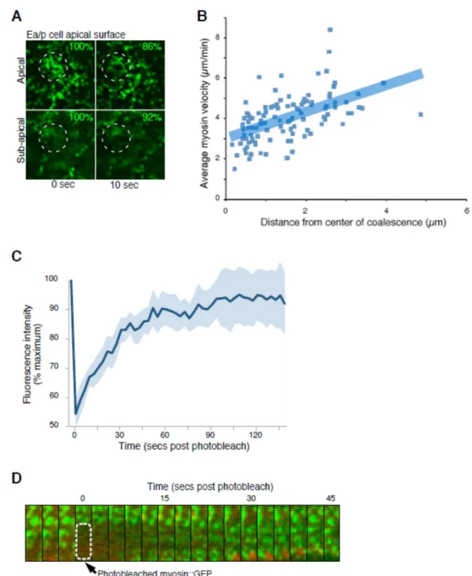
and myosin particles near the center of each coalescence moved at a slower speed than those further away (Fig. 3.7) as seen in other contracting actomyosin networks (Munro et al., 2004). Particle tracking and fluorescence recovery after
photobleaching (FRAP) experiments suggested that the networks were continuously remodeled by exchange of myosin molecules on and off particles as expected (Fig. 3.7).
To investigate how apical actomyosin networks shrink apical cell surfaces, we tracked the outlines of these surfaces, the apical cell-cell contact zones,
19
Our observations were not entirely consistent with a simple pattern of
uncoupled movements early and coupled movements later (Fig. 3.8); instead, some variation existed at each stage. Tracking movements by particle image velocimetry (PIV) demonstrated that in general, the myosin particles and contact zones moved increasingly in unison as time progressed (Fig. 3.1H). We confirmed this result by measuring the rates of individually tracked myosin particles and nearby contact zones, defining the difference between these two rates as a slipping rate (Fig. 3.1I). Actomyosin contractions appeared to drive contact zone movements with ~25% efficiency in early stages, increasing to ~81% efficiency near the end of Ea/p internalization, based on comparing measurements from cells with a computer simulation (Fig. 3.9). Labeling cell surfaces with Quantum Dots or a plasma
membrane marker demonstrated that cell surfaces moved in concert with underlying actomyosin network contractions; i.e. there may be strong frictional force or drag force between the actomyosin network and the overlying plasma membrane (Fig. 3.10). Thus, slipping between actomyosin and membrane occurred specifically at apical cell contact zones, and the relationship between cytoskeletal dynamics and cell shape change during apical constriction is more dynamic than existing models (Odell et al., 1981; Sawyer et al., 2010) predict.
20
toward or away from stationary membranes, and thus was not well connected to contact zone movements at first (Fig. 3.2B). One or more rounds of myosin
enrichment and dissipation occurred in most cells (89%; n=55) before apical profiles began to shrink (Fig. 3.2C–E). These early actomyosin contractions occurred
periodically, with a time interval of 75 ± 24s, similar to that previously measured just after this stage, during apical constriction (Martin et al., 2009). Some of the early contractions might contribute to cell surface flattening in Drosophila, because apical surfaces are not yet completely flattened at this stage (Dawes-Hoang et al., 2005), although many early contractions were not centripetally directed (Fig. 3.2B). Myosin moved at a faster rate than did nearby contact zones at first, and this difference was significantly reduced later, as also observed in C. elegans (Fig. 3.2F). Thus, the early activation of actomyosin contraction, before apical cell profiles begin to shrink, might be a conserved feature of apical constriction.
21
between early and late stages, suggesting little change in cortical tension or stiffness of the network over time (Mayer et al., 2010; Toyama et al., 2008) (Fig. 3.3B).
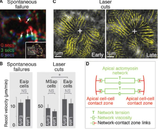
Second, we cut the cortical actomyosin network using a focused UV laser beam and measured initial recoil speed as a quantitative estimate of tension in the network (Fernandez-Gonzalez et al., 2009; Hutson et al., 2003; Kiehart et al., 2000; Martin et al., 2010; Rauzi et al., 2008; Solon et al., 2009). The cortical network recoiled rapidly from cuts in Ea/p (Fig. 3.3B,C), again with little change in initial recoil speed between early and late stages (Fig. 3.3B). Cutting a neighboring cell’s cortex produced a recoil that also did not change significantly over time, and that was slower than in Ea/p (Fig. 3.3B), suggesting that network tension is lower in this cell. Thus, the large difference in the degree of coupled movement between early and late stages is accompanied by little measurable difference in the viscoelastic properties of cortical networks. These results reveal that the cortical tension associated with apical
constriction (Fig. 3.3D) is established well before apical constriction begins, and suggest that the differences between early and late stages might be explained by a change in efficiency of actomyosin-contact zone connections alone.
These results support a picture in which a continuously coalescing apical actomyosin network adds little cortical tension as it begins to move apical cell
contact zones, i.e. the tension involved in coalescing the apical actomyosin network is great compared to the small additional tension required to pull contact zones. Although this model may appear counterintuitive, it is in fact consistent with
22
Our results build a model of apical constriction in which the relevant
cytoskeletal dynamics can run constitutively, transitioning to driving rapid cell shape change at a later time. We speculate that there may exist in this system a molecular clutch – a regulatable, molecular connection between actomyosin networks and contact zones, transmitting the forces generated by actomyosin contraction to the contact zones. Molecular clutches coordinate actin dynamics and adhesion
formation in migrating growth cones and cultured cells (Mitchison and Kirschner, 1988). Our results raise the possibility that there might be developmentally regulated clutches functioning in epithelial morphogenesis. Indeed, targeting a
cadherin-catenin complex and a Rac pathway prevented the transition to coupled movements, genetically separating coupled movements from contractions in this system (Fig. 3.4, Fig. 3.11, Fig 3.12, and Fig. 3.13). Thus, cadherin-catenin complex members, Rac pathway targets, or proteins that function alongside either might contribute to a clutch. Temporal regulation of actin nucleators at contact zones could also function as a clutch, if actin polymerized in a centripetal direction from contact zones
primarily at early stages. In either model, gradual engagement of a clutch would stabilize connections between a contracting actomyosin network and cell-cell
23
additionally by the rate at which apical membrane can be removed (Lee and Harland, 2010).
Recent work has highlighted a number of actomyosin-based mechanisms that drive cell shape changes in morphogenesis (Kasza and Zallen, 2011; Lecuit et al., 2011; Martin et al., 2010). Periodic contractions of actomyosin networks, flows of actomyosin, and an actomyosin-based ratchet make contributions to changing cell shapes (He et al., 2010; Martin et al., 2009; Rauzi et al., 2010; Solon et al., 2009). Here we found that the actomyosin contractions and cortical tension associated with a cell shape change are established even before the cell shape change begins. Thus, the immediate trigger for apical constriction is not the activation of actomyosin contractions or a change in cortical tension, which highlights the dynamic nature of the connections between the actomyosin cytoskeleton and the sites of cell-cell adhesion as a key area of interest for understanding morphogenesis mechanisms. Materials and Methods
Strains and worm maintenance
Nematodes were cultured and handled as described (Brenner, 1974). The following mutant and reporter strains were used: MT4417 ced-5(n1812) dpy-20(e1282) IV referred to here as ced-5; MS126 unc-119(ed4) III; irIs16
[tbx-35::NLS::GFP]; JJ1473 zuIs45 [nmy- 2::NMY-2::GFP; unc-119 (+)]; referred to here as NMY-2::GFP, JJ1317 zuIs3 [end- 1::GFP], OD70 ItIs44 [pie-1:: mCherry::PH domain of PLCdelta] (mCherry::PH) (Kachur et al., 2008), PF100 nnIs [unc-119(+) pie-1 promoter::GFP::Dm-moesin437–578 (amino acids 437–578 of D.
24
NMY-2::GFP. LP54 was constructed by crossing OD70 mCherry::PH males with JJ1473 NMY-2::GFP hermaphrodites. The NMY-2::GFP; ced-5 and mCherry::PH; NMY-2::GFP; ced-5 strains were constructed by crossing ced-5 hermaphrodites with NMY-2::GFP or mCherry::PH; NMY-2::GFP males, respectively. Imaging was
performed at 20°C–23°C for all strains. RNA interference (RNAi)
RNAi by injection was performed according to a standard protocol (Dudley et al., 2002). Double stranded RNA was injected at a concentration of 100 ng/ul. Embryos were analyzed 22- 25 hours later.
DIC and fluorescence microscopy
Embryos were mounted and DIC images were acquired as described
(McCarthy Campbell et al., 2009). Time-lapse images were acquired at 1 µm optical sections every 1 minute and analyzed with Metamorph software (Molecular
Devices). Gastrulation was scored by examination of whether the Ea/p cells were completely surrounded by neighboring cells at the time that Ea/p divided. If Ea/p divided without being completely surrounded, we scored gastrulation as having failed. Spinning disk confocal images were acquired and processed as described (Lee et al., 2006). Epifluorescent images to analyze cell fate were acquired and processed as described (Lee et al., 2006). Embryos expressing end-1::GFP or tbx-35::GFP were mounted laterally and GFP images were acquired at gastrulation stages.
25
Embryos were mounted onto poly-L-lysine-coated coverslips at a specific angle such that the ventral surface was facing the detection objective and the long axis of the embryo was perpendicular to the path of the Bessel beam. The sample chamber was filled with egg buffer (Hepes pH 7.2 25mM, NaCl 110mM, KCl 4mM, Mg Acetate 5mM, CaCl2 5mM). For timelapse movies, approximately forty 200nm thick optical sections were captured every three seconds with both 488nm and 561 nm linear excitation. For whole embryo renderings, 5-phase structured illumination was combined with Bessel beam plane illumination. Point spread functions were calculated, and images were translated and deconvolved as previously described (Planchon et al., 2011). Three-dimensional renderings were created using Amira software (Visage Imaging). Resolution for whole embryo is 194 nm, 238 nm, 419 nm in x, y, z respectively for myosin and 217 nm, 264 nm, 472nm for membrane.
Analysis of F-actin, myosin and membrane dynamics by spinning disk confocal microscopy
26
unless otherwise indicated, imaging in two planes as diagrammed in Fig. S2. Kymographs were made using Metamorph software. The MTrackJ ImageJ plugin was used to track myosin particles and calculate myosin velocities. To calculate slipping rates, ImageJ software was used to generate kymographs of individual myosin particles and nearby apical cell-cell contact zones. The velocities of myosin and membrane were each calculated. The difference in speed (along the axis of myosin movement) between myosin and membrane was determined. Quantum Dots (Molecular Probes) were applied to cell surfaces on devitellinized embryos, n=6 Quantum Dots in three embryos.
Imaging and analysis of Drosophila ventral furrow
27
planes of myosin were merged, and maximum fluorescence intensity was measured. Apical area and myosin intensity were then plotted as a ratio over the initial
measurement, over time before and during apical shrinking. Analysis of Ea/p apical constriction speeds
Three 2-micron steps of ventrally placed wild-type or cadherin/hmr-1 depleted embryos expressing mCherry::PH were taken every 5 seconds. The z-planes were merged, and the apical area was calculated every 25 seconds using ImageJ
software. An average radius was calculated based on the area. To calculate closure speed, the radius at each time point was subtracted from the average of the prior three time points. Early timepoints tended to show little or no decrease in area, contrary to a linear trend. We tested whether the data fit, or failed to fit, a linear trend, by fitting the data from all 10 4 recordings (in Fig. 1D) to a linear and then a quadratic trend by standard methods, via regressions with dependent errors, with the error process represented as a second order autoregressive model. We found that the fit was indeed best (the Akaike information criterion was minimized) for the quadratic trends vs. the linear trends in nine out of ten of the curves, and the
coefficient for the quadratic term was significant for each of these nine models. This result provides convincing evidence of a non-linear trend in the data, with early timepoints showing little or no decrease in apical area.
Computer simulation
28
is chosen at a distance from a contact zone, particles are drawn at randomized angles and distances from this center, and particles are moved toward the coalescence point at a speed proportional to their individual distances from the center, to simulate marks placed on a homogenously contracting two-dimensional sheet. A single contact zone is drawn and moved either not at all, or at a fraction of the speed that particles at the same distance would move equivalent to the percent efficiency of connection, or at the same speed as particles at that distance would move, to simulate 0 to 100% efficiency connection between the contact zone to the contracting network. The program reports the speed of movement of particles along one axis, just as we measured from cells. The speed of contact zone (membrane) movement in the same direction was subtracted from this, resulting in velocities near zero when particles and contact zones moved in concert, positive values when particles moved faster than a contact zone along the same direction, and the most negative values when they moved in opposite directions (for example, for particles on the opposite side of the coalescence point, the side further from the contact zone). Iterations were run with randomized distances between the coalescence center and the contact zone to generate the data graphed in Fig. S5. The y intercept for 0% coupling simulation data was assigned a value matching an average speed specifically for myosin particles near contact zones, which we measured in cells during the early stage, 4.70 µm/min.
Analysis of myosin dynamics during spontaneous network failures and after laser-cutting
For spontaneous failures, myosin images at ti (initial timepoint) and tf (final
29
which myosin particles moved during the meshwork failures was measured, and a speed was calculated. These myosin speeds were then plotted against the distance of myosin particles from the center of the failure. To measure half-life, rates for myosin punctae that were within 1 µm of the meshwork failure were measured for 3 time points. An exponential curve was fitted along the graph, resulting in an R2 value of 0.91 for the early stage and 0.99 for the late stage, and T1/2 was determined for
each. Laser-cutting of the cell cortex was performed using a UV laser as in reference 14 using a single 3-ns pulse in each case. Sudden outward movement of myosin particles and failure of cells to lyse upon focusing the laser on the cell cortex of NMY-2::GFP expressing embryos were interpreted as disruptions to the cell cortex that did not 5 similarly disrupt the plasma membrane. Recoil speed was calculated using radial kymographs centered at each cut site, tracking recoiling myosin
particles within 4.7 µm of each cut site.
Analysis of myosin and membrane movements
30
embryo and tracked at 3s intervals. Particles with lifetimes shorter than three intervals were discarded. Velocity was calculated by dividing the net displacement by the time elapsed. Directedness was calculated by dividing net displacement by the total path length of each particle. Myosin and membrane movements were also tracked by PIV using ImageTracker (http://www.cismm.org/downloads). Movements are represented by vectors, showing direction of movement, with the length of each vector proportional to the estimated speed. Vectors were summed over 2-minute periods to minimize the noise of apparently diffusional movements.
FRAP
Photobleaching of NMY-2::GFP was performed on a VT-HAWK (Visitech) microscope, equipped with an Orca R2 camera and a 100X VC Nikon objective. Images were taken every 5 seconds after photobleaching with the 491 nm and 561 nm 50 mW laser at 30% power. For photobleaching, the 488 nm laser was used at 100% transmission for 5 seconds on a region of interest. Nine cases with
exponential recovery out of eleven total were used to calculate T1/2 and percent recovery using Prism GraphPad software.
Labeling cell surfaces with Quantum Dots
31
32 Figures
Figure 3.1 Actomyosin contraction precedes the rapid shrinking of the apical surface
(A) Diagram of imaging method. (B) NMY-2::GFP coalescence (white arrowheads) in apical cortex of Ea/p cell. (C) Shrinking of apical surfaces during gastrulation
33
surface areas over 575 or 825 s (five embryos each) before closure of the apical surface. (Inset) Apical cell–cell contact zones (arrowheads) on Ep (asterisk). (E) Average radius of apical surfaces derived from area measurements. (F) Mean and 95% CI of radius and myosin particle rate over time. (Inset) Myosin directionality (net distance over total distance, vertical scale 0 to 1) over time (time scale: same as larger graph). (G) Movements of individual myosin particles (arrowheads) near contact zones (white dotted lines) in early or late stages of closure. Arrows at bottom indicate relative distances traveled by each. (H) PIV, three magnifications. Boxes indicate enlarged areas. Left to right are whole embryo at plane of Ea/p apical
34
Figure 3.2. Periodic actomyosin coalescence occurs before apical cell profiles shrink in Drosophila gastrulation
(A) Drosophila ventral furrow formation. Circles mark apical myosin enrichment seen before apical cell profiles began to shrink. (B) Kymograph of a cell (diagrammed) showing myosin (green) movement toward a stationary cell-cell boundary (red) before apical shrinking began. (C) Myosin coalesced (green arrowheads) and dissipated (gray arrowheads) before apical cell profiles began to shrink. This is shown quantitatively from one cell in (D) and from 11 cells chosen at random in (E). Heatmaps in (E) show local maxima of apical myosin levels (three-timepoint running averages of myosin level at each timepoint minus the average of 10 timepoints before and after, normalized to maximum and minimum). Green and gray
35
36
Figure 3.3. Cortical tension associated with apical constricton is established early and changes little as apical shrinking accelerates in C.
elegans
37
38
Figure 3.4. Targeting classical cadherin and Rac signaling prevents coupled movements but not actomyosin contraction
39
40
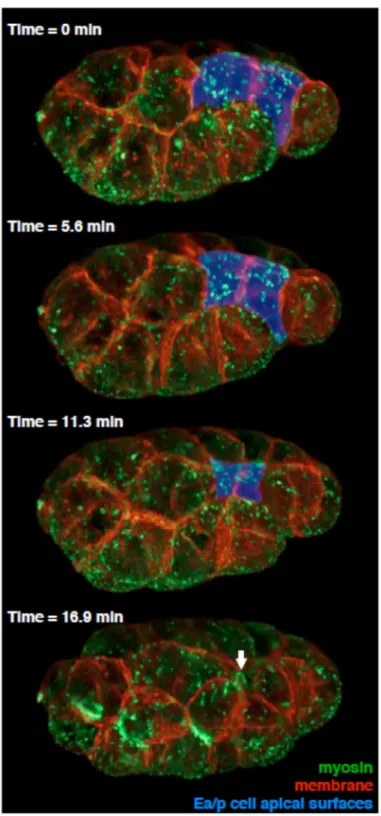
Figure 3.5. Images of myosin and plasma membrane at four timepoints in gastrulation, collected by Bessel beam structured plane illumination
41
Each of the four timepoints was built from 1510 raw images: 151 200-nm z-planes, 5- phase structured illumination, in two color channels. Exposed surfaces of Ea/p cells are pseudocolored blue. Ea/p cells fully internalize between the third and fourth timepoint. Times are min after the first frame shown. The site of closure of
42
Figure 3.6. Movements of myosin and F-actin
43
early stage (~12µm across). (B) Most myosin particles move centripetally. Graph shows distance of myosin particle from the center of the Ea/p apical surface at the end of a myosin track (tf) subtracted from the distance at the beginning of a myosin
track (ti). (C) New myosin particles form near contact zones. Myosin particles were
tracked for 30 secs, and particles were classified as pre-existing (present throughout the 30 secs) or newly-formed (appearing during the 30 secs) at the end of this
44
Figure 3.7. The actomyosin network is contractile and dynamic
45
46
47
Figure 3.8. Diagram of early and late stage movements
48
Figure 3.9. Estimating the efficiency of actomyosin network-contact zone connection by comparing data from a simulation to data from cells
49
50
Figure 3.10. Overlying cell surfaces appear to move centripetally, as the myosin particles do, during the early stage
51
actomyosin can flow in two opposing directions without accompanying movement of nearby membrane protrusions (Rauzi et al., 2010). Alternatively, the actomyosin network might be poorly connected specifically to the contact zones (A, position 2). (B) Diagram of coupled myosin and contact zone movements in the late stage. (C) To distinguish between these models, we used Quantum Dots (Jaiswal and Simon, 2004) as stably fluorescent fiduciary marks on cell surfaces. Quantum Dots applied to cell surfaces are presumed to associate nonspecifically with surface
52
marking PIP2-enrichment, in the apical plasma membrane. mCherry::PH-enriched spots (one is shown, circled in blue), interpreted as membrane invaginations
because they were seen in the apical plasma membrane and just below the plasma membrane, moved in concert with neighboring myosin particles. Lower right drawing shows tracings of first and last timepoints above.
53
Figure 3.11. Embryos deficient in cadherin-catenin complex proteins and Rac signaling have gastrulation defects
If contact zones become mechanically connected to preexisting actomyosin network contractions as we propose, then we predicted that it should be possible to
genetically separate contractions from coupled movements, by identifying genes required for coupled movements and not contractions. We began by examining the sole classical cadherin in C. elegans, HMR-1. HMR-1 is localized to cell-cell contact zones in C. elegans epithelia, it is required for F-actin attachments to contact zones at later stages (Costa et al., 1998), and it is known to function redundantly in cell-cell adhesion and gastrulation (Grana et al., 2010). We targeted cadherin/hmr-1 by RNAi and found that shrinking of Ea/p apical cell surfaces did not reach the speed
54
55
Figure 3.12. hmr-1(RNAi); ced-5(n1812) embryos appear to have normal endomesodermal cell fates and normal F-actin and myosin localization
Analysis of wild-type embryos (left panels) and hmr-1(RNAi); ced-5(n1812) embryos (right panels). Ea/p cells are marked by asterisks. Only those embryos that exhibited Ea/p cell internalization defects in hmr-1(RNAi); ced-5(n1812) embryos were
56
57
Figure 3.13. hmr-1(RNAi); ced-5(n1812) embryos failed to establish coupled movements during late stages
58
Figure 3.14. Centripetal myosin movements occurred in multiple cells
Why would actomyosin contractions begin so early in Ea/p? We speculate that the early actomyosin contractions in C. elegans might be a remnant of an actomyosin-based mechanism for capping apical proteins during apical-basal polarization at earlier stages. At the four- and eight-cell stages, actomyosin contractions have been implicated in redistributing apical
59
stage (A) Cell lineage. (B) Kymographs of myosin-GFP in E, MSa/MSp, Ea/Ep, and MSaa/MSap/MSpa/MSpp cells (top panels), with outlined kymographs, showing some centripetal myosin movement in all of these cells (bottom panels). See Fig. 2.15 for PIV analysis. Consistent with the lower amount of activated myosin in non-Ea/p apical cortexes compared to non-Ea/p apical cortexes [8], these movements appeared slower in a non-internalizing cell than in Ea/p cells during gastrulation (2.34 ± 0.33 µm/min in MSap cells, 3.19 ± 0.14 µm/min in Ea/p cells, p<0.0001 by 2-tailed t-test; Fig. 2.15; note that the myosin rate in MSap does not change
significantly over time, 2.20 ± 0.49, 2.61 ± 0.71, 2.24 ± 0.55 µm/min at 3 minutes before (n=26), 2 minutes after (n=23), and 8 minutes after MS daughters divide (n=27) respectively, p>0.35 for all pairwise 2-tailed t-tests). Our results suggest that the same actomyosin network movements that participate in apical-basal cell
polarization starting at the four-cell stage may be co-opted and upregulated in specific cells later in development to drive the internalization of cells, and that the transition between these two events may be mediated in part by connecting the actomyosin network efficiently to the contact zones in only specific cells.
60
61
Figure 3.15. PIV of Ea and MSap cells at early and late stages
62
REFERENCES
Brenner, S. (1974). The Genetics of Caenorhabditis Elegans. Genetics 77, 71–94. Cheeks, R.J., Canman, J.C., Gabriel, W.N., Meyer, N., Strome, S., and Goldstein, B. (2004). C. elegans PAR proteins function by mobilizing and stabilizing
asymmetrically localized protein complexes. Curr. Biol. CB 14, 851–862.
Copp, A.J. and Greene, N.D.E. (2010). Genetics and development of neural tube defects. J. Pathol. 220, 217–230.
Costa, M., Raich, W., Agbunag, C., Leung, B., Hardin, J., and Priess, J.R. (1998). A Putative Catenin–Cadherin System Mediates Morphogenesis of the Caenorhabditis elegans Embryo. J. Cell Biol. 141, 297–308.
David, D.J.V., Tishkina, A., and Harris, T.J.C. (2010). The PAR complex regulates pulsed actomyosin contractions during amnioserosa apical constriction in
Drosophila. Dev. Camb. Engl. 137, 1645–1655.
Dawes-Hoang, R.E., Parmar, K.M., Christiansen, A.E., Phelps, C.B., Brand, A.H., and Wieschaus, E.F. (2005). folded gastrulation, cell shape change and the control of myosin localization. Dev. Camb. Engl. 132, 4165–4178.
Dudley, N.R., Labbé, J.-C., and Goldstein, B. (2002). Using RNA interference to identify genes required for RNA interference. Proc. Natl. Acad. Sci. U. S. A. 99, 4191–4196.
Edgar, L.G. (1995). Blastomere culture and analysis. Methods Cell Biol. 48, 303– 321.
Fabry, B., Maksym, G.N., Butler, J.P., Glogauer, M., Navajas, D., and Fredberg, J.J. (2001). Scaling the microrheology of living cells. Phys. Rev. Lett. 87, 148102.
Fernandez-Gonzalez, R., Simoes, S. de M., Röper, J.-C., Eaton, S., and Zallen, J.A. (2009). Myosin II Dynamics Are Regulated by Tension in Intercalating Cells. Dev. Cell 17, 736–743.
Friedl, P. and Gilmour, D. (2009). Collective cell migration in morphogenesis, regeneration and cancer. Nat. Rev. Mol. Cell Biol. 10, 445–457.