THE EFFECT OF A SINGLE 30-‐MINUTE BOUT OF MODERATE INTENSITY INTERMITTENT EXERCISE ON THE INFLAMMATORY RESPONSE IN BREAST CANCER
SURVIVORS
Robert Coleman Mills III
A thesis submitted to the faculty of the University of North Carolina at Chapel Hill in partial fulfillment of the requirements for the degree of Master of Arts in the
Department of Exercise and Sport Science (Exercise Physiology).
Chapel Hill 2014
Approved by:
Claudio L. Battaglini
Anthony C. Hackney
Elizabeth S. Evans
©2014
ABSTRACT
Robert Coleman Mills III: The Effect of a Single 30-‐Minute Bout of Moderate Intensity Intermittent Exercise on the Inflammatory Response in Breast Cancer Survivors
(Under the direction of Claudio L. Battaglini)
This retrospective study examined the effect of an acute 30-‐minute bout of moderate intensity intermittent exercise on inflammatory markers (TNF-‐α, CRP, and IL-‐ 10) in breast cancer survivors and healthy controls. Relationships between
inflammatory markers and NK cell count were also explored. Eighteen subjects (9 breast cancer survivors, 9 healthy controls) completed the study. Markers of inflammation and NK cell count were analyzed from blood samples taken at pre-‐
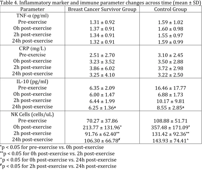
exercise, immediately post-‐exercise, 2 hours post-‐exercise, and 24 hours post-‐exercise. There were no significant changes in inflammatory markers across time for both groups (p > 0.05). There were no significant relationships between changes in inflammatory markers and NK cell across time (p > 0.05). In conclusion, both groups responded similarly to the acute bout of exercise relating to markers of inflammation, and from this sample changes in NK cell did not seem to be mediated by the inflammatory response.
ACKNOWLEDGEMENTS
I have had such a wonderful experience for the past two years in the exercise physiology graduate program at UNC-‐CH. I have learned so much about the research process which will help me in my future endeavors, and have been fortunate enough to become close with some amazing mentors along the way. I would like to thank my mother and father in always supporting me financially and pushing me in my studies. To Dr. Battaglini, words cannot explain how appreciative I am to have you as my advisor. I not only consider you to be an educator, but also a truly close friend who I will remain in touch with for the rest of my life. You have done so much for me in helping me grow as a student in the field of exercise physiology, and your support and encouragement has been really appreciated. To my committee members Dr. Hackney and Dr. Evans, thank you so much for all of your time and effort with helping me in this project. Dr. Hackney, you dedicated so much of your time in helping me with my assays, and I cannot thank you enough. I consider you to be such a role model for me to follow, and I really value your hard work ethic. Dr. Evans, thank you for helping me with all of the logistics of the project since you knew the ins and outs of the samples better than anyone else. Finally, I would like to thank the faculty of the EXSS department for providing me with a strong foundation, which I have used in this project and will continue to build on in the future.
TABLE OF CONTENTS
LIST OF TABLES ... vii
LIST OF FIGURES ... viii
Chapter I. INTRODUCTION ... 1
Statement of purpose ... 5
Research questions ... 6
Hypotheses ... 6
Operational definitions ... 8
Delimitations ... 10
Assumptions ... 11
Limitations ... 11
Significance of study ... 12
II. REVIEW OF LITERATURE ... 14
Breast cancer facts, treatment-‐related side effects, and general exercise benefits ... 14
Inflammation and immunology overview ... 17
Effects of exercise on markers of inflammation in healthy populations ... 23
Cancer and markers of inflammation ... 31
Summary ... 43
III. METHODOLOGY ... 45
Subjects ... 45
Instrumentation ... 46
Procedures ... 48
Data analysis ... 55
IV. RESULTS ... 58
Subjects ... 58
Metabolic Responses ... 60
Inflammatory Marker and Immune Response Changes ... 60
Hypotheses ... 62
V. DISCUSSION ... 67
Pre-‐exercise to Immediately Post-‐exercise ... 70
Pre-‐exercise to 2 hours Post-‐exercise ... 73
Pre-‐exercise to 24 hours Post-‐exercise ... 75
Relationships between markers of inflammation and the immune response ... 77
Inflammatory and immune response due to an acute bout of exercise ... 78
Conclusion ... 82
Recommendations for future research ... 82
APPENDIX A: Sample size calculation ... 84
LIST OF TABLES
Table
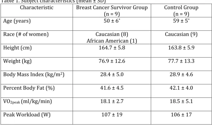
1. Subject characteristics ... 59
2. Cancer treatments received by breast cancer survivor group ... 59
3. Metabolic responses during the submax aerobic exercise session ... 60
4. Inflammatory marker and immune parameter changes across time ... 61
LIST OF FIGURES
Figure
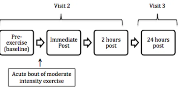
1. Blood sampling timeline ... 50
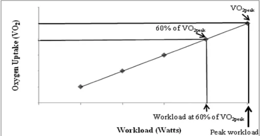
2. Example regression for determining workload corresponding to
60% of VO2peak ... 51
CHAPTER I
INTRODUCTION
Breast cancer is the second leading cause of cancer death in women after lung cancer, and is currently the most common form of cancer in women other than skin cancer in the United States (ACS, 2013). In the United States in 2013, a total of 232,340 new breast cancer cases and 39,620 new breast cancer deaths are projected to occur in women (Siegel et al., 2013). Surgery, chemotherapy, radiation, and hormone
replacement therapies are the most commonly used treatments for those with cancer (ACS, 2013). Treatment improvements and early diagnosis have increased survival rates, and more women are surviving longer than ever before. There have also been other factors contributing to the increased survival rate of cancer patients such as a higher awareness of the importance of self-‐examination, greater education, and advancements in modern technology (ACS, 2013).
and pain have been well documented in the literature (Dimeo et al., 1997; Herrero et al., 2005; Kim et al., 2006; Battaglini et al., 2006; De Backer et al., 2008; Hayes et al., 2012; Cormie et al., 2013).
Most recently, one of the areas that has sparked interest among researchers in exercise oncology is the effect of exercise on markers of inflammation (Cytokines) and immune response. One of the main reasons for the increased interest in this area of research is due to the important roles cytokines have in immune response. These roles include: coordination of inflammatory responses which permits communication
between immune cells, immune regulation through interactions with specific
membrane receptors, and activation of intracellular signaling transduction pathways to induce gene transcription and synthesis of new cellular proteins (Chaplin, 2010; Zhou, 2010). Cytokines are also mediators of innate and adaptive immunity, and are often involved in inflammation through their effects on cell recruitment, cell activation, and antigen presentation (Zhou, 2010). Cytokines have been shown to mediate the
proliferative and cytolytic activities of different cell types that target malignant cells such as NK cells, cytotoxic T cells, and monocytes (Abbas, 2005).
response presented in the current literature may be due to different study populations, different types of anti-‐cancer treatment, and the type, intensity, and duration of the exercise. In addition, the sensitivity of the assays used in different studies can affect the cytokine profile (Pederson, 2000).
An interesting observation when examining the current literature regarding the
effects of exercise on cytokines and immune marker responses in cancer patients is that all previous studies have only examined the immune and inflammatory marker
responses in relation to chronic exercise; in other words, these studies had cancer patients exercise train for a period of time, and cytokines and inflammatory markers were assessed at baseline and follow-‐up (prior to training and at the end of the exercise training period) (Fairey et al., 2005a; Hutnick et al., 2005; Gomez et al., 2011; Janelsins et al., 2011; Jones et al., 2012). The focus of these studies was to examine the effects of exercise training interventions on potential up-‐regulation of immune function through mediation of positive changes in inflammatory markers (é anti-‐inflammation while ê pro-‐inflammatory markers) (Fairey et al., 2005a; Peterson and Pedersen, 2005).
Cancer treatments may often have considerable effects on the immune system of cancer patients and survivors, particularly by decreasing cell number and function of immune cells including components that are associated with anti-‐cancer defense (NK Cells, neutrophils, macrophages, and T lymphocytes) (Fairey et al., 2002). In order to better understand how these treatments impact immune function and perhaps how exercise may alter the immune system in cancer patients, it is paramount that the examination of an acute bout of exercise on the immune response in cancer patients is investigated. Examining the acute effects of an exercise bout on markers of
inflammation and immune function in this population will allow for a better
understanding of the effect of exercise on the immune response of cancer patients and the role of inflammation in the regulation of immune function. This will also help identify the immune system recovery throughout the exercise timeline so more precise exercise prescriptions in regards to intensity, volume, and frequency of exercise
training can be devised for cancer patients. Studying these effects can help provide certainty for promoting positive immune adaptations while avoiding the potential of exercise to create an immune suppression response in this population.
The majority of breast cancer survivors have been shown to have higher levels of inflammation compared to healthy populations (Seruga et al., 2008). Exercise
prescriptions for breast cancer survivors are designed after those for healthy
completion of an exercise bout (recovery from exercise) so comprehensible exercise prescription guidelines for breast cancer survivors can be designed to be safer and promote optimal benefits while minimizing the potential issue of promoting immune suppression.
In a previous dissertation, Dr. Elizabeth Evans evaluated the immune response to an acute bout of exercise in breast cancer survivors and healthy controls. The current study is retrospective in nature, and will use stored blood samples from Dr. Evans’ dissertation to examine the inflammatory response between breast cancer survivors and healthy controls during one acute bout of moderate intensity intermittent exercise. With the goal of researching the pro and anti-‐inflammatory response, Tumor Necrosis Factor (TNF-‐α), C-‐Reactive Protein (CRP), and Interleukin 10 (IL-‐10) were the inflammatory markers selected for the current study. These markers were also
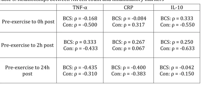
selected because they are commonly seen in research examining this population. NK cell count has already been measured in the previous dissertation of Dr. Evans. NK cells are highly associated with tumor progression, and they were selected for correlational purposes in order to investigate if inflammatory markers can potentially modulate immune function due to exercise in breast cancer survivors.
Statement of the purpose
A secondary purpose was to examine the relationship between selected markers of inflammation and natural killer cell count to explore if the potential mediation of the immune response was also influenced by the inflammatory response due to exercise in breast cancer survivors.
Research questions
R1. Does one bout of acute aerobic exercise alter the inflammatory response in breast cancer survivors and healthy controls immediately post-‐exercise?
R2. How long does the inflammatory response to an acute bout of exercise last in both the breast cancer and healthy control groups?
R3. Does the inflammatory response due to exercise in breast cancer survivors differ from healthy controls?
R4. Is there a relationship between the changes in inflammatory markers and the change in NK cell count after an acute bout of aerobic exercise in breast cancer survivors?
Hypotheses
H1: From pre-‐exercise to immediately post-‐exercise both TNF-‐α and CRP will be significantly reduced in both groups.
H1a: From pre-‐exercise to immediately post-‐exercise IL-‐10 will be significantly increased in both groups.
H2a: From pre-‐exercise to 2 hours post-‐exercise IL-‐10 will be significantly increased in both groups.
H3: From pre-‐exercise to 24 hours post-‐exercise both TNF-‐α and CRP will be significantly reduced in both groups.
H3a: From pre-‐exercise to 24 hours post-‐exercise IL-‐10 will be significantly increased in both groups.
H4: There will be a significant inverse correlation between changes in NK cell count and changes in both TNF-‐α and CRP from pre-‐exercise to immediately post-‐exercise in both groups.
H4a: There will be a significant inverse correlation between changes in NK cell count and changes in both TNF-‐α and CRP from pre-‐exercise to 2 hours post-‐ exercise in both groups.
H4b: There will be a significant inverse correlation between changes in NK cell count and changes in both TNF-‐α and CRP from pre-‐exercise to 24 hours post-‐ exercise in both groups.
H5: There will be a significant positive correlation between changes in NK cell count and changes in IL-‐10 from pre-‐exercise to immediately post-‐exercise in both groups. H5a: There will be a significant positive correlation between changes in NK cell count and changes in IL-‐10 from pre-‐exercise to 2 hours post-‐exercise in both groups.
Operational definitions
• Major Cancer Treatment: Surgery, chemotherapy, and radiation therapy. • Breast Cancer Survivors: Women between the ages of 40-‐70 years who had
confirmed diagnosis of Stages I-‐III invasive breast cancer and had completed all planned surgery, radiation therapy, and chemotherapy 3-‐6 months prior to participation in the study.
• Healthy Controls: Study group which included women 40-‐70 years of age who were
healthy, sedentary, and had never received treatment for cancer of any type. Healthy controls were matched to breast cancer survivors based on age.
• Sedentary: Not participating in regular physical activity for at least 1 year prior to
enrollment in the study. Regular physical activity would be considered as 30-‐ minutes of moderate-‐vigorous activity, 3 days per week.
• VO2peak: A subject’s peak aerobic capacity, measured during a maximal
cardiopulmonary exercise test (CPET).
• Acute Bout of Intermittent Exercise: The 30-minute exercise bout was comprised of ten
3-minute exercise intervals performed at 60% of VO2peak alternating with 1.5 minutes of
rest. A discontinuous exercise protocol was used to ensure that all subjects would be
capable of completing the exercise session.
• Baseline: The study time point that will occur immediately before commencing the
acute bout of moderate intensity exercise. Baseline is synonymous with pre-‐ exercise.
• Immediately post-‐exercise: The study time point that occurred immediately upon
• 2 hours post-‐exercise: The study time point that occurred 2 hours after completion
of the acute bout of moderate intensity exercise.
• 24 hours post-‐exercise: The study time point that occurred 24 hours after
completion of the acute bout of moderate intensity exercise.
• Innate immune system: The initial host response due to the broad number of
recognition molecules that act rapidly after an invading pathogen or toxin is encountered (Chaplin, 2010; Seruga et al., 2008).
• Inflammation: A complex reaction of the innate immune system in tissues involving
the accumulation and activation of plasma proteins and leukocytes at a site of cell injury, toxin exposure, or infection (Abbas, 2005).
• NK cells: Major cellular components of the innate immune response that attack
virally infected cells as well as tumor cells (Abbas, 2005).
• Cytokines: Proteins produced by macrophages and many other types of innate
immune system cells in response to microbes and other antigens that mediate inflammatory and immune reactions (Abbas, 2005). They help control various aspects of cellular growth and differentiation (Corwin, 2000)
• C-‐reactive protein: An acute phase protein produced in the liver that has pro-‐
inflammatory characteristics and is a highly significant marker of inflammatory disease (Abbas, 2005).
• TNF-‐α: A pro-‐inflammatory cytokine that is responsible for many of the systemic
complications of severe infections. TNF-‐α is also an important mediator of the acute inflammatory response to infectious microbes (Abbas, 2005).
those involving macrophages (Corwin, 2000). IL-‐10 has anti-‐inflammatory properties and is considered the most profound immunosuppressive cytokine (Smith, 2003).
Delimitations
• All subjects were women between the ages of 40 and 70 years old.
• All subjects in the breast cancer survivor group had completed their major cancer
treatments and surgeries 3-‐6 months prior to the time of participation in the study.
• All subjects in the control group had been sedentary for at least 1 year prior to
enrollment; i.e., they had not participated in regular organized physical activity within the past year.
• Subjects in the breast cancer survivor group were recruited from the Medical
Oncology clinic and the Radiation Oncology clinic at the NC Cancer Hospital on the campus of the University of North Carolina at Chapel Hill (UNC-‐Chapel Hill) as well as from the waitlist of the Get REAL and HEEL Breast Cancer Program in the
Department of Exercise and Sport Science at UNC-‐Chapel Hill.
• Subjects in the healthy control group were recruited from the faculty, staff, and
student populations at UNC-‐Chapel Hill, as well as from across the Triangle region of North Carolina.
• Baseline study time points occurred between 7:00-‐10:00am in order to control for
• All the subjects were cleared by their medical oncologist or primary physician prior
to enrolling in the study, and had no major health issues that precluded their ability to engage in low to moderate exercise training.
• Before the VO2peak assessment and the acute bout of exercise, subjects were asked to
refrain from eating 2 hours prior to testing and not to exercise for at least 12 hours prior to testing. Also, subjects were asked to refrain from caffeine use at least 12 hours prior to testing and not consume alcohol at least 48 hours prior to testing.
Assumptions
• All breast cancer survivors strictly adhered to all of the pre-‐assessment guidelines
given to them prior to reporting to all laboratory-‐testing sessions.
• The impact of different cancer treatments and drugs would result in similar side
effects experienced by the subjects in the breast cancer survivor group.
• All subjects accurately reported medical history, and abstained from using any
medications that may affect the response of the study variables.
• Breast cancer survivors did not alter their dietary practices during the study.
Limitations
• This is a retrospective study and all data collection procedures could not be altered
for this study.
• The results of this study may only apply to sedentary women between the ages of
able to be generalized to women of all ages, men, all cancer survivors, cancer patients currently undergoing major cancer treatment, or individuals who are exercise-‐trained.
• Cancer survivors’ exercise training prior to treatment could alter the inflammatory
and immune response.
• Changes in cytokines were measured only in the blood.
• The different treatment regimens that breast cancer survivors underwent prior to
the study could have influenced their exercise-‐induced response.
• The intermittent nature of the exercise bout may differently influence the immune
and inflammatory response compared to continuous exercise.
Significance of study
This study helped to clarify if inflammation is acutely altered after a bout of moderate intensity exercise in breast cancer survivors. Few studies have attempted to examine potential mediators of the exercise-‐induced immune response in breast cancer patients, such as inflammatory markers. Most studies in this area have only looked at the inflammatory response to long-‐term exercise training interventions in this
population. However, it is important to understand the acute inflammatory response to exercise in order to monitor the safety and efficacy of implemented exercise programs for this population. Since inflammation is a mechanism of innate immunity, the
immune system can potentially be helped if an acute bout of moderate intensity exercise can positively alter inflammatory markers (Bruunsgaard, 2005). Thus,
commonly seen in breast cancer survivors. In order to assess whether exercise may reduce the risk of illness in cancer patients, clinicians and researchers need to understand how exercise affects the inflammatory response in these patients. Consequently, more specific and relevant exercise prescriptive guidelines can be developed to allow these patients to achieve maximum health benefits without risking further illness (Jones et al., 2012). More precise aerobic exercise prescriptions for breast cancer survivors can be designed as a result of knowledge gained from this study, especially with regard to full recovery time needed from one bout of moderate intensity exercise to a subsequent one.
CHAPTER II
REVIEW OF LITERATURE
For the purpose of organization, Chapter II will be organized into the following sections: I. Breast cancer facts, treatment-‐related side effects, and general exercise benefits; II. Inflammation and immunology overview; III. Effects of exercise on markers of inflammation in healthy populations; IV. Cancer and markers of inflammation; V. Effects of exercise on markers of inflammation in breast cancer survivors; VI. Summary. Measures from the following reviewed articles have been delimited in relation to the measurements pertinent to the current study.
Breast cancer facts, treatment-‐related side effects, and general exercise benefits
Breast cancer facts
Breast cancer is defined as a malignant tumor starting in the cells of the breast that may metastasize to distant areas of the body or invade surrounding tissues. Except for skin cancers, breast cancer is the most common form of cancer in women in
America, and approximately 12% of American women will develop invasive breast cancer throughout their lifetime (ACS, 2013). In 2013, it is estimated that
Breast cancer treatment primarily consists of surgery, chemotherapy, radiation therapy, and hormone therapy. The method of administration depends on the type and stage of the breast cancer, and many of these treatments are combined according to the needs of the patient (NCI, 2013). Common surgical treatments are used to remove cancer from the breast and may include lumpectomy, partial mastectomy, or total mastectomy. Chemotherapy is a treatment that destroys cancer cells by killing them or stopping their division and is administered both orally and intravenously. Radiation is another breast cancer treatment that uses high-‐energy x-‐rays to destroy cancer cells or prevent them from growing larger. Hormone therapy treatment removes hormones by preventing their release from endocrine glands and blocking their action in order to help stop cancer growth (NCI, 2013). Targeted therapies are being developed that are tumor specific. These types of therapies are growing in number and include
trastuzumab also known as Herceptin, which is a monoclonal antibody given to breast cancer survivors who overexpress the protein called human epidermal growth factor receptor 2 (HER2/neu receptor) that is responsible for promoting the growth of cancer cells (ACSM’s Guide to Exercise and Cancer Survivorship, 2012).
Breast cancer treatment-‐related side effects
after the completion of treatment (ACSM’s Guide to Exercise and Cancer Survivorship, 2012). Pain, infection, tenderness, bleeding, and temporary swelling are among the side effects of surgical treatment for breast cancer (ACS, 2013). Chemotherapy side effects may include weight changes, nausea, hair loss, fatigue, vomiting, and an increased chance of infections (ACSM’s Guide to Exercise and Cancer Survivorship, 2012). Radiation treatments may cause patients to encounter soreness, fatigue, skin changes, and swelling (ACSM’s Guide to Exercise and Cancer Survivorship, 2012). Side effects of hormone therapy may involve hot flashes, fatigue, vaginal discomfort, and mood swings (ACSM’s Guide to Exercise and Cancer Survivorship, 2012).
Overall, usual side effects observed in cancer patients that have undergone treatment are depression, worry, pain, cachexia, dyspnea, nausea, and fatigue
(Battaglini et al., 2006). Studies have reported that 70 percent of patients undergoing chemotherapy and radiation have fatigue (Battaglini et al., 2007). Both radiation and chemotherapy have also been shown to cause necrotic death of cancer cells and surrounding tissues, which can result in increased inflammation in breast cancer patients (Grivennikov et al., 2010). In addition, cancer patients may experience depressed immune system parameters. Patients receiving chemotherapy have been shown to have suppressed NK cell counts compared to healthy populations (Shore et al., 1999).
Exercise benefits on treatment-‐related side effects
There is now strong evidence found in epidemiologic studies that exercise may affect breast cancer risk reduction. In a systematic review conducted by C. M.
physical activity reduces breast cancer risk by about 25% (Friedenreich, 2010).
Exercise may also improve overall general health, and studies have shown that exercise can be a helpful tool in attenuating the physiological effects associated with breast cancer treatment. Improvements in cardiorespiratory fitness, body composition, physical functioning, quality of life, and fatigue have been shown by systematic review evidence in cancer survivors who exercise (DeBacker et al., 2008).
Patients receiving cancer treatments in previous years were advised to rest and avoid activity known to further decrease energy levels. Exercise has been shown by scientific research to help alleviate the routine symptoms of cancer treatments such as pain, nausea, and fatigue. Possible benefits of exercise used to alter normal cancer treatment side effects include improved cardiovascular efficiency, increased
mobilization, muscle regeneration, energy production enhancement, and stimulation of erythrocyte, leukocyte, and thrombocyte cell production (Battaglini et al., 2006). Numerous studies have generally demonstrated that exercise does indeed reduce insulin resistance, endogenous estrogen levels, adiposity levels, and inflammation (Friedenreich, 2010).
Inflammation and immunology overview
Inflammation
Inflammation can be described as a complex reaction of the innate immune system in tissues involving the accumulation and activation of plasma proteins and leukocytes at a site of cell injury, toxin exposure, or infection (Abbas, 2005).
damage caused by physical agents such as trauma, surgery, or radiation. Other causes of inflammation may be due to infections by pathogens, either viral or bacterial (Zhou et al., 2010). The inflammatory process is considered to be homeostatic. In this process the host begins a complex series of humoral and cellular responses in an effort to limit tissue damage, isolate and destroy the invading organism, and activate the course of repair (Smith, 2003). Inflammation can serve as a protective mechanism in promoting tissue repair and controlling infections, but it can also cause tissue damage and disease (Abbas, 2005).
Cytokines
Cytokines are proteins produced by many different types of cells in response to microbes and other antigens that mediate inflammatory and immune reactions (Abbas, 2005). The production of cytokines can be from macrophages and lymphocytes as well as other cells such as fibroblasts and endothelial cells (Smith, 2003). Production of these proteins is increased in response to various stressors including injury, infection by microorganisms, normal growth demands, psychological distress, and inflammation (Corwin, 2000). At low concentrations, many cytokines have beneficial effects, whereas tissue damage and multiple organ failure are associated with higher concentrations (Smith, 2003). Cytokines are often involved in inflammation through their effects on cell recruitment, bone marrow differentiation, antigen presentation, cell activation, and cell adhesion molecule expression (Zhou et al., 2010). There are three broad classes of cytokines including lymphokines and monokines, growth factors, and colony-‐
Depending on the microenvironment, cytokines can be functionally separated into two categories: those that are inherently pro-‐inflammatory and anti-‐inflammatory. Pro-‐inflammatory cytokines are important in the inception and advancement of
inflammation. On the other hand, anti-‐inflammatory cytokines inhibit certain conditions of adaptive immunity (Abbas, 2005). Pro-‐inflammatory cytokines are especially of interest to clinicians since they guide the immune response by the stimulation of white blood cell proliferation and cytotoxicity. These cytokines can be controlled by anti-‐inflammatory cytokines through negative feedback (Corwin, 2000). Cytokines are regarded as communication molecules that can act at any range (autocrine, paracrine, endocrine), and are important in activating local
inflammatory/immune cells and regulating invasion of white blood cells into
traumatized tissue (Smith, 2003). Similar to classic hormones, cytokines act by binding to a receptor on a target cell to alter the function of the target cell (Corwin, 2000). They act as immune regulators through interactions with specific membrane receptors (Zhou et al., 2010). Cytokines elicit cellular responses by activating signal transduction
pathways within the cell. This is accomplished with the binding of cytokines to
transmembrane cell surface receptors and ultimately leads to the introduction of new gene transcription and the synthesis of new cellular proteins (Chaplin, 2010).
functional effects (Abbas, 2005). Virtually every system in the body is affected by cytokines. They also have an effect on all aspects of growth and development as well as every host response to injury, infection, and inflammation (Corwin, 2000).
Specific markers of inflammation
Tumor necrosis factor (TNF-‐α) is released from activated macrophages in response to increased concentrations of IL-‐1 and IL-‐2 and bacterial endotoxin (Corwin, 2000). This pro-‐inflammatory cytokine is responsible for many of the systemic
complications of severe infections. TNF-‐α is also an important mediator of the acute inflammatory response to infectious microbes (Abbas, 2005). This cytokine acts to induce expression of IL-‐1 and IL-‐2 and stimulates the production of IL-‐6, leading to increased signs of infection and inflammation as well as the production of acute phase proteins (Corwin, 2000). One of the main physiological functions of TNF-‐α is to stimulate the recruitment of monocytes and neutrophils to infection sites and activate these cells to eliminate microbes (Abbas, 2005).
contributes to sedimentation rate increases that frequently accompany infective and inflammatory disease. In some circumstances the measurement of CRP may be considered to be a key indicator of morbidity relating to infective and inflammatory diseases in general (Corwin, 2000).
Interleukin-‐10 (IL-‐10) is involved in controlling innate immunity reactions and cell-‐mediated immunity through the suppression of activated macrophages and
dendritic cells. IL-‐10 is produced by T cells and is an inhibitor of host immune responses, especially those involving macrophages (Abbas, 2005; Corwin, 2000). On the basis of cell-‐mediated immunity and macrophage function, IL-‐10 is considered the most profound immunosuppressive cytokine (Smith, 2003). IL-‐10 has an anti-‐
inflammatory property known to down-‐regulate inflammatory cytokine production including TNF-‐α, IL-‐1, and IL-‐6 (Zhou et al., 2010). IL-‐10 may suppress cytokine
synthesis by inhibiting the transcription of the genes of cells and by post-‐transcriptional mechanisms (Petersen and Pedersen, 2005).
Cytokines and immunity
Cytokines are considered the principal mediators of communication between cells of the immune system, functioning as key contributors to immunity and
This fully integrated immune response draws elements from multiple effector systems in order to modify a specific response to the invading pathogen (Chaplin, 2010). Immune function
The immune system is commonly divided into two main sectors including innate immunity and adaptive immunity. Both innate and adaptive mechanisms are used to detect and eliminate pathogenic microbes (Chaplin, 2010). NK cells as well as
monocytes/macrophages, T cytotoxic cells, and polymorphonuclear cells are involved in innate immunity. These cells help to form the “first line of defense,” and generally respond to any invading pathogen by rapidly recognizing common molecular patterns and indiscriminately attacking anything appearing to be foreign (Abbas, 2005). The innate system is the initial host response due to the broad number of recognition molecules that act rapidly after an invading pathogen or toxin is encountered (Chaplin, 2010; Seruga et al., 2008). NK cells are an important component of cell-‐mediated immunity, and they are the most sensitive cells to typical stress-‐induced
immunosuppression (Smith, 2003). NK cells are morphologically defined as large granular lymphocytes that recognize their targets using an elaborate accumulation of activating and inhibitory cell surface receptors (Chaplin, 2010). These cells develop in bone marrow and have no antigen-‐specific receptors. They can destroy target cells through antibody dependent cell-‐mediated cytotoxicity. In addition, NK cells have prominent anti-‐tumor effects and are potent killers of virally infected cells (Chaplin, 2010).
Effects of exercise on markers of inflammation in healthy populations
Exercise and cytokine response
Exercise modulates the immune system in healthy individuals and can work as a model of temporary immunosuppression (Pedersen and Hoffman-‐Goetz, 2000). The inflammatory regulation of exercise has been linked to the production of cytokines (Petersen and Pedersen, 2005). Cytokines are released at the site of inflammation as a result of exercise, infection, or tissue injury. The exercise-‐induced local inflammatory response is accompanied by a systemic response known as the acute phase response (Pedersen and Hoffman-‐Goetz, 2000; Ostrowski et al., 1999). This response includes the production of a large number of hepatocyte-‐derived acute phase proteins, such as CRP (Petersen and Pedersen, 2005).
Although many different cell types produce plasma cytokines, muscle cells are a major source during exercise (Zhou et al., 2010). Muscle contractions can induce a myokine response, which is characterized by the release of cytokines, such as IL-‐6, from working muscles. Regular muscle contraction can mediate signals with myokines that may suppress pro-‐inflammatory activities at both distant sites as well as within the skeletal muscle itself (Bruunsgaard, 2005).
systemic response with exercise may be due to exercise only resulting in a transient release of cytokines (Pedersen and Hoffman-‐Goetz, 2000). Increases in pro-‐
inflammatory cytokines such as TNF-‐α are minute due to exercise without muscle damage, and this indicates that in non-‐traumatic exercise, the cytokine cascade differs importantly from the classical acute-‐phase response in infectious systems
(Bruunsgaard, 2005). The fact that pro-‐inflammatory cytokines such as TNF-‐α may not increase with exercise provides evidence for the difference in the cytokine response to exercise compared to that of severe infections (Petersen and Pedersen, 2005). Also, anti-‐inflammatory cytokines, soluble receptors, and receptor antagonists can restrict the inflammatory response to exercise, potentially having an effect on exercise not producing a full systemic response (Ostrowski et al., 1999).
Numerous studies have shown that several cytokines can be found in plasma both during and after exercise. While acute exercise protocols result in short term changes in particular cytokines, chronic exercise training may result in decreased levels of many circulating cytokines (Zhou et al., 2010). Exercise may also induce an increase in the systemic levels of a number of cytokines with anti-‐inflammatory properties, and thereby can protect against chronic medical disorders associated with low-‐grade systemic inflammation (Petersen and Pedersen, 2005).
Exercise in relation to the inflammatory process
in the range of 20-‐60% compared with a sedentary lifestyle (Bruunsgaard, 2005). Even physical inactivity has been shown to be associated with low-‐grade systemic
inflammation in healthy subjects according to several cross-‐sectional studies (Petersen and Pedersen, 2005). In healthy individuals, elevated levels of CRP may be associated with increased body fat and sedentary lifestyles (Pierce et al., 2009b). Overall, multiple studies have found a strong inverse relationship between physical fitness and markers of inflammation (Markovitch et al., 2008).
More frequent physical activity may help reduce the levels of systemic
inflammation in the general population. Several cross-‐sectional reports using general population samples have indicated that increased levels of exercise are associated with decreased levels of markers of inflammation such as C-‐reactive protein (CRP)
(Abramson and Vaccarino, 2002). Abramson and Vaccarino (2002) found that a higher frequency of physical activity was independently associated with lower odds of having elevated inflammation levels, such as CRP and white blood cell count, among healthy middle-‐aged and older US adults. This association was found even after adjustments were made for potential confounding factors including measures of general obesity (body mass index) and central obesity (waist-‐to-‐hip ratio), which have been shown to influence levels of inflammation (Abramson and Vaccarino, 2002). Also, physical activity has been shown to be inversely associated with CRP concentrations in a
representative sample of healthy adults in the US, suggesting that physical activity may mitigate inflammation (Ford, 2002).
antioxidant effects of exercise may be involved in the relationship between exercise and decreasing levels of inflammation (Abramson and Vaccarino, 2002). It is possible that exercise training in general may reduce markers of inflammation, such as CRP, both directly and indirectly. Exercise may reduce CRP directly by reducing cytokine production in muscle, fat, and mononuclear cells and indirectly by reducing body weight, increasing insulin sensitivity, and improving endothelial function (Kasapis and Thompson, 2005). Multiple studies have found an association between exercise and inflammation, but the mechanisms through which physical activity influences the inflammatory process are not all currently known (Ford, 2002).
Even light to moderate physical activity has been shown to be associated with lower blood concentrations of inflammatory markers, especially in cross-‐sectional designs. Specifically, Pitsavos et al. (2003) found that leisure time physical activity was associated with 33% lower concentrations of CRP and 10% lower concentrations of white blood cell counts independent of age (Pitsavos et al., 2003). Also, only 30 minutes of moderate exercise on a regular basis may have the ability to facilitate an anti-‐
inflammatory environment represented by enhanced levels of cytokines such as IL-‐10 (Bruunsgaard, 2005). Very few studies have examined the inflammatory response due to acute moderate-‐intensity exercise, and the findings are controversial. While some studies have found increases in inflammatory markers due to acute moderate-‐intensity exercise, others have found no changes in these markers (Nieman et al., 2005;
Exercise-‐induced anti-‐inflammatory response
Several studies have shown that physical activity mediates strong anti-‐ inflammatory effects in skeletal muscle and fat tissue. Exercise has the ability to produce acute increases in various anti-‐inflammatory mediators (Kasapis and
Thompson, 2005). The anti-‐inflammatory effects of exercise have been attributed to mechanisms that involve the cytokine IL-‐6 (Bruunsgaard, 2005; Steensberg et al.,
2003). IL-‐6 can increase as much as 100-‐fold after strenuous exercise, and this increase is the earliest as well as the most prominent cytokine response to exercise (Kasapis and Thompson, 2005). There is controversy over whether IL-‐6 has pro-‐ or anti-‐
inflammatory properties, but some studies suggest IL-‐6 should be classified as an anti-‐ inflammatory cytokine due to the fact that IL-‐6 may stimulate the production of IL-‐10 and IL-‐1ra while inhibiting the production of TNF-‐α (Starkie et al., 2003; Steensberg et al., 2003). IL-‐6 has also been found to enhance the levels of both IL-‐10 and IL-‐1ra, independently of TNF-‐α (Steensberg et al., 2003).
Starkie et al. (2003) found that physical activity may mediate anti-‐inflammatory activity, and exercise-‐induced IL-‐6 production may help to mediate the effect of exercise on TNF-‐α production. Eight healthy subjects received an endotoxin bolus to induce low-‐grade inflammation after 2.5 hours into a 3-‐hour bout of exercise on a cycle
ergometer. Increases in TNF-‐α from the endotoxin bolus were totally attenuated due to the exercise, whereas the endotoxin induced a two-‐ to threefold increase in circulating levels of TNF-‐α during rest in the same subjects (Starkie et al., 2003). There are
has the ability to blunt the appearance of TNF-‐α production (Starkie et al., 2003). The finding that exercise suppresses TNF-‐α production has also been supported in animal models in which exercise normalized the overexpression of TNF-‐α in TNF-‐R knockout mice (Keller et al., 2004).
In a 2005 review article by Petersen and Pedersen, circulating levels of anti-‐ inflammatory cytokines were commonly found in relation to exercise. As a result of exercise, increases primarily in IL-‐6 followed by increases in IL-‐1ra and IL-‐10 have been thoroughly reported (Petersen and Pedersen, 2005). The production of IL-‐10 that has been shown with exercise can lead to inhibition of the synthesis of a large range of pro-‐ inflammatory cytokines by different cells, notably of the monocytic line of descent. Therefore, IL-‐10 may be able to inhibit the production of TNF-‐α by attenuating the surface expression of TNF-‐α receptors (Petersen and Pedersen, 2005; Steensberg et al., 2003). IL-‐10 may also be able to inhibit the production of IL-‐1α and IL-‐1β as well as the production of some chemokines. The anti-‐inflammatory effects of an acute bout of exercise may protect against chronic systemic low-‐grade inflammation, but a similar link between the acute effects of exercise and long-‐term benefits has yet to be
determined (Petersen and Pedersen, 2005).
(Pedersen and Hoffman-‐Goetz, 2000; Ostrowski et al., 1999). Therefore, there is a parallel anti-‐inflammatory counter-‐regulation that is also part of the acute phase response to exercise (Kasapis and Thompson, 2005; Pedersen, 2000).
Ostrowski et al. (1999) found a significant increase in TNF-‐α after a strenuous bout of exercise (marathon) in healthy adult males. Significant increases in the
inflammation responsive cytokine IL-‐6 were also found following the exercise with only a modest increase in plasma CRP. Both anti-‐inflammatory cytokines (IL-‐10) and
cytokine inhibitors were significantly increased due to the exercise, serving to balance the exercise-‐induced release of pro-‐inflammatory markers (Ostrowski et al., 1999). Contraction-‐induced IL-‐6 expression may be followed by a systemic anti-‐inflammatory response, providing a common underlying pathway by which pro-‐inflammatory
markers (TNF-‐α activity) are attenuated after a single bout of exercise (Bruunsgaard, 2005). Although strenuous exercise has been shown to result in positive changes in inflammatory markers, severe exercise or exercise in an immunosuppressed state may induce a negative inflammatory state as well as immunosuppression (Nieman et al., 1999; Smith, 2000). This phenomenon called the “open window theory,” describes a period of time after exercise in which an individual may be subject to increased risk of infection due to a depression in immune function. “Over-‐trained” states may result in the depression of key immune parameters, which have been linked to imbalances in cytokines (Nieman et al., 1999).
that acute moderate intensity exercise had no effect on pro-‐ or anti-‐inflammatory responses. Twelve sedentary men underwent 30 minutes of walking at 50% VO2max in which no significant changes were found in CRP, IL-‐6, or IL-‐10 concentrations over the 7 days following the single bout of exercise. The results from this study may suggest that the long-‐term anti-‐inflammatory effects that were previously reported with exercise of moderate-‐intensity must be explained by something other than a net anti-‐ inflammatory response to each exercise bout (Markovitch et al., 2008).
Variability found in the levels of inflammatory markers across studies
There have been many differences found between markers of inflammation due to exercise. Inconsistent findings have been reported for TNF-‐α and CRP in response to exercise (Ostrowski et al., 1999). There are several possible explanations for these variable results on cytokines in relation to exercise including: 1) the intensity and duration of the exercise as well as the type of physical activity; 2) the specificity and the sensitivity of the assays used in particular studies; 3) heterogeneity of subjects from sample to sample (Pedersen and Hoffman-‐Goetz, 2000; Pedersen, 2000). Increased cytokine levels have mainly been described due to eccentric training, but concentric exercise can induce cytokine production as well. Also, the magnitude of the increases found in markers of inflammation is closely related to the duration of the exercise (Pedersen and Hoffman-‐Goetz, 2000; Pedersen, 2000).
Cancer and markers of inflammation
Cancer and inflammation
Increased circulating levels of cytokines are known to be associated with cancer, and epidemiological findings have shown a strong association between chronic
inflammation and some types of cancer (Gomez et al., 2011; Seruga et al., 2008).
Inflammatory cells may have powerful effects on tumor development, and inflammation has been shown to act as a tumor promoter. Inflammation can affect tumor
development and progression in addition to the response to therapy (Balkwill and Mantovani, 2001; Grivennikov et al., 2010). Cytokines are mediators that govern a vast range of processes involved in the development of cancer, and markers of inflammation form a major part of the tumor microenvironment (Candido and Hagemann, 2013). Inflammatory cells as well as cytokines regulate the entire tumor organ, managing the migration, growth, and differentiation of all types of cells in the tumor
microenvironment (Coussens and Werb, 2002). These cytokines can also influence immunosuppression and tissue remodeling in the inflammatory microenvironment, which is a critical component of all tumors (Grivennikov et al., 2010; Seruga et al., 2008).