VEGAS as a Platform for Facile Directed
Evolution in Mammalian Cells
Justin G. English,1,5,*Reid H.J. Olsen,1Katherine Lansu,1Michael Patel,2Karoline White,3Adam S. Cockrell,4 Darshan Singh,1Ryan T. Strachan,1Daniel Wacker,1and Bryan L. Roth1,*
1Department of Pharmacology, University of North Carolina, Chapel Hill, NC 27514, USA
2Eshelman School of Pharmacy, University of North Carolina, Chapel Hill, NC 27514, USA
3Department of Biology, University of North Carolina, Chapel Hill, NC 27514, USA
4Department of Epidemiology, University of North Carolina, Chapel Hill, NC 27514, USA
5Lead Contact
*Correspondence:[email protected](J.G.E.),[email protected](B.L.R.)
rather than whole organisms, is called directed evolution (Arnold, 1998; Chen and Arnold, 1993). Directed evolution has been used to create novel binding proteins (Hanes and Plu¨ckthun, 1997; Xu et al., 2002), enzymes (Chen and Arnold, 1993; Kuchner and Arnold, 1997), chemogenetic tools (Armbruster et al., 2007), and fluorescent reporters (Campbell et al., 2002; Crameri et al., 1996) with broad scientific and industrial utility.
Directed evolution approaches typically use peptide display or microorganisms to screen large-scale DNA libraries that encode mutant proteins. ‘‘Hits’’ from these systems are isolated, muta-genized, and rescreened in an interrupted or iterative fashion. Iterative systems minimize evolution cycle time and omit user-biased ‘‘winner’’ selection by combining mutagenesis, selection, and heredity in parallel. Iterative systems have been improved using uninterrupted facile (McMahon et al., 2018) and continuous methods (Badran and Liu, 2015; Carlson et al., 2014; Esvelt et al., 2011). Although both methods have produced excellent results, these systems have been developed outside the context of the mammalian cell signaling environment. Consequently, incom-patibility of function when transferring evolved products from unicellular to mammalian systems frequently occurs (see
Armbruster et al., 2007for examples), wherein additional rounds of selection and focused mutagenesis must be performed. Addi-tionally, the currently available directed evolution systems omit classes of proteins that are usually incompatible with non-mammalian host systems—including G-protein coupled recep-tors (GPCRs).
GPCRs comprise one of the largest protein families in the hu-man genome with >900 unique protein coding genes ( Fredriks-son et al., 2003; Wacker et al., 2017a). GPCRs represent the largest class of druggable targets and are known to regulate most biological processes (Hauser et al., 2017). Despite their importance, GPCRs are largely omitted from directed evolution studies due to their functional incompatibilities with non-mammalian systems (although, see Armbruster et al., 2007; Sarkar et al., 2008; Schu¨tz et al., 2016). GPCRs are seven-trans-membrane receptors that transduce extracellular signals into biological responses via heterotrimeric G proteins andb -arrest-ins (Gilman, 1987; Pierce et al., 2002). GPCR signal transduction is accomplished via a network of interacting molecular switches (Wacker et al., 2017a), yielding an isomerizing landscape of conformations that evoke unique cellular signaling cascades https://doi.org/10.1016/j.cell.2019.05.051
SUMMARY
Directed
evolution,
artificial
selection
toward
de-signed
objectives,
is
routinely
used
to
develop
new
molecular
tools
and
therapeutics.
Successful
directed
molecular
evolution
campaigns
repeatedly
test
diverse
sequences
with
a
designed
selective
pressure.
Unicellular
organisms
and
their
viral
patho-gens
are
exceptional
for
this
purpose
and
have
been
used
for
decades.
However,
many
desirable
targets
of
directed
evolution
perform
poorly
or
unnaturally
in
unicellular
backgrounds.
Here,
we
present
a
sys-tem
for
facile
directed
evolution
in
mammalian
cells.
Using
the
RNA
alphavirus
Sindbis
as
a
vector
for
heredity
and
diversity,
we
achieved
24-h
selection
cycles
surpassing
10
3mutations
per
base.
Selec-tion
is
achieved
through
genetically
actuated
se-quences
internal
to
the
host
cell,
thus
the
system’s
name:
viral
evolution
of
genetically
actuating
se-quences,
or
‘‘VEGAS.’’
Using
VEGAS,
we
evolve
tran-scription
factors,
GPCRs,
and
allosteric
nanobodies
toward
functional
signaling
endpoints
each
in
less
than
1
weeks’
time.
INTRODUCTION
Spontaneousgeneticmutationsdiversifytraitsamonga popula-tionoforganismswhileselectivepressurecullsdiverse popula-tions. This enrichment of ultimately advantageous traits is a processknownasevolutionbymeansofnaturalselection( Dar-winandBynum,2009;Wallace,1855,1871). Humans can
accel-erate the development of organisms with desirable traits by guiding evolution through artificial selection. This technique canbetracedbacktotheearliestagriculturalcrops(Diamond, 2002;Wrightetal.,2005) and domesticated animals (Driscoll etal.,2009). As a biomedical laboratory tool, artificial selection
has been instrumental in understanding myriad processes ranging from the cell cycle (Hartwelletal.,1970) to bacterial
(De Lean et al., 1980; Kobilka and Deupi, 2007; Onaran and Costa, 2009). GPCR-directed pharmaceuticals stabilize subsets of this signaling landscape leading to stabilization of active (e.g., agonism) or inactive (Neubig et al., 2003; Wacker et al., 2017a) states. A directed evolution system capable of targeting these states, and the signaling pathways downstream of such targets, could provide key insights necessary to advance cell signaling biology and drug development.
Here, we present a system for the viral evolution of genetically actuating sequences, which we dub ‘‘VEGAS,’’ using a facile directed evolution platform in mammalian cells. Using the RNA alphavirus Sindbis for parallel mutagenesis, selection, and he-redity, we demonstrate the robust, directed, and functional evolution of both GPCRs and allosteric GPCR intrabodies in mammalian cell culture in less than 1 week.
RESULTS
Sindbis Virus for Directed Evolution in Mammalian Cell Culture
Mammalian cell-based directed evolution has had many suc-cesses (Armbruster et al., 2007; Berman et al., 2018; Buchholz et al., 1998; Chan et al., 2017; Hess et al., 2016; Maheshri et al., 2006), albeit using time consuming, costly, and specialized screening platforms. The use of engineered viruses has advanced the field (Berman et al., 2018), serving as vectors for library stor-age, delivery, and heredity; the use of viruses, however, has been limited to conventional iterative systems involving panning, ‘‘winner-picking,’’ and ex vivo mutagenesis. Such iterative directed evolution approaches sabotage the powerful evolu-tionary principles at play in competitive genetic populations (Huston, 1979). Here, we aimed to develop a mammalian directed evolution system where viral mutagenesis, selection, and heredity could operate simultaneously.
We required a mutagenic virus that could replicate freely at titers sufficient for constant reinfection of naive cells in culture, and for this, we focused on obligate RNA viruses, the most muta-genic viral class (Drake and Holland, 1999). Due to concerns related to laboratory safety and utility only a handful of RNA vi-ruses are feasible for routine use. Of those available, we focused our efforts on the Alphavirus Sindbis, from theTogaviridaefamily (Strauss et al., 1984; Xiong et al., 1989). Sindbis virus is a single-stranded RNA virus encoding an RNA-dependent RNA replicase targeted to the viral genome bycis-acting, conserved 5-30 se-quences (Frolov et al., 2001). These sequences are required to initiate replication and RNA templates, even those from related viral families, cannot be replicated by the Sindbis virus replicase, resulting in high selectivity between the replicase and the Sindbis virus genome (Frolov et al., 2001), which functions simulta-neously as a replication template and coding strand for viral protein translation. Sindbis virus has been engineered as a trans-gene delivery vector (Agapov et al., 1998; Schlesinger, 1993; Strauss and Strauss, 1994; Xiong et al., 1989); here, we further engineered Sindbis virus to control the packaging process using mammalian expression vectors.
We first determined that Sindbis virus can be continuously pack-aged in mammalian cell culture using an expression plasmid en-coding the Sindbis virus structural genome (Figure 1A, also refer
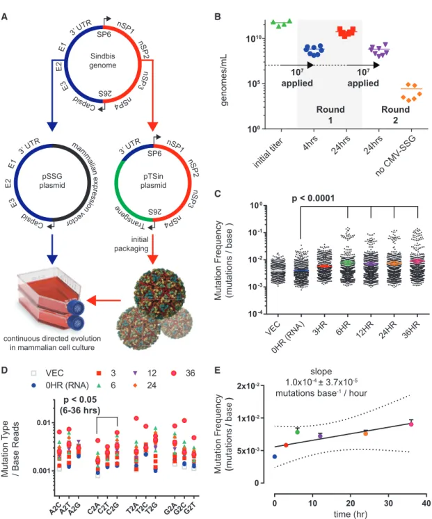
toData S1[VEGAS Supplemental Resource] andData S2[VEGAS Extended Protocol] for additional details). Transgenic Sindbis virus plasmid harboring green fluorescent protein (pTSin-EGFP) (STAR Methods) was packaged and titered at 5.4531011genomes/mL (Figure 1B) as determined by qRT-PCR targeting the Sindbis virus packaging signal sequence (STAR Methods). This initial titer was applied to 1 3 107 cells transfected with pCMV-SSG (Sindbis structural genome) (STAR Methods) at an MOI (multiplicity of infection) of 1. Harvesting and subsequent analysis of the culture media from these cells revealed high viral titer pro-duction, with 6.643 108 genomes/mL produced after 4 h and 5.5731010genomes/mL produced after 24 h (Figure 1B). The
24 h sample from round 1 was transferred to naive cells transfected with or without pCMV-SSG at an MOI = 1. After 24 h, pCMV-SSG transfected cells produced 6.373108genomes/mL while untrans-fected control cells produced 2.63105genomes/mL (Figure 1B). Fluorescent imaging of the infected culture over time confirmed passage of the EGFP transgene (Figure S1A). Transgene expres-sion is rapid, with EGFP detectable in as few as 4 h post infection. These experiments demonstrate that Sindbis virus can be used for sustained transgene packaging in mammalian cell culture using a plasmid-borne structural genome.
RNA viruses, such as Sindbis, are highly mutagenic, with no known proof-reading capability. Approximations of RNA virus mutation rates range from 105to 103mutations per base repli-cated (Drake et al., 1998; Morley and Turner, 2017; Sanjua´n et al., 2010; Schnell et al., 1996; Strauss and Strauss, 1994). As no prior study quantified the genetic stability of a non-essen-tial transgenic gene during Sindbis virus replication, we next determined the mutation frequency of our directed evolution sys-tem. We initiated packaging of pTSin-EGFP in pCMV-SSG trans-fected cells and collected supernatant after 3, 6, 12, 24, and 36 h. The EGFP transgenic segment, as well as the vector template and initially packaged RNA, was amplified and sequenced using an Illumina NextSeq500. The sequences were aligned (STAR Methods) and quantified for positional sequence integrity of EGFP (Figures S1B and S1C). A significant (p < 0.0001) (STAR Methods) time-dependent increase in average mutation fre-quency was observed when comparing the 0HR (initial RNA) sample versus the 6, 12, 24, and 36 h samples (Figure 1C). The number of observed insertions and deletions also increased with time (Figure S1D). Nucleotide substitution rates were rela-tively even, with the exception of a modest (p < 0.05) C > G pref-erence in samples 6–36 h (Figure 1D), and events occured pro-portionally across read lengths (Figures S1E–S1G). Linear regression analysis of mutation frequency versus time (Figure 1E) yielded an estimate of 1.0 3 104 ± 3.7 3 105 mutations base1/h—1 mutation per 1,000 bases replicated or >109total mutations per day at the observed viral production rates. This high mutation rate, coupled to accumulating insertions and dele-tions infrequently accessible to rational design platforms, makes Sindbis virus an ideal candidate for developing a mammalian-directed evolution platform.
Directed Evolution of Transcription Factors with Sindbis Virus
A B
C
D E
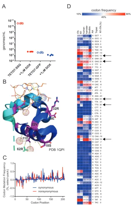
Figure 1. Sindbis Virus for Facile Directed Evolution in Mammalian Cell Culture Development of Sindbis virus for facile, mutagenic viral propagation in mammalian cell culture.
(A) Design of plasmids used for facile directed evolution with Sindbis virus. Artificial Sindbis genome; Girdwood, MF459683.1. pSSG plasmid; capsid, E3, E2, E1, and 30UTR moved to a mammalian expression vector. pTSin plasmid; the structural genome elements of the artificial Sindbis genome replaced by any transgene sequence (pTSin). Propagation and selection can then be performed in mammalian cell culture using pTSin packaged virus applied to cells transfected with pSSG. (B) qRT-PCR quantification of Sindbis virus production from cell culture. Data are represented as mean of individual biological replicates, N > 3.
(C) Mutations observed from Illumina paired-end sequencing of Sindbis packaged EGFP transgene over time. Mutation frequency is plotted as mutations observed per read at each nucleotide position across the transgene. Data are plotted for each individual replicate (N = 3; 24HR and VECTOR, N = 2) around mean±95% confidence interval.
(D) Base changes observed from sequencing of Sindbis packaged EGFP transgene over time. A, adenine, T, thymine, G, guanine, C, cytosine. Statistical comparison tested within base groups between each time point.
(E) Calculation of Sindbis mutation rate from sequencing of Sindbis packaged EGFP transgene over time. Data are represented as mean±SEM and as linear regression, dotted line highlights the 99% confidence interval band.
selective pressure must be applied. Each Sindbis viral particle requires 240 copies of each of the structural proteins E1, E2, and capsid to form a functional viral particle that can mature and propagate (Tang et al., 2011), and without this envelope, the virus is unable to mature and propagate. By engineering re-strictions on structural genome transcription, we developed a system to apply selective pressure on transgenic Sindbis virus. As proof of concept for this method, we placed the Sindbis virus structural genome under control of the tetracycline oper-ator sequence (pTETO7-SSG) (Das et al., 2004; Gossen et al., 1995; Orth et al., 2000;STAR Methods) and packaged trans-genic Sindbis virus with tetracycline transactivator (pTSin-tTA) (Gossen and Bujard, 1992; STAR Methods). We infected cells ±TETO7-SSG with viral pTSin-tTA or pTSin-EGFP and
then treated cells with either the tTA inhibitor doxycycline (DOX, 1mM) or vehicle at the time of infection. Virus was pack-aged at 3.5331010genomes/mL in the vehicle + TETO7-SSG
cell line, while <106genomes/mL were detected for all other conditions (Figure S2A).
Using the TS-tTA system, we sought to benchmark the capa-bilities of VEGAS by evolving tTA to be DOX-insensitive. To accomplish this, we packaged TS-tTA virus under non-selec-tive conditions (R0) and exposed it to constant rounds of selection using increasing concentrations of DOX (Figure 2A). Seven selection rounds, encompassing just 7 days of evolution, produced a large number of full-length tTA sequences (see
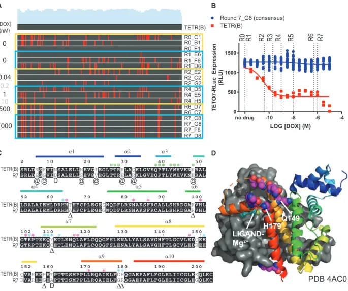
Table S1). By round 6, a consensus sequence dominated the observed coding sequence pool that was carried through to round 7. This consensus sequence, dubbed ‘‘R7,’’ was completely resistant to DOX (Figure 2B). R7 possessed twenty-two coding mutations spanning all functional domains of the protein (Figure 2C). We had predicted that mutations directly involved in ligand interaction (Figures 2C and 2D;Kisker et al., 1995; Orth et al., 1999a, 1999b) would be enriched in the final consensus. To our surprise, none of these residues were mutated in R7. Instead, mutations accumulated primarily adja-cent to key interacting residues for each functional domain (Figures 2C andS2B), many of which have been previously identified to reduce the effect of DOX on TETR-TETO interac-tion (Berens et al., 1992; Hecht et al., 1993; Mu¨ller et al., 1995; Orth et al., 1998; Scholz et al., 2004; Schubert et al., 2001; Smith and Bertrand, 1988; Urlinger et al., 2000; Wiss-mann et al., 1991; seeFigure 2C andTable S2for details). In addition, a cluster of negatively charged residues comprising helices 8 and 9 residing over the ligand binding pocket spanning Q149-H179 (Figure 2D) were converted to primarily positively charged residues (Figure 2C;Table S2). Among vary-ing bacterial species, the net charge, but not specific residues, of this loop is conserved, and this conserved charge landscape has been proposed to attract the tetracycline-Mg2+ inducer to the ligand binding pocket (Orth et al., 1998). The mutations observed in R7 increase the net charge of this loop by +3.19, concentrated near the ligand entry tunnel. This gain in local charge presumably repels the positively charged DOX-Mg2+.
Interestingly, in addition to augmenting the peptide sequence through directed evolution, our analysis of the nucleotide se-quences from each round revealed codon usage optimizations as well (Figures S2C and S2D). Non-synonymous mutations
acquired through tTA evolution converted rarely used codon se-quences for BHK21, derived fromMesocricetus auratus, to the more frequently used GAC (D, +13%), GAG (E, +29%), AAG (K, +43%), TTG (L, 30%), and CAG (Q, +19%).
Augmenting TETR ligand sensitivity has been attempted previ-ously using mammalian-directed evolution (Das et al., 2004), wherein 2 mutations were identified in 114 days. Our evolution of tTA generated an order of magnitude more functional muta-tions in 7 days thereby illustrating how our Sindbis virus system can be used for successful directed evolution of a transcription factor in mammalian cell culture. Key to the evolutionary compo-nent of this method is the actuation of a genetically encoded cir-cuit to unlock expression of the Sindbis structural proteins, capsid, E1, and E2. Consequently, we have named this Sindbis virus system viral evolution of genetically actuating sequences, or ‘‘VEGAS.’’
VEGAS for the Evolution of GPCRs
With VEGAS in hand to perform directed evolution in mammalian cells, we focused our efforts on GPCRs, a superfamily of trans-membrane receptors with substantial pharmacological and physiological importance (Hauser et al., 2017; Wacker et al., 2017a). Critical to the GPCR field is the mapping of interacting residues associated with the transition from an inactive to active receptor. Mapping these motifs can provide anchor points for homology modeling, evolutionary sequence analysis, and ligand design (Fan et al., 2009; Michino et al., 2015; Roth et al., 2017). Even among the best studied receptors, using extensive muta-tion campaigns and high resolumuta-tion crystal structures of inactive and active receptor conformations, the field has struggled to consistently identify key residues involved in state transition (Dror et al., 2011; Huang et al., 2015a; Latorraca et al., 2017). Class A GPCRs possess conserved binding pockets and trigger motif residues involved in the inactive to active state transition. However, many class A GPCRs lack conservation within these motifs, a disproportionate number of which are classified as understudied or orphan receptors (Figure S3A). Here, we used VEGAS to identify previously unknown constitutively active mu-tations for the understudied receptor MRGPRX2; our approach demonstrates how VEGAS can illuminate the complex confor-mational changes involved in GPCR activation even in the absence of structural information.
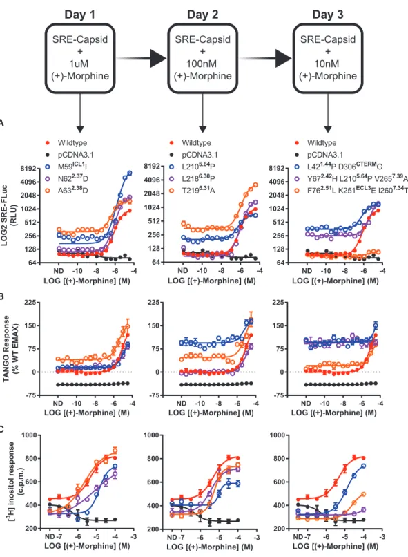
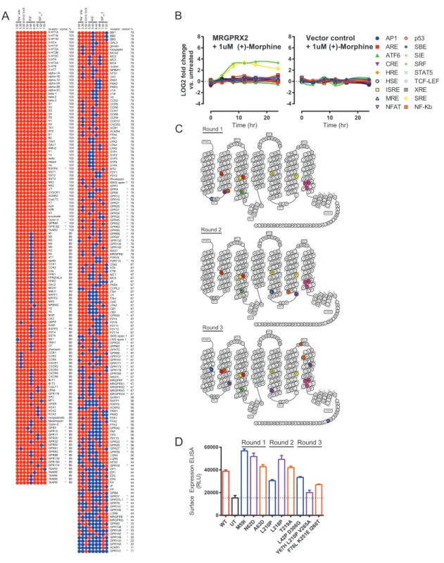
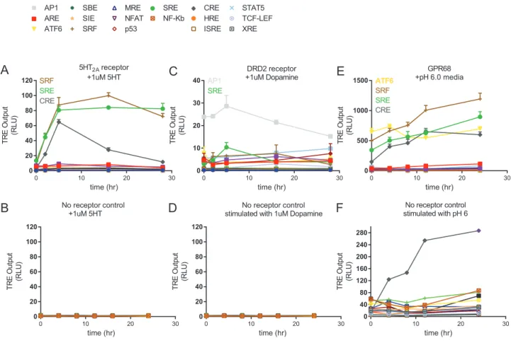
We screened each mutant for activity in SRE-luc2P (Figure 3A),
b-arrestin recruitment (Figure 3B), and phosphoinositide (PI) hy-drolysis (Figure 3C) functional assays. We also quantified surface receptor expression via ELISA to ensure proper trafficking and expression (Figure S3D). For SRE and TANGO assays, basal activity across the variants increased at each evolutionary gener-ation. TANGO basal activity reached 100% of wild type (+)-morphine stimulation for three independent mutants: L210P, Y67H+L210P+V265A, and L42P+D306G. PI hydrolysis,
A B
C D
Figure 2. Directed Evolution of Transcription Factors with Sindbis
Sindbis was used as a directed evolution platform to generate a doxycycline (DOX)-resistant variant of the transcription factor tTA.
(A) Nucleotide sequence alignment of TETR clones isolated from each round of selection to the wild type TETR(B) sequence. Each round is outlined in yellow or blue, applied concentration of DOX to the left, name of individual clones to the right. Gray DOX values indicate no clones were isolated from the round. Red lines in the alignment denote a sequence mismatch from wild type.
(B) TETO7-Rluc reporter assay with increasing concentrations of DOX. Dotted lines are selection round DOX concentrations, for reference. Data are represented as mean±SEM of individual biological replicates.
(C) Peptide sequence alignment of TETR(B) and the R7 consensus. Matching residues are shaded, mutations are unshaded. Alpha helices (a) are labeled and color coded to match with palettes in (D) andS2B. Exact residue (D), position (@), or subtype (D) substitutions previously published to enhance tTA activity in the presence of DOX as perTable S2. Residues (*) with direct involvement in DNA binding (green), ligand binding (magenta), and ligand entry (cyan) as perOrth et al. (2000)andSchubert et al. (2004).
(D) Crystal structure PDB: 4AC0 of TETR(B) in complex with minocycline-Mg2+
. Helix 8-9 ligand enclosure spanning Q149-H179 is displayed with spheres highlighting the residues for mutations Q149R, Q152R, K155R, R158G, T160A (no density), D178G, and H197R observed in R7.
See alsoFigure S2,Tables S1andS2, andData S1andS2.
genome(SRE-SSG).CellstransfectedwithSRE-SSGwere in-fected with transgenic Sindbis virus harboring MRGPRX2 (pTSin-MRGPRX2)andselectedwithdiminishingamountsof (+)-morphine over 3 days (Figure3). Resultant viral genomes
were isolated, and their MRGPRX2 transgenes were tested in subsequentassays.
A
B
C
FLuc
Figure 3. VEGAS for the Evolution of GPCRs
Using VEGAS multiple constitutively active mutants of the GPCR MRGPRX2 were produced in 3 days through application of decreasing concentrations of the MRGPRX2 agonist (+)-morphine. Mutations acquired in each round were tested functionally. Mutations are listed with their receptor residue position and Ballesteros-Weinstein annotation.
(A) Serum response element (SRE) reporter assay. FLuc production equates to relative receptor activation. ND, no drug. Data are represented as mean±SEM, N = 3.
(B) TANGO reporter assay. RLuc production equates to receptor-mediatedb-arrestin2 activation. ND, no drug. Data are represented as mean±SEM, N = 3. (C) Phosphoinositide hydrolysis assay. Accumulation of [3
H] inositol equates to receptor-mediated Gaqactivation. ND, no drug. Data are represented as mean±SEM, N = 3.
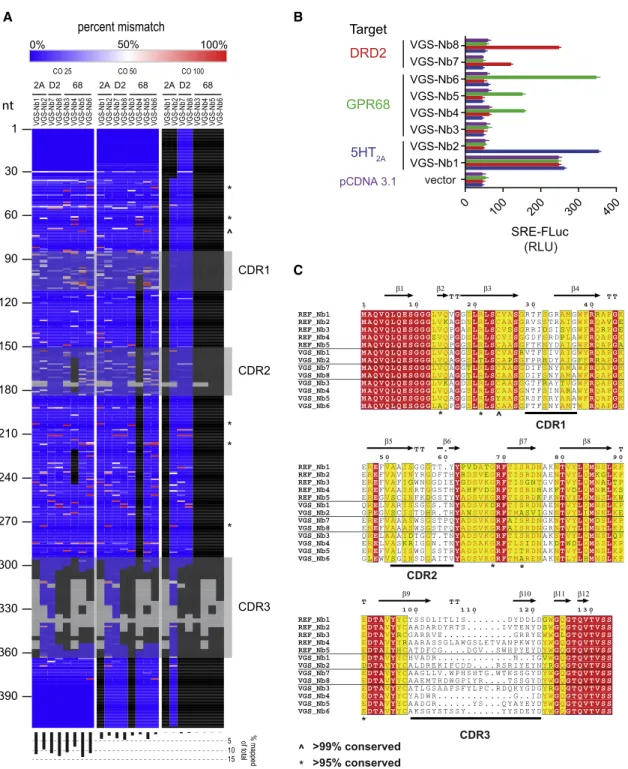
sequenced the clonal library using a NextSeq500 (STAR Methods). Stringent end-to-end alignment of the entire sequence pool (total reads equaled 20 million) was assessed over 3 independent score cut-offs. Reads aligning to the VEGAS-derived nanobodies are displayed in Figure 4A. Each VEGAS-derived nanobody possessed sequences that were not detected within or outside the complementarity-determining re-gions (CDRs). In addition, we compared reference nanobodies (REF_Nbs) cloned from the parent library and VEGAS isolates to the amino acid frequency distribution of 1,346 deposited
Llama glamaVhH sequences from >50 animals (Table S4). Five positions with >99% sequence conservation across populations were conserved in the REF_Nb sequences, but were mutated in the VEGAS evolved sequences (Figure 4C). Both sequence anal-ysis methods demonstrate that the VEGAS-derived nanobodies were not original to the library but evolved from that initial pool of nanobody cDNAs.
Positive Allosteric Modulation of GPCRs by VEGAS-Evolved Nanobodies
Using VEGAS, we produced 8 nanobodies targeted against 3 independent GPCRs in less than 1 week. Here, we interrogate their physical and molecular interactions with each target and provide a detailed characterization of the mechanism of VGS-Nb2, a positive allosteric modulator of the 5-HT2A serotonin
receptor.
First, we established whether the VEGAS-evolved nanobodies directly associated with their intended GPCR targets via biolumi-nescence resonance energy transfer (BRET). For BRET, GPCR-RLuc fusions and increasing concentrations of mVenus nano-body (mVenus-Nb) fusion proteins were co-transfected in to HEK293T cells. We observed a strong association between mVenus-VGS-Nb2 and 5-HT2A-RLuc, but no association to the
closely related serotonin 2B (5-HT2B) receptor (Figure 5A).
Addi-tion of the agonist serotonin (5-HT) at 1mM or above (Figures 5A andS5A) had no effect on VGS-Nb2 association to either 5-HT2A
or 5-HT2B. We also observed association between
mVenus-VGS-Nb6 and GPR68-RLuc at pH 8, but no association of the nanobody to 5-HT2A, the protein used to develop the initial
nano-body library for directed evolution (Figure 5B). GPR68 activity increases with increasing pH (Huang et al., 2015b), we therefore stimulated our BRET assay with a pH 6 buffer and observed an increased association between mVenus-VGS-Nb6 and GPR68-RLuc (Figure 5B). Low, non-specific interaction of the DRD2-targeted nanobodies VGS-Nb7 and VGS-Nb8 was also observed (Figures S5C and S5D). However, VGS-Nb7 and VGS-Nb8 both increase SRE activity in the presence of DRD2 (Figure S5J) through an unknown mechanism.
The serotonin 2A (HTR2A, 5-HT2A) receptor is a GPCR of
sig-nificant importance to mental health, disease, pharmacology, and homeostatic biology (McCorvy and Roth, 2015). Structures of 5-HT2A and closely related 5-HT2-family receptors 5-HT2B
(Wacker et al., 2013) and 5-HT2C(Peng et al., 2018) have yet to
be obtained for their active states. Using VEGAS we have iden-tified a nanobody that binds active 5-HT2A, but not 5-HT2B. We
therefore further characterized the 5-HT2Ananobody VGS-Nb2.
We first confirmed the interaction between 5-HT2Aand
VGS-Nb2 via coimmunoprecipitation (coIP), as analyzed by western a proxy for Gaq activity, detected decreased basal activity,
ligand potency, and efficacy for all mutants. Decreases in maximum agonist-induced Gaq activity correlated with
in-creases in constitutive TANGO and SRE activity.
VEGAS for the Evolution of Active-State Nanobodies
GPCRligandsstabilizesignal-state-specificreceptor conforma-tions (KobilkaandDeupi,2007;OnaranandCosta,2009; Stra-chan et al., 2014), and the development of novel ligands is enhanced by signal-state-specific GPCR crystal structures (Cheetal.,2018;Mangliketal.,2016;Wangetal.,2017). Nano-bodies, genetically encodable antigen recognition domains from dromedaries (Muyldermansetal.,2001), can be used to obtain
thesestabilizedactivestatestructures(Cheetal.,2018;Manglik etal.,2016;Rasmussenetal.,2007;Staus etal.,2016). The
nanobodies developed in these studies mimic the Gaprotein, displacing it. More desirable would be a nanobody that stabilizes the complex between GPCR and its transducer Ga protein. These nanobodies would be allosteric modulators capable of enhancing GPCR-Ga coupling. Using VEGAS, we can create allosteric nanobodies for multiple GPCR-Ga pairings in less than a week.
TocreateGPCRnanobodiesusingVEGAS,wefirstgenerated a GPCR-targeted nanobody library by immunizing a llama againsttheserotonin2A(5-HT2A)GPCRboundtothe
high-affin-ity agonist lysergic acid diethylamide (LSD). We isolated single-domainantibodiesfromtheperipheralbloodmononuclearcells of the immunized llama through amplification of the variable re-gion‘‘VHH’’ ofIgG (Pardonetal.,2014;STAR Methods).The
VHH ampliconwasusedto generateacDNA libraryof13
107colonies, which was subsequently packaged in Sindbis vi-rus.Thislibrarywasthenusedtoevolveintracellulartargeting nanobodies against 5-HT2A, as well as the dopamine-D2
(DRD2)andpH-sensingGPR68(Huangetal.,2015b)receptors. EachofthesereceptorscouplescanonicallytoadifferentGa protein (2A, Gaq; D2, Gai, 68, Gas). Developing nanobodies
to-ward each receptor serves to demonstrate the broad applica-bilityoftheVEGASsystem.
Toevolveactivestate-stabilizingnanobodiesforeachGPCR, we first screened each receptor for transcription factor coupling (FiguresS4A–S4F).Allthreereceptorsweredeterminedto acti-vate SRE with varying efficacy and we therefore chose to develop nanobodies that engage the SRE-signaling state for each receptor. Cells transfected with SRE-SSG and a GPCR wereinfectedwithanMOI=1oftheviralnanobodylibrary.To selectforSREactivatingnanobodies,5-HT2AandDRD2cultures
were incubated in the absence of ligand, while GPR68 was incu-batedatitsinactivepH8(Huangetal.,2015b).Day1viral parti-cles were harvested, the selection was repeated, and individual nanobodycloneswereisolatedfromtheday1and2titersand sequenced. Clones with N R 2 identity in the subcloned popula-tionwereselectedandscreenedforGPCR-dependentSRE acti-vation (Figure4B). Each evolution series produced nanobodies
deep-A B
C
SRE-FLuc
Figure 4. VEGAS for Evolution of Active-State Nanobodies
VEGAS was used to develop nanobodies that selectively activate diverse GPCR targets from a single cDNA library.
(A) Deep sequencing of the nanobody cDNA library used for VEGAS. 20 million reads were aligned to VEGAS-derived clones and plotted as % mismatch. Data was analyzed with score cut-offs (CO) of 25, 50, and 100 (STAR Methods). Grey blocks are gaps in alignment as per (C). Black blocks are regions with mapped reads <2,000 counts (<0.0001%). Symbols^and * mirror those on (C). Bottom histogram, percent total mapped reads for each alignment.
(B) Serum-response element (SRE) reporter assay. Nanobody:receptor:reporter transfection ratio of 5:1:1. FLuc production equates to relative receptor acti-vation. Data are represented as mean±SEM, N = 3.
(C) Amino acid sequence alignment of library (REF_NB#) and VEGAS-derived clones. Shading: 100% (red), >75% (yellow), <75% (white). Variations identified in VEGAS, but not reference sequence, derived clones at positions of high genetic conservation (seeTable S4) are annotated,^>99% conserved, * >95% conserved. Nanobody secondary structure annotated above, retrieved from PDB 3P0G, chain B. Complementarity determining regions (CDRs) annotated below.b, beta sheet. TT, strictb-turn.
A B
C D
E
2B
Figure 5. Positive Allosteric Modulation of GPCRs by VEGAS-Evolved Nanobodies VEGAS-derived nanobodies were tested for direct association and allosteric modulation of their targets.
(A) Bioluminescence resonance energy transfer (BRET) association assay between 5-HT2A-RLuc or 5-HT2B-5-HT2A-RLuc and mVenus-VGS-Nb2 at increasing transfection ratios of nanobody. Data are represented as mean±SEM, N = 3. Symbols for 5-HT2B-Rluc data underlie those for the +1mM 5-HT data.
(B) Bioluminescence resonance energy transfer (BRET) association assay between GPR68-RLuc or 5-HT2A-RLuc and mVenus-VGS-Nb6. Data are represented as mean±SEM, N = 3.
(C) Serum response element (SRE) reporter assay. RLuc production equates to relative receptor activation. Data are represented as mean±SEM, N = 3.
(D) Saturation radioligand binding assay. 5-HT2A-Gaqmembrane treated with vehicle or 5mM VGS-Nb2. 5-HT2A; Kd= 0.30 nM, Bmax= 1,333 fmol/mg. 5-HT2A+VGS-Nb2; Kd= 0.566 nM, Bmax= 1,993 fmol/mg. Data are represented as mean±SEM, N = 3, *p < 0.05.
(E) Competitive radioligand binding assay. 5-HT2A and 5-HT2A-Gaqmembrane treated with vehicle or 7.5mM VGS-Nb2 labeled with 1 nM [3H]ketanserin and increasing concentrations of DOI. Data are represented as total-count normalized means±
SEM, N = 3.
See alsoFigure S5,Table S5, andData S1andS2.
ysis that VGS-Nb2 does not stabilize, or lock, a transducer-coupled state. This aligns with the evolved purpose of this nanobody to act as a positive allosteric modulator (PAM) of 5-HT2A rather than
inhibit transducer cycling.
To further validate VGS-Nb2 as a 5-HT2APAM, we first assessed its ability to positively modulate
SRE signaling downstream of 5-HT2A. At Nb ratios
demon-strated to bind <50% of 5-HT2A (Figure 5A), VGS-Nb2
increased the agonist-mediated SRE response by up to 2-fold (Figure 5C). This SRE signal could originate from Gaqand/or b-arrestin pathways. We assessed the effect of the nanobody using calcium and arrestin recruitment assays, respectively. VGS-Nb2 allosterically enhanced 5-HT2Acalcium release ( Fig-ure S5F), a Gaq-mediated signal response. Conversely,
VGS-Nb2 diminished mVenus-b-arrestin2 recruitment to the 5-HT2A-RLuc fusion protein as a function of time (Figure S5G)
and agonist concentration (Figures S5H and S5I). From these experiments, we hypothesized that VGS-Nb2 stabilizes the high-affinity Gaq-coupled state of the receptor.
Unliganded receptors are rarely found in their active, or high-affinity, conformational state (Manglik et al., 2015). However, ra-diolabeled ligands can be used to probe and quantify high-affinity receptor sites. The number of these sites increases when allosteric effectors, such as Gaqor nanobodies, are bound
blot (FigureS5B) and mass spectrometry (FigureS5E). These
as-saysconfirmedastableinteractionbetweenthereceptorand nanobody. These assays were performed in the absence of ligand, confirming our previous BRET observation that the interaction is ligand-independent. This is consistent with our directedevolutionselectionparadigm,whichrequireda nano-bodycapableinducingreceptoractivityintheabsenceofligand. 5-HT2Acouples to Gaqand b-arrestin to transduce its signal in
cells (Wackeretal.,2017b). We tested whether VGS-Nb2
asso-ciationto5-HT2AwasGa-dependentusingGaqD/11D/sD knockout
cell lines (Alvarez-Curtoetal.,2016) in BRET recruitment assays
(FigureS5A),coIPbywesternblot(FigureS5B),andmass spec-trometry (FigureS5E). In all three studies, no appreciable
differ-ence in VGS-Nb2 association was detected. Notably, as with all epistasisexperiments, knockoutcelllinesfrequently adaptto gene loss by augmenting signaling pathways (Duncan etal., 2012;Luttrelletal.,2018).However,fromtheadditionalproteins identifiedinHEK-TandGaqD/11D/sD cells(TableS5),nocanonical
anal-to the recepanal-tor (Che et al., 2018; Staus et al., 2016; Strachan et al., 2014). To test whether VGS-Nb2 stabilizes the 5-HT2AGaq
-coupled active state, as predicted from our functional data, we first employed radioligand saturation binding using the partial agonist [3H]LSD (Figure 5D). Membranes from cells transfected
with 5-HT2Afused to its transducer Gaq(5-HT2A-Gq) were
incu-bated with increasing concentrations of [3H]LSD±5mM purified VGS-Nb2. As shown inFigure 5D, VGS-Nb2 increased labeled agonist binding sites by 50%. This increase in high-affinity agonist binding sites was additionally confirmed through competitive radioligand binding wherein 5-HT2A and 5-HT2A
-Gaqmembranes were incubated with±7.5mM purified
VGS-Nb2 (Figure 5E). In competition with the radiolabeled 5-HT2A
antagonist [3H]ketanserin, the selective agonist DOI bound the
5-HT2A receptor with a half maximal inhibitory concentration
(IC50) equal to 550 nM. Neither a local excess of Gaq (DOI
IC50= 307 nM, p = 0.1264) nor the addition of purified
VGS-Nb2 (DOI IC50= 746 nM, p = 0.3957) significantly affected DOI
binding. However, in the presence of both Gaqand VGS-Nb2, 50% of the available ligand binding sites were stabilized in the high-affinity conformation that bound DOI with an IC50= 0.15 nM.
We have therefore demonstrated the directed evolution of multiple functionally distinct nanobody sequences against GPCRs using VEGAS. Of these, we have characterized VGS-Nb2 as a Gaq-dependent positive allosteric modulator of 5-HT2A.
DISCUSSION
Here, we demonstrate the development of a system for facile directed evolution in mammalian cells: VEGAS. Leveraging the alpha virus Sindbis as a vector for heredity, mutagenesis, and se-lection, we succeeded in evolving context-dependent functions for three independent classes of proteins. Our evolution targets were primary (tTA), secondary (GPCR), and tertiary (Nbs) interac-tors to downstream outputs, demonstrating the ability of VEGAS to provide tools at multiple levels of cell signaling. Our primary system evolved tTA to engage with TETO7 in the presence
of >1 mM DOX. Our secondary system evolved the GPCR MRGPRX2 to constitutively activate the serum response element via endogenous signaling pathways. Our tertiary system evolved nanobodies to selectively activate GPCRs, which in turn acti-vated the serum response element via endogenous signaling pathways. Together, these applications showcase the ease and power of the VEGAS system as a tool for enabling directed evolution campaigns across a broad range of potential mamma-lian applications.
Directed evolution allows genetic sequences to evolve under selective pressure in an appropriate context. Through this pro-cess we are able to guide solutions to otherwise intractable biological problems (Hammer et al., 2017; Kan et al., 2016; Mat-sumoto et al., 2015; Shapiro et al., 2010). However, powerful sys-tems for directed evolution in a mammalian cell context have lagged behind unicellular systems. VEGAS offers three major advantages for the directed evolution of biomedical tools and therapies.
First, VEGAS evolves within the signaling framework of the host cell. Signaling proteins never operate in isolation, but
as interacting heteromeric complexes, to transfer information through the cell (Garrington and Johnson, 1999; Pawson and Scott, 1997; Purvis and Lahav, 2013; Varnaite and MacNeill,_ 2016). The timing, location, and kinetics of these interactions is critical to performance and cannot be easily replicated in non-native environments. In addition, we can take advantage of the negative feedback (Amit et al., 2007; Behar et al., 2007; English et al., 2015; Ferrell, 2002; Howell et al., 2012; Subramaniam et al., 1989) mechanisms built in to endogenous signaling pathways to encode viral selection—as was done for both MRGPRX2- and nanobody-directed evolution in this study.
Second, VEGAS is wholly dependent on the host cell for trans-gene maturation (Garcia-Moreno et al., 2013). Directed evolution performance can falter when transferring tools evolved in one context to another (Armbruster et al., 2007). This may be a consequence of improper trafficking, failed compartmentaliza-tion, incorrect protein maturacompartmentaliza-tion, or absence of non-native co-factors. With VEGAS, mammalian translation is a requirement of the evolved product.
Third, VEGAS selection is constant and highly mutagenic, enabling it to overcome many of the pitfalls inherent to complex fitness landscapes (Romero and Arnold, 2009; Tracewell and Arnold, 2009). To avoid dead-ends and early fitness bias, directed evolution systems must sample toward saturation whenever possible. This directed evolution paradigm helps to maintain diversity by preserving even poor performing early evo-lution variants, which may ultimately rise to the highest fitness peaks. This is achieved with VEGAS, in part, because each host cell operates as a closed system. This allows evolved solu-tions derived in each cell to compete in the subsequent rounds— even when vastly superior solutions may have arisen elsewhere within the same selection cycle.
There are many potential applications of the VEGAS system. Sindbis virus has a transgene packaging capacity of >6 kb (Huang and Summers, 1991), placing few limits on the potential targets for directed evolution. High value targets would include: Cas9 variants evolved to better engage endogenous se-quences (Doudna and Charpentier, 2014; Kleinstiver et al., 2015; Lee et al., 2018), fluorescent protein variants evolved for maturity time, photostability, brightness, or wavelength specificity in human tissue (Drobizhev et al., 2011; Piatkevich et al., 2017; Shaner et al., 2004, 2008), or designer receptors exclusively activated by designer drugs (DREADDs) for the che-mogenetic control of cell signaling (Armbruster et al., 2007; Roth, 2016).
biology, filling an essential need for facile directed evolution in a mammalian context.
STAR+METHODS
Detailed methods are provided in the online version of this paper and include the following:
d KEY RESOURCES TABLE
d LEAD CONTACT AND MATERIALS AVAILABILITY
d METHOD DETAILS
B Molecular Biology & Plasmid Construction
B General Cell Culture
B Sindbis Virus Production
B Sustained Passage of Sindbis for Directed Evolution and Transgene Isolation
B RNA Deep Sequencing
B Quantification of Viral RNA via qRT-PCR
B tTA Reporter Assay
B Transcription Factor Reporter Primary Screen
B SRE Reporter Assay
B TANGOb-arrestin recruitment assay
B Calcium flux assay
B Bioluminescence resonance energy transfer associa-tion assay
B Phosphoinositide hydrolysis assay
B Surface expression enzyme-linked immunosor-bent assay
B Nanobody Production
B Saturation and competitive radioligand binding assays
B Co-Immunoprecipitation Analysis
d QUANTIFICATION AND STATISTICAL ANALYSIS
d DATA AND CODE AVAILABILITY
Received: November 19, 2018 Revised: February 6, 2019 Accepted: May 23, 2019
Published: July 4, 2019; corrected online: July 19, 2019
REFERENCES
Agapov, E.V., Frolov, I., Lindenbach, B.D., Pra´gai, B.M., Schlesinger, S., and Rice, C.M. (1998). Noncytopathic Sindbis virus RNA vectors for heterologous gene expression. Proc. Natl. Acad. Sci. USA95, 12989–12994.
Albert, T.J., Dailidiene, D., Dailide, G., Norton, J.E., Kalia, A., Richmond, T.A., Molla, M., Singh, J., Green, R.D., and Berg, D.E. (2005). Mutation discovery in bacterial genomes: metronidazole resistance in Helicobacter pylori. Nat. Methods2, 951–953.
Alvarez-Curto, E., Inoue, A., Jenkins, L., Raihan, S.Z., Prihandoko, R., Tobin, A.B., and Milligan, G. (2016). Targeted Elimination of G Proteins and Arrestins Defines Their Specific Contributions to Both Intensity and Duration of G Protein-coupled Receptor Signaling. J. Biol. Chem.291, 27147–27159.
Amit, I., Citri, A., Shay, T., Lu, Y., Katz, M., Zhang, F., Tarcic, G., Siwak, D., La-had, J., Jacob-Hirsch, J., et al. (2007). A module of negative feedback regulators defines growth factor signaling. Nat. Genet.39, 503–512.
Armbruster, B.N., Li, X., Pausch, M.H., Herlitze, S., and Roth, B.L. (2007). Evolving the lock to fit the key to create a family of G protein-coupled receptors potently activated by an inert ligand. Proc. Natl. Acad. Sci. USA
104, 5163–5168.
Arnold, F.H. (1998). Design by Directed Evolution. Acc. Chem. Res. 31, 125–131.
Badran, A.H., and Liu, D.R. (2015). In vivo continuous directed evolution. Curr. Opin. Chem. Biol.24, 1–10.
Baym, M., Lieberman, T.D., Kelsic, E.D., Chait, R., Gross, R., Yelin, I., and Kishony, R. (2016). Spatiotemporal microbial evolution on antibiotic land-scapes. Science353, 1147–1151.
Behar, M., Hao, N., Dohlman, H.G., and Elston, T.C. (2007). Mathematical and computational analysis of adaptation via feedback inhibition in signal trans-duction pathways. Biophys. J.93, 806–821.
Berens, C., Altschmied, L., and Hillen, W. (1992). The role of the N terminus in Tet repressor for tet operator binding determined by a mutational analysis. J. Biol. Chem.267, 1945–1952.
Berman, C.M., Papa, L.J., 3rd, Hendel, S.J., Moore, C.L., Suen, P.H., Weick-hardt, A.F., Doan, N.-D., Kumar, C.M., Uil, T.G., Butty, V.L., et al. (2018). An Adaptable Platform for Directed Evolution in Human Cells. J. Am. Chem. Soc.140, 18093–18103.
Bourdon, D.M., Wing, M.R., Edwards, E.B., Sondek, J., and Harden, T.K. (2006). Quantification of Isozyme-Specific Activation of Phospholipase C-b2 by Rac GTPases and Phospholipase C-E by Rho GTPases in an Intact Cell Assay System. Methods Enzymol.406, 489–499.
Bredenbeek, P.J., Frolov, I., Rice, C.M., and Schlesinger, S. (1993). Sindbis vi-rus expression vectors: packaging of RNA replicons by using defective helper RNAs. J. Virol.67, 6439–6446.
Buchholz, F., Angrand, P.-O., and Stewart, A.F. (1998). Improved properties of FLP recombinase evolved by cycling mutagenesis. Nat. Biotechnol. 16, 657–662.
Campbell, R.E., Tour, O., Palmer, A.E., Steinbach, P.A., Baird, G.S., Zacharias, D.A., and Tsien, R.Y. (2002). A monomeric red fluorescent protein. Proc. Natl. Acad. Sci. USA99, 7877–7882.
Carlson, J.C., Badran, A.H., Guggiana-Nilo, D.A., and Liu, D.R. (2014). Nega-tive selection and stringency modulation in phage-assisted continuous evolu-tion. Nat. Chem. Biol.10, 216–222.
Chan, K.Y., Jang, M.J., Yoo, B.B., Greenbaum, A., Ravi, N., Wu, W.-L., Sa´nchez-Guardado, L., Lois, C., Mazmanian, S.K., Deverman, B.E., and Gra-dinaru, V. (2017). Engineered AAVs for efficient noninvasive gene delivery to the central and peripheral nervous systems. Nat. Neurosci.20, 1172–1179. SUPPLEMENTAL INFORMATION
SupplementalInformationcanbefoundonlineathttps://doi.org/10.1016/j. cell.2019.05.051.
ACKNOWLEDGMENTS
WethankourcolleagueGaryJohnsonforprovidingresourcesforRNA-seq analysis.WealsothankourcolleagueMarkHeiseforprovidingtheinitial Sind-bis virus vectorsused in thisstudy. This work was supported by NIH (1U01MH105892-01, 5U24DK116195-02, RO1MH112205, R37DA045657) andagrantfromtheUniversityofNorthCarolinaLinebergerComprehensive CancerCentertoB.L.R.
AUTHORCONTRIBUTIONS
Conceptualization, J.G.E.; Methodology, A.S.C., B.L.R., D.W., J.G.E., R.H.J.O.,andR.T.S.; Software,D.S. andJ.G.E.;Validation,J.G.E., K.L., R.H.J.O.,andR.T.S.;FormalAnalysis,J.G.E.andD.S.;Investigation,J.G.E., K.L.,R.H.J.O.,andR.T.S.;Resources,D.W.,J.G.E.,K.W.,andM.P.;Data Cu-ration,D.S.andJ.G.E.;Writing–OriginalDraft,J.G.E.;Writing–Review& Edit-ing,A.S.C.,B.L.R.,D.W.,J.G.E.,K.L.,R.H.J.O.,andR.T.S.;Visualization, J.G.E.;Supervision,B.L.R.andJ.G.E.;ProjectAdministration,B.L.R. and J.G.E.;FundingAcquisition,B.L.R.andJ.G.E.
DECLARATIONOFINTERESTS
Che, T., Majumdar, S., Zaidi, S.A., Ondachi, P., McCorvy, J.D., Wang, S., Mos-ier, P.D., Uprety, R., Vardy, E., Krumm, B.E., et al. (2018). Structure of the Nanobody-Stabilized Active State of the Kappa Opioid Receptor. Cell
172, 55–67.
Chen, K., and Arnold, F.H. (1993). Tuning the activity of an enzyme for unusual environments: sequential random mutagenesis of subtilisin E for catalysis in dimethylformamide. Proc. Natl. Acad. Sci. USA90, 5618–5622.
Crameri, A., Whitehorn, E.A., Tate, E., and Stemmer, W.P.C. (1996). Improved green fluorescent protein by molecular evolution using DNA shuffling. Nat. Bio-technol.14, 315–319.
Darwin, C., and Bynum, W.F. (2009). On the Origin of Species by Means of Nat-ural Selection: Or, the Preservation of Favored Races in the Struggle for Life (Penguin Classics).
Das, A.T., Zhou, X., Vink, M., Klaver, B., Verhoef, K., Marzio, G., and Berkhout, B. (2004). Viral evolution as a tool to improve the tetracycline-regulated gene expression system. J. Biol. Chem.279, 18776–18782.
De Lean, A., Stadel, J.M., and Lefkowitz, R.J. (1980). A ternary complex model explains the agonist-specific binding properties of the adenylate cyclase-coupledb-adrenergic receptor. J. Biol. Chem.255, 7108–7117.
Di Tommaso, P., Moretti, S., Xenarios, I., Orobitg, M., Montanyola, A., Chang, J.-M., Taly, J.-F., and Notredame, C. (2011). T-Coffee: a web server for the multiple sequence alignment of protein and RNA sequences using structural information and homology extension. Nucleic Acids Res.39, W13-7.
Diamond, J. (2002). Evolution, consequences and future of plant and animal domestication. Nature418, 700–707.
Doudna, J.A., and Charpentier, E. (2014). Genome editing. The new frontier of genome engineering with CRISPR-Cas9. Science346, 1258096.
Drake, J.W., and Holland, J.J. (1999). Mutation rates among RNA viruses. Proc. Natl. Acad. Sci. USA96, 13910–13913.
Drake, J.W., Charlesworth, B., Charlesworth, D., and Crow, J.F. (1998). Rates of spontaneous mutation. Genetics148, 1667–1686.
Driscoll, C.A., Macdonald, D.W., and O’Brien, S.J. (2009). From wild animals to domestic pets, an evolutionary view of domestication. Proc. Natl. Acad. Sci.
USA106(Suppl 1), 9971–9978.
Drobizhev, M., Makarov, N.S., Tillo, S.E., Hughes, T.E., and Rebane, A. (2011). Two-photon absorption properties of fluorescent proteins. Nat. Methods8, 393–399.
Dror, R.O., Arlow, D.H., Maragakis, P., Mildorf, T.J., Pan, A.C., Xu, H., Borhani, D.W., and Shaw, D.E. (2011). Activation mechanism of theb2-adrenergic re-ceptor. Proc. Natl. Acad. Sci. USA108, 18684–18689.
Duncan, J.S., Whittle, M.C., Nakamura, K., Abell, A.N., Midland, A.A., Zawis-towski, J.S., Johnson, N.L., Granger, D.A., Jordan, N.V., Darr, D.B., et al. (2012). Dynamic reprogramming of the kinome in response to targeted MEK inhibition in triple-negative breast cancer. Cell149, 307–321.
English, J.G., Shellhammer, J.P., Malahe, M., McCarter, P.C., Elston, T.C., and Dohlman, H.G. (2015). MAPK feedback encodes a switch and timer for tunable stress adaptation in yeast. Sci. Signal.8, ra5.
Esvelt, K.M., Carlson, J.C., and Liu, D.R. (2011). A system for the continuous directed evolution of biomolecules. Nature472, 499–503.
Fan, H., Irwin, J.J., Webb, B.M., Klebe, G., Shoichet, B.K., and Sali, A. (2009). Molecular docking screens using comparative models of proteins. J. Chem. Inf. Model.49, 2512–2527.
Ferrell, J.E., Jr. (2002). Self-perpetuating states in signal transduction: positive feedback, double-negative feedback and bistability. Curr. Opin. Cell Biol.14, 140–148.
Filizola, M., and Devi, L.A. (2013). Grand opening of structure-guided design for novel opioids. Trends Pharmacol. Sci.34, 6–12.
Fredriksson, R., Lagerstro¨m, M.C., Lundin, L.-G., and Schio¨th, H.B. (2003). The G-protein-coupled receptors in the human genome form five main fam-ilies. Phylogenetic analysis, paralogon groups, and fingerprints. Mol. Pharma-col.63, 1256–1272.
Frolov, I., Hardy, R., and Rice, C.M. (2001). Cis-acting RNA elements at the 50end of Sindbis virus genome RNA regulate minus- and plus-strand RNA synthesis. RNA7, 1638–1651.
Garcia-Moreno, M., Sanz, M.A., Pelletier, J., and Carrasco, L. (2013). Requirements for eIF4A and eIF2 during translation of Sindbis virus subgenomic mRNA in vertebrate and invertebrate host cells. Cell. Microbiol.
15, 823–840.
Garrington, T.P., and Johnson, G.L. (1999). Organization and regulation of mitogen-activated protein kinase signaling pathways. Curr. Opin. Cell Biol.
11, 211–218.
Gilman, A.G. (1987). G proteins: transducers of receptor-generated signals. Annu. Rev. Biochem.56, 615–649.
Gossen, M., and Bujard, H. (1992). Tight control of gene expression in mammalian cells by tetracycline-responsive promoters. Proc. Natl. Acad. Sci. USA89, 5547–5551.
Gossen, M., Freundlieb, S., Bender, G., Mu¨ller, G., Hillen, W., and Bujard, H. (1995). Transcriptional activation by tetracyclines in mammalian cells. Science
268, 1766–1769.
Hammer, S.C., Kubik, G., Watkins, E., Huang, S., Minges, H., and Arnold, F.H. (2017). Anti-Markovnikov alkene oxidation by metal-oxo-mediated enzyme catalysis. Science358, 215–218.
Hanes, J., and Plu¨ckthun, A. (1997). In vitro selection and evolution of functional proteins by using ribosome display. Proc. Natl. Acad. Sci. USA
94, 4937–4942.
Hartwell, L.H., Culotti, J., and Reid, B. (1970). Genetic control of the cell-division cycle in yeast. I. Detection of mutants. Proc. Natl. Acad. Sci. USA
66, 352–359.
Hauser, A.S., Attwood, M.M., Rask-Andersen, M., Schio¨th, H.B., and Gloriam, D.E. (2017). Trends in GPCR drug discovery: new agents, targets and indica-tions. Nat. Rev. Drug Discov.16, 829–842.
Hecht, B., Mu¨ller, G., and Hillen, W. (1993). Noninducible Tet repressor muta-tions map from the operator binding motif to the C terminus. J. Bacteriol.175, 1206–1210.
Hess, G.T., Fre´sard, L., Han, K., Lee, C.H., Li, A., Cimprich, K.A., Montgomery, S.B., and Bassik, M.C. (2016). Directed evolution using dCas9-targeted so-matic hypermutation in mammalian cells. Nat. Methods13, 1036–1042.
Howell, A.S., Jin, M., Wu, C.-F., Zyla, T.R., Elston, T.C., and Lew, D.J. (2012). Negative feedback enhances robustness in the yeast polarity establishment circuit. Cell149, 322–333.
Huang, M.J., and Summers, J. (1991). Infection initiated by the RNA prege-nome of a DNA virus. J. Virol.65, 5435–5439.
Huang, X.-P., Setola, V., Yadav, P.N., Allen, J.A., Rogan, S.C., Hanson, B.J., Revankar, C., Robers, M., Doucette, C., and Roth, B.L. (2009). Parallel func-tional activity profiling reveals valvulopathogens are potent 5-hydroxytrypta-mine(2B) receptor agonists: implications for drug safety assessment. Mol. Pharmacol.76, 710–722.
Huang, W., Manglik, A., Venkatakrishnan, A.J., Laeremans, T., Feinberg, E.N., Sanborn, A.L., Kato, H.E., Livingston, K.E., Thorsen, T.S., Kling, R.C., et al. (2015a). Structural insights intom-opioid receptor activation. Nature 524, 315–321.
Huang, X.-P., Karpiak, J., Kroeze, W.K., Zhu, H., Chen, X., Moy, S.S., Saddo-ris, K.A., Nikolova, V.D., Farrell, M.S., Wang, S., et al. (2015b). Allosteric ligands for the pharmacologically dark receptors GPR68 and GPR65. Nature
527, 477–483.
Huston, M. (1979). A General Hypothesis of Species Diversity. Am. Nat.
113, 81–101.
Isberg, V., de Graaf, C., Bortolato, A., Cherezov, V., Katritch, V., Marshall, F.H., Mordalski, S., Pin, J.-P., Stevens, R.C., Vriend, G., and Gloriam, D.E. (2015). Generic GPCR residue numbers - aligning topology maps while minding the gaps. Trends Pharmacol. Sci.36, 22–31.
Nakamura, Y., Gojobori, T., and Ikemura, T. (2000). Codon usage tabulated from international DNA sequence databases: status for the year 2000. Nucleic Acids Res.28, 292.
Neubig, R.R., Spedding, M., Kenakin, T., and Christopoulos, A.; International Union of Pharmacology Committee on Receptor Nomenclature and Drug Clas-sification (2003). International Union of Pharmacology Committee on Receptor Nomenclature and Drug Classification. XXXVIII. Update on terms and symbols in quantitative pharmacology. Pharmacol. Rev.55, 597–606.
Onaran, H.O., and Costa, T. (2009). Allosteric coupling and conformational fluctuations in proteins. Curr. Protein Pept. Sci.10, 110–115.
Orth, P., Cordes, F., Schnappinger, D., Hillen, W., Saenger, W., and Hinrichs, W. (1998). Conformational changes of the Tet repressor induced by tetracy-cline trapping. J. Mol. Biol.279, 439–447.
Orth, P., Schnappinger, D., Sum, P.E., Ellestad, G.A., Hillen, W., Saenger, W., and Hinrichs, W. (1999a). Crystal structure of the tet repressor in complex with a novel tetracycline, 9-(N,N-dimethylglycylamido)- 6-demethyl-6-deoxy-tetra-cycline. J. Mol. Biol.285, 455–461.
Orth, P., Saenger, W., and Hinrichs, W. (1999b). Tetracycline-chelated Mg2+ ion initiates helix unwinding in Tet repressor induction. Biochemistry38, 191–198.
Orth, P., Schnappinger, D., Hillen, W., Saenger, W., and Hinrichs, W. (2000). Structural basis of gene regulation by the tetracycline inducible Tet repressor-operator system. Nat. Struct. Biol.7, 215–219.
Pa´ndy-Szekeres, G., Munk, C., Tsonkov, T.M., Mordalski, S., Harpsøe, K., Hauser, A.S., Bojarski, A.J., and Gloriam, D.E. (2018). GPCRdb in 2018: adding GPCR structure models and ligands. Nucleic Acids Res.46(D1), D440–D446.
Pardon, E., Laeremans, T., Triest, S., Rasmussen, S.G.F., Wohlko¨nig, A., Ruf, A., Muyldermans, S., Hol, W.G.J., Kobilka, B.K., and Steyaert, J. (2014). A gen-eral protocol for the generation of Nanobodies for structural biology. Nat. Protoc.9, 674–693.
Pawson, T., and Scott, J.D. (1997). Signaling through scaffold, anchoring, and adaptor proteins. Science278, 2075–2080.
Peng, Y., McCorvy, J.D., Harpsøe, K., Lansu, K., Yuan, S., Popov, P., Qu, L., Pu, M., Che, T., Nikolajsen, L.F., et al. (2018). 5-HT2C Receptor
Structures Reveal the Structural Basis of GPCR Polypharmacology. Cell
172, 719–730.
Piatkevich, K.D., Suk, H.-J., Kodandaramaiah, S.B., Yoshida, F., DeGennaro, E.M., Drobizhev, M., Hughes, T.E., Desimone, R., Boyden, E.S., and Verkhu-sha, V.V. (2017). Near-Infrared Fluorescent Proteins Engineered from Bacterial Phytochromes in Neuroimaging. Biophys. J.113, 2299–2309.
Pierce, K.L., Premont, R.T., and Lefkowitz, R.J. (2002). Seven-transmembrane receptors. Nat. Rev. Mol. Cell Biol.3, 639–650.
Purvis, J.E., and Lahav, G. (2013). Encoding and decoding cellular information through signaling dynamics. Cell152, 945–956.
Rasmussen, S.G.F., Choi, H.-J., Rosenbaum, D.M., Kobilka, T.S., Thian, F.S., Edwards, P.C., Burghammer, M., Ratnala, V.R.P., Sanishvili, R., Fischetti, R.F., et al. (2007). Crystal structure of the humanb2adrenergic G-protein-coupled
receptor. Nature450, 383–387.
Robert, X., and Gouet, P. (2014). Deciphering key features in protein structures with the new ENDscript server. Nucleic Acids Res.42, W320-4.
Robinson, J.T., Thorvaldsdo´ttir, H., Winckler, W., Guttman, M., Lander, E.S., Getz, G., and Mesirov, J.P. (2011). Integrative genomics viewer. Nat. Bio-technol.29, 24–26.
Romero, P.A., and Arnold, F.H. (2009). Exploring protein fitness landscapes by directed evolution. Nat. Rev. Mol. Cell Biol.10, 866–876.
Roth, B.L. (2016). DREADDs for Neuroscientists. Neuron89, 683–694.
Roth, B.L., Irwin, J.J., and Shoichet, B.K. (2017). Discovery of new GPCR ligands to illuminate new biology. Nat. Chem. Biol.13, 1143–1151.
Sane, J., Kurkela, S., Levanov, L., Nikkari, S., Vaheri, A., and Vapalahti, O. (2012). Development and evaluation of a real-time RT-PCR assay for Sindbis virus detection. J. Virol. Methods179, 185–188.
Katritch,V.,Fenalti,G.,Abola,E.E.,Roth,B.L.,Cherezov,V.,andStevens, R.C.(2014).AllostericsodiuminclassAGPCRsignaling.TrendsBiochem. Sci.39,233–244.
Kisker,C.,Hinrichs,W.,Tovar,K.,Hillen,W.,andSaenger,W.(1995).The complexformedbetweenTetrepressorandtetracycline-Mg2+reveals mech-anismofantibioticresistance.J.Mol.Biol.247,260–280.
Kleinstiver,B.P.,Prew,M.S.,Tsai,S.Q.,Topkar,V.V.,Nguyen,N.T.,Zheng,Z., Gonzales, A.P.W., Li, Z., Peterson, R.T., Yeh, J.-R.J., et al. (2015). EngineeredCRISPR-Cas9nucleaseswithalteredPAMspecificities.Nature
523,481–485.
Kobilka,B.K.,andDeupi,X.(2007).Conformationalcomplexityof G-protein-coupledreceptors.TrendsPharmacol.Sci.28,397–406.
Kroeze,W.K.,Sassano,M.F.,Huang,X.-P.,Lansu,K.,McCorvy,J.D.,Gigue`re, P.M.,Sciaky,N.,andRoth,B.L.(2015).PRESTO-Tangoasanopen-source resourceforinterrogationofthedruggablehumanGPCRome.Nat.Struct. Mol.Biol.22,362–369.
Kuchner,O.,andArnold,F.H.(1997).Directedevolutionofenzymecatalysts. TrendsBiotechnol.15,523–530.
Lansu,K.,Karpiak,J.,Liu,J.,Huang,X.-P.,McCorvy,J.D.,Kroeze,W.K.,Che, T.,Nagase,H.,Carroll,F.I.,Jin,J.,etal.(2017).Insilicodesignofnovelprobes fortheatypicalopioidreceptorMRGPRX2.Nat.Chem.Biol.13,529–536.
Latorraca, N.R., Venkatakrishnan, A.J., and Dror, R.O. (2017). GPCR Dynamics:StructuresinMotion.Chem.Rev.117,139–155.
Lee,J.K.,Jeong,E.,Lee,J.,Jung,M.,Shin,E.,Kim,Y.H.,Lee,K.,Jung,I.,Kim, D.,Kim,S.,andKim,J.S.(2018).DirectedevolutionofCRISPR-Cas9to in-creaseitsspecificity.Nat.Commun.9,3048.
Luttrell,L.M.,Wang,J.,Plouffe,B.,Smith,J.S.,Yamani,L.,Kaur,S., Jean-Charles,P.-Y.,Gauthier,C.,Lee,M.-H.,Pani,B.,etal.(2018).Manifoldroles ofb-arrestinsinGPCRsignalingelucidatedwithsiRNAandCRISPR/Cas9. Sci.Signal11,eaat7650.
Maheshri,N.,Koerber,J.T.,Kaspar,B.K.,andSchaffer,D.V.(2006).Directed evolutionofadeno-associatedvirusyieldsenhancedgenedeliveryvectors. Nat.Biotechnol.24,198–204.
Manglik,A.,Kim,T.H.,Masureel,M.,Altenbach,C.,Yang,Z.,Hilger,D.,Lerch, M.T.,Kobilka,T.S.,Thian,F.S.,Hubbell,W.L.,etal.(2015).Structuralinsights into thedynamic process ofb2-adrenergic receptorsignaling. Cell 161, 1101–1111.
Manglik,A.,Lin,H.,Aryal,D.K.,McCorvy,J.D.,Dengler,D.,Corder,G.,Levit, A.,Kling,R.C.,Bernat,V.,Hu¨bner,H.,etal.(2016).Structure-baseddiscovery ofopioidanalgesicswithreducedsideeffects.Nature537,185–190.
Matsumoto,Y.,Chen,R.,Anikeeva,P.,andJasanoff,A.(2015).Engineering intracellularbiomineralizationandbiosensing byamagnetic protein.Nat. Commun.6,8721.
McCorvy,J.D.,andRoth,B.L.(2015).StructureandfunctionofserotoninG protein-coupledreceptors.Pharmacol.Ther.150,129–142.
McMahon,C.,Baier,A.S.,Pascolutti,R.,Wegrecki,M.,Zheng,S.,Ong,J.X., Erlandson,S.C.,Hilger,D.,Rasmussen,S.G.F.,Ring,A.M.,etal.(2018).Yeast surfacedisplayplatformforrapid discoveryofconformationally selective nanobodies.Nat.Struct.Mol.Biol.25,289–296.
Michino,M.,Beuming,T.,Donthamsetti,P.,Newman,A.H.,Javitch,J.A.,and Shi,L.(2015).Whatcancrystalstructuresofaminergicreceptorstellusabout designingsubtype-selectiveligands?Pharmacol.Rev.67,198–213.
Morley,V.J.,andTurner,P.E.(2017).DynamicsofmolecularevolutioninRNA viruspopulationsdependonsuddenversusgradualenvironmentalchange. Evolution71,872–883.
Mu¨ller,G.,Hecht,B.,Helbl,V.,Hinrichs,W.,Saenger,W.,andHillen,W.(1995). Characterizationofnon-inducibleTetrepressormutantssuggests conforma-tionalchangesnecessaryforinduction.Nat.Struct.Biol.2,693–703.
Sanjua´n, R., Nebot, M.R., Chirico, N., Mansky, L.M., and Belshaw, R. (2010). Viral mutation rates. J. Virol.84, 9733–9748.
Sarkar, C.A., Dodevski, I., Kenig, M., Dudli, S., Mohr, A., Hermans, E., and Plu¨ckthun, A. (2008). Directed evolution of a G protein-coupled receptor for expression, stability, and binding selectivity. Proc. Natl. Acad. Sci. USA105, 14808–14813.
Schlesinger, S. (1993). Alphaviruses–vectors for the expression of heterolo-gous genes. Trends Biotechnol.11, 18–22.
Schnell, M.J., Buonocore, L., Whitt, M.A., and Rose, J.K. (1996). The minimal conserved transcription stop-start signal promotes stable expression of a foreign gene in vesicular stomatitis virus. J. Virol.70, 2318–2323.
Scholz, O., Henssler, E.-M., Bail, J., Schubert, P., Bogdanska-Urbaniak, J., Sopp, S., Reich, M., Wisshak, S., Ko¨stner, M., Bertram, R., and Hillen, W. (2004). Activity reversal of Tet repressor caused by single amino acid ex-changes. Mol. Microbiol.53, 777–789.
Schubert, P., Schnappinger, D., Pfleiderer, K., and Hillen, W. (2001). Identifica-tion of a stability determinant on the edge of the Tet repressor four-helix bundle dimerization motif. Biochemistry40, 3257–3263.
Schubert, P., Pfleiderer, K., and Hillen, W. (2004). Tet repressor residues indi-rectly recognizing anhydrotetracycline. Eur. J. Biochem.271, 2144–2152.
Schu¨tz, M., Scho¨ppe, J., Sedla´k, E., Hillenbrand, M., Nagy-Davidescu, G., Ehrenmann, J., Klenk, C., Egloff, P., Kummer, L., and Plu¨ckthun, A. (2016). Directed evolution of G protein-coupled receptors in yeast for higher functional production in eukaryotic expression hosts. Sci. Rep.6, 21508.
Shaner, N.C., Campbell, R.E., Steinbach, P.A., Giepmans, B.N.G., Palmer, A.E., and Tsien, R.Y. (2004). Improved monomeric red, orange and yellow fluo-rescent proteins derived from Discosoma sp. red fluofluo-rescent protein. Nat. Biotechnol.22, 1567–1572.
Shaner, N.C., Lin, M.Z., McKeown, M.R., Steinbach, P.A., Hazelwood, K.L., Davidson, M.W., and Tsien, R.Y. (2008). Improving the photostability of bright monomeric orange and red fluorescent proteins. Nat. Methods5, 545–551.
Shapiro, M.G., Westmeyer, G.G., Romero, P.A., Szablowski, J.O., Ku¨ster, B., Shah, A., Otey, C.R., Langer, R., Arnold, F.H., and Jasanoff, A. (2010). Directed evolution of a magnetic resonance imaging contrast agent for noninvasive im-aging of dopamine. Nat. Biotechnol.28, 264–270.
Smith, L.D., and Bertrand, K.P. (1988). Mutations in the Tn10 tet repressor that interfere with induction. Location of the tetracycline-binding domain. J. Mol. Biol.203, 949–959.
Smith, T.F., and Waterman, M.S. (1981). Identification of common molecular subsequences. J. Mol. Biol.147, 195–197.
Staus, D.P., Wingler, L.M., Strachan, R.T., Rasmussen, S.G.F., Pardon, E., Ahn, S., Steyaert, J., Kobilka, B.K., and Lefkowitz, R.J. (2014). Regulation of
b2-adrenergic receptor function by conformationally selective single-domain intrabodies. Mol. Pharmacol.85, 472–481.
Staus, D.P., Strachan, R.T., Manglik, A., Pani, B., Kahsai, A.W., Kim, T.H., Wingler, L.M., Ahn, S., Chatterjee, A., Masoudi, A., et al. (2016). Allosteric nanobodies reveal the dynamic range and diverse mechanisms of G-protein-coupled receptor activation. Nature535, 448–452.
Strachan, R.T., Sun, J.P., Rominger, D.H., Violin, J.D., Ahn, S., Rojas Bie Thomsen, A., Zhu, X., Kleist, A., Costa, T., and Lefkowitz, R.J. (2014). Diver-gent transducer-specific molecular efficacies generate biased agonism at a G protein-coupled receptor (GPCR). J. Biol. Chem.289, 14211–14224.
Strauss, J.H., and Strauss, E.G. (1994). The alphaviruses: gene expression, replication, and evolution. Microbiol. Rev.58, 491–562.
Strauss, E.G., Rice, C.M., and Strauss, J.H. (1984). Complete nucleotide sequence of the genomic RNA of Sindbis virus. Virology133, 92–110.
Subramaniam, M., Schmidt, L.J., Crutchfield, C.E., 3rd, and Getz, M.J. (1989). Negative regulation of serum-responsive enhancer elements. Nature
340, 64–66.
Tang, J., Jose, J., Chipman, P., Zhang, W., Kuhn, R.J., and Baker, T.S. (2011). Molecular links between the E2 envelope glycoprotein and nucleocapsid core in Sindbis virus. J. Mol. Biol.414, 442–459.
Thorvaldsdo´ttir, H., Robinson, J.T., and Mesirov, J.P. (2013). Integrative Genomics Viewer (IGV): high-performance genomics data visualization and exploration. Brief. Bioinform.14, 178–192.
Toprak, E., Veres, A., Michel, J.-B., Chait, R., Hartl, D.L., and Kishony, R. (2011). Evolutionary paths to antibiotic resistance under dynamically sustained drug selection. Nat. Genet.44, 101–105.
Tracewell, C.A., and Arnold, F.H. (2009). Directed enzyme evolution: climbing fitness peaks one amino acid at a time. Curr. Opin. Chem. Biol.13, 3–9.
Urlinger, S., Baron, U., Thellmann, M., Hasan, M.T., Bujard, H., and Hillen, W. (2000). Exploring the sequence space for tetracycline-dependent transcrip-tional activators: novel mutations yield expanded range and sensitivity. Proc. Natl. Acad. Sci. USA97, 7963–7968.
van der Kant, R., and Vriend, G. (2014). Alpha-bulges in G protein-coupled re-ceptors. Int. J. Mol. Sci.15, 7841–7864.
Varnait_e, R., and MacNeill, S.A. (2016). Meet the neighbors: Mapping local pro-tein interactomes by proximity-dependent labeling with BioID. Proteomics16, 2503–2518.
Volkers, G., and Hinrichs, W. (2012). 4AC0: TETR(B) in complex with minocy-cline and magnesium.https://www.rcsb.org/structure/4AC0.
Wacker, D., Wang, C., Katritch, V., Han, G.W., Huang, X.-P., Vardy, E., McCorvy, J.D., Jiang, Y., Chu, M., Siu, F.Y., et al. (2013). Structural fea-tures for functional selectivity at serotonin receptors. Science 340, 615–619.
Wacker, D., Stevens, R.C., and Roth, B.L. (2017a). How Ligands Illuminate GPCR Molecular Pharmacology. Cell170, 414–427.
Wacker, D., Wang, S., McCorvy, J.D., Betz, R.M., Venkatakrishnan, A.J., Levit, A., Lansu, K., Schools, Z.L., Che, T., Nichols, D.E., et al. (2017b). Crystal Struc-ture of an LSD-Bound Human Serotonin Receptor. Cell168, 377–389.
Wallace, A.R. (1855). XVIII.—On the law which has regulated the introduction of new species. Ann. Mag. Nat. Hist.16, 184–196.
Wallace, A.R. (1871). Contributions to the Theory of Natural Selection (Macmillan).
Wang, S., Wacker, D., Levit, A., Che, T., Betz, R.M., McCorvy, J.D., Venkatak-rishnan, A.J., Huang, X.-P., Dror, R.O., Shoichet, B.K., and Roth, B.L. (2017). D4dopamine receptor high-resolution structures enable the discovery of
se-lective agonists. Science358, 381–386.
Wissmann, A., Baumeister, R., Mu¨ller, G., Hecht, B., Helbl, V., Pfleiderer, K., and Hillen, W. (1991). Amino acids determining operator binding specificity in the helix-turn-helix motif of Tn10 Tet repressor. EMBO J.10, 4145–4152.
Wright, S.I., Bi, I.V., Schroeder, S.G., Yamasaki, M., Doebley, J.F., McMullen, M.D., and Gaut, B.S. (2005). The effects of artificial selection on the maize genome. Science308, 1310–1314.
Xiong, C., Levis, R., Shen, P., Schlesinger, S., Rice, C.M., and Huang, H.V. (1989). Sindbis virus: an efficient, broad host range vector for gene expression in animal cells. Science243, 1188–1191.
STAR
+
METHODS
KEY RESOURCES TABLE
REAGENT or RESOURCE SOURCE IDENTIFIER
Antibodies
Donkey polyclonal anti-rabbit IgG HRP Jackson ImmunoResearch 711-035-152; RRID: AB_10015282
Horse polyclonal anti-mouse IgG HRP Cell Signaling 7076S; RRID: AB_330924
Mouse monoclonal anti-FLAG M2-Peroxidase (HRP) Sigma-Aldrich A8592; RRID: AB_439702
Mouse polyclonal anti-FLAG-M2 Sigma-Aldrich F1804; RRID: AB_262044
Rabbit polyclonal anti-GFP Novus Biologicals NB600-308; RRID: AB_10003058
Bacterial and Virus Strains
One Shot Stbl3 Chemically CompetentE. coli Thermofisher C737303
Chemicals, Peptides, and Recombinant Proteins
(+)-Morphine base NIDA Drug Supply 9300-012
[3H]Ketanserine PerkinElmer NET791025
[3H]-myo-inositol PerkinElmer NET1177001MC
[N-Methyl-3H]-Lysergic Acid Diethylamide ([3H]-LSD) PerkinElmer NET638250UC
1-(4-iodo-2,5-dimethoxyphenyl)propan-2-amine HCl (DOI) Tocris 2643
1,2-dipalmitoyl-sn-glycero-3-phosphocholine (DPPC) Avanti Polar Lipids 850355C
10x Hank’s Buffered Saline Solution (HBSS) Life Technologies 14065-056
3x FLAG Peptide Sigma-Aldrich F4799
4% paraformaldehyde Fisher AAJ19943K2
4-(2-Aminoethyl)benzenesulfonyl fluoride hydrochloride (AEBSF) Sigma-Aldrich A8456
Aprotinin Sigma-Aldrich A1153
Bovine Serum Albumin (BSA), fatty-acid free Akron Biotech AK8909
Bright-Glo Promega E2620
Carbenicillin Gold Bio C-103-25
cholesteryl hemisuccinate (CHS) Sigma-Aldrich C6512
Coelenterazine h Promega S2011
Decyl Maltose Neopentyl Glycol (DMNG) Anatrace NG322
Dimethyl sulfoxide (DMSO) Sigma-Aldrich 276855
Dopamine HCl Tocris 3584
Doxycycline HCl (DOX) Sigma-Aldrich D3447
D-Phosphate Buffered Saline (D-PBS), Ca2+/Mg2+free ThermoFisher 14190144
E-64 Sigma-Aldrich E3132
GppNHp Abcam ab146659
Hygromycin B KSE 98-923
imidazole Sigma-Aldrich I5513
iodoacetamide Sigma-Aldrich I6125
Leupeptin Sigma-Aldrich L2884
Lipid A Sigma-Aldrich L5399
lysergic acid diethylamide synthetic Wacker et al., 2017b
Methiothepin mesylate salt Sigma-Aldrich M149
n-dodecyl-b-D-maltopyranoside (DDM) Anatrace D319
n-Octyl-b-D-Glucopyranoside Anatrace O311
nuclease-free water (H2O) NEB B1500
Penicillin/Streptomycin ThermoFisher 15140122
Phenoxybenzamine HCl Sigma-Aldrich B019