Multisociety Consensus Quality Improvement
Guidelines for Intraarterial Catheter-directed
Treatment of Acute Ischemic Stroke, from the
American Society of Neuroradiology, Canadian
Interventional Radiology Association, Cardiovascular
and Interventional Radiological Society of Europe,
Society for Cardiovascular Angiography and
Interventions, Society of Interventional Radiology,
Society of NeuroInterventional Surgery, European
Society of Minimally Invasive Neurological Therapy,
and Society of Vascular and Interventional Neurology
David Sacks, MD, Carl M. Black, MD, Christophe Cognard, MD, John J. Connors III, MD,
Donald Frei, MD, Rishi Gupta, MD, Tudor G. Jovin, MD, Bryan Kluck, MD,
Philip M. Meyers, MD, Kieran J. Murphy, MD, Stephen Ramee, MD, Daniel A. Ru¨fenacht, MD,
M.J. Bernadette Stallmeyer, MD, PhD, and Dierk Vorwerk, MD
ABSTRACT
Purpose:
In this international multispecialty document, quality benchmarks for processes of care and clinical outcomes are defined.
It is intended that these benchmarks be used in a quality assurance program to assess and improve processes and outcomes in acute
stroke revascularization.
Materials and Methods:
Members of the writing group were appointed by the American Society of Neuroradiology, Canadian
Interventional Radiology Association, Cardiovascular and Interventional Radiological Society of Europe, Society of Cardiac Angiography and
Interventions, Society of Interventional Radiology, Society of NeuroInterventional Surgery, European Society of Minimally Invasive
Neurological Therapy, and Society of Vascular and Interventional Neurology. The writing group reviewed the relevant literature from 1986
through February 2012 to create an evidence table summarizing processes and outcomes of care. Performance metrics and thresholds were
then created by consensus. The guideline was approved by the sponsoring societies. It is intended that this guideline be fully updated in 3 years.
From the Department of Interventional Radiology (D.S., M.J.B.S.), Reading Hospital and Medical Center, West Reading; Center for Neuroendovascular Therapy (T.G.J.), University of Pittsburgh Medical Center Stroke Institute, Pittsburgh; The Heart Care Group (B.K.), Allentown, Pennsylvania; Depart-ment of Radiology (C.M.B.), Utah Valley Regional Medical Center, Provo, Utah; Departments of Radiology, Neurological Surgery, and Neurology (J.J.C.), Vanderbilt University Medical Center, Nashville, Tennessee; Depart-ment of Neurointerventional Surgery (D.F.), Radiology Imaging Associates and Swedish Medical Center, Denver, Colorado; Department of Neurology (R.G.), Emory Clinic, Atlanta, Georgia; Department of Neurological Surgery (P.M.M.), Columbia University College of Physicians and Surgeons, New York, New York; Department of Interventional Cardiology (S.R.), Ochsner Medical Center, New Orleans, Louisiana; Diagnostic and Therapeutic Neuror-adiology Service (C.C.), Centre Hospitalier Universitaire de Toulouse, H ˆopital Purpan, Toulouse, France; Department of Medical Imaging (K.J.M.), Univer-sity of Toronto, Toronto, Ontario, Canada; Neuroradiology Division (D.A.R.), Swiss Neuro Institute Clinic Hirslanden, Zu¨rich, Switzerland; and Institute for Diagnostic and Interventional Radiology (D.V.), Klinikum Ingolstadt, Ingolstadt,
Germany. Received November 23, 2012; final revision received and accepted November 28, 2012. Address correspondence to D.S., Department of Interventional Radiology, Reading Hospital and Medical Center, 6th and Spruce Sts., West Reading, PA 19612; E-mail: [email protected] C.C. is a consultant for Stryker, Microvention, Covidien and Codman. D.F. is a consultant for MicroVention and is a shareholder and receives funding from Penumbra. R.G. is a consultant for Stryker, Covidien, and Reverse Medical. None of the other authors have identified a conflict of interest.
An evidence table (Appendix) is available online atwww.jvir.org.
&SIR, 2013
J Vasc Interv Radiol 2013; 24:151–163 http://dx.doi.org/10.1016/j.jvir.2012.11.028
Results:
In this international multispecialty document, quality benchmarks for processes of care and clinical outcomes are defined.
These include process measures of time to imaging, arterial puncture, and revascularization and measures of clinical outcome up to
90 days.
Conclusions:
Quality improvement guidelines are provided for endovascular acute ischemic stroke revascularization procedures.
ABBREVIATIONS
D2B = door to balloon, INSTOR = Interventional Stroke Therapy Outcomes Registry, MI = myocardial infarction, mRS = modified Rankin Scale, NIHSS = National Institute of Health Stroke Scale, SICH = symptomatic intracerebral hemorrhage, SITS-MOST = Safe Implementation of Thrombolysis in Stroke Monitoring Study, TIMI = thrombolysis in myocardial infarction, TICI = thrombolysis in cerebral infarction, tPA = tissue plasminogen activator
INTRODUCTION
Stroke is the fourth leading cause of adult death and disability in the United States and the third leading cause of death in Canada, Europe, and Japan. Throughout the developed world, stroke systems of care are under development. High-level evidence exists concerning efficacy for intrave-nous (IV) fibrinolysis using recombinant tissue plasminogen activator (tPA) for treatment of ischemic stroke. The evidence for efficacy of endovascular therapy is less robust, but data for endovascular stroke procedures using fibrinolytic drugs or mechanical devices show promise. Evidence evaluating endovascular treatment of acute ischemic stroke now includes randomized trials subjected to metaanalysis (1–4), as well as registry data and case series (5–14). Techniques for revascularization use pharmacologic lysis, mechanical clot extraction, and clot displacement. Sometimes these strategies are used in combination. Research reporting standards for stroke revascularization procedures should serve as a guide to trial design and technical reporting (15). Although research in stroke revascularization is ongoing, and the advantages of mechanical devices compared with pharmacologic lysis are yet to be proven in randomized trials, these procedures have been incorporated into usual clinical practice in many locations. For this reason, quality assurance guidelines for this care are relevant and necessary.
In this international multispecialty document, quality benchmarks for processes of care and clinical outcomes are defined. It is intended that these benchmarks be used in a quality assurance program to assess and improve processes and outcomes in acute stroke revascularization. Most of the metrics apply to the interventional physician, regardless of specialty or board certification, but stroke care requires a broad multidisciplinary process involving care that ranges from emergency dispatch of paramedics through acute hospital care and posttreatment subacute rehabilitation. Therefore, although it is not the intention of this document to assess in detail the quality of facilities, some of the metrics also apply to institutional policies and procedures for stroke care.
MATERIALS AND METHODS
A literature search was performed using PubMed based on article titles from 1986 to September 2010 (all languages) that included the word ‘‘stroke’’ plus any one of the following words: ‘‘intraarterial,’’ ‘‘endovascular,’’ ‘‘revascu-larization,’’ ‘‘concentric,’’ or ‘‘penumbra.’’ Additional articles were then solicited from writing group members. An evidence table (Appendix; available online at www.jvir.org) was constructed using articles that included at least 25 patients. From the evidence table, metrics were chosen that were believed to be important markers of quality of care. Thresholds for metrics were then chosen by consensus of the writing group based on review of the evidence table. The evidence table was then updated using the same search terms, as well as the additional search terms of ‘‘Stentriever,’’ ‘‘Trevo,’’ or ‘‘Solitaire,’’ in February 2012 at the time of completion of the draft of the document to allow updating of the metrics if appropriate.
DEFINITIONS
Outcomes will depend on the definition of a good outcome or a complication and the time at which patients are assessed for these
outcomes, as many patients show gradual improvement following an ischemic stroke. Varying definitions have been used in most trials. The definitions used in this document were derived from review of these trials and then consensus of the writing group.
Door
The term ‘‘door’’ is used to determine the time of onset of medical care, as in ‘‘door to time of computed tomography (CT) imaging.’’ It is defined as the time of arrival in the emergency department for an outpatient or the time first discovered to have a stroke for an inpatient. When patients are transferred, ‘‘door’’ refers to the arrival time at the receiving facility.
Start of Revascularization
The start of revascularization is considered to represent the start of lytic infusion or first pass of mechanical device in the target vessel.
Revascularization
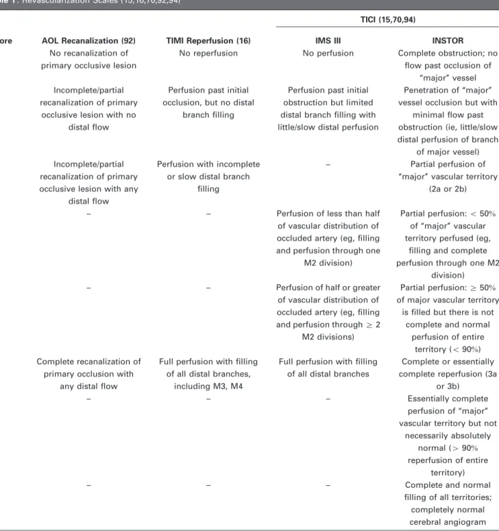
Revascularization is considered to represent Thrombolysis in Myocardial Infarction (MI; TIMI) (16) or Thrombolysis in Cerebral Infarction (TICI) (15) grade 2 or 3 flow through the previously occluded vessel segment (Table 1).
Parenchymal Hematoma Type II
Parenchymal hematoma type II is a space-occupying hematoma of more than 30% of the infarct zone with substantial mass effect attributable to the hematoma (17) (Table 2) (18).
Symptomatic Intracranial Hemorrhage
Symptomatic intracranial hemorrhage (SICH) is a parenchymal hematoma type II or subarachnoid hemorrhage with neurologic deterioration leading to an increase in National Institute of Health Stroke Scale (NIHSS)44 or leading to death within 36 hours of treatment (19). Because of the risk of vessel perforation during endovascular procedures, subarachnoid hemorrhage has been added as a cause of intracranial hemorrhage to the Safe Implementation of Thrombolysis in Stroke Monitoring Study (SITS-MOST) definition (18,20).
Good Clinical Outcome
A good clinical outcome is a measure of neurologic functional outcome with a score of 0–2 on the modified Rankin Scale (mRS) (21) assessed 90 days after treatment.
INDICATIONS AND CONTRAINDICATIONS
Endovascular treatment of acute stroke with intraarterial (IA) thrombolytic agents or mechanical thrombectomy is a consideration in patients in whom IV tPA fails or is considered likely to fail, who are excluded from IV tPA treatment, and/or who present with a large vessel occlusion.
Failure to respond to IV tPA after the 1-hour infusion of drug is completed has been assessed (22,23). Patients in whom this treatment is
Table 2. Definition of Postprocedure Bleeding Incidents (20)
Category Definition
HI 1 Small petechiae along margins of infarct
HI 2 More confluent petechiae within infarct area but without space-occupying effect PH 1 Blood clot(s)r30%of infarct area with some mild space-occupying effect PH 2 Blood clots430%of infarct area with significant space-occupying effect
PHr 1 Small or medium-sized clots located remote from actual infarct; mild space-occupying effect could be present PHr 2 Large confluent dense blood clots in area remote from actual infarct; significant space-occupying effect may be present HI¼hemorrhagic infarct, PH¼parenchymal hematoma, PHr¼parenchymal hematoma remote.
Table 1. Revascularization Scales (15,16,70,92,94)
Score AOL Recanalization (92) TIMI Reperfusion (16)
TICI (15,70,94)
IMS III INSTOR
0 No recanalization of
primary occlusive lesion
No reperfusion No perfusion Complete obstruction; no flow past occlusion of
‘‘major’’ vessel
1 Incomplete/partial
recanalization of primary occlusive lesion with no
distal flow
Perfusion past initial occlusion, but no distal
branch filling
Perfusion past initial obstruction but limited distal branch filling with little/slow distal perfusion
Penetration of ‘‘major’’ vessel occlusion but with
minimal flow past obstruction (ie, little/slow distal perfusion of branch
of major vessel)
2 Incomplete/partial
recanalization of primary occlusive lesion with any
distal flow
Perfusion with incomplete or slow distal branch
filling
– Partial perfusion of
‘‘major’’ vascular territory (2a or 2b)
2a – – Perfusion of less than half
of vascular distribution of occluded artery (eg, filling and perfusion through one
M2 division)
Partial perfusion:o50% of ‘‘major’’ vascular territory perfused (eg,
filling and complete perfusion through one M2
division)
2b – – Perfusion of half or greater
of vascular distribution of occluded artery (eg, filling and perfusion throughZ2
M2 divisions)
Partial perfusion:Z50% of major vascular territory
is filled but there is not complete and normal
perfusion of entire territory (o90%) 3 Complete recanalization of
primary occlusion with any distal flow
Full perfusion with filling of all distal branches,
including M3, M4
Full perfusion with filling of all distal branches
Complete or essentially complete reperfusion (3a
or 3b)
3a – – – Essentially complete
perfusion of ‘‘major’’ vascular territory but not
necessarily absolutely normal (490% reperfusion of entire
territory)
3b – – – Complete and normal
filling of all territories; completely normal cerebral angiogram AOL = arterial occlusive lesion, IMS = Interventional Management of Stroke, INSTOR = Interventional Stroke Therapy Outcomes Registry, TIMI = Thrombolysis in Myocardial Infarction, TICI = Thrombolysis in Cerebral Infarction.
considered likely to fail are those with large clot burdens associated with large intracranial vessel occlusions that can be detected with noncontrast CT, CT angiography, magnetic resonance (MR) angiography, transcranial Doppler imaging, or catheter angiography (24–27), or indirectly by a high NIHSS (ie,410) (28,29). Cases of occlusion of the carotid terminus or middle cerebral artery are associated with low recanalization rates of 4% and 30%, respectively, after IV tPA (30), and only a small minority of patients receiving IV tPA show a marked rapid improvement (31,32). Although IV tPA is the standard of care in patients arriving in an appropriate IV tPA time window, waiting for tPA to lyse the thrombus may significantly delay IA treatment, and it is reasonable to simulta-neously proceed to IA treatment. This approach is being evaluated in several trials (33–35).
Indications and contraindications for IA revascularization are derived from clinical trials and case series. Clinical trials tend to have more restrictive criteria whereas case series represent more of a ‘‘real-world’’ experience. Selection criteria are based on stroke severity, time (duration of symptoms), imaging, clot location, age, and medical comorbidities.
Stroke Severity
Clinical indications for endovascular treatment in multiple trials include new onset of a significant neurologic deficit (NIHSSZ8 [1,11,36]) or severe aphasia likely to be caused by a large artery occlusion. Patients with an NIHSS greater than 25 have traditionally been excluded from IV tPA trials (18,37) but can be treated effectively by endovascular methods, particularly those with posterior circulation stroke (38). Some, but not all, trials of IA therapy have also excluded patients with very high NIHSS (1), whereas some case series have included such patients (38). Higher NIHSS before IA treatment is associated with worse clinical outcomes.
Time
Trials of IA lytic agents and mechanical revascularization devices have historically required start of treatment as long as 6 hours or 8 hours, respectively (1,11,14,36), for anterior circulation strokes. The strongest level of evidence for IA treatment is for patients with middle cerebral artery occlusion treated with lytic agents within 6 hours (2,39,40). More rapid time to reperfusion has been linked to improved clinical outcomes and is therefore an important consideration in patient selection (41). Patients presenting at later time points with favorable imaging findings likely have more developed collateral vessels and thus different physiology. Case series that involved advanced imaging (CT angiography/CT perfusion or MR angiography/imaging) for patient selection describe acceptable outcomes regardless of time duration (5,42,43), but this approach is not uniformly accepted. Vertebrobasilar occlusions have been treated at extended times from symptom onset, sometimes more than 24–48 hours after symptom onset (44,45).
Imaging
Noncontrast CT has been an essential component of patient selection in randomized trials of IV and IA revascularization for treatment of acute stroke (1,11,31,36,37). Absolute contraindications to endovascular treat-ment based on noncontrast CT are similar to those for IV thrombolytic agents, and include the presence of acute intracranial hemorrhage or a significant developed infarct (31). Infarct size can be quantified on noncontrast CT using the Alberta Stroke Programme Early CT Score (46,47). Larger infarcts (scoreo7) are associated with an increased risk of thrombolysis-related hemorrhage (48) and worse clinical outcomes. The hyperdense middle cerebral artery sign can alert clinicians to the presence of a large vessel occlusion with a high degree of specificity. Clot length on noncontrast CT of more than 8 mm has been associated with unlikely recanalization after IV tPA administration (49). The hyperdense sign can be best seen with thin-cut reconstructions of a standard noncontrast CT at 0.625 mm (49).
Many hospitals proceed directly from noncontrast CT to angiography and potentially to endovascular treatment, as was done in the Prolyse in Acute Cerebral Thromboembolism II trial (1). This approach may have the
potential benefit of being more time-efficient, but may not be sensitive enough to optimally select patients. The effectiveness of selecting patients based on advanced imaging has not yet been confirmed in randomized trials; however, it is becoming common to also perform advanced imaging with CT angiography/perfusion or MR imaging/angiography to confirm large vessel occlusion, better detect completed infarct, and estimate ischemic tissue at risk that may progress to infarct (50–52).
Exclusion criteria that have been reported using advanced imaging include a core infarct larger than 100 cm3(53), and penumbra less than 20% larger than the core infarct (54,55). Recently in a small cohort of patients (56), it was demonstrated that a pretreatment diffusion-weighted MR imaging volume greater than 70 cm3 was associated with a poor clinical outcome regardless of successful reperfusion.
Clot Location
Arterial occlusions arising more proximally are associated with poorer outcomes. Most notably, internal carotid artery ‘‘T-lesions’’ have the poorest outcomes among anterior circulation strokes (24,57). Proximal M1 occlusions have worse outcomes than distal M1 occlusions as a result of occlusion of lenticulostriate arteries and basal ganglia infarction, with an increased risk of reperfusion hemorrhage (58).
Age
Increased age is associated with worse clinical outcomes following revascularization (59,60). This may be a result of a combination of factors including more difficult catheterization, decreased cerebral reserve, and greater medical comorbidities. Additionally, elderly patients have a higher prevalence of dementia. Prestroke dementia before endovascular reperfusion has been linked with a low probability of achieving a good clinical outcome (61). Some trials have therefore excluded patients older than 80 years of age (33).
Medical Comorbidities
There are numerous relative contraindications to stroke revascularization, including recent head trauma, MI, gastrointestinal/genitourinary bleeding, arterial punctures in noncompressible sites, recent surgery, uncontrollable hypertension, International Normalized Ratio greater than 1.7, platelet count lower than 100,000, seizure at stroke onset, and very low or high blood glucose levels. Because of the ability to use no or low doses of thrombolytic agent, the strong or absolute contraindications for IV lytic agent use in view of the risk of systemic bleeding are only relative contraindications for IA therapy. However, compared with IV treatment, IA treatment poses a consistently higher risk of SICH (1,36,62), which may be secondary to more effective reperfusion (63) of infarcted brain. Diabetes and atrial fibrillation have also been associated with worse outcomes (64–66).
The combination of stroke severity, age, and medical comorbidities has been used to create scoring systems to assess the likelihood of a good outcome after IA therapy (66,67). More complete scoring systems that also include duration of symptoms, clot location, collateral flow, and advanced imaging do not as yet exist. The criteria chosen to select patients for treatment will affect outcomes. Patients at higher risk are more likely to do poorly with or without treatment, but selection of only patients at low risk will deny clinical benefit to a large number of severely ill patients. Because published selection criteria vary, there is no single ‘‘correct’’ list of inclusion and exclusion criteria. Based on published data and desired ratio of benefit and risk, each institution will need to create and follow its own indications and contraindications.
Metrics. At least 90% of patients treated with IA therapy should meet the institutional selection criteria (indications/contraindications).
PROCESS AND OUTCOMES METRICS
In general, previously published endovascular stroke therapy metrics (68) were designed to measure aggregate performance of a hospital or clinical outcomes. They were neither designed nor intended to define individual physician performance. In contrast, the present document provides requirements for performance criteria for the individual practitioner. The
purpose of these metrics is to define the minimum mandatory standards for performance of endovascular procedures to rescue patients from stroke.
The likelihood of achieving a good clinical result depends on patient selection and procedural performance, such as time to treatment and successful revascularization. A powerful factor for patient selection for rescue is the amount of still-viable brain versus brain already infarcted. Currently, there is no consensus concerning how best to distinguish ischemic from infarcted brain, but there is consensus that imaging provides vital information to answer this question.
DATA COLLECTION
Unlike many other areas of quality assurance/morbidity and mortality that concentrate on specific anecdotal events—typically errors in care or complications—the endovascular treatment of stroke must have a mini-mum level of positive outcomes to be clinically beneficial. The measure of benefit from endovascular stroke therapy is not based on single or isolated cases; it is rather expressed as a percentage of patients treated who can function independently by 3 months after the intervention. This requires entering all patients and their procedural, process, and clinical outcomes into a database, trial, or registry (39,68–70). Without the denominator of ‘‘all patients,’’ success measures/percentages are meaningless. These data allow comparison against benchmarks for individual procedural perfor-mance, risk-adjusted clinical outcomes, and individual and institutional process measures. As stated in an earlier document concerning compre-hensive stroke centers (68), it is advantageous to collect data in a standardized fashion to avoid redundant efforts. Examples of such data collection tools for treatment of acute ischemic stroke may include the Interventional Stroke Therapy Outcomes Registry (INSTOR) (70) and Get with the Guidelines–Stroke (71). The mandatory threshold for collection of the defined elements is 100%.
Data concerning demographics are used to identify various patient subgroups, whereas other data points are pertinent for risk adjustment and are necessary for evaluation of procedural and clinical outcomes. These would include factors specific to the individual case (eg, location of occlusion, time from onset) or factors specific to patient subgroups (eg, age, sex). Collection of these data points is necessary for an appropriate evaluation of patient risk factors and also for study of institutional factors that could influence overall patient outcomes and therefore have a bearing on evaluation of operator performance.
At a minimum, these data should include age, sex, race, NIHSS, location of occlusion, various time points and intervals described later, blood pressure, presence or absence of diabetes, presence or absence of atrial fibrillation, and type of occlusion (embolic vs atherosclerotic). Other data elements may be helpful and may become evident with further research.
Metrics. 100% of patients have the required process and outcomes data entered into a national database, trial, or registry.
Time Intervals
Emergency endovascular stroke treatment is one of the most complex multidisciplinary functions a medical institution chooses to undertake. Reperfusion treatment (IV or endovascular) achieved within the shortest period of time is widely accepted as a prerequisite for optimal clinical outcomes (41,72,73). It is estimated that, for every 30-minute delay in time to revascularization, there is a 10% decrease in the likelihood of a good outcome from endovascular stroke therapy (41). There are many steps from stroke onset to completion of treatment, and optimal execution of each of these steps is necessary to achieve the stated goal. Numerous opportunities exist to minimize the time needed for each step from the time of the acute stroke to patient arrival to the hospital and then until reperfusion is achieved.
Process improvement for emergency stroke treatment should be an ongoing component of all stroke systems of care and should focus on all the tasks and activities in this complex sequence of events. These data are then used for quality assessment/assurance and process improvement, and thus directly relate to the eventual clinical outcome of the patients being
treated by the operator. To judge satisfaction of these performance goals in regard to expeditious delivery of care, time points and intervals are the units of measurement.
At a minimum, all time points and intervals specified in this document should be tracked in all cases. Institutions may choose to measure additional time points. The more time points that are recorded, the more exactly deficiencies might be identified. For instance, delays in obtaining a CT scan may result from delay in ordering the study, delay in response by CT staff (eg, because of multiple other procedures being requested at the same time), or delay caused by transportation.
Acknowledgment of the critical importance of time to reperfusion for obtaining favorable outcomes in myocardial reperfusion treatments has led to the formation of initiatives such as Door to Balloon (D2B): An Alliance for Quality (74), an international effort organized by the American College of Cardiology in partnership with the American Heart Association and 37 other organizations to rapidly translate research about how best to achieve outstanding D2B times for patients with ST-segment elevation MI. The key metrics recommended by this initiative, which has enrolled more than 1,000 hospitals, were achievement of a D2B time of less than 90 minutes for at least 75% of patients presenting directly to the treating hospital (74). Key strategies chosen by the D2B Alliance include having the emergency medicine physician activate the catheterization laboratory with a single call, having the team prepared within 20–30 minutes of the call, rapid data feedback, a team-based approach, and administrative support (74). Such initiatives have resulted in dramatic reductions in the times required from presentation to the hospital to procedure initiation. A recent study (75) found that D2B times have decreased nationally from a median of 96 minutes in 2005 to a median of 64 minutes in 2010.
The impressive results in shortening the time to myocardial reperfusion for acute MI obtained by such initiatives provided an impetus for launching similar initiatives related to IV tPA for stroke. Target Stroke is a national initiative that has hitherto enrolled more than 1,200 hospitals in the United States (76). It is organized by the American Heart Association/American Stroke Association in partnership with other organizations and aims to assist hospitals in increasing the proportion of IV tPA-treated patients in whom guideline-recommended door-to-needle times are achieved. The initial goal is to achieve a door-to-needle time no more than 60 minutes for at least 50% of patients with acute ischemic stroke. The Joint Commission has set a more ambitious goal of 80% of patients treated within 1 hour at primary stroke centers (77). The 10 key strategies chosen by Target: Stroke include emergency medical service prenotification, activating the stroke team with a single call, rapid acquisition and interpretation of brain imaging, use of specific protocols and tools, premixing tPA, a team-based approach, and rapid data feedback. Many of these strategies apply to the endovascular approach for stroke as well.
The realities of endovascular stroke practice have yet to achieve the level of refinement that the acute MI process has realized by implementation of guidelines. Miley et al (78) reported a mean time of 174 minutes⫾60 from noncontrast head CT to microcatheter placement in the thrombus of patients with acute stroke caused by large vessel intracranial occlusions across three academic centers performing endovascular acute stroke treatment in the United States. This report does not contain detailed breakdown of times required for each step, for example, from CT to additional imaging (if performed), transportation to the angiography suite, general anesthesia, and groin puncture to microcatheter insertion. Costalat et al (10) described a more detailed analysis of these steps in a group of patients with acute stroke selected with MR imaging and treated with endovascular therapy at a single center in Europe. In this report (10), the mean time to MR imaging was 59 minutes, the mean MR imaging duration was 22 minutes, the mean time from MR imaging to groin puncture was 81 minutes, and the mean time from groin puncture to reperfusion was 54 minutes. Adding all these times would yield mean times of 162 minutes from door to groin puncture and 216 minutes from door to reperfusion. Similar times can be derived from recommendations regarding initiation of IA therapy (groin puncture) set forth by the
Interventional Management of Stroke III executive committee (50) suggesting that groin puncture should occur within 90 minutes of starting IV therapy. In combination with a door–to–IV tPA time of 60 minutes, this would lead to a door-to-puncture time of as long as 150 minutes for combined IV/IA therapy.
As a result of the need for imaging and, in many places, anesthesia services in addition to the emergency medicine and interventional components, patients with acute stroke referred for endovascular therapy require at least an additional 1 hour compared with acute MI (and, in some hospitals, closer to 2 h) for initiation of treatment. Although rapid response mechanisms aiming to result in initiation of revascularization therapies within the minimum amount of time can be modeled according to the MI experience, it should be recognized that acute stroke treatment, especially endovascular therapy, requires a far more complex infrastruc-ture. Notwithstanding that, it is clear that, similar to the cardiology model, major improvements in door-to-treatment times need to take place to increase the proportion of favorable outcomes for patients treated with endovascular therapy for acute stroke.
Key Time Intervals
The time-interval metrics should be applicable regardless of the time of day and regardless of whether the patient presents on a weekday versus a weekend. These metrics represent maximum recommended times. Because of ample evidence that, the shorter the time to reperfusion, the higher the likelihood of a favorable outcome, all centers should strive to initiate endovascular therapy within the shortest possible time frame. Although IV tPA administration should not represent a justification for excessive delays in initiation of endovascular therapy, IV thrombolysis may be associated with some delays in initiation of endovascular therapy.
Door to Imaging. Most hospitals will use a noncontrast CT, but some hospital protocols may use MR imaging as the first imaging study. This study should be performed within 25 minutes (68,79) and interpreted within 45 minutes (79,80). The present document also requires that the interpretation must be sufficient to make decisions for patient care and the interpretation and time of interpretation be documented in the medical record. Because of the difficulty in defining exactly when an order might have been given, this document is in agreement with the American Stroke Association recommendations (68) that these time intervals be measured from arrival to start of imaging rather than from time of order to completion of imaging.
Metrics. At least 80% of patients with acute stroke being evaluated for revascularization should have a noncontrast head CT or MR imaging study within 25 minutes from time of arrival.
Metrics. At least 80% of patients with acute stroke being evaluated for revascularization should have a noncontrast head CT or MR imaging study interpreted within 45 minutes from time of arrival.
Use of CT Angiography/Perfusion or MR Imaging/Angiography (for Centers that Perform MR Imaging) as First-step Imaging. Randomized trials of IV and IA stroke revascularization have selected patients based on noncontrast head CT (1,31,37). Currently, no randomized trials confirm the superiority of advanced imaging (CT angiography/perfusion or MR imaging/angiography) as a selection tool for endovascular therapy versus plain CT alone. However, previously published guidelines on imaging in patients with acute stroke (81) recommend that noninvasive vascular imaging be routinely performed. Such imaging must not unduly delay therapy with IV tPA or delay time from door to arterial puncture beyond 2 hours (as detailed further later).
Metrics. At centers whose institutional protocols require noninvasive vascular and parenchymal imaging (CT angiography/perfusion or MR imaging/angiography) before intervention, at least 80% of all patients potentially eligible for endovascular treatment should undergo these studies.
Door to Puncture. The majority of time from door to revascularization comes from the steps from door to puncture, rather than from puncture to revascularization (10,78). Therefore, the largest opportunities to reduce delays and improve outcomes will come from
reducing door-to-puncture times. Regardless of clinical evaluation and imaging details, the recommended time from patient arrival to start of procedure (arterial puncture) is 2 hours or less. This is more rapid than reported in previous trials (10,78), but it is the consensus of the writing group that this time metrics are necessary and achievable, and consistent with the improvement in D2B times that have been achieved for acute MI.
Metrics. At least 75% of patients treated with endovascular therapy should have a door to puncture time of less than 2 hours.
Puncture Time to Start of Revascularization. The start of revascularization is defined as the start of IA infusion of a thrombolytic drug or the first pass of a recanalization device (68).
Metrics. At least 50% of patients should have a time from puncture to start of lytic infusion or first pass of mechanical device in the target vessel of less than 45 minutes.
Puncture Time to Revascularization. These metrics of time from puncture to revascularization assess the efficiency of the interventional physician and team. Given the rapid advancements in endovascular treatment modalities, these recommendations are likely to change. In the Mechanical Embolus Removal in Cerebral Ischemia registry, the largest prospective endovascular database to date reflecting procedural outcomes across a large variety of stroke centers in the United States, the median time from groin puncture to end of procedure was 90 minutes. Newer technologies such as stentrievers have been noted to achieve significantly shorter procedural times (median of approximately 50 min) (10,12,14,82,83). Although the time to final angiography is easily measured, it may be variable depending on the need to perform thrombolysis of peripheral branch occlusions after recanalization of the proximal occlusion. For this reason, puncture time to initial revascularization to TIMI grade 2 or TICI grade 2a was chosen as a metric. Although this is not proven, it is the consensus of the writing group that more complete revascularization is likely to lead to improved clinical outcomes, albeit at some increased procedural risk, and therefore TICI grade 2a reperfusion may not be the intended endpoint of revascularization. It is possible that further time will be necessary to achieve more complete revascularization.
Metrics. At least 50% of patients should have TIMI grade 2 or TICI grade 2a revascularization within 90 minutes of arterial puncture.
Recanalization/Reperfusion
Recanalization and reperfusion are not necessarily the same, but both are measures of revascularization. Recanalization typically pertains to the original occlusion site whereas reperfusion pertains to the distal capillary bed. Endovascular methods of recanalizing vessels have been studied for more than 30 years. Numerous trials and series have demonstrated efficacy and clinical benefit for recanalization of cerebral vessels for acute ischemic stroke (1,3,10,84–88). Incomplete recanalization may lead to reocclusion, with clinical deterioration (84,89). Recanalization is a measure of technical success and thought to be a surrogate for subsequent clinical benefit, and has been accepted as such by the Food and Drug Administration (11,36,90). Recanalization of the occluded vessel has been achieved in 70%–89% of patients by using mechanical devices in large series (11,12,14,36), but this high rate may not correspond to effective reperfusion of the vascular bed. Recanalization rates in the middle cerebral artery from thrombolysis alone average 65% (2). Recanalization/ reperfusion rates can be affected by clot source, location, and size, with higher rates of failed recanalization and poor clinical outcomes for larger and/or more proximal occlusions (eg, carotid T-lesions) (91).
There are several methods to measure recanalization, including arterial occlusive lesion (92), TIMI (16), Thrombolysis in Brain Ischemia (93), and TICI (15,70,94) (Table 1). Several studies and series have used these various means to describe recanalization rates (13,95,96). Simplistic evaluations of recanalization of the major vascular occlusion (eg, arterial occlusive lesion) are thought to be less informative than data concerning reperfusion of the entire vascular bed. Under the heading of ‘‘TIMI Scale,’’ recent stroke clinical trials (94,97) have actually used very
different brain-adapted versions, hampering comparisons and understand-ing of trial findunderstand-ings. The TICI scale is a commonly used revascularization measure that was developed in 2003 in an effort to standardize reporting of revascularization efforts and scores range from 0 (ie, total occlusion) to 3 (ie, completely normal angiogram) (15). TICI is currently used in the Interventional Management of Stroke trial (33) and in the ongoing stroke registry INSTOR (70). For the purposes of the present document, TIMI or TICI is suitable for evaluating the success of recanalization at the end of the procedure.
The metric for revascularization includes all clot locations (in anterior and posterior circulations) and is therefore lower than the reported rates for middle cerebral artery occlusions alone. As technology and performance improve, the threshold for recanalization may increase. The ultimate goal of revascularization is to improve patient outcomes. However, there is a risk that persistent attempts to recanalize an occlusion may lead to more complications. The combined metrics for SICH, recanalization, and mRS score of 0–2 measure these risks and benefits.
Metrics. At least 60% of patients should have TIMI grade 2 or TICI grade 2/3 recanalization for all clot locations at procedure completion.
Postprocedure CT/MR
Certain vital information concerning procedural success or failure requires postprocedure cross-sectional imaging, and this is typically done 24–36 hours after finishing the case (98). Postprocedure imaging is necessary to identify acute subarachnoid hemorrhage, intraparenchymal hemorrhage or contrast staining, parenchymal hematoma, overall extent of new stroke, and other findings. CT or MR imaging within 36 hours after the intervention should be performed in all stroke cases. Although some patients may receive CT or MR imaging immediately after the procedure, imaging performed the next day provides additional valuable information. It is recognized that there are certain circumstances that might render follow-up imaging difficult or impossible to perform. Therefore, the threshold for this imaging is 90%, with the expectation that a goal of 100% is desired.
Metrics. At least 90% of patients should have a brain CT or MR imaging study within 36 hours of the end of the procedure.
Symptomatic Intracerebral Hemorrhage
The most common major risk of endovascular treatment of acute ischemic stroke is SICH. The reported incidences of SICH (as defined by individual studies) following IA revascularization range from 5% to 12% (1,3,7,11,13,14,36,60,65,66,87,90,99–114), with an average of approxi-mately 10% in a metaanalysis of randomized trials (2). Several definitions have been used, starting with the National Institute of Neurological Disorders and Stroke trial (31) and in the SITS-MOST (19) and INSTOR registries (70). The definition chosen for this metric is based on that used by SITS-MOST (20), and is parenchymal hematoma type II or subarachnoid hemorrhage with neurologic deterioration leading to an increase in NIHSS44 or leading to death within 36 hours of treatment (Table 2) (98,115,116). SICH is not only an ‘‘end-result’’ evaluation of clinical judgment in the realm of patient selection and technical skill, but also a reflection of timing, procedural execution, and expeditious completion of task. For these reasons, tracking SICH is mandatory.
Subarachnoid hemorrhage is a unique complication of endovascular therapy, and is not typically seen with IV therapy with tPA alone. Intraprocedural subarachnoid hemorrhage can be rapidly fatal and is typically technique-related, and is therefore an entity that deserves special scrutiny (117). Although it is reported infrequently in the Interventional Management of Stroke trial (110) and thought to result from wire perforation, it may be more likely to occur with mechanical clot retriever use and/or rescue angioplasty (11,118–120).
Metrics. 100% of cases with SICH are reviewed for educational purposes. No more than 12% of treated patients should develop SICH.
Clinical Outcomes
The standard definition of a good clinical outcome from IA therapy is an mRS score of 0–2 at 90 days as assessed by a certified examiner
independent of the interventional physician (1–4,11,21,36,90,121–124). Clinical outcomes of stroke revascularization depend on patient selection and the technical judgment and skill of the operator. Variables such as patient age, stroke severity, time from onset, hypertension, diabetes, atrial fibrillation, infarct size, location of occlusion, and collateral arterial supply, among others, will affect the likelihood of a good outcome (60,66,67,91,125,126).
Reported incidences of good clinical outcome resulting from endovascular stroke therapy are approximately 40% (1,3,5,7–9,14, 60,78,91,101,107–114,127–134), but many of the reported trials and case series included patients with only M1 or M2 occlusions and time to treatment of no more than 6–8 hours, and excluded patients based on, for example, comorbidities, stroke severity, age, or clot location. In addition, these studies have disparate criteria for stroke onset–to–treatment time. Recent studies include diverse locations of occlusions, and the time to treatment is broad, which may be responsible for variable clinical results (11,90). Because of the difficulty in stratifying good outcomes based on selection and treatment variables, there was consensus to use a single global threshold (rather than exclude a subcategory such as T-occlusions), and the intent is to update the document when data obtained with the use of risk-adjusted thresholds become available.
A universal outcome threshold is difficult to define. There may be differences in patient selection between clinical trials and routine clinical practice. If 40% of patients with stroke seen at experienced trial centers have good clinical outcomes on average, this means that some centers achieve better than a 40% incidence of mRS scores of 0–2 at 90 days, yet some excellent centers achieve worse outcomes. Consequently, a 40% incidence of mRS scores of 0–2 may not represent an appropriate quality threshold based on statistical variability. Many facilities will treat small numbers of stroke cases with endovascular revascularization. Most studies report on relatively small numbers of patients, limiting the statistical significance of subgroup analysis. The consensus of the present writing group was to combine outcomes for a ‘‘standard’’ patient included in most trials, including anterior and posterior circulation strokes, and results from pharmacologic or mechanical revascularization.
Physicians who choose to treat sicker patients may achieve less favorable outcomes and may not meet the benchmarks specified in this document. This does not mean such physicians are providing a lower quality of care, but rather that such physicians have chosen to treat a different patient mix from those patients entered into the trials used to generate these benchmarks. The specific characteristics of patients who are treated will need to be documented in this circumstance. If physicians choose to treat patients who would not have been included in the trials used to generate the benchmarks of this article, this should be justified.
IA acute ischemic stroke revascularization is a resource-intensive procedure. Treatment of large numbers of patients at high risk may lead to a high percentage of patients with futile recanalization, meaning that the revascularization procedures performed do not lead to improved patient outcomes. Futile recanalization can also lead to a higher complication rate and potentially worse outcomes compared with the natural history of acute ischemic stroke without endovascular intervention (130,135–137). For this reason, the consensus of the authors of the present document is to recommend adherence to the 90-day mRS score of 0–2 outcome threshold to confirm appropriate selection of patients who are likely to benefit from therapy, as well as to prove adequate procedural performance.
The writing group’s consensus established a threshold of a 30% incidence of mRS score of 0–2 at 90 days for all strokes treated by endovascular methods. The outcomes of posterior circulation stroke revascularization may not be as favorable as the outcomes of anterior circulation revascularization, but there are insufficient data to generate a separate threshold for posterior circulation strokes. It is likely that a practice that achieves an acceptable rate of good outcomes for anterior strokes will also do so for posterior strokes. As better data on anterior and posterior circulation stroke revascularization become available, separate thresholds may be set. As technology and performance improve, the threshold for mRS score of 0–2 at 90 days will likely increase.
Metrics. At least 30% of patients with strokes treated by endovas-cular methods should have an mRS score of 0–2 at 90 days.
Death within 72 Hours of Treatment
Death within 72 hours of stroke is typically not a result of the stroke itself. The authors clearly acknowledge that every case is unique and that each instance needs to be reviewed in its entirety with the understanding that there are circumstances (eg, MI) that lead to death in the short term and are unrelated to operator factors. Death soon after a procedure in and of itself does not imply or indicate a quality problem. However, all deaths within 72 hours are a trigger for review and should be subject to immediate or focused inquiry. The threshold for death within 72 hours is 0%.
Metrics. 100% of cases with death within 72 hours of the end of the procedure are reviewed.
QUALITY IMPROVEMENT
Ongoing Quality Improvement
As IA treatment of acute ischemic stroke becomes a mainstream offering at many centers, an IA-specific multidisciplinary quality improvement
process should be established in all programs that offer IA treatment options. These endovascular cases and procedural techniques are innova-tive and can offer improved clinical outcomes, but should be monitored in a continuous and ongoing fashion.
A peer-review committee should be formed that involves personnel from the several backgrounds that have expertise in stroke care and a vested interest in quality of care and good outcomes. This committee should provide an open and transparent forum for process and case review. Transparency will optimize confidence in the process and its positive impact on patient care. Although there may be potential for conflict or disagreement among various participants, it is vital that the process be viewed as a nonpolitical, nonpunitive instrument for care process improvement.
In keeping with standards established under the Health Care Quality Improvement Act of 1986 (138), peer-review meetings and minutes are generally protected from legal inquiry in most states as long as the review is conducted under the auspices of the facility quality improvement program. The Health Care Quality Improvement Act established standards for profes-sional review actions. If a profesprofes-sional review body meets these standards, neither the professional review body nor any person acting as a member or staff to the body will be liable in damages under most federal or state laws
Table 3. Intraarterial Therapy Quality Improvement Case Review Triggers and Process Metrics
Category Metrics
Indications for intraarterial treatment Z90%of patients treated with intraarterial therapy should meet institution selection criteria (indications/contraindications)
Data collection 100%of patients have required process and outcomes data
entered into national database, trial, or registry Key time intervals
Door to imaging At least 80%of patients with acute stroke being evaluated for
revascularization should have noncontrast head CT or MR study within 25 min from time of arrival, completed and interpreted
within 45 min from time of arrival Use of CT/MR (for centers that perform MR imaging) as
first-step imaging
At centers whose institutional protocols require noninvasive vascular and parenchymal imaging (CT angiography/perfusion or MR imaging/angiography) before intervention,Z80%of all patients potentially eligible for endovascular treatment should
undergo these studies
Door to puncture Z75%of patients treated with endovascular therapy should
have a door-to-puncture timeo2 h
Puncture time to start of revascularization Z50%of patients should have a time from puncture to start of lytic infusion or first pass of mechanical device in the target
vesselo45 min
Puncture time to revascularization Z50%of patients should have TIMI grade 2 or TICI grade 2a revascularization within 90 min of arterial puncture Outcome metrics
Recanalization/reperfusion Z60%of patients should have TIMI grade 2 or TICI grade 2/3 recanalization for all clot locations at procedure completion
Postprocedure CT/MR Z90%of patients should have brain CT or MR within 36 h of
the end of procedure
SICH 100%of cases with SICH are reviewed for educational
purposes;r12%of treated patients should develop SICH
Clinical outcomes At least 30%of patients with strokes treated by endovascular
methods should have an mRS score of 0–2 at 90 d Death within 72 h of treatment 100%of deaths within 72 h of the end of procedure are
reviewed
mRS ¼ modified Rankin Scale, NIHSS ¼ National Institute of Health Stroke Scale, SICH ¼ symptomatic intracerebral hemorrhage, TIMI¼thrombolysis in myocardial infarction, TICI¼thrombolysis in cerebral infarction, tPA¼tissue plasminogen activator.
with respect to the action (138–141). All associated quality improvement documents should include routine annotation, which establishes the purpose of the document and that its content is protected under applicable federal or state law. The program should operate under the local facility umbrella established for all facility quality improvement and peer-review initiatives.
Peer Review Team
It is recommended that under the oversight of the stroke team medical director a predetermined multidisciplinary subgroup consisting of medical personnel with familiarity and expertise in IA therapy be established to address issues specifically relating to IA treatment. Although a stroke neurologist is generally in the best overall position to objectively assess overall process deficiencies and outcomes, for technical and procedural issues, the interventionist’s perspective must be considered. Care should be taken to avoid the inclusion of bias or review by individuals who are not familiar with the technical aspects of IA revascularization and its related potential complications. Ideally, the IA oversight team should be directed by a highly qualified, observant, compassionate, prompt, and unbiased physician, such as a noninterventional vascular neurologist. Depending on the institution, the IA quality improvement peer group could include a variable combination of interventionists, vascular neurologists, cerebrovas-cular neurosurgeons, intensivists, and diagnostic neuroradiologists. Addi-tional members might include hospital representative(s) from the quality assurance/improvement or risk management departments as well as possibly the stroke coordinator or other data personnel and secretarial support staff.
Review Process
The IA quality improvement meeting should occur at least quarterly, and, depending on volume, may need to occur more frequently to provide adequate assessment and review. The number of IA revascularization cases at a given institution is generally not of such magnitude as to preclude a review of every case, regardless of outcome. All endovascular stroke cases should be reviewed, and, as noted earlier in the section on data collection, entered into a trial, database, or registry with national participation (39,68,69).
The interventionist who performed the specific case under review should be present to offer his/her observations and perspective. The focused IA peer review should routinely include assessment of technical factors such as device choice, supplemental lytic infusion parameters, and equipment inventory assessment. Process elements such as on-call notification, response time, procedure table setup, and overall communication should also receive routine attention. It is also the role of the IA quality improvement review to assure that regular interventional support personnel receive routine in-services. Perfor-mance review is not limited to the treating endovascular physician, but should also include personnel from the emergency department and neurology and neurointensive care units; interventional technologists; nursing staff; and personnel from other related service areas as indicated.
Triggers for Review
Any event that might affect quality should be reviewed. Specific triggers for IA review include unmet process benchmarks, death, and symptomatic postprocedure hemorrhage. Some complications or process delays may be unavoidable, whereas others may reflect significant errors in judgment or process deficiencies. A determination must be made if the patient was harmed. Process problems such as delays or inadequate communication increase the risk of harm. Therefore, complications and events that increase the risk of poor outcomes need to be reviewed as a means of improving quality. There must also be differentiation between clearly procedure-related complications such as perforation and/or dissection, distal dislodgment of thrombus that remains unreachable, air embolus, embolus to a previously unaffected territory, and immediate SICH following the procedure from those that might be related to the primary ischemic event itself such as infarction, cerebral edema, and hemorrhagic
transformation. Predisposing underlying vascular disease and comorbid-ities must also be considered.
Physicians who choose to treat sicker patients may have poorer outcomes and may not meet established benchmarks. These cases should not be considered in isolation, as a poor outcome does not necessarily indicate that such physicians are providing a lower quality of care, but rather that they have a different patient mix from the trials that were used to create the benchmarks (130,135–137). IA quality improvement case review triggers and key process metrics are summarized in Table 3.
In addition to these morbidity and mortality markers, it is incumbent on the institution and the quality assurance/improvement and peer-review committee to also assess the ‘‘good outcomes.’’ A certain percentage of good outcomes are necessary for there to be sufficient benefit to the overall patient population. This document also defines minimal recanalization rates as well as improved clinical outcomes that should be attained.
Performance and Process Improvement
The committee should be equipped to deal with poor performance in a supportive, constructive, and collegial manner. In cases in which negative trends and deficiencies become apparent, improvement may require one-on-one mentoring, additional education, or supplemental training. IA stroke quality improvement review of problematic cases should generate a specific course of action to remedy recognized problems and prevent future occurrences. Individual assignments should be tracked with accountability reports scheduled for subsequent meetings. In addition, process improvement is a continuing activity that, along with individual performance improvement, will significantly impact clinical outcomes (142).
REFERENCES
1. Furlan A, Higashida R, Wechsler L, et al. Intra-arterial prourokinase for acute ischemic stroke. The PROACT II study: a randomized controlled trial. Prolyse in Acute Cerebral Thromboembolism. JAMA 1999; 282: 2003–2011.
2. Lee M, Hong KS, Saver JL. Efficacy of intra-arterial fibrinolysis for acute ischemic stroke: meta-analysis of randomized controlled trials. Stroke 2010; 41:932–937.
3. Ogawa A, Mori E, Minematsu K, et al. Randomized trial of intraarterial infusion of urokinase within 6 hours of middle cerebral artery stroke: the middle cerebral artery embolism local fibrinolytic intervention trial (MELT) Japan. Stroke 2007; 38:2633–2639.
4. Fields J, Khatri P, Nesbit G. Meta-analysis of randomized intra-arterial thrombolytic trials for the treatment of acute stroke due to middle cerebral artery occlusion. J Neurointervent Surg 2011; 3:151–155. 5. Jovin TG, Liebeskind DS, Gupta R, et al. Imaging-based endovascular
therapy for acute ischemic stroke due to proximal intracranial anterior circulation occlusion treated beyond 8 hours from time last seen well: retrospective multicenter analysis of 237 consecutive patients. Stroke 2011; 42:2206–2211.
6. Rouchaud A, Mazighi M, Labreuche J, et al. Outcomes of mechanical endovascular therapy for acute ischemic stroke: a clinical registry study and systematic review. Stroke 2011; 42:1289–1294.
7. Belisle JG, McCollom VE, Tytle TL, et al. Intraarterial therapy for acute ischemic strokes. J Vasc Interv Radiol 2009; 20:327–333.
8. Abou-Chebl A, Lin R, Hussain MS, et al. Conscious sedation versus general anesthesia during endovascular therapy for acute anterior circulation stroke: preliminary results from a retrospective, multicenter study. Stroke 2010; 41:1175–1179.
9. Josephson SA, Saver JL, Smith WS. Comparison of mechanical embolectomy and intraarterial thrombolysis in acute ischemic stroke within the MCA: MERCI and Multi MERCI compared to PROACT II. Neurocrit Care 2009; 10:43–49.
10. Costalat V, Machi P, Lobotesis K, et al. Rescue, combined, and stand-alone thrombectomy in the management of large vessel occlusion stroke using the solitaire device: a prospective 50-patient single-center study: timing, safety, and efficacy. Stroke 2011; 42:1929–1935.
11. Penumbra Pivotal Stroke Trial Investigators. The penumbra pivotal stroke trial: safety and effectiveness of a new generation of mechanical devices
for clot removal in intracranial large vessel occlusive disease. Stroke 2009; 40:2761–2768.
12. Machi P, Costalat V, Lobotesis K, et al. Solitaire FR thrombectomy system: immediate results in 56 consecutive acute ischemic stroke patients. J Neurointervent Surg 2012; 4:62–66.
13. Gupta R, Tayal AH, Levy EI, et al. Intra-arterial thrombolysis or stent placement during endovascular treatment for acute ischemic stroke leads to the highest recanalization rate: results of a multicenter retro-spective study. Neurosurgery 2011; 68:1618–1623.
14. Tarr R, Hsu D, Kulcsar Z, et al. The POST trial: initial post-market experience of the Penumbra system: revascularization of large vessel occlusion in acute ischemic stroke in the United States and Europe. J Neurointerv Surg 2010; 2:341–344.
15. Higashida RT, Furlan AJ, Roberts H, et al. Trial design and reporting standards for intra-arterial cerebral thrombolysis for acute ischemic stroke. Stroke 2003; 34:e109–e137.
16. The Thrombolysis in Myocardial Infarction (TIMI) trial. Phase I findings. TIMI Study Group. N Engl J Med 1985; 312:932–936.
17. Hacke W, Kaste M, Fieschi C, et al. Randomised double-blind placebo-controlled trial of thrombolytic therapy with intravenous alteplase in acute ischaemic stroke (ECASS II). Second European-Australasian Acute Stroke Study Investigators. Lancet 1998; 352:1245–1251.
18. SITS-MOST protocol. Available at: http://sitsinternational.org/. Accessed September 4, 2011.
19. Wahlgren N, Ahmed N, Eriksson N, et al. Multivariable analysis of outcome predictors and adjustment of main outcome results to baseline data profile in randomized controlled trials: Safe Implementation of Thrombolysis in Stroke-MOnitoring STudy (SITS-MOST). Stroke 2008; 39:3316–3322.
20. Wahlgren N, Ahmed N, Davalos A, et al. Thrombolysis with alteplase for acute ischaemic stroke in the Safe Implementation of Thrombolysis in Stroke-Monitoring Study (SITS-MOST): an observational study. Lancet 2007; 369:275–282.
21. van Swieten JC, Koudstaal PJ, Visser MC, Schouten HJ, van Gijn J. Interobserver agreement for the assessment of handicap in stroke patients. Stroke 1988; 19:604–607.
22. Christou I, Alexandrov AV, Burgin WS, et al. Timing of recanalization after tissue plasminogen activator therapy determined by transcranial doppler correlates with clinical recovery from ischemic stroke. Stroke 2000; 31:1812–1816.
23. Saqqur M, Molina CA, Salam A, et al. Clinical deterioration after intravenous recombinant tissue plasminogen activator treatment: a multicenter transcranial Doppler study. Stroke 2007; 38:69–74. 24. Barreto AD, Albright KC, Hallevi H, et al. Thrombus burden is
asso-ciated with clinical outcome after intra-arterial therapy for acute ischemic stroke. Stroke 2008; 39:3231–3235.
25. Christou I, Felberg RA, Demchuk AM, et al. Intravenous tissue plasminogen activator and flow improvement in acute ischemic stroke patients with internal carotid artery occlusion. J Neuroimaging 2002; 12: 119–123.
26. Kimura K, Iguchi Y, Shibazaki K, Aoki J, Uemura J. Early recanalization rate of major occluded brain arteries after intravenous tissue plasmino-gen activator therapy using serial magnetic resonance angiography studies. Eur Neurol 2009; 62:287–292.
27. Puetz V, Dzialowski I, Hill MD, et al. Malignant profile detected by CT angiographic information predicts poor prognosis despite thrombolysis within three hours from symptom onset. Cerebrovasc Dis 2010; 29: 584–591.
28. Fink JN, Selim MH, Kumar S, et al. Is the association of National Institutes of Health Stroke Scale scores and acute magnetic resonance imaging stroke volume equal for patients with right- and left-hemisphere ischemic stroke? Stroke 2002; 33:954–958
29. Fischer U, Arnold M, Nedeltchev K, et al. NIHSS score and arterio-graphic findings in acute ischemic stroke. Stroke 2005; 36:2121–2125. 30. Bhatia R, Hill MD, Shobha N, et al. Low rates of acute recanalization
with intravenous recombinant tissue plasminogen activator in ischemic stroke: real-world experience and a call for action. Stroke 2010; 41: 2254–2258.
31. Tissue plasminogen activator for acute ischemic stroke. The National Institute of Neurological Disorders and Stroke rt-PA Stroke Study Group. N Engl J Med 1995; 333:1581–1587.
32. Alexandrov AV, Demchuk AM, Felberg RA, et al. High rate of complete recanalization and dramatic clinical recovery during tPA infusion when continuously monitored with 2-MHz transcranial Doppler monitoring. Stroke 2000; 31:610–614.
33. Khatri P, Hill MD, Palesch YY, et al. Methodology of the Interventional Management of Stroke III Trial. Int J Stroke 2008; 3:130–137. 34. Assess the Penumbra System in the Treatment of Acute
Stroke (THERAPY). Available at: http://clinicaltrials.gov/ct2/show/ NCT01429350?term=therapyþandþTPA&rank=1. Accessed March 14, 2012.
35. Trial and Cost Effectiveness Evaluation of Intra-arterial Thrombectomy in Acute Ischemic Stroke (THRACE). Available at: http://clinicaltrials.gov/ ct2/show/NCT01062698. Accessed March 19, 2012.
36. Smith WS, Sung G, Saver J, et al. Mechanical thrombectomy for acute ischemic stroke: final results of the Multi MERCI trial. Stroke 2008; 39: 1205–1212.
37. Hacke W, Kaste M, Bluhmki E, et al. Thrombolysis with alteplase 3 to 4.5 hours after acute ischemic stroke. N Engl J Med 2008; 359: 1317–1329.
38. Egan R, Clark W, Lutsep H, Nesbit G, Barnwell S, Kellogg J. Efficacy of intraarterial thrombolysis of basilar artery stroke. J Stroke Cerebrovasc Dis 1999; 8:22–27.
39. Meyers PM, Schumacher HC, Alexander MJ, et al. Performance and training standards for endovascular ischemic stroke treatment. J Stroke Cerebrovasc Dis 2009; 18:411–415.
40. Blackham KA, Meyers PM, Abruzzo TA, et al. Endovascular therapy of acute ischemic stroke: report of the Standards of Practice Committee of the Society of NeuroInterventional Surgery. J Neurointervent Surg 2012; 4:87–93.
41. Khatri P, Abruzzo T, Yeatts SD, Nichols C, Broderick JP, Tomsick TA. Good clinical outcome after ischemic stroke with successful revasculariza-tion is time-dependent. Neurology 2009; 73:1066–1072.
42. Michel P, Odier C, Rutgers M, et al. The Acute STroke Registry and Analysis of Lausanne (ASTRAL): design and baseline analysis of an ischemic stroke registry including acute multimodal imaging. Stroke 2010; 41:2491–2498.
43. Turk A, Magarik JA, Chaudry I, et al. CT perfusion-guided patient selection for endovascular treatment of acute ischemic stroke is safe and effective. J Neurointervent Surg 2012; 4:261–265.
44. Brandt T, von Kummer R, Muller-Kuppers M, Hacke W. Thrombolytic therapy of acute basilar artery occlusion. Variables affecting recanaliza-tion and outcome. Stroke 1996; 27:875–881.
45. Lin DD, Gailloud P, Beauchamp NJ, Aldrich EM, Wityk RJ, Murphy KJ. Combined stent placement and thrombolysis in acute vertebrobasilar ischemic stroke. AJNR Am J Neuroradiol 2003; 24:1827–1833.
46. Barber PA, Demchuk AM, Zhang J, Buchan AM. Validity and reliability of a quantitative computed tomography score in predicting outcome of hyperacute stroke before thrombolytic therapy. ASPECTS Study Group. Alberta Stroke Programme Early CT Score. Lancet 2000; 355: 1670–1674.
47. Pexman JH, Barber PA, Hill MD, et al. Use of the Alberta Stroke Program Early CT Score (ASPECTS) for assessing CT scans in patients with acute stroke. AJNR Am J Neuroradiol 2001; 22:1534–1542. 48. Cucchiara B, Kasner SE, Tanne D, et al. Factors associated
with intracerebral hemorrhage after thrombolytic therapy for ischemic stroke: pooled analysis of placebo data from the Stroke-Acute Ischemic NXY Treatment (SAINT) I and SAINT II trials. Stroke 2009; 40: 3067–3072.
49. Riedel CH, Zimmermann P, Jensen-Kondering U, Stingele R, Deuschl G, Jansen O. The importance of size: successful recanalization by intra-venous thrombolysis in acute anterior stroke depends on thrombus length. Stroke 2011; 42:1775–1777.
50. Mackey J, Khatri P, Broderick JP. Increasing use of CT angiography in interventional study sites: the IMS III experience. AJNR Am J Neuror-adiol 2010; 31:E34.
51. Rai AT, Raghuram K, Domico J, Hobbs G, Carpenter J. Pre-intervention triage incorporating perfusion imaging improves outcomes in patients undergoing endovascular stroke therapy: a comparison with the device trials. J Neurointervent Surg 2012, 10.1136/neurintsurg-2011-010189.
52. Thomalla G, Cheng B, Ebinger M, et al. DWI-FLAIR mismatch for the identification of patients with acute ischaemic stroke within 4.5 h of symptom onset (PRE-FLAIR): a multicentre observational study. Lancet Neurol 2011; 10:978–986.
53. Souza LC, Yoo AJ, Chaudhry ZA, et al. Malignant CTA collateral profile is highly specific for large admission DWI infarct core and poor outcome in acute stroke. AJNR Am J Neuroradiol 2012; 33:1331–1336.