Tio
2Mediated Photocatalytic Inactivation of Positive And
Gram-Negative Bacteria Using Fluorescent Light
Amrita Pal a, Simo O. Pehkonen b, Liya E. Yu b, Madhumita B. Ray c, 1*
a
Department of Chemical and Biomolecular Engineering
b
Division of Environmental Science & Engineering
a, b
National University of Singapore, 10 Kent Ridge Crescent, Singapore 119260
c
Department of Chemical and Biochemical Engineering, Thompson Engineering Building, Room 443, Faculty of Engineering, The University of Western Ontario, London, Ontario, Canada, N6A 5B9
Abstract
Photocatalytic inactivation of six different species of bacteria using fluorescent light and TiO2 was conducted. Up to five surface loadings of TiO2 varying from 234-8662 mg/m2, impregnated on membrane filters were used with fluorescent light of constant illuminance of 3900 Lux for the inactivation of four ATCC bacteria (E. coli K-12, Pseudomonas fluorescens, Bacillus subtilis
and Microbacterium sp.) and two other species of bacteria (Microbacteriaceae str. W7 and Paenibacillus sp. SAFN-007) collected from outdoor air in Singapore. A Gram-negative
bacterium E. coli K-12 was the most effectively inactivated, while Gram-positive Bacillus subtilis exhibited the least response to the photocatalytic treatment. The inactivation rate
increased with an increase in the TiO2 loading, the maximum inactivation of most bacteria was achieved at an optimum TiO2 loading of 1116-1666 mg/m2. 100% of the E. coli K-12 was inactivated after 30 minutes of treatment at a TiO2 loading of 1666 mg/m2, while inactivation of one log10 was obtained for Microbacterium sp., Paenibacillus sp. SAFN-007 and
Microbacteriaceae str. W7 after two hours. Preliminary experiments indicate that the
photocatalytic inactivation using Degussa P25 is 1.83-5.41 times higher than that of Hombikat UV-100.
Keywords: Photocatalytic inactivation; Gram-positive and Gram-negative Bacteria; Fluorescent light inactivation; Optimum TiO2 loading
1.0 Introduction
Indoor air pollution due to biological contaminants (bacteria, viruses, fungi, etc.) is receiving increasing attention as a public health problem as people spend 80-90% of their time indoors. In tropical countries like Singapore, hot and humid climate enhances the proliferation and the growth of the biological contaminants in indoor environment, many of which may cause asthma and other respiratory illnesses and may transmit diseases like tuberculosis, cough and cold, mumps, measles, rubella, pneumonia, meningitis, Legionaries, influenza etc. [1].
isolation by pressurization control, inactivation using low-level ozonation and ultraviolet germicidal irradiation (UVGI) [2]. The effect of ultraviolet radiation on damaging bacterial cells and its application in water disinfection have been established in [3, 4]. Heterogeneous photocatalysis, that uses UV-A of 320-400 nm coupled with TiO2 catalyst, is a potential alternative as the process does not involve any expensive oxidizing chemicals, uses atmospheric oxygen, and produces hydroxyl radical and reactive oxygen species which are indiscriminate and powerful oxidizing agents and have the potential of causing inactivation in most of the microorganisms [5]. Heterogeneous photocatalysis has been proved to be successful in the treatment of water to a great extent [6-11]. Application of photocatalysis to inactivate air-borne bacteria is relatively new and most of the earlier studies reported the inactivation of E. coli using
either a TiO2 suspension [12, 13] or TiO2 immobilized on supports, such as glass [1, 11, 14] or quartz disc [8]. Moreover, the bactericidal efficiency of heterogeneous photocatalysis using has been tested on various bacterial species like E. coli K-12 [9, 7, 10, 1], E. coli [6, 5, 12, 11], Bacillus subtilis [8], Staphylococcus aureus, Enterococcus faecium, Candida albicans [11], Enterobacter cloacae [9], Pseudomonas aeruginosa [9, 11], and Salmonella typhimurium [9].
Though photocatalytic inactivation using UV-A and TiO2 can be an effective method, the inactivation efficiency of bacteria using fluorescent light and TiO2 requires extensive studies, since indoor environments (such as commercial and office premises) have TiO2 as the key constituent of wall paints, and are commonly illuminated by fluorescent light. A fluorescent lamp is essentially a low-pressure mercury lamp with the inner surface coated with various types of phosphors to absorb the 254 nm radiation and emit longer wavelengths [15] and hence can emit a very small fraction of UV-A [16]. Although the glass envelope surrounding the lamp absorbs all far-UV emission, the commonly used daylight or cool white lamps radiate appreciable amounts at 313, 334, and 365 nm of the mercury lines. A much stronger emission at these wavelengths is typical of the blacklight lamps, which are sometimes used in rooms to provide a fluorescent effect. To optimize the utilization of existing lighting in indoor environments, and to minimize additional energy consumption, this study systematically examines the effect of inactivation of bacteria using fluorescent light and TiO2 photocatalysts. To the best of our knowledge, the inactivation of bacteria using TiO2 catalyst irradiated by fluorescent light has not been reported in the literature. The effect of TiO2 loading on the inactivation efficiency of six different bacterial strains under fluorescent irradiation has been evaluated.
2.0 Materials and Methods
2.1 Materials
The photocatalyst used was non-porous titanium dioxide (TiO2, P25, Degussa AG, Germany). It had a primary particle diameter of 21 nm, specific surface area of 50 ± 15 m2/g, and a crystal distribution of 80% anatase and 20% rutile. TiO2 suspensions in deionised water at nine different concentrations were prepared and autoclaved for the following inactivation experiments. The following bacterial strains were used for the inactivation studies: Escherichia coli K-12 (ATCC
former four species were purchased from ATCC, while the latter two species were collected from outdoor air in Singapore using a six stage sampler (Andersen, location, USA) and identified to their respective closest relatives. Escherichia coli K-12 and Pseudomonas fluorescens are
Gram-negative bacteria, while the rest are Gram-positive.
In the photocatalytic experiments, an 18 W fluorescent lamp (NEC 6700K, TRI-PHOSPHOR T8, Japan), was used as the light source and was clamped at 8-9 cm above the surface of the filter samples. Such lamp, with a wavelength range of 400-700 nm, is commonly used for room illumination. The fluorescent illuminance (3900 Lux) was monitored using a Luxmeter in the experiments. A digital radiometer was used to determine the intensity of the UV-A light emitted from the fluorescent lamp which was measured to be 0.013 mW/cm2 at 365 nm on the surface of the filer. Figure 1 shows the schematic setup of the batch inactivation system.
Figure 1: Schematic diagram of the batch experimental set-up
2.2 Bacterial culture and membrane filter preparation
The bacterial cells were inoculated in 10 ml of Luria-bertani broth and incubated for 16 hours at 121 rpm in a rotating water shaker at 260C and 370C for Pseudomonas fluorescens and the other
strains, respectively. The cultured bacteria were centrifuged at 4,000 rpm for 5 minutes and washed with an autoclaved 0.9% sodium-chloride solution twice and re-suspended in 50 ml of an autoclaved 0.9% sodium chloride solution.The bacterial solution was then diluted to 107 or 108 times through consecutively re-suspending in a 0.9% sodium chloride solution and separated into individual 50 ml aliquots. Between each dilution, the bacterial suspension was well stirred using a vortex mixer to ensure uniformity of the suspension. A 50-ml aliquot of this bacterial solution was filtered through a cellulose acetate membrane filter (with an average pore size of 0.45 µm and a diameter of 47 mm) and the filter was placed in a sterile Petri dish, for the control experiments without TiO2. In the case of the inactivation experiments with TiO2, 50 ml of the autoclaved TiO2 solution at a required concentration was first filtered, followed by immobilization of the bacterial suspension onto the TiO2-loaded filters. In order to determine the amount of TiO2 coated on each membrane filter, the membrane filters were weighed before and after the TiO2 impregnation process. Five tests showed an average TiO2 loading ranging from 234 - 8662 (mg/m2), depending on the initial TiO
2 suspension employed (Table 1). The pore size of the coated membrane filters is expected to be reduced to less than 0.45 µm, which can ensure complete capture of the bacteria (≥1 µm) on the filters. Assuming a uniform distribution, the
Fluorescent lamp clamped to support Petri
dishes containing sample membrane
thickness of the TiO2 coating on each membrane ranged from 62-2279 nm for individual TiO2 loadings (last column, Table 1).
Table1. TiO2 loading and the resulting thickness of the TiO2 coating on the membrane. The error
is based on a replicate of five sets of data.
TiO2 Concentration
(in suspension) mg/la
Loading on the membrane filter surface (mg/m2)b
Error in the loading (%)
Thickness of the TiO2
coating on the
membrane (nm)c
10 234 5.12 62
20 511 6.56 134 30 840 14.82 221
40 1116 5.35 294
60 1666 6.49 438
80 2297 2.03 605
120 3490 1.13 919
200 5778 1.04 1521
300 8662 0.61 2279
a.The amount of the TiO2 solution impregnated on each membrane is 50 ml
b.The surface area of the membrane on which TiO2 is being loaded, is 17.35 cm2 c. The specific gravity of TiO2 has been taken as 3.8
2.3 Bacteria inactivation using fluorescent light irradiation
Photocatalytic inactivation was carried out at six irradiation durations of 0, 15, 30, 45, 60 and 120 minutes. Triplicate measurements were taken for each experiment. The temperature and the relative humidity in the black-box were measured before and after individual experiments and the values were constant throughout the experiment. After irradiation, membrane filters were immediately removed from the Petri dishes and placed face-down on agar plates. For E. coli
K-12 and Pseudomonas fluorescens, eosin methylene blue (EMB) agar plates were used, while
tryptic soy agar (TSA) was used for Microbacterium sp., Paenibacillus sp. SAFN-007 and Bacillus subtilis and R2A agar was used for Microbacteriaceae str. W7. All the plates were
sealed with parafilm tapes and placed in an incubator at 260C and 370C for
Pseudomonas fluorescens and the other bacterial strains, respectively. The colonies were then counted using a
colony counter daily on days 2 through 5, and on the 10th day, during which they were regularly checked for any regrowth of the colonies. For all the species the growth of colonies was complete within 3 days of incubation but in just one among an average of six experiments, E. coli and Microbacterium sp. showed new growth of colonies in 2-3 of the 18 Petri dishes on the
light source, and the second type of control experiment was conducted with light, but in the absence of TiO2. The bacterial inactivation efficiency followed 1st order kinetics with respect to bacterial colony count (Nt), which is shown by Equation (1),
ln (Nt/N0) = - kt (1)
Here, Nt = the number of CFUs after irradiation for t min. N0 = the number of CFUs at 0 min.
k = the inactivation rate constant. Nt/N0 = survival ratio
The survival ratio was calculated by normalizing the resultant CFUs on any plate to that on the plate without exposure to light. This ratio was compared under different durations of exposure to photocatalytic treatment and catalyst loadings to determine the inactivation efficiencies.
3.0 Results and Discussion
3.1 Bacteria inactivation under fluorescent light irradiation without TiO2
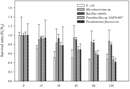
In the control experiments carried in absence of fluorescent light, the six immobilized bacterial strains were exposed to 1116 mg/m2 of TiO2 to a dark environment up to two hours. It was found that the average colony counts for all the bacterial strains varied insignificantly with little deviation (standard deviation of 0-10 %). Hence, in the absence of fluorescent light, bacteria impregnated on a membrane surface seemed to be insensitive to TiO2. The control experiments carried in absence of TiO2, showed that the fluorescent irradiation alone inactivated various strains of bacteria, results of which are shown in Figure 2. Figure 2 shows that in two hours, around 40-50% of E. coli K-12, Pseudomonas fluorescens and Paenibacillus sp. SAFN-007 were
inactivated. Although Microbacteriaceae str. W7 showed negligible inactivation (not shown in
Fig. 2), 20% of Bacillus subtilis and only 13% of Microbacterium sp. were inactivated. The
Time of exposure to fluorescent light (minutes)
0 15 30 45 60 120
Survi
val
rat
io
(N
t
/N0
)
0.0 0.2 0.4 0.6 0.8 1.0 1.2 1.4
1.6 E. coliMicrobacterium sp.
Bacillus subtilis
Paenibacillus sp. SAFN-007 Pseudomonas fluorescens
Figure 2. Survival ratios (Nt/N0) as a function of exposure duration (without TiO2) for E. coli K-12,
Pseudomonas fluorescens, Microbacterium sp., Paenibacillus sp. SAFN-007 and Bacillus
subtilis. The data shown in this figure are the averages of three replicates. Here, Nt = the
number of CFUs after irradiation for t minute and N0 = the number of CFUs at 0 minute.
3.2 Bacteria inactivation under fluorescent light irradiation and TiO2
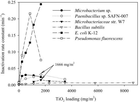
The inactivation of bacteria in the presence of TiO2 irradiated by fluorescent light exhibited first order reaction kinetics and the rate of inactivation increased with increased exposure to fluorescent light for all the bacteria. However, the variation in the inactivation rate constant with an increase in the TiO2 loading was different for the different bacteria. Figure 3 shows the inactivation rate constants of the six bacterial strains at different TiO2 loadings. Gram-negative bacteria E. coli K-12 and Pseudomonas fluorescens showed the highest inactivation rate
(0.0078-0.2442 min-1) while the Gram-positive bacteria appeared to be more resistant to the photocatalytic inactivation (Figure 3). This agrees well with other studies that the inactivation efficiency of Gram-negative bacteria, such as E. coli, was higher than that of Gram-positive
bacteria, such as S. aureus, E. faecium [11], and L. helveticus [12]. Although the exact
TiO2 loading (mg/m2)
0 2000 4000 6000 8000
Inactivation rate constant
(min
-1 )
0.00 0.05 0.10 0.15 0.20 0.25
Microbacterium sp.
Paenibacillus sp. SAFN-007
Microbacteriaceae str. W7
Bacillus subtilis E. coli K-12
Pseudomonas fluorescens
1666 mg/m2
Figure 3. Inactivation rate constant vs. the TiO2 loading for Gram-negative and Gram-positive
bacteria.
Table 2. Survival ratios (Nt/N0) and errors with respect to irradiation time (min) and TiO2 loading (mg/m2) for (a) Microbacterium sp., (b) Paenibacillus sp. SAFN-007, (c) Microbacteriaceae str. W7, (d) Bacillus subtilis (e) Pseudomonas fluorescens and
(f) E. coli.
(a)
(b)
TiO2 loading
511 (mg/m2) 1116 (mg/m2) 1666 (mg/m2) 3490 (mg/m2) Time
(min)
Nt/N0 Error Nt/N0 Error Nt/N0 Error Nt/N0 Error
0 1.0 0.12 1.0 0.07 1.0 0.11 1.0 0.12
15 0.86 0.12 0.60 0.05 0.69 0.08 0.64 0.09 TiO2 loading
234 (mg/m2) 511 (mg/m2) 840 (mg/m2) 1116 (mg/m2) 1666 (mg/m2) Time
(min)
Nt/N0 Error Nt/N0 Error Nt/N0 Error Nt/N0 Error Nt/N0 Error 0 1.0 0.13 1.0 0.00 1.0 0.40 1.0 0.63 1.0 0.00 15 0.80 0.14 0.48 0.13 0.93 0.36 0.44 0.23 0.89 0.12
30 0.76 0.17 0.48 0.06 0.74 0.30 0.71 0.35 0.80 0.24
45 0.55 0.14 0.49 0.06 0.91 0.33 0.18 0.08 0.61 0.13
60 0.33 0.04 0.30 0.16 0.47 0.16 0.11 0.09 0.20 0.15
30 0.62 0.05 0.44 0.12 0.52 0.09 0.47 0.06
45 0.40 0.08 0.27 0.08 0.31 0.04 0.41 0.04
60 0.24 0.10 0.18 0.06 0.17 0.05 0.29 0.06
120 0.12 0.06 0.07 0.04 0.02 0.03 0.16 0.02
(c)
TiO2 loading
1116 (mg/m2) 1666 (mg/m2) 2297 (mg/m2) 3490 (mg/m2) Time
(min)
Nt/N0 Error Nt/N0 Error Nt/N0 Error Nt/N0 Error
0 1.0 0.26 1.0 0.25 1.0 0.12 1.0 0.0
15 0.86 0.24 0.83 0.23 0.76 0.09 0.94 0.17
30 0.77 0.16 0.66 0.14 0.72 0.06 0.79 0.12
45 0.67 0.15 0.51 0.14 0.59 0.08 0.64 0.03
60 0.51 0.20 0.38 0.24 0.50 0.07 0.50 0.24
120 0.01 0.01 0.02 0.01 0.03 0.00 0.01 0.00
(d)
TiO2 loading
234 (mg/m2) 511 (mg/m2) 1116 (mg/m2) 2297 (mg/m2) 3490 (mg/m2) 5778 (mg/m2) Time
(min)
Nt/N0 Error Nt/N0 Error Nt/N0 Error Nt/N0 Error Nt/N0 Error Nt/N0 Error
0 1.0 0.105 1.0 0.04 1.0 0.10 1.0 0.10 1.0 0.08 1.0 0.03
15 0.92 0.09 0.88 0.04 0.87 0.10 0.76 0.06 0.896 0.09 0.94 0.03
30 0.88 0.07 0.85 0.04 0.73 0.06 0.77 0.12 0.73 0.08 0.90 0.09
45 0.86 0.07 0.83 0.03 0.71 0.08 0.70 0.10 0.67 0.06 0.77 0.08
60 0.80 0.06 0.80 0.03 0.70 0.07 0.68 0.09 0.64 0.05 0.57 0.04
(e)
TiO2 loading
234 (mg/m2) 840 (mg/m2) 1666 (mg/m2) Time
(min)
Nt/N0 Error Nt/N0 Error Nt/N0 Error
0 1.0 0.51 1.0 0.14 1.0 0.25
15 0.17 0.09 0.04 0.05 0.38 0.13
30 0.06 0.11 0.00 0.00 0.10 0.08
45 0.02 0.01 0.00 0.00 0.00 0.00
60 0.00 0.00 0.00 0.00 0.00 0.00
120 0.00 0.00 0.00 0.00 0.00 0.00
(f)
* Error = (N
t/N0)×(((a/Nt)^2 + (b/N0)^2)^0.5), where Nt and N0 are experimental variables whose standard deviations
are a and b, respectively.
In case of E. coli K-12, under a high TiO2 loading of 1666 mg/m2, over 96% of the bacteria were inactivated within 15 minutes of exposure, and all the bacteria were inactivated after a 30-minute or longer exposure (Table 2-f). This is encouraging because a UV-A intensity of only 0.013 mW/cm2 available in the fluorescent irradiation yielded an inactivation efficiency comparable with that reported by Huang et al., 2000 [21] who reported the damage of cell walls of E. coli
within 20 minutes of exposure to UV-A light at 0.8 mW/cm2 with the presence of TiO
2. Since a higher TiO2 loading may enhance the generation of reactive oxygen species (ROS), causing damage to the cell wall, the cytoplasmic membrane, and other intracellular components, the resultant inactivation rate was substantially increased. This is consistent with previous studies that higher inactivation of E. coli was observed when higher TiO2 concentrations were adopted with UV-visible radiation longer than 380 nm [18], or under UV-A irradiation [19].
The inactivation rate constant of Gram-positive bacteria Paenibacillus sp. SAFN-007 and Microbacteriaceae str. W7 reached a maximum at a TiO2 loading of 1666 mg/m2 (Figure 3). This loading corresponds to a thickness of the TiO2 coating on the membrane of 438 nm (Table 1). Since the wavelength of UV-A light is in the range of 320-400 nm with peak wavelength of 365 nm, a thin TiO coating of 62 and 134 nm may incompletely absorb light of 365 nm [20],
TiO2 loading
234 (mg/m2) 511 (mg/m2) 840 (mg/m2) 1116 (mg/m2) 1666 (mg/m2) Time
(min)
Nt/N0 Error Nt/N0 Error Nt/N0 Error Nt/N0 Error Nt/N0 Error
0 1.0 0.70 1.0 0.24 1.0 0.16 1.0 0.48 1.0 0.02
15 0.44 0.29 0.11 0.08 0.12 0.04 0.04 0.04 0.03 0.03
30 0.07 0.05 0.01 0.02 0.02 0.04 0.01 0.01 0 0
45 0.07 0.05 0.01 0.02 0 0 0 0 0 0
60 0.01 0.01 0.01 0.00 0 0 0 0 0 0
whereas a thicker TiO2 coating in the range of 134-438 nm (Table 1), could completely absorb the incoming UV-A. Nevertheless, further increases in the TiO2 loading with agglomeration of TiO2 on the filter surface could reduce activation efficiency because increases in the TiO2 concentration can cause terminal reactions (shown as reactions (3) and (4) below), which can form less reactive hydroperoxyl radicals (HO2·) and decrease the bacterial inactivation efficiency [10].
·OH + ·OH → H2O2 (3) H2O2 + ·OH → H2O + HO2· (4)
Interestingly, unlike other Gram-positive bacteria shown in Table 2, the inactivation rate constant of Bacillus subtilis reached a plateau at a loading higher than 2297 mg/m2 (Figure 3). The
stronger resistance of Bacillus subtilis to inactivatioin could be due to its transformation into
endospores and becoming insensitive to the changes in the environment (e.g., increases in the TiO2 loading). Although in the absence of TiO2, two hours of exposure to fluorescent light inactivated ~ 21% of the Bacillus subtilis (Figure 2), the presence of TiO2 with loadings up to 8662 mg/m2 only inactivated around 53% of the bacteria (data not shown) suggesting that
Bacillus subtilis were little affected by either a small amount of UV-A irradiation or increased
TiO2 loading. This can be supported by Kuhn et al. [11], who reported that the spores of Bacillus
subtilis were well resistant to 60 minutes of UV-A photocatalytic treatment. For Microbacterium
sp. and Microbacteriaceae str. W7, one log10 inactivation was obtained after 2 hours of light exposure at all TiO2 loadings, while for Paenibacillus sp. SAFN-007, the same inactivation occurred in the TiO2 loading range of 1116-1666 mg/m2, while 100% inactivation of
Pseudomonas fluorescens was obtained after 45 minutes of exposure to light at a TiO2 loading of 840-1666 mg/m2 (Table 2 a-e).
Limited experiments were carried out using another TiO2 photocatalyst, Hombikat UV-100 for Gram-positive bacterium Paenibacillus sp. SAFN-007, at the TiO2 loadings of 234, 511, 1116 and 1666 mg/m2. Hombikat UV-100 is less active than Degussa P25 and the difference is more significant at higher TiO2 loading. At the TiO2 loading larger than 1116 (mg/m2), the photocatalytic activity of Degussa P25 is 1.83-5.41 times higher than that of Hombikat UV-100. Rincon and Pulgarin (2003)[10] reported that a mixture of anatase and rutile showed better photocatalytic activity than anatase or rutile alone. Since Degussa P25 is a mixture of 80% anatase and 20% rutile, it is not surprising that Degussa P25 exhibited a higher inactivation efficiency than Hombikat UV-100 (100% anatase).
4.0 Conclusion
The results show that TiO2 mediated inactivation of bacteria is possible in the presence of fluorescent light, commonly used as room lighting. Experiments on six strains of bacteria including four Gram-positive and two Gram-negative bacteria have shown that E. coli was the
most effective and Bacillus subtilis was the least effective in photocatalytic inactivation. Of the
fluorescent light at TiO2 loading of 1666 mg/m2 (438 nm of TiO2 coating). This thickness of TiO2 can be applied on indoor wall commonly illuminated with fluorescent lighting to induce sufficient inactivation of the indoor bacteria. The study also indicates that reaction with OH radical and reactive oxygen species is less significant for Gram-positive bacteria in comparison to Gram-negative bacteria.
Acknowledgement
The authors acknowledge the financial support from National University of Singapore, grant RP-279-000-131-112.
References
[1] Jacoby, W.A., P.C. Maness, E.J. Wolfrum, D.M. Blake, J.A. Fennell. 1998. Mineralization of bacterial cell mass on a photocatalytic surface in air. Environmental Science & Technology 32 (17), pp.2650 – 2653. [2] Kowalski, W.J. and W. Bahnfleth. 1998. Airborne respiratory diseases and mechanical systems for control of
microbes. Heating, Piping and Air conditioning Engineering. Vol. 70, Issue 7, pp.34-47.
[3] F. Taghipour. 2004. Ultraviolet and ionizing radiation for microorganism inactivation. Water Research. Vol. 38, Issue 18, pp. 3940-3948.
[4] Hijnen, W.A.M., E.F. Beerendonk and G.J. Medema. 2006. Inactivation credit of UV radiation for viruses, bacteria and protozoan (oo)cysts in water: A review. Water Research. Vol. 40, Issue 1, pp.3-22.
[5] Huang, N., Z. Xiao, D. Huang and C. Yuan. 1998. Photochemical disinfection of Escherichia coli with a TiO2
colloid solution and a self-assembled TiO2 thin film. Supramolecular Science 5, pp.559-564.
[6] Bekbolet, M., C.V. Araz. 1996. Inactivation of E. coli by photocatalytic oxidation. Chemosphere 32 (5),
pp.959 –965.
[7] Maness, P.C., S. Smolinski, D.M. Blake, Z. Huang, E.J. Wolfrum, and W.A. Jacoby. 1999. Bactericidal
activity of photocatalytic TiO2 reaction: toward an understanding of its killing mechanism. Applied and
Environmental Microbiology 65, pp.4094 – 4098.
[8] Wolfrum, E.J., J. Huang, D.M. Blake, P. Maness, Huang Z and J. Fiest. 2002. Photocatalytic oxidation of bacteria, bacterial and fungal spores, and model biofilm components to carbon dioxide on titanium dioxide- coated surfaces. Environmental Science and Technology, 36, pp.3412-3419.
[9] Ibanez, J.A., M.I. Litter, R.A. Pizarro. 2003. Photocatalytic bactericidal effect of TiO2 on Enterobacter
cloacae: Comparative study with other Gram (-) bacteria. Journal of Photochemistry and Photobiology A:
Chemistry 6283, pp.1 – 5.
[10] Rincón, A.G., C. Pulgarin. 2003. Photocatalytical inactivation of E. coli: effect of (continuous–intermittent)
light intensity and of (suspended–fixed) TiO2 concentration. Applied Catalysis B: Environmental 44, pp.263–
284.
[11] Kuhn, K.P., I.F. Chaberny, K. Massholder, M. Stickler, V.W. Benz, H. Sonntag and L. Erdinger. 2003. Disinfection of surfaces by photocatalytic oxidation with titanium dioxide and UVA light. Chemosphere 53, pp.71-77.
[12] Liu, H. and T. C. Yang. Photocatalytic inactivation of Escherichia coli and Lactobacillushelveticus by ZnO
and TiO2 activated with ultraviolet light. Process Biochemistry, 39, pp.475-481. 2003.
[13] Matsunaga, T., R. Tomoda, T. Nakajima and H. Wake. 1985. Photoelectrochemical sterilization of microbial cells by semiconductor powders. FEMS Microbiology Letters 29, pp.211-214.
[14] Sunada, K., T. Watanabe and K. Hashimoto. 2003. Studies on photokilling of bacteria on TiO2 film. Journal
of Photochemistry and Photobiology A: Chemistry 156, pp.227-233.
[15] Bolton, J. 2002. Fundamentals of ultraviolet light. First Asia Regional Conference on Ultraviolet Technologies for Water, Wastewater & Environmental Applications.
[17] Oguma, K., H. Katayama and S. Ohgaki. 2002. Photoreactivation of Escherichia coli after low- or
medium-pressure UV disinfection determined by an endonuclease sensitive site assay. Applied And Environmental Microbiology, pp.6029-6035.
[18] Wei, C., W. Lin, Z. Zainal, N. Williams, K. Zhu and A. P. Kruzic. 1994. Bactericidal activity of TiO2
photocatalyst in aqueous media: toward a solar-assisted water disinfection system. Environmental Science and Technology, 28, pp.934-938.
[19] Cho, M., H. Chung., C. Wonyong and J. Yoon. 2004. Linear correlation between inactivation of E. coli and
OH radical concentration in TiO2 photocatalytic disinfection. Water Research 38, pp.1069-1077.
[20] Chen, D., F. Li and A. K. Ray. 2001. External and internal mass transfer effect on photocatalytic degradation. Catalysis Today 66, pp.475-485.