Bidder’s organ – structure, development and function
RAFAL P. PIPREK*
,1, MALGORZATA KLOC
4,5and JACEK Z. KUBIAK
2,31Department of Comparative Anatomy, Institute of Zoology, Jagiellonian University, Krakow, Poland, 2CNRS, UMR 6290, Institute of Genetics and Development of Rennes, Cell Cycle Group, France, 3Université Rennes 1, UEB, UMS
Biosit, Faculty of Medicine, Rennes, France, 4Department of Surgery, The Houston Methodist Hospital, Houston, USA and 5The Houston Methodist Research Institute, Houston, USA
ABSTRACT Bidder’s organ is an ovary-like structure, which develops from the anterior part of the gonadal ridge in anuran amphibians belonging to the Bufonidae family. Bidder’s organs form in both males and females. Because Bidder’s organ contains female germ cells (oocytes), the bufonid males are de facto hermaphrodites. Due to similarity with the undeveloped ovary, Bidder’s organ was, in early literature, described, inaccurately, as a structure present only in males. Due to the fact that Bidder’s organ is a unique structure present only in Bufonidae, it is not well studied and its function still remains a mystery. Here we describe the development and structure of Bidder’s organs, summarize the knowledge on gene expression and steroidogenic activity in these organs, and present hypotheses regarding Bidder’s organ function.
KEY WORDS:
Bidder’s organ, testis, ovary, oocytes, sex hormones
Introduction
Bidder’s organs are ovarian-like structures present in males and females of majority of bufonid species. Bidder’s organs develop, irrespectively of the genetic sex, from the anterior tips of the go-nads. The germ cells present in Bidder’s organs enter shortened oogenesis and, in the larvae, the diplotene oocytes become arrested in previtellogenesis (Brown et al., 2002). Despite the fact that Bid-der’s organs have been studied for two centuries their function still remains vague. Here we review available information on Bidder’s organ structure, development and its proposed functions.
Short history of Bidder’s organ in Bufonidae
Bidder’s organ was discovered in 18th century. In 1758 Roesel
von Rosenhof presented in his Historia Naturalis Ranarum Nos-tratium the first ever drawing of the pair of lobes at the anterior tips of gonads in Bufo calamita. In 1825 Jacobson noted that these peculiar structures are the vestigial ovaries characteristic for bufonids (Takahashi, 1923). In 1846 Bidder described these structures as a part of the testis. In 1876 Spengel used, for the first time, a term ‘Bidder’s organ’, to describe this structure in bufonid gonads. Despite the fact that the presence of Bidder’s organ has been described in many bufonid species, and that the removal of testes leads to transformation of Bidder’s organs into ovaries, which indicates that Bidder’s organs represent developmentally inhibited
www.intjdevbiol.com
*Address correspondence to: Rafal P. Piprek. Department of Comparative Anatomy, Institute of Zoology, Jagiellonian University, Gronostajowa 9, 30-387 Krakow,
Poland. Tel: +48126645059. e-mail: [email protected]
Accepted: 24 July 2014.
ISSN: Online 1696-3547, Print 0214-6282
© 2014 UBC Press Printed in Spain
Abbreviations used in this paper: BO, Bidder’s organ.
ovaries, the role of these organs is still unclear (Table 1; Harms, 1923; Ponse, 1924; Tanimura and Iwasawa, 1986; Duellman and Trueb, 1994; Abramyan et al., 2010, Piprek et al., 2013). Although commonly accepted belief is that Bidder’s organs are rudimentary or vestigial structures, some investigators believe that they are fully functional steroidogenic organs important for reproduction control (Davies, 1936; Colombo and Colombo-Belvedere, 1980; Abramyan et al., 2010; Scaia et al., 2011).
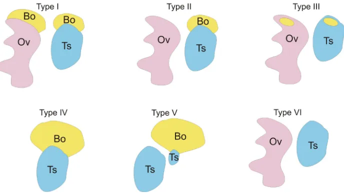
Bidder’s organs are present in the majority of studied spe-cies from Bufonidae family with the exception of South American Melanophryniscus setiba and Melanophryniscu stelzneri (Peloso, 2012) and two known species of Truebella from Peru (Fig. 1. Type VI) (Graybeal and Cannatella, 1995). This indicates that Bidder’s organ is not a synapomorphy of Bufonidae but only of a part of the family (Frost, 2006). It is commonly accepted that the absence of Bidder’s organ is a primitive feature for Anura, and its presence in Bufonidae is a derived feature (Duellman and Trueb, 1994).
in Bufo bufo, Bufo (=Anaxyrus) americanus, Bufo (=Anaxyrus) terrestris, Bufo (=Anaxyrus) quercicus, Bufo (=Rhinella, Chaunus) ictericus, Pseudophryne bibronii (Fig. 1. Type I) (Stohler, 1931; Witschi, 1933). However, Rau and Gaterby (1923) indicated the absence of Bidder’s organ in P. bibronii. This discrepancy between the different investigators’ data may result from the analysis of different age individuals. The presence of Bidder’s organs only in males was reported for Bufo (=Pseudepidalea) viridis, Bufo (=Rhi-nella) marinus, Bufo lentiginosus, Bufo (=Anaxyrus) canorus, Bufo (=Anaxyrus) fowleri, Bufo (=Poyntonophrynus) vertebralis, Bufo (=Capensibufo) rosei, Bufo (=Vandijkophrynus) angusticeps, Bufo (=Vandijkophrynus) gariepensis, and Nectophrynoides viviparus (Fig. 1. Type II) (King, 1908; Stohler, 1931; Witschi, 1933; Vos, 1935; Brown et al., 2002). Bidder’s organ has been found also in Nectophryne, Nectophrynoides, Pseudophryne, Pelophryne, and Pseudobufo (Davies, 1936; Ponse, 1924; Stohler, 1931). In the adult males and females of some species Bidder’s organs are not visible macroscopically, which suggests that they became incor-porated into the gonad proper. Such a situation was described in adult males of Asian Bufo (=Duttaphrynus) melanostictus in which Bidder’s organ is hidden within the testis (Koch, 1934). Similarly, in adult females of Bufo fowleri, Bidder’s organs do not degenerate but become incorporated into ovarian tissue, however, in juvenile females and all males Bidder’s organs and gonads remain as the separate structures. In the European common toad (B. bufo), Bid-der’s organs are present in adults of both sexes, however Dubois (1947), reported singular, 7 cm long, old female toad (B. bufo) lacking Bidder’s organs, which suggests that Bidder’s organ can degenerate over time. In Bufo arenarum, atresia of Bidder’s organs
of Bidder’s organs.
There is a great variability in the degree of Bidder’s organ de-velopment among individuals, which may depend on significant fluctuations of gene expression in developing Bidder’s organs (Abramyan et al., 2010). This variability has also been shown to be associated with environmental pollutants and seasonal activity (Scaia et al., 2008, 2011). There are also examples of individuals with well-developed Bidder’s organs and small remnants of testes (Bufo woodhousii; Pancak, 1987). In B. arenarum some males have one Bidder’s organ and two testes, and the testis connected to Bidder’s organ is smaller than the other (Fig. 1. Type V). In B. woodhousii and B. bufo some males from natural population have enlarged Bidder’s organs, which may represent the functional ovaries (Fig. 1. Type IV; Witschi, 1933; Pancak, 1987). The naturally occurring hermaphroditic toads frequent among North American hybrids of Bufo (=Anaxyrus) microscaphus and B. woodhousii have large ovaries (presumably functional Bidder’s organs) and small testes, and their gonads produce eggs and sperm (Sullivan et al., 1996).
The structure of Bidder’s organ
In bufonids the female gonad has two regions: Bidder’s organ and the proper ovary (connected via the retrobidderian region) (Fig. 2F). In males there is a specific intermediate region between Bidder’s organ and testes. In this intermediate region there is a gradual wave of oogenesis, which resembles the pattern of oo-genesis in the anterior part of the ovary proper (Zaccanti et al., 1971). Interestingly, the morphology of oocytes in male and female Bidder’s organs is the same (Farias et al., 2002, 2004; Tanimura
Fig. 1. The diversity of the external morphology of Bidder’s organs and gonads among bufo-nids. (Type I) The Bidder’s organs (Bo) are present at the anterior poles of ovaries (Ov) and testes (Ts). The presence of Bidder’s organs in both adult males and females was described in Bufo bufo,
Bufoamericanus, Bufoterrestris, Bufoquercicus, Bufo ictericus, Pseudophryne bibronii. (Type II)
The Bidder’s organs presence only in males, and its disappearance in adult females is characteristic for Bufo viridis, Bufo marinus, Bufo lentiginosus, Bufocanorus, Bufofowleri, Bufovertebralis, Bufo rosei, Bufoangusticeps, Bufogariepensis, and Nectophrynoides viviparus. (Type III) The Bidder’s organ incorporation into the adult ovaries and testes was described in Bufo melanostictus, Bufo fowleri, Nectophrynoidesoccidentalis. (Type IV) In some B. woodhousii males the Bidder’s organs are well-developed and resemble the ovaries. (Type V) In some B. arenarum males the testes with well-developed Bidder’s organs are smaller than the testes lacking Bidder’s organs. (Type VI) In
Melanophryniscus and Truebella the Bidder’s organs are absent.
occurs in females during third winter after metamorphosis and the final resorption of Bidder’s organs takes place during the fourth summer (Echeveira, 1990). The males of B. arenarum have one or two Bidder’s organs, which undergo a cyclic pattern of growth and abatement during a year (Echeveira, 1990). Thus, Bidder’s organ cyclicity may contribute to its size variability between individuals of the same species.
and Iwasawa, 1992).
Vitale-Calpe (1969) described Bidder’s organ of young males of South American Bufo arenarum as a thin strands of connective tissue accompanied by a loose plexus of blood vessels separating different groups of cells. This Bidder’s organ has oocyte-containing follicles, enclosed by squamous follicular cells and separated by collagen fibers, capillaries and slender stromal cells resembling fibroblasts (Vitale-Calpe, 1969). The author did not observe the presence of Balbiani body or annulated lamellae (characteristic for ovarian oocytes) in the oocytes of Bidder’s organ, however, these structures had been found in Bidder’s organ oocytes of B. bufo (Gurrieri et al., 1964).
Farias and colleagues (2002, 2004) described the structure of Bidder’s organs in South American Bufo ictericus. Here Bidder’s organ has a typical ovarian structure such as cortex and medulla. The cortex contains oocytes at different stages of oogenesis, which are enclosed by a monolayer of follicular cells. Oocytes have large round nuclei, diffuse chromatin, and prominent nucleoli. The cytoplasm of oocytes shows a well-developed smooth ER, numerous mitochondria, many peroxysomes, poorly developed Golgi apparatus and occasional lysosomes and lipid droplets. There is a discrepancy in the literature concerning the presence of vitellogenic oocytes in Bidder’s organs. The majority of stud-ies have shown that the vitellogenic oocytes appear in Bidder’s organ only after orchidectomy (testis removal) (Table 1; Ponse, 1927; Pancak-Roessler and Norris, 1991), however, some au-thors described the presence of vitellogenic oocytes in Bidder’s organs of Bufo marinus or Bufo arenarum during a reproductive season when these organs grow (McCoy et al., 2008; Scaia et al., 2011). Moreover, the lampbrush chromosomes, characteristic of intensively RNA-synthesizing anuran oocytes, were described in the oocytes of Bidder’s organs in B. marinus, Bufo terrestris, Bufo woodhousii, which indicates high metabolic activity of bidderian oocytes (Eichler, 1976). In summary, the ultrastructure of bidderian oocytes suggests that Bidder’s organs are not vestigial ovaries, but rather organs that are able to produce functional female germ cells (Farias et al., 2002).
Interestingly, the binuclear oocytes had been found in Bidder’s organ in Bufo melanostictus (Gulhati, 1963). Such binuclear oocytes are also present in B. bufo and B. viridis, where they constitute about 20% of all diplotene oocytes of Bidder’s organs (Fig. 2E; Piprek, unpublished data).
The development of Bidder’s organ
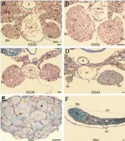
Early studies suggested that Bidder’s organ is a residual ovarian structure that develops from progonad i.e. from the most anterior part of the genital ridge (early gonadal anlage) (Ponse, 1951; Pisano and Pizarro, 1958). Newer studies showed that in anuran amphibians the progonad differentiates into the fat body, which suggests that Bidder’s organ develops from the most cranial part of the proper gonad (medial part of the genital ridge) instead (Viertel and Richter, 1999). Figure 2 depicts the structure and development of Bidder’s organs during the larval life in B. viridis. Witschi (1933) stated that Bidder’s organ (which does not have medullar cavity characteristic for ovary) develops from anterior gonomeres (meta-meres of gonad), which do not have typical medullar cavity. The lack of medullar cavity in Bidder’s organ of Bufo japonicus would support this hypothesis (Tanimura and Iwasawa, 1993; Moriguchi
and Iwasawa, 1987; Moriguchi et al., 1991). There is also no distinct medulla in Bidder’s organs in B. viridis (Fig. 2 A-F). However, there are several instances of the medulla being present in the center of Bidder’s organs (Beccari, 1925; Vannini and Busetto, 1945; Talluri and Padoa, 1953). Farias and coworkers (2002) showed a small mass of somatic cells and capillaries immersed in collagen fibers and glycoproteins within Bidder’s organ, which they believe represents the gonadal medulla. In fact, this could be a gonadal stroma that is located between the gonadal cortex and medulla and is invaded by capillaries. Falconi and coworkers (2007) showed the presence of small medulla only at the beginning of Bidder’s organ development.
Bidder’s organs develop at early stage of larval development (during the onset of hindlimb formation) (Beccari, 1925; Ponse, 1925; Ponse and Dovaz, 1943; Falconi et al., 2007). These stud-ies showed that in B. bufo and B. viridis at the onset of gonadal development at Gosner stage 29 (the hind limb bud development stage) the germ cells are larger and more abundant in the anterior part of genital ridge, i.e. in the region of future Bidder’s organ, than
Fig. 2. The structure of developing Bidder’s organ in Bufo viridis. (A-E) Cross sections through Bidder’s organs at subsequent Gosner stages (GS32 to metamorphosis, MET). (D) Cross section through the ovary (left gonad) and the Bidder’s organ (right gonad). (E) Cross section through the Bidder’s organ just after metamorphosis containing diplotene oocytes; asterisks indicate binuclear oocytes. (F) Longitudinal section through the Bidder’s organ (Bo) and the ovary (ov); diplotene oocytes (D) are present in the Bidder’s organ, oogonia (og) in the ovary, and the zygotene oocytes (Z) in the transitional retrobidderian region between the Bidder’s organ and the ovary. During the development of the Bidder’s organ in tadpoles, the germ cells enter diplotene and bidderian follicles form. a - aorta, dm - dorsal mesentery, ms - mesonephros, sc - secondary cavity in the ovaries, vc - vena cava. Debreuill staining, scale bar is equal to 30 mm.
B
C
D
E
F
in the caudal region (Fig. 2F). The general structure of anterior and caudal region is similar; they both contain the gonadal cortex built of germ and somatic cells and the primary gonadal cavity lined with the basal lamina in the center.
At Gosner stage 34 (the toe differentiation stage) Bidder’s organ has three layers: peripheral layer containing somatic cells and germ cells at various developmental stages, intermediate layer contain-ing follicles with diplotene oocytes, and the central layer – medulla (Falconi et al., 2007). Similar to the ovary proper the oogonia are present only in the peripheral layer. At Gosner stage 46 (metamor-phosis complete) Bidder’s organ contains only two layers: peripheral and intermediate. In contrast to the ovary proper, the medulla of Bidder’s organ does not develop further and as a consequence Bidder’s organs lack the secondary cavity (Fig. 2 A-F). The oocytes of Bidder’s organs are enclosed by follicular cells (granulosa) and the theca interna. The theca externa that develops from medullar cells in the proper ovaries is absent in Bidder’s organs (Witschi, 1933). The lack of medulla and the high ratio of germ to somatic cells found in Bidder’s organ have been suggested to be the cause of irreversible determination of the anterior end of the bufonid gonad toward the ovarian fate and the formation of Bidder’s organ (Witschi, 1933; Lepori, 1980). At Gosner stage 34, Bidder’s organ contains synaptic and diplotene oocytes (Fig. 2B). In the ovary proper, the first oocytes appear later (around metamorphosis) than in Bidder’s organ. At metamorphosis there are two different size (small and large) diplotene oocytes in Bidder’s organs, which reflects the two waves of oogenesis during Bidder’s organ development. The first wave begins at Gosner stage 34 and the second wave occurs at the end of metamorphosis (Falconi et al., 2007). The first wave of bidderian oogenesis is so short and fast that some researchers postulated that there is a direct passage of primary oognia into diplotene oocytes without transient synaptic stage (Ponse and Dovaz, 1943; Ponse, 1949). During the first wave of oogenesis the diplotene oocytes grow faster in females than in males, however, there is no size difference in the second wave. Bidder’s organ oocytes stop growing at previtellogenesis (stage I of oogenesis according to Dumont; Brown et al., 2002).
The number of germ cells in Bidder’s organ increases constantly, in males more slowly than in females (Falconi et al., 2007). In B. bufo the differentiation of testes, which begins about Gosner stage 41 (forelimb appearance stage), influences Bidder’s organ formation in males: the number of germ cells (oogonia and diplotene oocytes) and the size of diplotene oocytes of first oogonial wave decrease (Falconi et al., 2007). The second wave of oogenesis in Bidder’s organ ends, at least in the females, with the degeneration of large previtellogenic oocytes (Zaccanti and Gardenghi, 1968; Zaccanti et al., 1971). Similar to the proper ovary the waves of oogenesis in Bidder’s organs occur each year and they proceed from the peripherally located oogonia towards the central part of gonad.
Our studies showed that in Bufo viridis and Bufo bufo the ante-rior region of genital ridge grows more rapidly than the rest of the developing gonads due to the increase of germ cell number at the early stage of gonadogenesis (Gonser stage 29). This prolifera-tion of germ cells in the future Bidder’s organ leads to the club-like shape appearance of the developing gonads. Because in bufonids the development of Bidder’s organ correlates with the appearance of the oocytes in the anterior tip of developing gonad, we hypoth-esized that the meiotic entry is the major determinant of Bidder’s organ development (Piprek et al., 2013). Previously it was shown that retinoic acid (carotenoid compound) induces meiosis in mouse (Bowles et al., 2006; Koubova et al., 2006). We showed previously that inhibition of retinoic acid synthesis by citral as well as blockage of retinoic acid receptors by BMS453 (4-[(1E)-2-(5,6-dihydro-5,5-dimethyl-8-phenyl-2-naphthalenyl)ethenyl]-benzoic acid) results in decrease or lack of diplotene oocytes in in vitro developing larval Bidder’s organs (Table 1; Piprek et al., 2013). This study indicated that the retinoic acid induces meiosis both in Bidder’s organ and in developing ovaries. We also showed that there is a higher level of Raldh2 (retinoic acid synthesizing enzyme) in somatic cells of the developing Bidder’s organ than in developing ovaries and testes. In contrast, the level of Cyp26b1 (retinoic acid-degrading enzyme) is lower in the developing Bidder’s organ at Gosner stage 33 (the time of bidderian oocytes appearance and toe differentiation stage) than in the rest of the gonad (Piprek et al., 2013). This suggests that
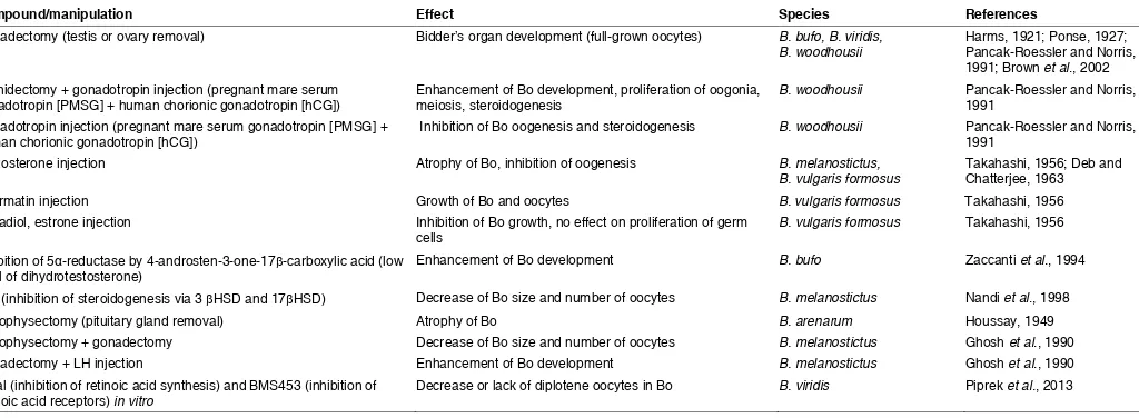
Compound/manipulation Effect Species References
Gonadectomy (testis or ovary removal) Bidder’s organ development (full-grown oocytes) B. bufo, B. viridis,
B. woodhousii Harms, 1921; Ponse, 1927; Pancak-Roessler and Norris, 1991; Brown et al., 2002 Orchidectomy + gonadotropin injection (pregnant mare serum
gonadotropin [PMSG] + human chorionic gonadotropin [hCG]) Enhancement of Bo development, proliferation of oogonia, meiosis, steroidogenesis B. woodhousii Pancak-Roessler and Norris, 1991 Gonadotropin injection (pregnant mare serum gonadotropin [PMSG] +
human chorionic gonadotropin [hCG]) Inhibition of Bo oogenesis and steroidogenesis B. woodhousii Pancak-Roessler and Norris, 1991
Testosterone injection Atrophy of Bo, inhibition of oogenesis B. melanostictus,
B. vulgaris formosus Takahashi, 1956; Deb and Chatterjee, 1963
Spermatin injection Growth of Bo and oocytes B. vulgaris formosus Takahashi, 1956
Estradiol, estrone injection Inhibition of Bo growth, no effect on proliferation of germ
cells B. vulgaris formosus Takahashi, 1956
Inhibition of 5α-reductase by 4-androsten-3-one-17β-carboxylic acid (low
level of dihydrotestosterone) Enhancement of Bo development B. bufo Zaccanti et al., 1994
LiCl (inhibition of steroidogenesis via 3 βHSD and 17βHSD) Decrease of Bo size and number of oocytes B. melanostictus Nandi et al., 1998
Hypophysectomy (pituitary gland removal) Atrophy of Bo B. arenarum Houssay, 1949
Hypophysectomy + gonadectomy Decrease of Bo size and number of oocytes B. melanostictus Ghosh et al., 1990
Gonadectomy + LH injection Enhancement of Bo development B. melanostictus Ghosh et al., 1990
Citral (inhibition of retinoic acid synthesis) and BMS453 (inhibition of
retinoic acid receptors) in vitro Decrease or lack of diplotene oocytes in Bo B. viridis Piprek et al., 2013
TABLE 1
the high level of retinoic acid in the anterior part of genital ridges is responsible for the formation of oocytes and Bidder’s organ.
The expression of genes involved in control of gonadogenesis was studied, using qRT-PCR, in developing Bidder’s organ of Bufo marinus (Abramyan et al., 2010). These studies showed that the gene expression in Bidder’s organ is organ specific and more similar to developing ovaries than testes (Table 2). The main genes involved in control of gonad development in amphibians, i.e. Dmrt1, Sox9, Sf1, Dax1 and p450arom, were expressed at different levels in the ovaries, testes and Bidder’s organs. The level of Dmrt1 (doublesex and mab-3 related transcription factor 1 involved in male sex deter-mination) expression was lower in Bidder’s organs than in testes and ovaries. The expression of Sf1 (steroidogenic factor 1, nuclear receptor involved in male sex determination) was higher in testes than in Bidder’s organs and ovaries. There was low expression of Sox9 (SRY-box 9, transcription factor involved in male sex determina-tion) in Bidder’s organ and ovary, and in the female Bidder’s organ there was a peak of Sox9 expression after metamorphosis. Dax1 (dosage-sensitive sex reversal, adrenal hypoplasia critical region, on chromosome X, gene 1, a nuclear receptor involved in female sex determination) and p450arom (aromatase enzyme) expression was the highest in Bidder’s organ, lower in the ovaries and the lowest in the testes. In female, but not in male Bidder’s organ, Dax1 had a second peak of expression after metamorphosis. In male and female Bidder’s organs a down-regulation of Dax1 expression correlated with the increase of p450arom and Sf1 expression. Because Dax1 inhibits p450arom expression, the high level of Dax1 expression in the male Bidder’s organ may be required to prevent estradiol synthesis. This may protect the testes from the feminizing effect of estrogens. Afterwards, when the testes are already differentiated, Dax1 is down regulated, and Sf1 and p450arom are up regulated in the male Bidder’s organs, which enable the synthesis of estradiol. There were large fluctuations in the expression level of genes in developing Bidder’s organs among individuals after gonadal dif-ferentiation into testes or ovaries, which may be associated with a high variability of Bidder’s organs structure. It is interesting that, in spite of structural similarities between ovaries and Bidder’s organs, the expression of steroidogenesis genes and sex-determining genes differ between these two organs.
Sex hormones and surgical experiments
Harms (1921) first demonstrated that in B. bufo (former Bufo vulgaris) Bidderian oogenesis in males could be enhanced by orchidectomy (testis removal). He stated that although the or-chidectomized male toads produce the full-grown oocytes within
their Bidder’s organ, they develop only rudimentary oviducts and therefore cannot lay eggs (Harms, 1921; Pancak-Roessler and Norris, 1991). However, it is interesting whether Bidderian oocytes are fertilizable. It can be tested via oocyte transplantation of marked bidderian oocytes into an appropriate female host. Figure 3 depicts the effects of various experiments on Bidder’s organs development and the hormonal interactions between the organs.
Ponse (1927) showed that gonadectomy in females and males of Bufo viridis leads to the development of Bidder’s organs into functional ovaries with vitellogenic oocytes (Table 1). Other authors also described the development of Bidder’s organs into functional ovaries after orchidectomy (Witschi, 1933, Pancak, 1987, Pancak-Roessler and Norris, 1991; Brown et al., 2002). Brown and coworkers (2002) described in the detail the resump-tion of Bidder’s organ development after orchidectomy. Bidder’s organs of orchidectomized individuals become highly vascular-ized, Bidderian oocytes grew (from 100 to 580 mm), accumulated yolk (onset of vitellogenesis), advanced to Dumont stages II-III, and within 1 month post-orchidectomy the whole Bidder’s organ increased considerably in size. During the growth of bidderian oocytes after testes removal, the lamina-associated polypeptide 2 (LAP2 b) located under the nuclear envelope is replaced by LAP2ω (Brown et al., 2002). Similar lamina isoform replacement takes place during oocyte growth in the ovary proper; small oocytes and somatic cells of the ovary proper contain LAP2b isoform, which during oocyte growth is replaced by LAP2ω isoform. This isoform replacement is probably associated with the increase in oocyte transcriptional activity.
Pancak-Roessler and Norris (1991) showed that in Bufo wood-housii the administration of gonadotropins (pregnant mare serum gonadotropin [PMSG] + human chorionic gonadotropin [hCG]) after orchidectomy enhances growth of Bidder’s organ and stimulates proliferation of oogonia and development of oocytes towards later stages of oogenesis and vitellogenesis (Table 1). In addition, the orchidectomy enhances steroidogenic activity (3 beta-hydroxys-teroid dehydrogenase [3 beta-HSD] and 17 beta-HSD) in Bidder’s organ. Importantly, in the presence of testes (in sham-operated toads) Bidderian oogenesis remains inhibited despite the high level of gonadotropins in blood plasma. Thus, the high level of gonadotropins does not overcome the inhibitory effects of testes. The administration of testosterone in Bufo melanostictus caused atrophy of Bidder’s organs, which indicates that testes inhibit Bid-der’s organ development (oogenesis) via androgens (Deb and Chatterjee, 1963). This, in turn, indicates that the testes (being the major source of the androgens) are responsible for the inhibi-tion of oogenesis and the maintenance of Bidder’s organ (Fig. 3).
Takahashi (1956) showed that the administration of testosterone in Bufo vulgaris formosus led to diminution of Bidder’s organ size, inhibition of bidderian oocyte growth and inhibition of oogonia pro-liferation, but never to Bidder’s organ disappearance or reversal of its sex (Table 1). Spermatin (a substance present in semen, allied to alkali albumin and mucin) administration led to the growth of Bidder’s organ and increase of oocyte size and number. Administra-tion of estradiol and estrone inhibited the growth of Bidder’s organ, however, did not inhibit the proliferation of oogonia. All these results show that both androgens and estrogens produced by testes or ovaries inhibit the development of Bidder’s organ.
Also more active form of androgens – dihydrotestosterone – has inhibitory effect on Bidder’s organ. Zaccanti and colleagues (1994) Gene Testes Male Bidder’s organs Ovaries Female Bidder’s organs
Dmrt1 +++ + +++ +
GENE EXPRESSION IN DEVELOPING TESTES, OVARIES AND BIDDER’S ORGANS IN BUFO MARINUS
(BASED ON ABRAMYAN et al., 2010).
showed that inhibition of 5a-reductase, that converts testosterone into dihydrotestosterone (DHT), via 4-androsten-3-one-17b -car-boxylic acid, caused resumption of Bidder’s organ development in B. bufo (Table 1). The decrease of DHT synthesis resulted in the increase of Bidder’s organ volume and oocyte growth.
Petrini and Zaccanti (1998) showed that administration of tes-tosterone to the tadpoles’ water or inhibition of aromatase lead to the differentiation of the gonads into testis in the genetic females (sex reversal). Interestingly, they showed the strong tendency of Bidderian germ cells to develop toward oogenic fate regardless of sex genotype and steroid treatment. Neither the exogenous androgens nor the inhibition of estradiol synthesis (inhibition of aromatase) are able to alter the differentiation of germ cells pres-ent in Bidder’s organ into oogenic cells.
The influence of steroid hormones on Bidder’s organ was shown in B. melanostictus via subcutaneous injection of LiCl that inhibits steroidogenetic enzymes – 3bHSD and 17bHSD, important for androgen and estrogen production (Fig. 3) (Nandi et al., 1998), and in the in vitro study of Bidder’s organs cultured in the medium containing LiCl (Nandi et al., 1999). Both studies showed that the inhibition of androgen and estrogen production results in decrease of size and number of bidderian follicles (Table 1). This proves that balanced level of sex hormones are important for development of Bidder’s organ and its inhibition in appropriate stage of gonad development.
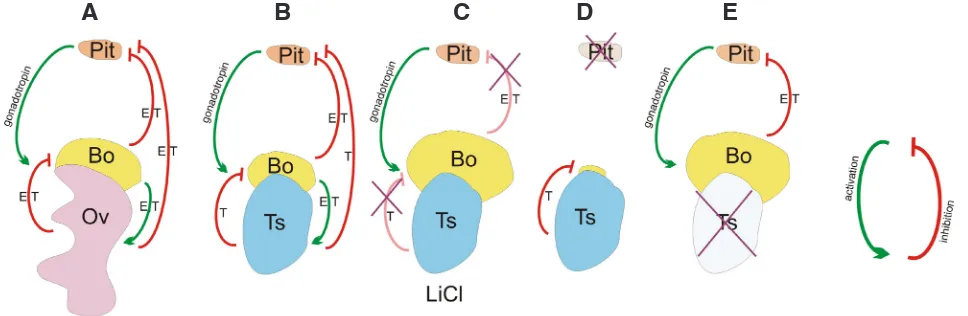
Bidder organ development and maintenance depends not only on gonadal hormones but also on the hypothalamus-pituitary-gonadal pathway of endocrine system, which is depicted in Figure 3. The removal of pituitary gland (hypophysectomy) in B. arenarum leads to the atrophy of Bidder’s organs (Fig. 3; Table 1; Houssay, 1949). Bidder’s organ does not enlarge in the specimens with removed both gonads and pituitary gland. Bidder’s organ resumes its growth and the vitellogenesis in its oocytes only when the pituitary gland is intact or if the toads are injected with solution of the pars distalis of pituitary gland. Ghosh and colleagues (1990) showed that in
B. melanostictus the removal of gonads (gonadectomy) led to the increase of Bidder’s organ weight, number of bidderian follicles and corpora lutea. However, the removal of both gonads and pituitary gland led to decrease in Bidder’s organ weight, number of bidderian follicles and corpora lutea (Table 1). Administration of luteinizing hormone (LH, pituitary hormone) led to the resumption of Bidder’s organ development. Thus, pituitary hormones are necessary for development of Bidder’s organs into functioning ovaries but the gonads suppress the inhibitory influence of pituitary hormones towards Bidder’s organs (Fig. 3).
Studies also showed that Bidder’s organs are a source of steroid hormones and can have important role in regulation of seasonal reproduction. The enzymes responsible for synthesis of androgens and estrogens (3bHSD, 17bHSD) from cholesterol are present in the ooplasm of bidderian oocytes and in the follicular cells in B. arenarum, B. melanostictus and B. woodhousii (Ghosh, 1982; Pancak-Roessler and Norris, 1991; Scaia et al., 2011). Bidder’s organ is capable of converting pregnenolone or progesterone into several steroid hormones such as 17-hydroxyprogesterone, andro-stendione, testosterone, 17b-estradiol and estrone (Colombo and Colombo-Belvedere, 1980). In B. arenarum the enzyme converting cholesterol into pregnenolone was detected in follicular cells only in few follicles, however, aromatase was present in all bidderian follicles (Scaia et al., 2011). In bufonids B. marinus, B. bufo, B. arenarum testes do not produce estradiol, however, the estradiol synthesis was detected in their Bidder’s organs (Kime and Hews, 1978; Colombo and Colombo Belvedere, 1980; Canosa et al., 1998). Orchidectomy in B. woodhousii led to decrease in androgens but did not influence the level of estradiol (Pancak-Roessler and Nor-ris, 1991). Thus, Bidder’s organ is a source of estradiol in females and males. There is a correlation between the weight of Bidder’s organ and level of plasma estradiol (Scaia et al., 2013). Estradiol is necessary for the regulation of cyclic reproduction activity and proliferation of spermatogonia in post-reproductive season. The removal of Bidder’s organs disturbed spermatogenesis in bufonid
Fig. 3. The hormonal interactions between Bidder’s organs, gonads and pituitary gland.(A) In the females the Bidder’s organs (Bo) produce estradiol (E) and testosterone (T) that stimulate (green arrow) ovaries (Ov) and together with E and T synthesized in the ovaries inhibit (red arrow) the pituitary gland (Pit). E and T produced in the ovaries inhibit the stimulating effect of gonadotropins on the Bidder’s organs, which blocks their development and ovarian fate differentiation. (B) In the males, the mechanism of Bidder’s organ regulation is similar to the females, however, the inhibition of the pituitary gland and thus the Bidder’s organ development is due to the great load of testosterone produced by testes (Ts). (C) The block of steroidogenesis by LiCl administration leads to derepression of stimulating effect of gonadotropins on the Bidder’s organs and thus to derepression of their development.
(D) The removal of pituitary gland (hypophysectomy, cross) eliminates the stimulating effect of gonadotropins and the gonadal hormones and leads to the atrophy of the Bidder’s organs. (E) The removal of the gonads (gonadectomy) eliminates the inhibitory effects of gonadal hormones on the Bidder’s organs and the stimulating effect of gonadotropins, which results in the development of the Bidder’s organs.
B
C
D
E
males, however, administration of estradiol to such males normal-ized the spermatogenesis. Thus, estradiol derived from Bidder’s organ is necessary for proper spermatogenesis. In Rana esculenta (Ranidae) high plasma and testicular level of estradiol was found in males (Fasano et al., 1989). The synthesis of the estradiol in the testes may explain abnormalities in the male gonad structure in this species and the presence of oocytes (testisova) in their testes. It seems that in bufonids the production of estrogens had been shifted from testes to Bidder’s organs, and this may prevent the exposure of testes and spermatogonia to high levels of female hormones. This may be one of the putative roles of Bidder’s organ.
The steroidogenesis in Bidder’s organ is independent of the gonadal steroidogenesis; the destruction of Leydig cells in the testes via administration of ethane 1,2-dimethane sulphonate, has no effect on steroidogenesis or oogenesis in Bidder’s organ (Scaia et al., 2011). Presumably, in addition to the steroid hormones secreted by gonads that inhibit differentiation of Bidder’s organ into functional ovaries, other factors may influence development of Bidder’s organs. Although the glucocorticoids do not influence Bidder’s organ development, the environmental pollutants have a significant effect on its presence and weight (Scaia et al., 2008). McCoy with colleagues (2008) showed that in B. marinus Bidder’s organ abnormalities increase in a dose-dependent fashion with the increase of the pollutant level. This suggests that the variability of the structure of Bidder’s organs observed in nature may result from the presence of environmental pollutants.
Seasonal changes in Bidder’s organ
Already early studies described the seasonal changes in Bidder’s organs (Alexander, 1932; Roessler et al., 1990; Pancak-Roessler and Norris, 1991). Recently, the correlation between steroidogen-esis and seasonal changes of Bidder’s organ size was described in details for South American Bufo arenarum (Scaia et al., 2011). Bidder’s organ size (weight) and the number of vitellogenic and atretic oocytes are the highest in the reproductive season (from September to December) and the lowest in the pre-reproductive season (from May to August) and post-reproductive season (from January to April) in B. arenarum. The testes are also larger during the reproductive season, however, the T/B ratio (testis/Bidder’s organ ratio) is lower in this season due to the significant growth of Bidder’s organ. Echeveria (1990) described the growth of Bidder’s organ during reproductive season and its degeneration during non-reproductive season in B. arenarum. In contrast, in B. wood-housii Bidder’s organ diminishes during the reproductive season (Calisi, 2005). There is correlation between the weight of Bidder’s organs and testes in B. woodhousii (Calisi, 2005), but not in B. arenarum (Scaia et al., 2011). The difference between these two bufonid species may result from the fact that in B. woodhousii the reproduction takes place when the level of androgens is high and in B. arenarum when the level of androgens is low. This indicates that Bidder’s organ size is influenced not by the reproductive or non-reproductive season but by the testosterone level.
In B. arenarum, there is increase of estradiol concentration dur-ing the reproductive season and its decrease in pre-reproductive season (Scaia et al., 2013). In contrast, the level of testosterone is low during the reproduction and high in pre-reproductive season. The same changes in sex hormones level occur in amphibians from temperate zone: the low level of testosterone during summer
(repro-duction season) correlates with proliferation of spermatogonia and formation of spermatocytes. The high level of testosterone during winter (non-reproductive season) correlates with spermiogenesis and inhibition of Bidder’s organ. Estradiol (high level in summer) is required for spermatogonia proliferation. Thus, in B. arenarum in the pre-reproductive season, when the expression of aromatase is low, the level of estradiol is low and the level of testosterone is high, the weight of Bidder’s organ is low, however, in the reproductive and post-reproductive season when the aromatase expression is high, estradiol load is high, and the level of testosterone is low, the weight of Bidder’s organ is high.
Similar to all amphibians where the sex hormones control the reproductive activity, in male bufonids the production of estradiol had been shifted from the testes to Bidder’s organ. The frequent total disappearance of Bidder’s organs in bufonid females may be associated with the fact that ovaries produce sufficient amount of estradiol.
In spite of extensive literature on Bidder’s organs develop-ment and structure their function still remains a mystery. Some researchers suggested that Bidder’s organ is a by-product of the evolutionary loss of hyperfecundity among bufonids (Roessler et al., 1990). These authors proposed that the bufonids are in the transitory state of evolutionary transformation from extensive, hyperfecund ovaries to smaller, less fecund ones. In this scenario, some members of Bufo retain hyperfecundity, while other such as derived bufonid species show a spectrum of ovarian reduction. In the latter cases Bidder’s organs, by performing steroidogenic functions, may compensate for the reduced ovaries.
Conclusions
Bidder’s organs are present in males and females of the major-ity of bufonid species. They usually disappear in females during the lifetime.
Melanophryniscus and Truebella are basal bufonid species with (probably) primary absence of Bidder’s organs. The Nec-tophrynoides show gradual reduction (to total disappearance) of Bidder’s organs.
Bidder’s organs develop, in both males and females, during the early gonadogenesis from the anterior parts of the gonads.
The gene expression profile in Bidder’s organ is organ specific, different than in ovaries or testes. The expression of genes involved in sex determination and gonadogenesis in Bidder’s organ is more similar to the expression in ovaries than in testes.
The analysis of retinoic acid synthesis and degradation suggests that the higher production of retinoic acid in the anterior ends of genital ridges in bufonids may be responsible for the formation of Bidder’s organs.
Bidderian oocytes are produced in waves and become arrested in the previtellogenic stage.
Bidder’s organs contain small and temporary medulla and lack of secondary ovarian cavity.
Bidder’s organs grow and resume oogenesis after the removal of testes. Bidder’s organs grow when the testosterone level is low, which occurs depending on the species during a reproductive or non-reproductive season.
The pituitary hormones are necessary for the development of Bidder’s organs.
The role of Bidder’s organ still remains unknown. Proposed role are the steroidogenesis and a by-product of the loss of hyperfe-cundity in Bufonidae.
References
ABRAMYAN J, WILHELM D, KOOPMAN P (2010). Molecular characterization of Bidder’s organ in the cane toad (Bufo marinus). J Exp Zool B Mol Dev Evol 314: 503-513.
ALEXANDER G (1932). Bidder’s organ in Bufo melanostictus Schneider. Copeia 1932: 78-80.
BECCARI N (1925). Ovogenesi larvale, organo di Bidder e differenziamento dei sessi nel Bufo viridis. Arch Anat Ital Embriol 22: 483-549.
BOWLES J, KNIGHT D, SMITH C, WILHELM D, RICHMAN JM, MAMIYA S, YAS-HIRO K, CHAWENGSAKSOPHAK K, WILSON MJ, ROSSANT J, HAMADA H, KOOPMAN P (2006). Retinoic signaling determines germ cell fate in mice. Science 312: 596-600.
BROWN FD, DEL PINO EM, KROHNE G (2002). Bidder’s organ in the toad Bufo marinus: effects of orchidectomy on the morphology and expression of lamina-associated polypeptide 2. Dev Growth Differ 44: 527-535.
CALISI RM (2005). Variation in Bidder’s organ volume is attributable to reproductive status in Bufo woodhousii. J Herpetol 39: 656-659.
CANOSA LF, POZZI AG, CEBALLOS NR (1998). Pregnenolone and progesterone metabolism by the testes of Bufo arenarum. J Comp Physiol B 168: 491-496. COLOMBO L, COLOMBO-BELVEDERE P (1980). Steroid hormone biosynthesis by
male Bidder’s organs of the toad Bufo bufo bufo. Gen Comp Endocrinol 40: 320-321. DAVIES DD (1936). The distribution of Bidder’s organ in the Bufonidae. Publ Field
Mus Nat Hist Zool Ser 20: 87-92.
DEB C, CHATTERJEE A (1963). Histochemical studies on the nature of Bidder’s organ in toad (Bufo melanostictus). Endokrinologie 44: 291-296.
DUBOIS R (1947). On the distribution of Bidder’s organ in bufonids. Zool Meded 28: 275-279.
DUELLMAN WE, TRUEB L (1994). Biology of Amphibians. The Johns Hopkins University Press Ltd. Baltimore, pp. 407-408.
ECHEVEIRA DD (1990). Organogenesis y ciclos estacionales del organo de Bidder en las larvas y los juveniles de Bufo arenarum (Anura, Bufonidae). Cuad Herp 5: 1-9. EICHLER VB (1976). The occurrence of lumpbruch chromosomes in Bidder’s organ
oocytes of male toads (Anura: Bufonidae). Herpetologica 32: 58-59.
FARIAS CF, CARVALHO-E-SILVA SP, de BRITO-GITIRANA L (2002). Bidder’s organ of Bufo ictericus: a light and electron microscopy analysis. Micron 33: 673-679. FARIAS CF, de BRITO-GITIRANA L, CARVALHO-E-SILVA SP (2004). Bidder’s
organ in the female of Bufo ictericus: morphological characterization. Contrib Zool 73: 317-319.
FALCONI R, DALPIAZ D, ZACCANTI F (2007). Morphological aspects of gonadal morphogenesis in Bufo bufo (Amphibia Anura): Bidder’s organ differentiation. Anat Rec 290: 801-13.
FASANO S, MINUCCI S, DI MATTEO L, DANTONIO M, PIERANTONI R (1989). Intratesticular feedback mechanisms in the regulation of steroid profiles in the frog, Rana esculenta. Gen Comp Endocrinol 75: 335-342.
FROST DR, GRANT T, FAIVOVICH J, BAIN RH, HAAS A, HADDAD CFB, DE SÁ RO, CHANNING A, WILKINSON M, DONNELLAN SC, RAXWORTHY CJ, CAMP-BELL JA, BLOTTO BL, MOLER P, DREWES RC, NUSSBAUM RA, LYNCH JD, GREEN DM, WHEELER WC (2006). The amphibian tree of life. Bull. Am. Mus. Nat. Hist. 297: 1-370.
GHOSH PK, GHOSH AK, BISWAS NM (1982). Effect of cadmium chloride on steroidogenic enzymes in Bidder’s organ of the toad (Bufo melanostictus). Experientia 40: 91-92.
GHOSH AK, GHOSH D, GHOSH PK, BISWAS NM (1990). Influence of pituitary on histology of Bidder’s organ in castrated toad Bufo melanostictus. Ind J Exp Biol 28: 790-791.
GRAYBEAL A, CANNATELLA DC (1995). A new taxon of Bufonidae from Peru, with descriptions of two new species and a review of the phylogenetic status of supraspecific bufonid taxa. Herpetologica 51: 105-131.
GULHATI KS (1963). Occurrence of binuclear oocytes in Bidder’s organ of male toad,
Bufo melanostictus (Schneid.). Naturwissenschaften 51: 93.
GURRIERI M, GRILLI A, VALDRE E (1964). Electron microscopic observations on Bidder’s organ of Bufo bufo. Boll Soc Ital Biol Sper 40: 766-8.
HARMS JW (1921). Verwandlung des Bidderschen Organs in ein Ovarium beim Mannchen von Bufo vulgaris Laur. Zool Anzeiger 53: 253-265.
HARMS JW (1923). Untersuchungen uber das Bidderschen Organ der mannlichen und weiblichen Kroten. 2. Die Physiologie des Bidderschen Organs und die experimentell-physiologishe Umdifferenzierung von Mannchen in Weibchen. Zeitsch f Anat u Entwicklungsgesch 69: 598-629.
HOUSSAY BA (1949). Hypophyseal functions in the toad Bufo arenarum. Quart Rev Biol 24: 1-27.
KIME DE, HEWS EA (1978). Androgen biosynthesis in vitro by testes from Amphibia. Gen Comp Endocrinol 35: 280-288.
KING HD (1908). The structure and development of Bidder’s organ in Bufo lentigi-nosus. J Morphol 19: 439-465.
KOCH M (1934). Uber das Urogenitalsystem der Bufoniden, im besonderen uber die Histologie des Bidderschen Organs. Jen Zeitschrift Naturw, vol. 11.
KOUBOVA J, MENCKE DB, ZHOU Q, CAPEL B, GRISWOLD MD, PAGE DC (2006). Retinoic acid regulates sex-specific timing of meiotic initiation in mice. Proc Natl Acad Sci USA 103: 2474-2479.
LAMOTTE M, GLAÇON R, XAVIER F (1973). Recherches sur le développement embryonnaire de Nectophrynoides occidentalis Angel amphibien anoure vivipare. 11Le développement des gonades. Ann Embryol Morph 6: 271-296.
LEPORI NG (1980). Sex differentiation, hermaphroditism and intersexuality in verte-brates including man. Piccin Medical Books, Padova. p 345.
MC COY KA, BORTNICK LJ, CAMPBELL CM, HAMLIN HJ, GUILLETTE LJ, ST MARRY CM (2008). Agriculture alters gonadal form and function in the toad Bufo marinus. Environ Health Perspect 116: 1526-1532.
MORIGUCHI Y, IWASAWA H (1987). Annual changes in male reproductive organs in Bufo japonicas formosus: histological observation. Gen Endocrinol 6: 115-120. MORIGUCHI Y, TANIMURA A, IWASAWA H (1991). Annual changes in Bidder’s organ
of the toad Bufo japonicus formosus: histological observation. Sci. Rep. Niigata Univ. S. D. Biol. 28: 11-17.
NANDI DK, GHOSH D, DEBNATH J (1998). Effect of lithium chloride on steroido-genic enzymes in Bidders organ, and on the blood level of testosterone, in the toad. Med Sci Res 26: 599-601.
NANDI DK, DEBNATH JM, GHOSH D (1999). Direct effects of lithium chloride on the activities of delta 5-3 beta and 17 beta-hydroxysteroid dehydrogenase in the testis and Bidder’s organ of the adult toad (Bufo melanostictus) - in vitro study. Folia Biol (Krakow) 47: 73-6.
PANCAK MK (1987). Studies on the endocrine regulation of oogenesis in Bidder’s organs of the male toad Bufo woodhousii. J Exp Zool 260: 323-336.
PANCAK-ROESSLER MK, NORRIS DO (1991). The effects of orchidectomy and gonadotropins on steroidogenesis and oogenesis in Bidder’s organs of the toad Bufo woodhousii. J Exp Zool 260: 323-336.
PELOSO PLV, FAIVOVICH J, GRANT T, GASPARINI JL, HADDAD CFB (2012). An extraordinary new species of Melanophryniscus (Anura, Bufonidae) from Southeastern Brazil. Am Mus Novit 3762: 1-32.
PETRINI S, ZACCANTI F (1998). The effects of aromatase and 5a-reductase inhibi-tors, antiandrogen, and sex steroids on Bidder’s organs development and gonadal differentiation in Bufo bufo tadpoles. J Exp Zool 280: 245-259.
PIPREK R, PECIO A, LASKOWSKA-KASZUB K, KLOC M, KUBIAK JZ, SZYMURA JM (2013). Retinoic acid homeostasis regulates meiotic entry in developing anuran gonads and in Bidder’s organ through Raldh2 and Cyp26b1 proteins. Mech Dev 130: 613-627.
PISANO A, PIZARRO N (1958).Observations of the development of the gonads of Bufo arenarum. Rev Soc Argent Biol 34: 175-84.
PONSE K (1924). L’organe de Bidder et le determinisme des caracteres sexuels secondaires du crapaud (Bufo vulgaris). Rev Suisse de Zoologie 31: 7-20. PONSE K (1925). Ponte et developpement d’oeufs provenant de l’organe de Bidder
d’un crapaud male feminise. CR Seanc Soc Biol 92: 582-583.
PONSE K (1927). Les hypotheses concernant la signification de l’organe de Bidder du Crapaud. CR Séanc Soc Biol 96: 777-778.
Lau-sanne: Rouge.
PONSE K (1951). La genetique du sexe chez les batraciens avec un apercu des travaux de RR Humphrey. Colleges Internat du Centre Nat de la Recherche Sci 31: 1-32. PONSE K, DOVAZ R (1943). Sur l’existence d’une mesogonade de type ovarien
chez les crapauds. CR Soc Phis Hist Nat Geneve 60: 158-161.
RAU SA, GATERBY JB (1923). Notes on the distribution, morphology and cytology of the organ of Bidder. J Royal Micr Soc London 43: 19-34.
ROESEL von ROSENHOF AJ (1758). Historia Naturalis Ranarum Nostratium. Germany, Nürnberg.
ROESSLER MKP, SMITH HM, CHISZAR D (1990). Bidders organs bufonid by-products of the evolutionary loss of hyperfecundity. Amphibia-Reptilia 113: 225-235. SCAIA MF, REGUEIRA E, CEBALLOS NR (2008). Bidder’s organ in the male toad
Bufo arenarum. Comp Biochem Physiol 151: S5.
SCAIA MF, REGUEIRA E, CEBALLOS NR (2013). Bidder’s organ in the male toad Bufo arenarum. Comp Biochem Physiol 151: S2-S10.
SCAIA MF, REGUEIRA E, SASSONE AG, VOLONTERI MC, CEBALLOS NR (2011). Bidder’s organ of the toad Rhinella arenarum (Amphibia, Anura). Presence of steroidogenic enzymes. J Exp Zool 315: 439-446.
SCAIA MF, REGUEIRA E, VOLONTERI MC, CEBALLOS NR (2013). Estradiol production by Bidder’s organ of the toad Rhinella arenarum (Amphibia, Anura). Seasonal variations in plasma estradiol. J Exp Zool 319A: 355-364.
SPENGEL FW (1876). Das Urogenitalsystem der Amphibien. Arb. Zool. Inst. Würzburg. Apud Vitale-Calpe, R., 1969, The Fine Structure of the Organ of Bidder in the Newly Differentiate Male of Bufo arenarum. Zeit. Anat. Entwicklungsgeschl. 129: 1-13. STOHLER R (1931). Das Vorkommen des potentiellen Ovars bei den Bufoniden.
Verh Naturf Gesellsch Basel, vol. 42.
SULLIVAN BK, PROPPER CR, DEMLONG MJ, HARVEY LA (1996). Natural her-maphroditic toad (Bufo microscaphus x Bufo woodhousii). Copeia 2: 470-472. TAKAHASHI H (1956). The effects of some sex hormones on the development of
Bidder’s organ in Bufo vulgaris formosus Boulenger. J Fac Sci Hokkaido Univ Ser VI, Zool 12: 297-308.
TALLURI MV, PADOA E (1953). L’organo del Bidder nei girini di Bufo viridis trattati con testosterone. Monitore Zoologie Italia 61: 186-197.
TANIMURA A, IWASAWA H (1986). Development of gonad and Bidder’s organ in Bufo japonicus formosus: Histological observation. Sci Rep Niigata Univ S D Biol 23: 11-21.
TANIMURA A, IWASAWA H (1992). Ultrastructural Observations of the Ovary and Bidder’s Organ in Young Toad, Bufo japonicus formosus. Sci Rep Niigata Univ S D Biol 29: 27-33.
TANIMURA A, IWASAWA H (1993). Ultrastructural studies on the development of amphibian gonad: disproof Witschi’s cortico-medullary antagonistic theory. Sci Rep Niigata Univ S D Biol 30: 1-23.
VANNINI E, BUSETTO I (1945). Origine interrenale del tessuto midollare e della go-nade e sviluppo dell’organo di Bidder nel Bufo bufo (L.) e nel Bufo viridis Laur. Atti Ist Veneto 104: 631-680.
VIERTEL B, RICHTER S (1999). Anatomy: Viscera and Endocrine. In Tadpoles: The Biology of Anuran Larvae (Eds RW McDiarmid and R Altig) University of Chicago Press, Chicago, pp. 92-148.
VITALE-CALPE R (1969). The fine structure of the organ of Bidder in the newly differentiate male of Bufo arenarum. Zeit Anat Entwicklungsgeschl 129: 1-13. VOS CM DE (1935). Bidder’s organ in South African species of Bufo. South Afr J
Sci 32: 396-403.
WAKE MH (1980). The reproductive biology of Nectophrynoides malcolmi (Amphibia: Bufonidae), with comments on the evolution of reproductive modes in the genus Nectophrynoides. Copeia 2: 193-209.
WITSCHI E (1933). Studies on sex differentiation and sex determination in amphibians: VI, the nature of Bidder’s organ in the toad. Am J Anat 52: 461-515.
ZACCANTI F, DI GRANDE F, GARDENGHI G (1971). Bidder’s organ development and gonad differentiation in young postmetamorphic Bufo bufo toads. Mon Zool Ital 5: 11-23.
ZACCANTI F, GARDENGHI G (1968). Osservazioni citometriche su ovociti ovarici e bidderiani di Bufo bufo durante il ciclo sessuale annuale. Boll Zool 36: 165-175. ZACCANTI F, PETRINI S, RUBATTA ML, STAGNI AM, GIORGI PP (1994).
Ac-celerated female differentiation of the gonad by inhibition of steroidogenesis in amphibia. Comp Biochem Phys A 107: 171-179.
Sexual dimorphism of AMH, DMRT1 and RSPO1 localization in the developing gonads of six anuran species
Rafal P. Piprek, Anna Pecio, Katarzyna Laskowska-Kaszub,Jacek Z. Kubiak and Jacek M. Szymura
Int. J. Dev. Biol. (2013) 57: 891-895
Dual embryonic origin of the hyobranchial apparatus in the Mexican axolotl (Ambys-toma mexicanum)
Asya Davidian and Yegor Malashichev Int. J. Dev. Biol. (2013) 57: 821-828
Clonal analyses in the anterior pre-placodal region: implications for the early lineage bias of placodal progenitors
Sujata Bhattacharyya and Marianne E. Bronner Int. J. Dev. Biol. (2013) 57: 753-757
Amphibian interorder nuclear transfer embryos reveal conserved embryonic gene transcription, but deficient DNA replication or chromosome segregation
Patrick Narbonne and John B. Gurdon Int. J. Dev. Biol. (2012) 56: 975-986
Origins of Cdx1 regulatory elements suggest roles in vertebrate evolution
Stephen J. Gaunt and Yu-Lee Paul Int. J. Dev. Biol. (2011) 55: 93-98
Reptile scale paradigm: Evo-Devo, pattern formation and regeneration
Cheng Chang, Ping Wu, Ruth E. Baker, Philip K. Maini, Lorenzo Alibardi and Cheng-Ming Chuong
Int. J. Dev. Biol. (2009) 53: 813-826
Proteomics analysis of regenerating amphibian limbs: changes during the onset of regeneration
Michael W. King, Anton W. Neff and Anthony L. Mescher Int. J. Dev. Biol. (2009) 53: 955-969