Some historical aspects of understanding placental
development, structure and function
LAWRENCE D. LONGO*
,1and LAWRENCE P. REYNOLDS
21Center for Perinatal Biology, Departments of Physiology and Obstetrics-Gynecology, Loma Linda Univer-sity, School of Medicine, Loma Linda, California and 2Center for Nutrition and Pregnancy, Department of
Animal and Range Sciences, North Dakota State University, Fargo, North Dakota, USA
ABSTRACT Ideas concerning the placenta, its development, structure and function can be traced to antiquity. An issue of importance that was only resolved in the late-eighteenth to early-nineteenth century is that of the separate nature of the maternal and fetal placental circulations. Other vital issues include the placentas of twins, the structure of placental villi and their covering, the classification of placental types, the existence of the intervillous space, and the relation of maternal to fetal blood flows in the placenta. Contemporary challenges in placental biology include understanding the basic mechanisms of numerous aspects of metabolism, endocrinology, immunology, epigenetics, and the role of specific placental proteins in embryonic/fetal develop-ment.
KEY WORDS:
placental development, placental circulation, fetus, history
Introduction
…neither is there occasion for returning and refining this blood [of the fetus] in the lungs of the mother, because that office is sufficiently performed in the placenta until the foetus is delivered, when its own lungs are put to their proper use.
(William Smellie, 1752, p 140)
I was born with a caul, which was advertised for sale, in the newspapers, at the low price of fifteen guineas.
(Charles Dickens, The personal life of David Copperfield, 1850, p 1)
The exact cooperation of the embryo’s allantoic vasculature and the trophoblast with the mother’s endometrial vascula-ture and glands in producing placental strucvascula-tures designed for both efficient interchange and barrier is one of the greatest biological marvels.
(Harland W. Mossman, 1987, p 60)
The placenta, a fetomaternal organ characteristic of mammalian pregnancy, differs from other organs in being formed by the interaction of both fetal and maternal tissues, shows extreme diversity in its structure among the species, and is of limited
BIOLOGY
www.intjdevbiol.com*Address correspondence to: Lawrence D. Longo. Center for Perinatal Biology, Departments of Physiology and Pharmacology, and Obstetrics and Gynecology, Loma Linda University, School of Medicine, Loma Linda, California 92350, USA. Fax: +1-909-558-4029. e-mail: [email protected]
Final author-corrected PDF published online: 3 November 2009.
ISSN: Online 1696-3547, Print 0214-6282 © 2009 UBC Press
Printed in Spain
lifespan. A history of placental biology might be considered from a number of perspectives. These include: embryology, variation by species, morphologic form, microscopic cellular organization, maternal and fetal circulations, the interface between maternal and fetal exchange surfaces, respiratory gas and nutrient ex-change, metabolic, endocrine, and immunologic functions and others. Quite obviously, considerations of these subjects in detail would require a multivolume treatise far beyond a simple histori-cal discussion.
Rather, our goal is to present an overview of the development of ideas that relate to the form and function of the human placenta, the interrelations of the maternal and fetal circulations, the pla-centas of twins, the delineation of placental villi and their struc-ture, and the exchange properties and functions. Perhaps surpris-ingly, some concepts such as the anatomically separate nature of the maternal and fetal placental circulations were resolved only over several centuries. In our present era, details of subjects such as placental metabolism and exchange, endocrinology, immunol-ogy, and other functions are being sorted out, as the powerful tools of contemporary biology are being brought to bear on these issues.
1935). In early Egypt, the placenta was believed to be the seat of the “External Soul.” A sculpture on an Egyptian ceremonial slate from Hierakonpolis depicts a Pharaoh, in what appears to be a ceremonial procession, preceded by five attendants, one of whom is bearing a standard interpreted as representing the Royal placenta with umbilical cord – the Pharaoh’s “soul” or “secret helper” (Seligman and Murray, 1911). The success and prosper-ity of the kingdom was held to be dependent upon the well-being of the sovereign, and the preservation of his soul or “Bundle of Life” (Murray, 1930). The Hebrew scriptures include several references to the placenta, sometimes referred to as the “Bundle of Life” and “External Soul” (For instance, I Samuel 25:29) (Stirrat, 1998). In the folklore of many peoples of the Pacific Islands, Australasia and Africa, the placenta is variously regarded as a sibling of the infant, a companion, or soul, or otherwise possess-ing supernatural properties (Longo, 1963). For instance, in Africa today, one tribe with which one of us is familiar, the Yoruba of
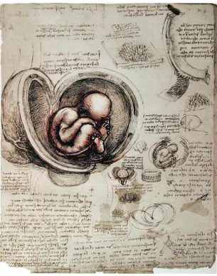
Fig. 1. The human fetus as depicted by Leonardo da Vinci, circa 1510. Shown is the gravid uterus with its blood vessels, fetus in the breech position, fetal membranes, and umbilical cord with vessels. Although Leonardo depicted a single placental disc beneath the fetus, he mistakingly showed the placental attachment as being cotyledonary, interdigitating with maternal crypts as in a ruminant (upper right portion of drawing). (See Text for details).
portions of the history of which have been reviewed by others (Boyd & Hamilton, 1970; Corner, 1963; DeWitt, 1959; Steven, 1975). In addition to the specific citations, we point to a number of reviews on various aspects of the subject, to which the reader can refer.
Pre-Eighteenth Century and the interrelation of
mater-nal and fetal placental blood vessels
Since earliest times, the placenta has been recognized as being of great importance, and at the same time quite mysterious – even somewhat mystical. For example, in many cultures the placenta has been held as an alter ego [my second self], a symbol for the preservation of health and good fortune, and as a talisman in case of danger. In some societies, a sympathetic animism exists between the placenta and the future adult (Ploss & Bartels,
Western Nigeria, buries the placenta near the en-trance to the home, so that the child “will always look back to its father” (Longo, 1964).
principal organs], the brain, heart, and liver; a third stage in which other structures developed; and finally further growth and matu-ration of the embryo/fetus (Needham, 1934). In the goat, Galen also described fetal movements, including those that followed compression or ligation of the umbilical cord (Duckworth, 1962). If there was a single dispute that captured the core of placental developmental biology from the early sixteenth through the eigh-teenth centuries, it was the debate over the degree to which the maternal and fetal placental blood vessels were interconnected. Another major controversy in embryology during this era was that of preformation; i.e., that all parts of the embryo and future organism exist completely formed in the germ cell (in the case of a male, the homunculus [diminutive of man] in the sperm, as so clearly illustrated by Nicolas Hartsoeker (1656-1725) in his Essay de dioptrique, 1694; Gall, 1966, pp 24-25), and develop only by increasing in size. This contrasts with the concept of epigenesis; i.e., that an individual develops by structural elaboration from an unstructured egg, rather than by simple enlargement of a pre-formed entity. This latter area of disagreement is far beyond the scope of the present essay (for an extended discussion, see Needham, 1934).
The study of anatomy experienced an awakening during the Renaissance, the humanistic revival and rebirth of classical art, literature, and learning, with its spirit of observation and inquiry, that originated in 14th-century Italy and spread throughout Eu-rope. Leonardo da Vinci (1492-1519), who combined science with aesthetics in his anatomical studies, prepared magnificent illus-trations of the human body for a volume that never was published (these art works now reside in the Royal Library of Windsor Castle, UK). Included among these illustrations is a human fetus in the breech position within the uterus of an ungulate. As shown in Fig. 1, Leonardo depicted the uterus with its blood vessels, as well as the fetal membranes and umbilical cord with its vessels. Although he also showed a single placental disc (upon which the fetus is sitting; the coiling umbilical cord can be seen just under the placental disc), he mistakenly depicted the placental attachment as being cotyledonary, as in an ox, with the fetal cotyledons interdigitating with the maternal crypts. Presciently, Leonardo observed, “Just as the fingers of the hand are interwoven, one in the interval of the other … so the fleshy villi of these little sponges [cotyledons] are interwoven like burrs, one half with the other.” He also noted, “the vessels of the infant do not ramify in the substance of the uterus of its mother, but in the secundines [placenta], which take the place of a shirt in the interior of the uterus which it coats,
and which it is connected (but not united) by means of the cotyledons …” (da Vinci, 1911-1916; O’Malley & Saunders, 1952, pp 472-476). As noted by Joseph Needham (1900-1995), da Vinci stated correctly that the fetal vasculature is not continuous with that of the mother (Needham, 1934, p 78).
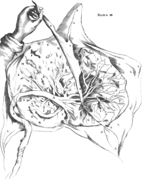
In his monumental work on the “Fabric of the human body…,” one of the most seminal volumes in the development of modern medicine and science, Andreas Vesalius (1514-1564) described the human uterus consisting of a single chamber, rather than having two horns as in many mammals, and its rich vasculature. Vesalius upheld the Galenic doctrine of anastomosis between maternal and fetal vessels. As illustrated in Fig. 2, he mistakingly illustrated the placenta as being zonary or girdle-like, as in the dog (Vesalius, 1543), but corrected this in the second edition (Fig. 3) (Vesalius, 1555). In his compendium on anatomy, Matteo Renaldo Colombo [Columbus] (circa 1510-1559), a pupil of Vesalius and his successor as professor at the University of Padua, first used the term placenta (Colombo, 1559). Several years later, Giulio Cesare Aranzi [Arantius] (1530-1589), of Bologna, in his work on the human fetus, described his dissection of the gravid uterus of several species, including ruminants, sows, and dogs. Here, he contradicted Galen’s claim of major vessels directly connecting mother and child, maintaining that the maternal and fetal vascu-lature were separate, an issue only settled two centuries later. Aranzi also demonstrated that the human placenta was not cotyledonary, and conceived of the organ as a hepar uterinum [liver of the uterus] functioning to purify blood for fetal nutrition. Nonetheless, in the Galenic tradition, Aranzi erred in believing that “vital spirits” supplied by the uterine arteries mingled with blood brought by the uterine veins to supply the “purified” blood to the fetus (Aranzi, 1564). Several decades later, André Du Laurens [Laurentius] (1558-1609) reaffirmed the Galenic view that the placental vessels of the fetus connect directly to those of the mother (Du Laurens, 1598).
The distinguished anatomist Girolamo Fabrizio [Fabricius ab Aquapendente] (1533-1619), of Padua, who wrote at length on embryology, attacked Aranzi for questioning the Galenic doctrine
Fig. 2. The human placenta as depicted by Andreas Vesalius in De humani corporis fabrica (1543). The original edition of 1543 shows a canine zonary placenta.
regarding the confluent fetal and maternal vascular channels. Because Fabricius mistook the cotyledon crypts of the sheep placenta as the opening of blood vessels, which match equivalent openings in the uterine caruncles, he concluded that there must be vascular continuity, so that the fetal vessels were “plugged into” those of the mother. Pointing out this mistake, however, should not lead the reader to discount his many contributions to comparative embryology and placentology (Fabrizio, 1604; 1621). An additional notable work of this period was De formato foetu by Adriaan van der Spieghel [Spigelius] (1578-1625), also of Padua, which included plates of the gravid uterus prepared some years previously for Giulio Casseri [Casserius] (circa 1561-1616) by an unknown artist (Spieghel, 1626). Spigelius held that an important function of the placenta was to prevent more than minimal blood loss at birth, as would be the case if the two circulations were joined by a single large vessel. He rejected the teaching of Aranzi, however, maintaining that the maternal and fetal vessels were united within the fleshy substance of the placenta, so that blood flows directly from the uterine vessels into the ends of the umbilical vein (Spieghel, 1626).
It was the celebrated William Harvey (1578-1657) who, in unraveling the mystery of the circulation of the blood and the function of the heart, and being the first to apply the scientific method to solve a biological problem (Harvey, 1628), in his De generatione animalium…, considered the circulation of the fetus and its relation to that of the mother (Harvey, 1651). To help grasp the significance of Harvey’s monumental achievement, one must
consider the traditional Galenic concept of the heart, blood vessels, and their contents that was the dogma at this time. Briefly, this view held that blood originated in the liver, which was the dynamic organ that provided vascular pulsations. The heart, in contrast, was believed to be a fibrous sac that dilated or collapsed passively as a consequence of the motion of the blood. In the liver, venous blood was mixed with an imaginary essence, the “natural spirits” to become a vegetative fluid, the elixir of nourishment, that was distributed to the several organs of the body. Venous blood within the right side of the heart was believed to seep through “pores” in the septum dividing the two sides of the heart, to mix on the left side with air to produce arterial blood. (Also at this time, most workers believed that the heart consisted of only two chambers, the ventricles, and that the atria were but exten-sions of the veins joining the heart). These fluids were imbued with a second essence, “vital spirits” or “pneuma” that entered the lungs with respiration, and passed via the pulmonary vein to the left ventricle, before being distributed to various organs. In the brain, “animal spirits” were added to pass through the nerves. Interaction of the “vital” and “animal” spirits was believed to provide movement, such as muscular activity. Rather than circu-lating, the two varieties of blood surged back and forth in the veins and arteries, as the tide
In regards to the placental circulation, Harvey stated, “the Extremities of the Umbilical vessels, are no way conjoined to the Uterine vessels by an Anastomosis; nor do extract blood from them…” (1653, p 439). By logic based on his knowledge of the circulation, he held the maternal and fetal circulations to be separate, each following in an opposite direction to the placenta by way of the arteries and returning by the veins. He also noted that the “Embryo is no other manner sustained in the Uterus, then [sic] the chicken in the Egge” (1653, p 440). Based on knowledge that in utero the fetus can grow and mature in the absence of air, however, at delivery it dies if the umbilical cord is compressed and it does not breathe, Harvey postulated that substances absorbed by the umbilical cord stimulated organ development, while the fetus received its main nourishment from the amniotic fluid. Following Aranzi, he also referred to the placenta as the hepar uterinum [uterine liver] and mamma uterina [uterine breasts]. It was not until a decade later that Marcello Malpighi (1628-1694) first described the capillary bed connecting arteries and veins (Malpighi, 1661), and made possible an understanding of the anatomical basis of regional circulation. Lacking this knowledge, Harvey could not understand completely certain details of the circulatory system in either the adult or the fetus. Jean Claude De La Courveé (1615-1664), a contemporary of Harvey’s and one of the first adherents to Harvey’s De generatione… (1651), gave considerable insight into the role of the placenta in fetal nutrition. Although supporting Harvey’s views on embryogenesis, he held that the fetus breathes in utero, and is nourished by the amniotic fluid. He also believed the placenta to be loosely connected to the uterine wall, rather than being tightly united with it. For reference, contemporary seventeenth and eighteenth century views on the source of fetal nutrition included the menstrual blood, amniotic fluid that the fetus either drank or passed through pores in its skin, drinking uterine milk, or via the blood of the umbilical cord (La Courveé, 1655; see Needham, 1934, pp 158-160).
In this work Harvey raised related question of fundamental importance to developmental biology and life in utero, that is of the
vessels that radiate into the substance of the chorion, where they form protuberances called placentulae that approximate uterine wall structures called glandulae or cotyledons (caruncles of present day terminology). Hoboken delineated the chorion as completely surrounding the outer membranes, not being pen-etrated by maternal vessels. Incorrectly, he held that the chorion and amnion were without vasculature. Figure 4 depicts his illus-tration of a human placenta. As shown in Fig. 5, Hoboken gave the original description of the intraluminal circular “valves” within the umbilical arteries and the semilunar folds or projections within the vein (Hoboken, 1669; 1675). Walter Needham (1631?-1691?), a Cambridge medical graduate who studied anatomy at Oxford and was the first to report chemical experiments on the develop-ing mammalian embryo, maintained that the uterine arteries must supply nutrients to the uterus and developing fetus. He also refuted the idea that “uterine milk” is identical with lymph. In Chapter Two of his Disquisitio anatomica de formato foetu [Dis-quisition on the anatomy of the formation of the fetus, 1667], Needham considered the anatomy of the placenta, its compara-tive anatomy among species, and its function. By careful dissec-tion, he demonstrated the chorion frondosum [leafy membrane] and chorion leave [disappearing membrane], and confirmed that the placenta consists of separate maternal and fetal portions. Importantly, he supported Harvey’s doctrine regarding the indi-vidual maternal and fetal placental circulations. In his De mulierum organis generationis… [The organs of a woman that serve gen-eration, 1672], which primarily was devoted to the female repro-ductive organs, Regner de Graaf (1641-1673) included several illustrations of the placenta, but did not consider the organ in detail.
At about this time, John Mayow (1643-1679), of Oxford, who first established the origin of “animal heat” in the skeletal muscles, identified a fraction of the air, nitro-aërial vapor or particles (later identified as oxygen), as being essential to both the burning of a candle and respiration in animals. In his De respiratione foetus in utero et ovo, Mayow asked, since this element is so essential for sustaining life, “… how it happens that the foetus can live though imprisoned in the straits of the womb and completely destitute of air.” After rejecting theories of his predecessors, Mayow affirmed, “we maintain that the blood of the embryo, conveyed by the umbilical arteries to the placenta or uterine carunculae, brings not only nutritious juice, but along with this a portion of nitro-aërial particles to the foetus for its support, so that it seems that the blood of the infant is impregnated with nitro-aërial particles by its circulation through the umbilical vessels quite in the same way as in the pulmonary vessels. And therefore I think that the placenta should no longer be called a uterine liver but rather a uterine lung” (Mayow, 1674). The Anglican divine and Cambridge Don, John Ray (1627-1705) in his The wisdom of God manifested in the works of the creation… expressed similar thoughts in respect to the placenta providing the fetus with air for the feeding of “that vital flame” (Ray, 1691). A century later, Joseph Priestley (1733-1804) isolated oxygen and showed it to be essential for animal life (Priestley, 1772), and Antoine Laurent Lavoisier (1743-1794) demonstrated the interchange of oxygen and carbon dioxide in the lungs and recognized the role of oxygen in respiration and supporting combustion (Lavoisier, 1778). Shortly thereafter, in his Zoonomia… Erasmus Darwin (1731-1802), grandfather of Charles Robert Darwin (1809-1882), wrote regarding respiration in the placenta serving as the lung for the fetus.
How does it happen that the foetus continues in its mother’s womb after the seventh month? Seeing that when expelled after this epoch, not only does it breathe, but without respiration cannot survive one small hour; whilst, as I have before stated, if it remains in utero, it lives in health and vigour more than two months longer without the aid of respiration at all. (Harvey, 1651, p 263).
When it was raised, Harvey’s question, as it came to be known, aroused the interest of both philosophers and experimentalists. However, with no clear understanding of respiration or metabo-lism, and without knowing of the existence of oxygen (the discov-ery of which did not occur until the following century), Harvey could only speculate on this question. Many details of the path of this discovery two centuries later by a young Swiss obstetrician, Paul Zweifel (1848-1927; Zweifel, 1876) have been given by Donald Henry Barron (1905-1993; Barron, 1978).
In his treatise on the anatomy of the placenta and fetal membranes in humans and several mammals, Nicolaas Hoboken (1632-1678) stated that in the cow, during the early months of gestation, the embryo was nourished by gelatinous juices se-creted by glands lining the uterus, and these were absorbed by pores in the chorionic membrane. He noted that, later, the umbilical vessels from the fetus divide into 60 to 80 smaller
placenta:
It appears that the basis of atmospheric air, called oxygene, is received by the blood through the membranes of the lungs; and that by this addition the colour of the blood is changed from a dark to a light red.… the placenta consists of arteries carrying the blood to its extremities, and a vein bringing it back, resembling exactly in structure the lungs and gills … and that blood changes its colour from a dark to light red in passing through these vessels. (Darwin, 1794-1796, vol.1, p 476).
These “enlightened views” of Erasmus Darwin on placental res-piration have been reviewed recently (Pijnenbrog & Vercruysse, 2007).
Eighteenth and early Nineteenth Centuries, and more
on the placental circulations
Although in the early eighteenth century the continuity of the maternal and fetal circulations was still held to be true (Cheselden, 1713), in his 1734 essay on fetal nutrition Alexander Monro primus (1697-1767), professor of anatomy at the newly estab-lished University of Edinburgh, stated clearly that there was no vascular continuity between the uterus and placenta. After his examination of the bodies of five women who had died while pregnant, he observed that in each he had found a thick, fungous, succulent, cellular substance between the muscular part of the womb and its villous coat, through which numerous thin-coated vessels passed, and in which the sinuses lay. Excepting its sinuses, this resembled the internal coat of the intestines, and was similar to tissue seen in the glandulae of the cow. Monro observed that he was ignorant of this structure when he dissected the first woman, and, therefore, when he had cut through the myometrium and saw this substance, he imagined it to be the placenta. He stated that he was surprised to find the cohesion of this supposed placenta to the womb so firm, but persisted in attempting to separate it from the uterine wall until, having torn some of this substance, he observed the smooth, tense chorion, from which the fungous substance separated most easily, and it did likewise from the placenta by gently pressing the ovum with one hand, and raising the womb with the other. Monro states that in dissecting the impregnated uteri from the other four women, which he examined afterwards, he avoided this mistake (Monro, 1734). Monro concluded that the tips of the fetal vessels extend beyond the base of the placenta passing through the uterine decidua to tap into the maternal blood vessels (Monro, 1734). Several years later, Wilhelm Noortwyck (ca. 1712-1778) of Leiden, and a pupil of the anatomist Bernhard Siegfried Albinus (1697-1770), who created impressive preparations of injected organs (Albinus, 1737-1747), apparently was the first to inject the uterine vessels of a young woman who had died near term. In this preparation, he believed, incorrectly, that some of the vessels were fetal in origin, and joined with those of the uterus (Noortwyck, 1743).
In his magnificent obstetric atlas, The Gravid Uterus, the anatomist-obstetrician William Hunter (1718-1783) gave “a so-phisticated description” (DeWitt, 1959, p 367) of the human placenta and associated membranes (Hunter, 1774). In magnifi-cent folio-sized, copper-plate engravings, the illustrations of which were executed by the Dutch artist Jan van Rymsdyk (fl.
1750-1788) over a twenty year period, he illustrated a number of aspects of placental and fetal development. Hunter, a Scotsman, had been a pupil of Alexander Munro primus at Edinburgh, and no doubt had read the latter’s 1734 essay. In Plate XXIV of The Gravid Uterus, Hunter illustrates the opened uterus of a woman at six months of gestation. The uterine arteries and veins had been injected with red and blue wax, respectively, and these with the umbilical vessels on the fetal placental surface, which contained no wax, are observed. Here and elsewhere, Hunter noted the “convoluted” (i.e., spiral) arteries of the uterine vasculature, and the separate circulation of the mother from that of the fetus (Hunter, 1774) (see Figs. 6,7). In his full Anatomic Description, published a decade after his death by his nephew Mathew Baillie (1761-1823), Hunter described the placenta as a blending of two components – a continuation of the fetal umbilical vessels and the uterine (decidual) aspect. He described in some detail his experi-ments of filling the uterine arteries and veins with liquid wax of different colors (Hunter, 1794, p 47), and also of injecting the umbilical vessels (Hunter, 1794, p 48), and concluded that the circulations were quite separate. He summarized his experience:
From all these experiments and observations which have been often repeated and diligently attended to, with no other desire than to discover truth, it seems incontestable that the human placenta, … is composed of two distinct parts, though blended together, viz, an umbilical, which may be considered as part of the foetus, and an uterine, which belongs to the mother; that each of these parts has its peculiar system of arteries and veins, and its peculiar circulation, receiving blood by its arteries, and returning it by its veins; that the circulation through these two parts of the
placenta differs in the following manner: in the umbilical portion the arteries terminate in the veins by a continuity of canal, whereas in the uterine portion there are intermediate cells into which the arteries terminate, and from which the veins begin. Though the placenta be completely filled with any injection thrown into the uterine vessels, none of the wax finds it way into any of the umbilical vessels; and in the same manner fluids injected into the umbilical vessels never can be pushed into the uterine, except by rupture or transu-dation. (Hunter, 1794, p 48).
In his account, William Hunter noted the occasional absence of one umbilical artery (Hunter, 1794, p 33), a condition now appre-ciated to be assoappre-ciated frequently with fetal malformation (Benirschke & Brown, 1955; Benirschke & Dodds, 1967; Faierman, 1960). An alternative view on resolving the problem of the sepa-rate fetal and maternal placental circulations has credited John Hunter (1728-1793) with that discovery. According to his account, in 1754 Colin Mackenzie (d. 1775), an assistant to the London obstetrician/anatomist William Smellie (1697-1763), had made an unusually careful dissection of the gravid uterus after injecting melted wax into the uterine arteries and veins of a pregnant woman who had died; this apparently in an attempt to confirm or deny the report of Noortwyck. John Hunter recorded that he was asked by Mackenzie to examine the dissection, and he wrote, “The facts being now ascertained and universally acknowledged, I consider myself as having a just claim to the discovery of the structure of the placenta, and to its communication with the uterus, together with the use arising from such structure and communication, and of having first demonstrated the vascularity of the spongy chorion” (J. Hunter, 1780). As a consequence, the brothers William and John disagreed on priority of this discovery, they went their separate ways, and it was not until near William’s death in 1783 that they were reconciled (Simmons, 1783).
Despite the convincing demonstrations of the Hunter brothers and Mackenzie, however, not all agreed with the idea of the complete separateness of the maternal and fetal placental circu-lations. In the latter part of the eighteenth century, Ebenezer Sibly (1751-1880), a physician who was entranced with astrology and the occult, mistakingly described the placenta as allowing vascu-lar communication between the mother and fetus, with the uterine arteries discharging “their blood into the branches of the umbilical vein. So that, after quickening, the blood of the mother is con-stantly passing in at one side of the placenta, and out again at the other, for the nourishment of the child” (Sibly, 1794, p 116). Perhaps surprisingly, as late as the mid-nineteenth century, doubts remained concerning the maternal and fetal circulations in the placenta. For instance, John Reid (1809-1849), anatomist of Edinburgh, wrote that in the half century since the Hunters’ publications, the separateness of maternal and fetal circulations had been questioned by eminent investigators, “…but there cannot be a doubt that this opinion is erroneous, and ought now to be totally abandoned” (Reid, 1841, p 2). In his Anatomical letters and memoir…, Filippo Civinini (1805-1844), of Pisa and Pistoia, confirmed that there is no direct connection between the placental circulations of the mother and the fetus. He presented autopsy reports and accounts of experiments carried out on dissected human and animal cadavers. Civinini established his assertion by injecting colored liquids into the placenta using an apparatus he devised for this purpose (Civinini, 1859). At this
time, as might be expected, controversy continued concerning the placenta and its relation to the circulation of blood in the fetus. For instance, in his textbook of midwifery of 1833, William Campbell (1788-1848), of Edinburgh, described in “Peculiarities of the Foetus at Birth,” the fetal circulation including that of the placenta. He observed, “… It is still a disputed point whether the pulmonary arteries convey blood to the lungs” (Campbell, 1833, p 128). He continued presenting arguments for some admixture of blood from the fetal venous circulation with that arterialized blood returning from the placenta via the umbilical vein. Although writing some sixty years after the discovery of oxygen as a gas, it was four years later that Heinrich Gustav Magnus (1802-1870) first estab-lished by quantitative analysis that arterial blood contains more oxygen than that in veins (Magnus, 1837). In this regard, Campbell noted, “Though denied by the most celebrated physiologists of the day, that any difference exists between the blood in the arteries and that contained in the veins, yet I must still … agree … that in colour at least, they are dissimilar” (Campbell, 1833, p 129).
In addition to his many other contributions, William Hunter presented the first definitive description of the decidua [from the Latin deciduus, falling off], the uterine endometrial lining that, except for its deepest layer, is shed at parturition. He illustrated this in Plates XXVIII, XXIX, and XXV of The Gravid Uterus,
distinguishing between a parietal lining membrana decidua vera [true decidua] from the capsular membrana decidua reflexa [decidua that is turned back] (Hunter, 1774). William’s brother John, a surgeon and comparative anatomist of note, had ob-served the decidua lining the uterus in cases of tubal (ectopic) pregnancy, and in the non-pregnant horn of animals with bicor-nate uteri in cases of unilateral pregnancy.
From this time forward, knowledge of the development of the placenta, including the villi, preceded pari passu with an under-standing of embryology. Essential to this development of ideas was the description of the ovum by Regner de Graaf (1641-1673) (Graaf, 1672), the fallopian tubes by Gabriele Falloppio (1523-1562) (Falloppio, 1561), and identification of the mammalian ovum by Carl Ernst von Baer (1792-1876) (Baer, 1827). In concert with these discoveries were advances in microscopy, and in the understanding of the implantation of the blastocyst. This is not the place to review in detail the technical aspects of microscopy and its development; however, after the studies of Robert Hooke (1635-1703) (Hooke, 1665) and Antonj van Leeuwenhoek (1632-1723) (Leeuwenhoek, 1693-1718), several individuals contrib-uted to the development of the achromatic compound micro-scope, which made possible more sophisticated studies of ana-tomic structure (Amici, 1818; Deijl, 1807; Goring, 1827; Lister, 1830; and others).
In the early nineteenth century, two theories claimed attention regarding the decidua and the implantation of the blastocyst. The first was the Einstülpung [inversion] theory of Karl Friedrich Burdach (1776-1847) (Burdach, 1814), in which the decidua was believed to cover the uterine orifices of the fallopian tubes, so that, without entering the uterine cavity, the ovum could mechanically insinuate itself between the layers of the decidua. An alternative view, that of Einssat [propulsion or a sowing], conceived by William Hunter and supported by Gabriel Gustav Valentin (1810-1883), held that the blastocyst “pushed aside” the preimplantation decidua to establish itself (Valentin, 1835). This concept is similar to the “histolytic” mechanism, with overgrowth of the decidua and imbedding of the ovum held by contemporary embryologists (Sadler, 1985).
It was only over a period of several decades that the morphol-ogy and vascular structure of chorionic villi came to be appreci-ated, as their structure could not be described properly until, as noted above, microscope optics improved. In his Gravid Uterus, William Hunter recorded the findings in several abortion speci-mens, describing, “The larger and more crowded branches of the shaggy vessels which shoot from the external surface of one part of the chorion, to mix with the decidua, or uterine part, to form the placenta” (Hunter, 1774, Plate XXXIII). In his Handbuch der Anatomie des Menschen of 1832, Ernst Heinrich Weber (1795-1878) first illustrated the fine branching of fetal vessels enclosed within teased out villi (Weber, 1832), thus negating the view held at that time that fetal vessels dipped freely into maternal blood. Weber’s illustrations later were published by Rudolph Wagner (1805-1864) in his Textbook of Physiology (1842). The villous structure soon was confirmed by others (Eschricht, 1837). Soon thereafter, the villi were described in considerable detail by John Dalrymple (1804-1854) of London, who, observing their epithelial character, stated, “the membrane enclosing the vessels and capillaries is studded on the exterior by nucleated cells, resem-bling an irregular epithelium” (Dalrymple, 1842, p 23-24). Dalrymple
noted further:
A single tuft or collection of villi, well injected, and laid flat under an … object glass, appears at first sight an inextri-cable confusion of curiously-contorted capillary vessels; but separated by needles, and a single villus detached, or expanded beneath a high magnifying power, this seeming confusion is reduced to order, and the true anatomy of these vessels explained…. The enclosed tufts, or capillaries, nowhere anastomose with other than foetal or umbilical vessels…. The villi are not connected together by cellular tissue, but the mass of the placenta is made up by vascular divisions and subdivisions, and by the tufts or bouquets of capillaries; the interstices are everywhere free, and commu-nicate with each other.… If the maternal blood is extrava-sated into the spongy mass of the placenta by the “curling arteries”, as supposed by John Hunter, or enters it in any other fashion, then the feotal tufts become, in function, absorbent villi; they take up the necessary or nutrient part of that fluid, which is then carried to the foetus by the umbilical vein … Hence, while the blood, or nutrient material of the blood, brought by the uterine arteries, and previously aërated by the mother, enters by endosmose [that is, endosmosis] the absorbent capillaries of the foetal villi, that portion of the foetal blood that requires the action of oxygen escapes by exosmose [that is, exosmosis] and returns by the uterine sinuses and veins to the maternal heart. Thus the lungs of the mother are in fact the lungs of the foetus… (Dalrymple, 1842, p 23-27).
At this time, the phenomena of gaseous diffusion, and that of fluid transfer by osmosis and ultrafiltration across semipermeable membranes (the latter terms called exosmosis and endosmosis, respectively, Dutrochet, 1827-1835) in physiologic systems, were understood (Dutrochet, 1824; 1827-1835; Magendie, 1816-1817). Thus, the question arises why Dalrymple believed it necessary to postulate that a substance must diffuse from the fetal blood into that of the mother to be “aerated” in her lungs and then return to the fetus, rather than being transferred directly from the mother’s blood to the fetus? Dalrymple may not have been aware of these studies; however, we simply do not know why he postulated this alternative mechanism.
Twins and placentation
Since the earliest times, the birth of multiple infants has inspired awe and admiration. In many cultures, twins were be-lieved to be the offspring of the gods. Examples include in ancient Egypt Isis and Nephthys, in Greek mythology Castor and Polydeuces [Latin, Pollux], and in Rome Remus and Romulus (Stevenson, 1941). As is well known, fetal complications of twinning include: intrauterine growth discordance, growth restric-tion, congenital anomalies, twin to twin transfusion syndrome, and twin emobolization syndrome. In the Hebrew Torah, Esau and Jacob may be examples of the twin transfusion syndrome, as Esau was ruddy and his whole body like a “hairy garment” (Genesis 25:24-26).
sex could not occupy the intrauterine cavity together because of horror incestus [dread or terror of defilement/unchaste]. In 1670 however, Joachim Georg Elsner (1642-1676) illustrated a case of male and female twins that helped to dispel this idea, and showed the separate placentas (Elsner, 1670). A dispute that continued for several centuries, however, was whether the placenta of twins was single or double, with most writers favoring the latter view. We now know that although approximately two-thirds of twin placen-tas are monochorionic/diamniotic and about one-third dichori-onic/diamniotic, a small percentage are monochorionic/ monoamniotic. As noted above, in his De re anatomica of 1559, Realdo Colombo [Columbus] coined the term placenta. Colombo also described the septum on its fetal surface that in many cases of twins separated the fetal sacs, holding that this prevented their growing together to form a double-bodied monster (Colombo, 1559; Moes & O’Malley, 1960). Two centuries later, Johann Ernst Hebenstreit (1703-1757) confirmed this observation, and main-tained that separate fetal compartments prevented both umbilical cord entanglement as well as the simultaneous birth of both infants (Hebenstreit, 1737).
Importantly, in the late-seventeenth century, it was established that vascular communications could exist between the placentas of twins. For instance, in cases of twin delivery Paul Portal (1630-1703) cautioned his pupils to knot the severed umbilical cord of the first twin that delivered, to prevent exanguination of the second twin (Portal, 1685). Soon thereafter Cornelis Stalpart van der Wiel (1620-1702) described a vascular anastomosis, “a third umbilical cord,” between twin placentae (Stalpart van der Wiel, 1687). In the mid-eighteenth century, in his Treatise… on Mid-wifery William Smellie wrote:
When two or more children are included in the Uterus, at the same time, each has a separate Placenta, … sometimes these Placent [sic] are altogether distinct, and at other times they form but one cake. Yet, by an instance that lately fell under my observation, it appears that sometimes twins have but one Placenta in common: whether or not there were two sets of membranes, I could not discover, because they had been tore off by the gentleman who delivered the woman; but, when the artery in one of the navel-strings was injected, the matter flowed out at one of the vessels belonging to the other; and the communication between them is still visible, though they are separated at the distance of three or four inches. (Smellie, 1752, p 122).
Other accoucheurs recorded similar vascular communications in the placentae of twins (Levert, 1766; Spence, 1784). Following his observation of Samuel Thomas Soemmering (1755-1830) injecting the placental vessels of twins to demonstrate their anastomosis, Friedrich Benjamin Osiander (1757-1822) recorded his surprise that the second twin did not become exanguinated (Osiander, 1787). In the mid-nineteenth century, Carl Christoph Hueter (1803-1857) reviewed the literature that described such anastomosis to that date (Hueter, 1845). Despite several reports of second twins that bled to death because the umbilical cord of the first twin was not ligated (Brachet, 1824; Spaeth, 1860; 1861-1862), until the mid-nineteenth century a number of writers denied twin-to-twin vascular anastomosis (see Hueter, 1845; Strong & Corney, 1967).
In addition, several authors described cases in which, after the delivery of a healthy child, the second twin was a “monster”. For
instance, John Clarke (1761-1815), an early writer on the dis-eases of children (Clarke, 1815), described such a case:
This monster was voided after the delivery of a healthy child. It was enclosed in a distinct bag of membranes, composed of decidua, chorion and amnios; and had a placenta belong-ing to it, the side of which was attached to the placenta of the healthy child…. Before the internal structure was examined, the navel-string of the perfect foetus was injected, from whence the injection readily passed through both placen-tae, viz that of itself and that of the monster; and then to the substance of the monster also, as appeared by the redness of the skin. (Clarke, 1793).
Sir Astley Paston Cooper (1768-1841), a noted London surgeon, described a similar case reported to him (Hodgkin & Cooper, 1836). In 1860, Joseph Späth (1823-1896), of Vienna, reviewed the placental findings in 185 sets of twins that occurred in 14,880 births (an incidence in one per 80). Of these, 30 (24 percent) of the placentas and membranes were monochorionic, two were monoamniotic, and by injection of wax or fluid he demonstrated vascular anastomosis was present in 17 (60 percent) of the 28 diamniotic placenta, but not in the 46 fused dichorionic placentae. Späth cautioned, however, that, because in his initial studies he experienced difficulty with the injections, the true incidence of anastomosis in monochorionic placentae probably exceeded 60 percent (Späth, 1860; 1861-1862).
The Viennese anatomist Joseph Hyrtl (1810-1894) also pub-lished a major work on the blood vessels of the placenta, with their many variations. In addition to vascular anastomosis on the fetal surface of the placenta, he described those that were deeply embedded in the placental parenchyma. In this work, he empha-sized the requirement for extreme care in performing such injec-tions to avoid being misled by incorrect findings (Hyrtl, 1870). Friedrich Schatz (1841-1920), an obstetrician-gynecologist of Rostock, also wrote extensively on the placenta of twins, and great variations in the volume of amniotic fluid of the two fetuses (Schatz, 1887; 1900).
Latter Nineteenth and Early Twentieth Centuries. The
intervillous space, the structure of villi and
classifica-tion of placentae
1859, p 719). Other investigators wrote of maternal blood filling this space (Bumm, 1890; Leopold, 1877; Turner, 1872; 1876a; Wagner, 1851-1859; Waldeyer 1887; 1890). However, despite these demonstrations, many could not accept the concept of maternal blood flowing through an open cavity without discrete surrounding walls. In part, this was because nothing like this existed in the placentas of other mammals that had been studied. (Soon thereafter, it was demonstrated that such a vascular arrangement exists in old world primates; Turner, 1872; 1876a; 1876b; 1877). Although many workers accepted the presence of maternal blood in this space, a common view was that, rather than being in direct contact, a layer of maternal endothelial membrane separated that blood from the fetal villi (see Boyd & Hamilton, 1970; Corner, 1963; Pijnenborg & Vercruysse, 2008). As may be appreciated, firm evidence of this circulation of maternal blood through the intervillous space awaited both more advanced microscopy and the cineradiographic studies a century later.
It should be noted that even at this time physiologic functions of the placenta were poorly understood. For instance, in summa-rizing contemporary knowledge, William (later Sir William) Turner (1832-1916) of the University of Edinburgh wrote:
The foetal placenta possesses an absorbing surface; the maternal placenta a secreting surface. The foetus is a parasite, which is nourished by the juices of the mother…. As there are, therefore, two sets of secreting structures in the gravid maternal mucosa, the Glands and the Crypts… it may be a matter of consideration how far the secreting organs perform similar or different functions in foetal nutri-tion…. The current doctrine that the nutrition of the foetus is provided for by the simple percolation or diffusion of mate-rials through the walls of the vessels from the maternal blood to the foetal blood can no longer be accepted. (Turner, 1876, p 114ff).
Despite this confusing view of placental function, Sir William Turner made important contributions to an understanding of placental structure and its comparative anatomy (Haig, 1997; Magee, 2003; Turner, 1872; 1876a; 1876b). Another issue of disagreement at this time was the nature of the outermost cells of the placental villi in the human. Theodor Langhans (1839-1915), when serving as professor of pathology at the University of Berne, correctly identified the villous covering of chorion frondosum [chorion villosum] and chorion laeve [chorion avillosum] as form-ing a continuous layer from the early stages of development, and as being of fetal origin (Langhans, 1870). Several years later, Langhans demonstrated that this membrane comprised two lay-ers, the outermost Chorionepithel [chorionic epithelium] of con-tinuous cells, and underlying Zellschicht [cell layer] of large, individual, epithelial cells, with cellular boundaries/membranes. We now know these as the syncytiotrophoblast and cytotropho-blast, or “Langhans” layer, respectively (Langhans, 1877; 1882). Langhans noted that the epithelium covering the outer surface of the chorion and villi forms a uniform layer of rather homogenous cytoplasm containing nuclei, but without a clear division into individual cells. Particularly in younger ova, he observed lines that might represent boundaries of cells of highly variable form, with large, often multiple finely granular protoplasm, and nuclei, the diameter of which were several times that of those in the overlying chorionic epithelium (Langhans, 1877).
A related contribution was that of Ambrosius Arnold Willem
Hubrecht (1853-1915), professor of zoology at the University of Utrecht, who, in contributing to an understanding of the process of implantation, introduced the term trophoblast [nutrition + germ] to indicate that portion of the blastocyst not contributing to formation of the embryo per se but rather forming the placental villi for the nourishment of the embryo (Hubrecht, 1888; 1889). Hubrecht appreciated the double cellular layer, with the outer layer of nuclei in “nests” not demonstrating mitosis, and the innermost layer of cylindrical cells. Although in his original de-scription, trophoblast had limited morphologic significance, be-fore long it came to apply to the epithelial derivatives of the outer layer of the blastocyst, the two cell layers described by Langhans. Later it became recognized that the outermost layer was a syncytium of cells, the syncytiotrophoblast [cells together + tro-phoblast], and the innermost Langhans layer, the cytotrophoblast [cellular + trophoblast] (Bonnet, 1903; Boyd & Hamilton, 1966). In his detailed studies of placental comparative anatomy, morphol-ogy, and histolmorphol-ogy, Mathias Marie Duval (1844-1907) described the invasion by trophoblast cells into the maternal spiral arteries (Duval, 1892, p 640), confirming limited evidence provided by Carl Friedlander (1847-1887), two decades earlier, of arterioles, in addition to more definitive documentation of “uterine sinuses,” being filled with these cells (Friedlander, 1870; see Pijnenborg & Vercruysse, 2008).
Further technical and conceptual contributions to understand-ing placental morphology were those of Charles Sedgwick Minot (1852-1914), who in 1886 invented the automatic rotary micro-tome for cutting ultra-thin tissue sections. Minot suggested the term trophoderm to describe the mature placental cells; however, this never became widely accepted. Nonetheless, he presented a definitive account of the microscopic structure of the human placenta, in one instance describing a section through the uterus and placenta in situ at seven months gestation, with amnion, chorion, villus trunk, sections of villi in the substance of the placenta, decidua, muscularis, uterine blood-vessels opening into the placenta, and so forth (Minot, 1889; 1891). In a later review, Minot noted that the chorion is separated by a dense forest of villi from the decidua, that the termini of some of the villi touch and are imbedded in the decidual tissue, and that the decidua is divided into two strata. He described the section passing through a wide tube, a vein containing blood, that opened into the interior of the placenta (Minot, 1900-1904). These contri-butions did much to help clarify the nature of the fetal-maternal (i.e., placental) barrier. In his Age, Growth and Death Minot stated the “law of cytomorphosis” (which we now term morphogenesis) by which developmental processes result in a systematic change in protoplasm from more elemental to highly differentiated cells and tissues (Minot, 1908).
(1857-1920), of Giessen, who categorized placenta as those Vollplacenten [complete placentae], in which at birth the maternal blood space is opened and part of the decidua sloughs off, and Halbplacenten [semi placentae], in which the cavity of maternal blood remains intact (Strahl, 1902; 1908). In turn, the anatomist-embryologist Arthur Robinson (1862-1948), of Edinburgh and London, suggested the term “apposed placentae” for those in-stances in which the chorionic membrane is closely applied to the uterine decidua, and “conjoined placentae” for those cases in which the layers are fused (Robinson, 1904). Another nomencla-ture suggested was that of placentae plicate, with a rather uncomplicated chorionic epithelium, and placentae cumulatae, with a complex trophoblastic structure that included lacunae for maternal blood (Assheton, 1906).
The classification of placental types most well known to con-temporary students of the subject is that of Otto Grosser (1873-1951), professor of anatomy at the University of Prague. Grosser classified the various chorioallantoic placental types on the basis of the number of tissue layers and cell types that, late in gestation, are interposed between the maternal and fetal blood streams, e.g., epithelio-chorial, syndesmo-chorial, endothelio-chorial, hemo-chorial, and hemo-endothelial (Grosser, 1908; 1909; 1910). Re-garding Grosser and his contributions, the embryologist George Washington Corner (1889-1981) stated, “… more than any other investigator, [he] has taught us to see in all that [placental] complexity a basically similar pattern in the relation of the mater-nal to fetal bloodstreams, throughout the mammalian order” (Corner, 1963, p 417). Nonetheless, several considerations have pointed to conceptual problems with Grosser’s classification. These include the complexity of the placental barrier in terms of differing mechanisms of exchange, the lack of consideration of the yolk sac, and other aspects of placentation early in gestation (Enders, 1965a; 1965b; Wislocki, 1955). Importantly, contempo-rary studies using the electron microscope have contributed greatly to understanding placental fine structure and its complex-ity (Wislocki & Dempsey, 1955; Wislocki & Padykula, 1961). As Emmanuel Ciprian Amoroso (1901-1982) of London (Amoroso, 1959; Lawn et al., 1969), Allen C. Enders (Enders, 1965a; 1965b), and others (Björkman, 1968), have emphasized, use of the Grosser classification critically depends upon an understanding of its limitations. For instance, electron microscopic studies have disclosed at least four subdivisions of Grosser’s hemo-chorial placental type, e.g., hemo-trichorial, hemo-dichorial, labyrinthine hemo-monochorial and villous hemo-monochorial (Amoroso, 1955; Enders, 1965a; 1965b). Recently, Enders has considered other subtleties of placental development (Enders, 2009).
The early twentieth century saw a number of contributions to implantation and early embryonic development, many of which led to a greater understanding of placental biology (Bryce, 1908; 1924; Bryce & Teacher, 1908; Webster, 1901). In recent years, however, it has come to be appreciated that, unfortunately, much of the material on which these studies were based was pathologic and/or poorly fixed.
Mid-Twentieth Century to the Present, placental fine
structure and function
At mid-century, several issues were paramount in terms of thinking regarding placental development and function. An
impor-tant contributor to an understanding of placental morphology and its early development was Harland Winfield Mossman (1898-1991) of the Department of Embryology of the Carnegie Institute of Washington, located in Baltimore. In his 1937 monograph on mammalian fetal membranes, Mossman surveyed the placenta and its membranes from both a developmental and comparative standpoints (Mossman, 1937). Fifty years later, he expanded this treatise into his Vertebrate fetal membranes.. (Mossman, 1987). From the standpoint of physiology of the placenta and fetal membranes, Mossman’s work is important for its presentation of morphologic correlates for biological function. Mossman has described some aspects of these contributions and their impact (Mossman, 1991).
Amoroso also contributed to knowledge of this subject of early placental development in a number of species (Amoroso 1952, 1955, 1959), as did Gordon Bourne (Bourne, 1962) and James Dixon Boyd (1906-1968) with William James Hamilton (1903-1975) in the human (Boyd & Hamilton, 1970). For instance, by examining these tissues in situ attached to the decidua and uterine wall in humans, Boyd and Hamilton traced development of the placenta from the time of implantation to the end of pregnancy. They demonstrated the lack of evidence for Spanner’s concept of maternal blood flow through the intervillous space, presented evidence for the morphologic changes in the uterine spiral arter-ies as they course through the decidua, with their invasion of trophoblastic cells, the development of stromal trophoblastic buds and giant cells, and specialized aspects of chorionic syncytium, and the development of interlobular septa. These workers also commenced a statistical analysis of placental and fetal growth (also see Boyd & Boyd, this volume and Glenister, 1976). In his Campbell oration to the Ulster Medical Society, Boyd predicted that,
… surely, the placenta will deserve … increasing attention, for it is the essential structural basis of the prenatal relation-ship between mother and child. … Derived by differentiation from cells that possess the potentiality of living for seventy, or more, years, its constituents sacrifice themselves after ten lunar months. Built up of disparate cytological elements derived from two heterozygous individuals, it has functions so diverse as to overlap those carried out, in the adult, by lungs, liver, intestinal tract, kidneys, and endocrine glands. … For any satisfying explanation of the relation of the unborn child to its mother the darkness of the intra-uterine workmanship must first be made visible and the inscrutabil-ity replaced by biological answers to rational questions. (Boyd, 1959, p 45).
1941; 1942; 1943a; 1943b; 1944a; 1944b; Rock & Hertig, 1942-1943; Wislocki, 1955; 1956; Wislocki & Dempsey, 1955; Wislocki & Padykula, 1961).
For the physiologist who investigates function at a systems level, each individual organ presents distinct problems. The placenta is unique in many respects. Essentially every known substance (except many macromolecules) exchange across the placenta by passive diffusion, facilitated diffusion, active trans-port, endocytosis, or other mechanisms. In addition, because of its numerous endocrinologic and immunologic functions, for the developing fetus the placenta serves as a lung, liver, kidney, and so forth. One of the challenges in placental function is that of understanding the mechanisms by which its nutrient exchange is matched to the requirements of fetal growth (Longo, 1985). The placenta also presents challenges in terms of the Heisenberg uncertainty principle (named for Werner Heisenberg (1901-1976)), in that the act of quantifying a given function alters the value of the parameter being measured. An example concerns the transfer of specific compounds, ions, carbohydrates, proteins, and so forth from the blood of the mother to that of the fetus (see Dancis, 1959; Longo, 1972; Snoeck, 1958; Villee, 1960). A pioneer in these studies was Louis Barkhouse Flexner (1902-1996) who, with his colleagues, first used radioactive isotopes to study placental transfer, and, in fact, presented one of the earliest reports of the use of radioisotopes for any biological studies (Flexner & Roberts, 1939). In part, to correlate the Grosser classification of placental layers to function, which proved not to be possible, Flexner demonstrated in the rat an increased transfer of sodium and water per gram of placenta as gestation proceeds, until a sharp de-crease occurred just before term. They observed an inverse correlation between the quantity of radioactive sodium exchanged and total fetal mass during the last four days of gestation (Flexner & Roberts, 1939; Hellman et al., 1948). The authors concluded, “There is good reason to believe that in the concentrations used here radioactive substances behave like their more common isotopes. This, together with the direct approach which it provides to physiological problems … makes the use of radioactive iso-topes of unique value in the study of many phases of placental permeability. The method, in addition, is the more valuable because of the ease and exactness of quantitative determina-tions” (Flexner & Roberts, 1939, p 157; see also Flexner, 1955). Despite increased understanding of placental fine structure, the issue of uterine circulation in the intervillous space remained somewhat of an enigma. Following their continued investigation of early embryonic-placental development (Ramsey, 1937; 1938), a group of Carnegie Institution investigators commenced a series of studies in their primates using cine-angio-radiography to ex-plore this conundrum. Leaders in this endeavor were Elizabeth Mapelsden Ramsey (1906-1993) and Samuel Robert Means Reynolds (1903-1982), and their colleagues. Corner has re-counted his participation in these studies, and in sorting out the mysteries remaining since the time of the Hunters and Mackenzie two centuries earlier, of the “… unexplored maze of the human placenta” (Corner, 1963, pp 417-418). The Carnegie group inves-tigated questions including uterine blood flow regulation and whether, in the human placenta, maternal and fetal blood flow in a countercurrent exchange pattern, as had been suggested in studies of the domestic cat (Tafani, 1887). This became a topic of interest in comparative placentology, thanks primarily to
histo-logic studies of Harland Winfield Mossman, who demonstrated in the labyrinthine placenta of small rodents and rabbits that mater-nal and fetal microcirculations appear to run in opposite direction (Mossman, 1926).
The physiological relevance of these observations is important as a countercurrent exchange pattern is highly efficient, allowing fetal blood exiting the placental capillaries to exchange respira-tory gases with maternal arteriolar blood. As a consequence, the oxygen partial pressure of umbilical venous blood would ap-proach that of uterine arterial blood. However, as the human placenta is not labyrinthine, uterine arteries do not carry oxygen-ated blood all the way to the fetal surface of the organ; rather, oxygenated blood is ejected into the intervillous space from the spiral arterial openings in the basal plate. With this arrangement, nonetheless, it would be possible for maternal arterial blood ejected under pressure deep into the fetal surface of the placenta, to be reflected toward the venous openings on the basal plate, creating a stream of blood that runs in opposite direction to that of the fetal blood flowing toward the umbilical vein from the tips of the chorionic villi, which in effect is a countercurrent exchange mecha-nism (Ramsey, 1973).
In visualizing the intervillous flow pattern in the rhesus monkey, the cineradioangiographic studies of Ramsey and colleagues demonstrated intermittent functioning, with blood being propelled by the vis a tergo [driving force] toward the chorionic villi as arteriolar “spurts” into the intervillous space, occurring as a somewhat stochastic process (Martin et al., 1964; Martin et al., 1966; Ramsey et al., 1960; Ramsey et al., 1963; Ramsey et al., 1967), a “winking and blinking” circulation, as she called it (Longo & Meschia, 2000). In contrast to studies in the rabbit (Faber & Hart, 1966) and guinea pig (Moll & Kastendieck, 1977), physi-ological data from the rhesus monkey (Parer & Behrman, 1967) and human (Pardi et al., 1992) have failed to provide evidence of countercurrent placental exchange. Another model suggested by histologic studies is the so-called “multi-villous” pattern of intervil-lous space perfusion (Bartels & Moll, 1964). Here, gas exchange has intermediate effectiveness between the countercurrent and concurrent systems, and such a model appears appropriate for the human and other primate placentas.
In concert with these studies, a related consideration was that of understanding the anatomic/morphologic relations of maternal and fetal blood flow in the placental exchange area. Among those working on this was Samuel R.M. Reynolds, of Chicago. Reynolds integrated his and the findings of others of the cotyledonary “structure,” with maternal blood from the spiral arteries spurting into the center of an “implanted crown” of anchoring villi, and surrounding third-order umbilical vessels in free villi forming a tambour (Bøe, 1953; Wilkin, 1958). Reynolds also correlated knowledge to that time of the day-by-day and week-by-week morphogenic changes with their functional implications (Reynolds, 1966).
Po2 and oxyhemoglobin saturation values fell dramatically as term approached (Bartels et al., 1962). In part, this also gave rise to the view of the fetal state being “Mount Everest in utero” (Eastman, 1954), and the “first breath of the newborn” being the “dying gasp of the fetus”. Subsequent studies using carbon monoxide to measure the placental diffusing capacity (a measure of exchange) indicated that the mean maternal to fetal Po2 difference equals only 5 to 6 Torr (Longo et al., 1967; 1969). This would allow virtual equilibration of maternal and fetal O2 tensions within the placental microcirculation, and suggested that the placenta is not a functional barrier for respiratory gas exchange (Longo, 1987).
A related issue of fundamental interest is that of uneven perfusion of the placenta, somewhat similar to that in the lung. Such nonuniform perfusion can result in unequal oxygenation of fetal blood in portions of the placental microcirculation, so that the blood returning to the fetus via the umbilical vein results from the mixing of blood streams with different oxyhemoglobin saturations. Given the shape of the oxyhemoglobin saturation curve, this mixing would yield an oxygen partial pressure in the collecting vein that is biased toward the lowest Po2 values in the mixture (Power & Longo, 1969; Longo & Power, 1969). Ramsey’s visual demonstration of nonuniform “winking-blinking” placental perfu-sion, has been substantiated by the use of radioactive microaggregates and of microspheres into the maternal and fetal sheep circulations to show a nonuniform distribution of both maternal and fetal placental blood flows and of the maternal/fetal blood flow ratio (Power et al., 1967; 1972; 1981; Power & Jenkins, 1975). The evolution of ideas about respiratory gas exchange in the placenta has been characterized by a continuous effort to identify the factors of major importance in oxygenation of the fetus. Without doubt, the critical factors are uterine-placental and umbilical blood flows, maternal arterial oxygen levels, and placen-tal diffusion characteristics (Longo, 1987). Several other aspects of placental exchange and their interpretation have been ad-dressed (Young et al., 1981).
Additional contributions during the past several decades, have been on nuances of placental development (Kaufmann & Frank, 2004), and the importance of placental hormone synthesis and metabolism. These, with knowledge of the critical dependence upon, and interactions among, the fetal adrenal gland and other organs, the maternal endocrine system and the placenta, have led to the concept of the functional “maternal-placental-fetal unit” or “complex” (Diczfalusy, 1964). These hormonal relationships are important in terms of fetal growth and development (Evseenko et al., 2007; Kingdom et al., 2000), the regulation of maternal blood volume in pregnancy (Longo, 1983), the role of the fetal-placental unit as a factor in the initiation of labor (Beshay et al., 2007; Challis et al., 2001; 2005) and other topics, many of which are considered in this Festschrift.
Some challenges for the future
Clearly, advances in microscopy will allow further understand-ing of the ultrastructure of the human placenta and that of other species. Advances in genomics and proteomics will see greater understanding of the metabolic, hormonal, immunologic, and other aspects of placentology, and the role these functions play in embryonic and fetal development. Only as endocrinologists,
physiologists, biochemists, cellular and molecular biologists, and other scientists work to untangle the “Placental Maze” (Corner, 1963), from the nuances of implantation to full development, will we come to a deeper understanding of the mysteries and wonders of the development and function of that wondrous “Bundle of Life” and “External Soul.”
Acknowledgements
We thank Jimin Suh for assistance in the preparation of this manu-script.
References
ADAMS, F. (1858). On the construction of the human placenta. A. Brown,
Aber-deen.
ADELMANN, H.B. (1966). Marcello Malpighi and the evolution of embryology. 5
vols. Cornell University Press, Ithaca, New York.
ALBINUS, B.S. ([1737]-1747). Tabulae sceleti et musculorum corporis humani.
J&H Verbeek, Lugduni Batavorum.
AMICI, G.B. (1818). De microscopj catadiottrici memoria. Presso la società
tipografica, Modena.
AMOROSO, E.C. (1952). Placentation. In Marshall’s Physiology of Reproduction.
(Ed. A.S. Parkes). Longmans, Green, London, pp 127-311.
AMOROSO, E.C. (1955). The comparative anatomy and histology of the placental barrier. In Gestation: Transactions of the First Conference March 9, 10 and 11, 1954, Princeton, N.J. (Ed. L.B. Flexner). Josiah Macy, Jr. Foundation, New
York, pp 119-224.
AMOROSO, E.C. (1959a). The biology of the placenta. In Gestation: Transactions of the Fifth Conference March 11, 12 and 13, 1958, Princeton, N.J. (Ed. C.A.
Villee). Josiah Macy, Jr. Foundation, New York, pp 15-76.
AMOROSO EC (1959b). Comparative anatomy of the placenta. Ann N Y Acad Sci
75: 855-872.
ARANZI, G.C. (1564). De humano foetu libellus…. Ex officina Joannis Rubrii,
Bononiae.
ARISTOTLE. (1831-1870). Opera, edidit Academia Regia Borussica. 5 vols.
Reimer, Berolini.
ASSHETON R (1906). The morphology of the ungulate placenta, particularly the development of that organ in the sheep, and notes upon the placenta of the elephant and hyrax. Philos Trans R Soc Ser B 198: 143-220.
BAER, K.E. VON. (1827). De ovi mammalium et hominis genesi. sumpt L. Vossii,
Lipsiae.
BAILLIE, M. (Ed.) (1794). An anatomical description of the human gravid uterus, and its contents, by William Hunter. Johnson & Nicol, London.
BALLANTYNE, J.W. (1902). Manual of antenatal pathology and hygiene; the foetus. Green & Sons, Edinburgh.
BARRON DH (1946). The oxygen pressure gradient between the maternal and fetal blood in pregnant sheep. Yale J Biol Med 19: 23-27.
BARRON, D.H. (1960). The placenta as the fetal lung. In The placenta and fetal membranes. (Ed. C.A. Villee). Williams & Wilkins Company, Baltimore. pp.
63-70.
BARRON, D.H. (1978). A history of fetal respiration: From Harvey’s Question (1651) to Zweifel’s Answer (1876). In Fetal and Newborn Cardiovascular Physiology. Volume 1, Developmental aspects. (Eds. L.D. Longo and D.D.
Reneau). Gardland STPM Press, New York, pp 1-32.
BARRON DH, ALEXANDER G (1952). Supplementary observations on the oxygen pressure gradient between the maternal and fetal bloods of sheep. Yale J Biol Med 25: 61-66.
BARTELS H, MOLL W (1964). Passage of inert substances and oxygen in the human placenta. Pflugers Arch Gesamte Physiol Menschen Tiere 280:
165-177.
BARTELS H, MOLL W, METCALFE J (1962). Physiology of gas exchange in the human placenta. Am J Obstet Gynecol 84: 1714-1730.