A SEMI-LETHAL MUTATION I N FOWL AFFECTING LENGTH OF T H E UPPER BEAK AND OF
T H E LONG BONES WALTER LANDAUER
Stows Agricultural Experiment Station, Storrs, Connecticut
Received January 29, 1941
N 1938 from matings of polydactylous fowl we found newly-hatched
I
chicks which had an abnormally short upper beak. I n some of these chicks the beak gradually became normal, while others developed a cross- beak. The latter type has been referred to in an earlier publication (LAND- AUER 1938, Postscript) as probably representing a new hereditary form of cross-beak in fowl.Further studies with this material revealed that the hatched chicks with abnormal beaks are the occasional survivors of a semi-lethal mutation. A
report on the morphological expression and the hereditary transmission of this mutation is given in the following pages.
MORPHOLOGY
I n late stages of embryonic development and subsequent to hatching, the upper beak of normal chicks protrudes at its tip slightly over the lower beak, the outer edge of the lower mandible fitting into the inner edge of the upper mandible. I n our material the upper beak was shortened. The amount of shortening varied greatly. In the least abnormal cases the upper beak was only approximately one millimeter shorter than the lower one and appeared normal in other respects, while in the most abnormal cases the upper beak was shortened by at least half its length, and its tip was rolled under. There were all intermediate stages between these two ex- tremes (fig. I). The beak abnormality could be distinguished as early as the ninth day of development, but not in six-day embryos.
As will be shown later, the majority of the “short upper beak” embryos died toward the end of the incubation period. A few, however, hatched. Among these abnormal newly-hatched chicks again all stages were found from a very slight to an extreme shortening of the upper beak. The beak abnormality in itself, then, appears not to be responsible for the failure of most of these chicks to hatch. The viability of the hatched “short upper beak” chicks was below normal; those with more extreme beak deformities usually died soon after hatching, presumably on account of difficulties in consuming sufficient amounts of food. In 1939, out of 23 hatched chicks with a short upper beak 18 died during the first month of life, and only three survived to maturity; in 1940, under better brooding conditions, three out of 18 died during the first month after hatching, and six survived
FIGURE I (above).-The heads of four homozygous “short upper beak” embryos, showing different degrees of shortening of the upper beak.
FIGURE 2 (left).-A mature, homozygous “short upper beak” cock (c7 1824). The upper beak is short and also crossed toward the right side.
to maturity (table I). Only in rare instances did adult birds or chicks which were two months of age or older show a shortening of the upper beak sim- ilar to the condition present a t hatching. In nearly half of the cases the beak gradually became normal, while in most of the remaining chicks a cross-beak developed (fig. 2 , 3 ) . It is interesting to note that, as in the cases of hereditary cross-beak reported earlier, twisting of the upper beak toward the right side was much more common than twisting toward the left side. Among a total of eleven cases for which we have records, 9 showed a cross-
TABLE I
NUMBER OF NUMBER CONDITION OF BEAK SUBSEQUENT TO MORTALITY
DURING
“SHORT WHICH SECOND MONTH
YEAR UPPER BEAK” SURVIVED SHORT CROSS-BEAK
FIRST NORMAL
CHICKS TO UPPER UPPER TO UPPER TO MONTH
HATCHED MATURITY BEAK RIGHT LEFT
‘939 23 I8 3 2 I 3 0
*
940 I 8 3 6 6 I 6 2beak toward the right and only two a cross-beak toward the left side (table I).
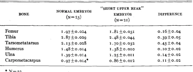
It was observed that some of the embryos and chicks with a short upper beak also had short legs. This led to a study of the length of long bones in this material. Unhatched but living normal and “short upper beak” em- bryos from the same matings were removed from the shells a t the end of the twenty-second day of incubation. They were preserved in formalde- hyde, and the long bones were dissected out and measured. The results of these measurements are shown in table 2 . It may be seen that (I) the mean
length of all long bones was reduced in the “short upper beak” embryos as compared with long bone length of normal embryos from the same matings; ( 2 ) in the “short upper beak” embryos the long bones of the legs were short-
TABLE 2
Average lengths of long bones i n cm, together with their standard errors.
BONE
“SHORT UPPER BEAK” NORMAL EMBRYOS
EMBRYOS DIFFERENCE ( N = 2 3 ) ( N = 2 I )
Femur I . 9 7 + 0 . 0 2 4 1 . 8 1 ~ 0 . 0 3 1 0 . 1 6 k 0.04 Tibia 2 . 8 7 f 0 . 0 2 9 2.48 + o . o u 0 . 3 9 f 0 . 0 5 Tarsometatarsus 2.13-10.028 1 . 7 0 f 0 . 0 3 2 0 . 4 3 * 0 . 0 4 Humerus I . 4 8 f 0 . 0 1 4 1 . 3 8 f 0 . 0 1 0 O . I O f O . 0 2
SEMI-LETHAL MUTATION I N FOWL 429
ened to a greater extent than those of the wings; ( 3 ) in the leg a proximo- distal gradient existed, the most proximal bone (femur) being least re- duced, while the most distal one (tarsometatarsus) showed the greatest degree of shortening.
As a check on these results, measurements were taken also of long bone length of normal and “short upper beak” embryos which had died between the eighteenth and twenty-second day of incubation. This material came from the same matings as that shown in table 2 . The following average measurements in centimeters were obtained for 23 normal and 40 “short upper beak” embryos:
NORMAL ‘‘SHORT UPPER BEAK”
Femur 1.85+0.029
Tibia 2.61k0.04j
Humerus 1.36k0.018
Ulna 1.28+_0.014
Carpometacarpus 0.89+0.012 Tarsometatarsus 1.99k0.030
I .84+o.org 2.jok0.021 1.68+0.01j 1.38+0.010 r . z g + o . o r o 0.88 f 0.009
Since the exact age of these embryos a t death was not known, no direct comparison of normal and “short upper beak” embryos was justified. However, it is interesting to compare these values with those in table 2. It may be seen that for the “short upper beak” embryos the averages of bone length of the living and dead embryos agree very closely. For the normal embryos, on the other hand, the long bones of the embryos which had died were all shorter than those of the embryos which were still alive a t the end of the incubation period. This was to be expected because of the younger age of the dead embryos. The fact that no such difference existed between the two groups of “short upper beak” embryos may be taken as an indication that little or no growth in bone length occurred in these embryos toward the end of incubation.
TABLE 3
Average indices of long bone length, together with their standard errors.
INDEX “SHORT UPPER BEAK” EMBRYOS
NORMAL EMBRYOS DIFFERENCE
Tarsometatarsusx IOO
Femur 108.0 k 0 . 5 4 9 4 . 2 2 1 . 2 3 1 3 . 8 k I . 3 4 Tibia X I 00
Femur 1 4 5 . 8 f 0 . 7 8 137.1 f I .27 8 . 7 2 I .49
TarsometatarsusX IOO
Tibia CarpometacarpusX I 00
Humerus
74.2 f o . 3 6 68.8 k 0 . 9 3 5 . 4 * 0.99
6 5 . 8 k 0 . 7 4 62.5 k 0 . 8 4 3 . 3 k I . I 2
Ulna X I 00
Humerus 93.9ik0.61 90.6 2 0.80 3.3ik I . 0 1 CarpometacarpusX IOO
Ulna 7 0 . 2 f 0 . 9 2 69. I k I .09 1
.
I f I .43It was of interest to know whether or not a correlation exists between the degree of abnormality of the upper beak and the extent to which the long bones were shortened. I n order to obtain information on this question the embryos were grouped into two classes according to the more or less extreme reduction of their upper beak, and the average length of the leg bones was then calculated for each of the two groups. The differences in bone length between the two groups were very slight and not consistently in one direction. Hence, it does not appear that there is a correlation be- tween the degree to which beak and long bones deviated from normal.
The progenies in which the “short upper beak” embryos occurred showed also segregation for polydactyly. The presence or absence of the poly- dactylous condition apparently had no influence on the degree to which the long bones of the legs were shortened.
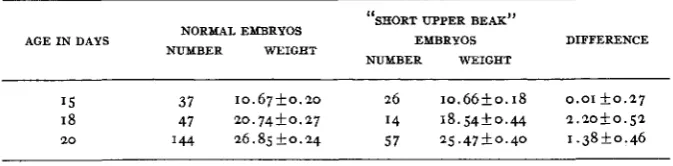
Determinations of body weight were made for embryos of 15, 18, and 2 0 days of incubation. The averages obtained are given in table 4. These data
TABLE 4
Average body weights of embryos in grams, together with their standard errors.
“SHORT UPPER BEAK”
DIFFERENCE NORMAL EMBRYOS
NUMBER WEIGHT
AGE I N DAYS EMBRYOS
NUMBER WEIGHT
I 5 37 1 0 . 6 7 k 0 . 2 0 26 1 0 . 6 6 f o . 1 8 0 . 0 1 k o . 2 7 I8 47 2 0 . 7 4 f 0 . 2 7 I4 1 8 . 5 4 f 0 . 4 4 2.2oiko.52
SEMI-LETHAL MUTATION I N FOWL 431
indicate that toward the end of incubation there was a tendency for the average weight of the “short upper beak” embryos to fall somewhat below that of normal embryos from the same matings. This agrees well with our observations on long bone growth reported above.
TABLE 5
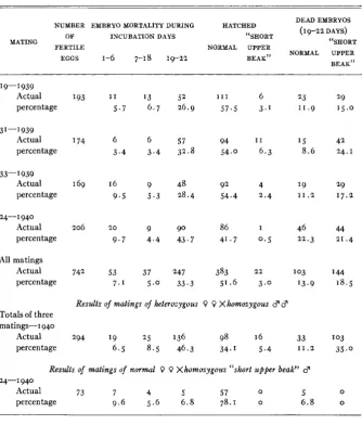
Results of matings o j heterozygous birds inter se.
DEAD EMBRYOS NUMBER EMBRYO MORTALITY DURING HATCHED
(I 9-22 DAYS)
OF INCUBATION DAYS “SHORT
MATING “SHORT
FERTILE NORMAL UPPER
NORMAL UPPER
EGGS 1-6 7-18 19-22 BEAK”
BEAK” 19-1939 Actual percentage 3 1-3939 Actual percentage 33-1939 Actual percentage
24-1 940 Actual percentage
All matings Actual percentage
I93 1 1 5.7
I74 6 3 . 4
169 16 9 . 5
206 20
9 . 7
742 53 7 . 1
13 52 6 . 7 26.9
6 57 3 . 4 32.8
9 48 5 . 3 28.4
9 90 4 . 4 43.7
37 247 5 . 0 33.3
I l l 57.5 94 54.0 92 54.4 86 41.7 383 51.6
6 23 3.1 11.9
I 1 15 6 . 3 8.6
4 19 2.4 1 1 . 2
I 46 0 . 5 22.3
22 103 3 .o 13.9
Results of malings o j heterozygous 0 9 Xhomozygous d 3
Totals of three matings-1940
Actual 294 19 25 136 98 16 33
percentage 6 . 5 8.5 46.3 34.1 5.4 1 1 . 2
Results of matings of normal 0 0 Xhomozygous “short upper beak” d
24-1 940
Actual 73 7 4 5 57 0 5
percentage 9.6 5 . 6 6.8 78.1 o 6 . 8 29
1 5 . 0
42 24. I
29 17.2 44 21.4 I44 18.5
I 03 35.0
0 0
432 WALTER LANDAUER
of 133 normal twenty-day embryos 1 1 1 or 83.5 percent had pierced the
inner shell membrane, while out of 56 “short upper beak” embryos of the same age only ten or 17.9 percent had done so.
INHERITANCE
Female progenies of crosses in which “short upper beak” embryos had cccurred were bred to males which were known to give such offspring. The results for those females which produced “short upper beak” em- bryos among their progenies are shown in table 5. It is unnecessary to dis- cuss each mating separately. There was a total of 742 fertile eggs, and of these 405 or 54.6 percent hatched. Among the embryos which had died be- tween the eighteenth and twenty-second day there were 144 or 18.5 per- cent with a short upper beak. In addition, there were 2 2 or 3.0 percent of
hatched chicks with an abnormal upper beak. Of all embryos which had survived to the eighteenth day of incubation (N =652), 166 or 25.5 percent had shortened upper beaks. From females which did not produce ab- normal embryos or chicks when bred to a heterozygous “short upper beak” cock, a total of 675 fertile eggs were obtained. Embryo mortality during the three incubation periods amounted to 5.0, 4.6, and 14.5 percent, re- spectively, and there were 75.9 percent hatched chicks. These segregation data agree well with the assumption that the shortening of the upper beak (and the other abnormalities associated with this trait) are caused by a re- cessive mutation which has a semi-lethal effect.
During 1940 it was possible to make crosses of females which were known to be heterozygous for the “short upper beak” mutation and males which had had a short upper beak a t hatching time and hence, presumably, were homozygous for the trait. The results of these crosses are given in table 5 .
Two hundred ninety-four fertile eggs were obtained, of which 114 or 39.5 percent hatched. Among the embryos which died between the eighteenth and twenty-second day there were 103 or 35.0 percent with a short upper beak; 16 or 5.4 percent of such specimens hatched. Out of a total of 2 5 0 embryos which survived to the end of the eighteenth day 119 or 47.6 per- cent had a short upper beak. This result agrees sufficiently well with the expected 50 percent of abnormal embryos and chicks to serve as verifica- tion of the assumption that the “short upper beak” trait is due to a single recessive gene difference.
A homozygous “short upper beak” cock was mated to hens which were known not to carry the mutation (table 5). Seventy-three fertile eggs were obtained, and 57 of these or 78.1 percent hatched. No abnormal embryos or chicks were produced. Hence, we are dealing with an autosomal muta- tion.
SEMI-LETHAL MUTATION IN FOWL 433
entia1 mortality of “short upper beak” embryos up to the end of the eight- eenth day of incubation. Further observations indicate that about one third of the “short upper beak” embryos which survived the eighteenth day were still alive when the unhatched eggs were opened a t the end of the twenty-second day, while most of the remaining embryos apparently died on the twenty-first and twenty-second day. The hatchability of those “short upper beak” embryos which survived the eighteenth day amounted to 13.3 percent ( 2 2 out of 166) in the matings of heterozygous parents and to 13.4 percent (16 out of 119) in the matings of heterozygous females by homozygous males.
I n the three matings of heterozygous females by homozygous males fer- tility of the eggs was low. The actual data are as follows.
- -
TOTAL EGGS NUMBER OF FERTILITY PEN
SET INFERTILE EGGS PERCENTAGE
1-1940
12-1 940
25- I940
58 618 474
36 241 405
37.9
61 .o 1 4 . 6
At the end of the regular hatching season the cocks in pens I and 2 5 were used for artificial insemination. Although both animals readily responded to stimulation, only small quantities of sperm were obtained. It would be interesting to know whether this low fertility is due to the beak abnor- mality (all three males had cross-beaks) and a consequent deficiency in food consumption or whether it is more directly a physiological concomi- tant of the “short upper beak” mutation. No effect on fertility was ob- served in our earlier breeding experiments with hereditary cross-beak of fowl. In two matings of normal females by cross-beak males‘ the fertility of eggs amounted to 62.5 percent (477 eggs set) and to 76.0 percent (367 eggs set), respectively. More critical information, however, is needed to settle this problem.
DISC US S I O N
A number of semi-lethal mutations exist in which occasional survivors reach the adult stage; in most instances such individuals are sterile. In fact, except for Drosophila, no matings seem to have been reported involv- ing homozygous semi-lethal animals. In Drosophila, a t least two sex- linked semi-lethal mutations are known (MORGAN 1929; BONNIER and
NORDENSKIOLD 1936) in which homozygous semi-lethals of one or both sexes have been bred. The only autosomal semi-lethal of Drosophila for which fertility has been reported is “curly” wing (WARD 19z3), though in this case no segregation ratios are on record.
434
The stock in which the “short upper beak” mutation was found traces back to Houdan ancestry, but they were not pure Houdans. DR. R. J. BUSHNELL kindly informed the writer that in pure Houdans he has ob- served embryos with the same kind of beak abnormality. It is likely, then, that the mutation with which we worked was derived from Houdan fowl. This is of interest because, as in the Cornish breed, Houdans are required by the standard of breeders to have rather short legs. Though we have no metric data as yet, there is some evidence suggesting that the effect which the “short upper beak” mutation has on long bone length may have a slight degree of expression in the heterozygous condition. If this is verified, we shall have another instance in which, as in Creeper and Cornish fowl, a reduction in length of the legs is maintained, a t least in part, a t the ex- pense of propagating a lethal mutation.
The morphological features of the “short upper beak” mutation are of great interest in relation to the other lethal mutations of fowl. There are a t present seven chicken lethals on record for which the morphological ex- pression is known. No less than five of these seven mutations affect simul- taneously head and extremities. These are ( I ) the early and late stages of the homozygous Creeper mutation (LANDAUER and DUNN 1930; LAND- AUER 1933); (2) the Cornish lethal (LANDAUER 1935); (3) the lethal pro- ducing extreme polydactyly (COLE 1939); (4) a new lethal which according to a brief communication by ASMUNDSON (1939) causes a deformity of the lower beak, curving of the upper beak, and shortening of the legs; and ( 5 )
the semi-lethal mutation described in the present report. After X-ray treat- ment of sperm (testes), KRAYEVOY (1938) frequently found F1 embryos in which abnormalities of head and extremities existed together.
A similar situation is met with among non-hereditary variations. Thus, association of head and appendicular abnormalities is shown by sporadic chondrodystrophy of chicken embryos (LANDAUER 1927; DUNN 1927); by embryos and chicks produced on manganese-deficient diets (BYERLY, TITUS, ELLIS, and LANDAUER 1935; LANDAUER 1936; CASKEY and NORRIS
1940) ; and by embryos developing in riboflavin-deficient eggs (ROMANOFF
1940). The simultaneous occurrence of malformations of head and extremi-
ties is also the most common type of embryonic abnormality produced by the ingestion of selenium by laying hens (LANDAUER 1939, c). I n mammals, including man, considerable evidence is also on record for the occurrence of similar syndromes, involving head and extremities, of hereditary and of non-genetic nature.
A shortening of the upper beak occurs commonly as a non-hereditary abnormality (phenocopy) under the influence of unfavorable incubation conditions, but it is not known whether or not in this case the shortening
SEMI-LETHAL MUTATION IN FOWL 43 5 The occurrence of abnormalities of this type is not limited to fowl ASMUNDSON (1939), for instance, found a recessive mutation among Bour- bon turkeys which brings about a reduction in length of the lower beak and of the legs. Among pigeons, the first two lethals were discovered recently. One of these mutations produces polydactyly and shortening of the upper beak (semi-lethal), and the other gives rise to micromelia (chondrodys- trophy ?) and abnormal conformation of the head.* Again, it is scarcely coincidence that the first two lethal mutations to be found in this genus should both show associated abnormalities of head and extremities. Inter- esting in this connection also are observations made by DARWIN (1876) on tumbler pigeons. He found in a comparison of different breeds of tumbler pigeons that a correlation exists between length of the (whole !) beak and length of the foot. Unfortunately, nothing is known concerning the develop- mental or genetic aspects of this correlation.
It cannot be assumed that all these different hereditary and non-genetic agencies interfere in a specific and identical manner with developmental processes involved in the formation of head and extremities. Alternatively, it seems reasonable to conclude that these embryonic parts are especially susceptible to disturbances of the most varied nature, and this high sus- ceptibility may have its basis in the intense growth activity of these parts during early developmental stages. Toward the end of development, growth of the whole body also falls below normal.
Another point of interest relates to the differential effect on length of different long bones. Measurements are available for three of the chicken lethals which affect long bone length (Creeper, Cornish, “short upper beak”), and in all three it was found that (I) the leg bones are more in- volved than the wing bones, and that ( 2 ) a proximo-distal gradient exists
within the legs, the femur being least reduced and the tarsometatarsus most. A similar situation is encountered in the non-hereditary micromelia produced in embryos and chicks by manganese deficiency of the eggs. These similarities of morphological response to three independent muta- tions and to a nutritional deficiency must have their causes in some funda- mental developmental pattern of the chicken embryo.
Such mechanisms, however, may operate also subsequent to hatching. In dwarfism of fowl produced by hereditary thyroid abnormality (LAND- AVER 1929; UPP 1934) the same type of gradient of shortening of theleg bones is found.
It has been shown by LERNER (1936, 1937) that a growth gradient exists in the leg of fowl, “with the distal bone (tarsometatarsus) growing at a more rapid rate in relation to the rate of body growth, the median bone (tibiotarsus) presenting an intermediate rate of growth, and the proximal
43 6 WALTER LANDAUER
bone (femur) the slowest.” These relations, of course, imply the existence of corresponding differences in growth intensity of the three bones, irre- spective of body weight. It has also been shown by LERNER that in these bones essentially the same growth relations hold for the embryonic and post-hatching period. There can be little doubt that this normal growth gradient and the differential response of the leg bones to the shortening effect of the hereditary and non-genetic agencies under discussion have a common material basis.
Even more illuminating, especially with reference to our observations on “short upper beak” embryos, are data on the relative growth of nestling house wrens recorded by HUGGINS (1940). Some of the growth constants (a) which she found are as follows: bill 1.3205, body 1.0167, tail 1.0551, femur I .2863, tibiotarsus 1.4106, tarsometatarsus 1.5778,humerus 1.3270, radius-ulna 1.4914, manus 1.2150. The beak and the long bones show much higher growth constants than the body proper. These are the parts which are affected by the “short upper beak” mutation of fowl. The long bones of the leg in the wrens, as in chickens, show a proximo-distal gradient of increasing growth intensity. It has been pointed out already that a similar gradient exists with reference to the amount of reduction in length of the long bones under the influence of the mutations in question. No
clear-cut proximo-distal gradient was found in the wing of the house wren, and this again agrees with the less clear-cut differential effect of the “short upper beak” mutation on the wing bones. HUGGINS’S data on relative growth of wren nestlings, however, do not furnish a basis for explaining the much lesser effect of the “short upper beak,” Cornish, and Creeper mutations on wing bones as compared with leg bones. This apparent dis- crepancy is probably due to the fact that a t the beginning of the post- hatching period the growth rate of the wing bones becomes much greater, relative to the leg bones, than it is during the embryonic period (STREICH and SWETOSAROW 1937), and it is, of course, growth during embryonic de- velopment which interests us with regard to the effect of the lethal muta- tions and of other factors interfering with normal development.
The foregoing evidence indicates clearly that the responses of parts of the embryo to the several mutations are influenced, a t least in certain re- spects, by the inherent normal growth characteristics of these parts. Do such differential responses lead only to size differences or are they responsi- ble also for structural peculiarities?
SEMI-LETHAL MUTATION I N FOWL 43 7 embryo as a whole has been abandoned. It has been pointed out instead that “the localized effeEts are due merely to quantitative differences in physiological needs of certain parts a t definite periods; these differences depending on such factors as differential growth activity, involving differ- ential needs for certain enzymes and other substances.”
In a discussion of his transplantation results with limb primordia of normal, heterozygous, and homozygous Creeper embryos, HAMBURGER (1941) admits the universal nature of the action of the Creeper mutation but thinks that its effect is probably due to “some deficiency in a general metabolic or respiratory mechanism” which “might concern a specific reaction or substance involved in such a general activity.” It seems to the present writer that the two interpretations are in virtual agreement.
HAMBURGER takes exception to the writer’s continuing emphasis on growth processes as probably being an especially important agency in calling forth differential responses. However, his failure to obtain Creeper phenocopies by the transplantation of normal limb buds cannot be taken as proof against such an interpretation. Growth is the result of the complex inter- action of many different processes. Interference with any of a great number of physiological reactions contributing to growth may result in. dispropor- tionate reduction of the size of certain parts but may a t the same time, according to differential needs of these parts, result in specific structural alterations in each instance.
43 8 WALTER LANDAUER
SUMMARY
A new lethal mutation in fowl is reported which produces a shortening of the upper beak and of the long bones of the extremities.
The mutation is recessive, autosomal, and semi-lethal. The majority of homozygous embryos die near the end of the incubation period, but about 13 percent of all homozygotes hatch.
The degree of shortening of the upper beak and of the long bones is variable, and there is apparently no correlation between the extent to which the two structures are affected.
In many of the hatched “short upper beak” chicks the beak becomes normal during growth. In others, a cross-beak develops, the curvature of the upper beak being more frequently toward the right than toward the left side. In a few, the shortness of the upper beak persists to maturity.
Among the hatched homozygotes there is a high mortality, presumably on account of difficulties in eating. Adult “short upper beak” cocks give low fertility.
The shortening of the long bones affects the wing bones less than those of the legs. Within the leg the shortening of the individual bones follows a proximo-distal gradient, the femur being least affected and the tarso- metatarsus most.
The shortening of the upper beak has been seen in embryos as early as nine days, but not in six-day embryos.
In late stages of development body weight of the homozygotes is below that of normal embryos. There are other signs that the rate of development falls below normal toward the end of incubation (delayed resorption of the yolk sac, frequent failure of embryos to pierce the inner shell membrane). These late manifestations of the “short upper beak” mutation presumably are another expression of the fundamental disturbances which occur early in development.
. The developmental aspects of the “short upper beak” mutation are dis- cussed, with special reference to similarities with the Creeper mutation. It is concluded that in both instances the mutations are responsible for aberrations in physiological mechanisms which are common to the whole embryo.
ACKNOWLEDGMENT
The foundation stock for the studies reported here was obtained from DR. D. C. WARREN, Manhattan, Kansas. I wish to record my appreciation of his generosity.
LITERATURE CITED
ASMUNDSON, V. S., 1939 Some factors affecting hatchability of eggs. Poult. Sci. 18: 399.
BONNIER, G., and M. NORDENSKIOLD, 1936 A semi-lethal causing exceptional sex-ratios in h o -
SEMI-LETHAL MUTATION I N FOWL 43 9 BYERLY, T. C., H . W. TITUS, N. R. ELLIS, and W. LANDAUER, 1935 A new nutritional disease of CASKEY, C. D., and L. C. NORRIS, 1940 Micromelia in adult fowl caused by manganese deficiency
COLE, R. K., 1939 A new autosomal lethal in the fowl. Poult. Sci. 18: 403-404.
DARWIN, C., 1876 The variation of animals and plants under domestication. 2d ed. New York. DORRIS, F., 1938 Differentiation of the chick eye in vitro. J. Exp. Zool. 78: 385-407.
DUNN, L. C., 1927 The occurrence of chondrodystrophy in chick embryos. Arch. Entw. Mech.
FELL, H. B., and W. LANDAUER, 1935 Experiments on skeletal growth and development in vitro HAMBURGER, V., I 941 Transplantation of limb primordia of homozygous and heterozygous
HUGGINS, S. E., 1940 Relative growth in the house wren. Growth 4: 225-236.
IRICHIMOWITSCH, A. I., 1936 Die Gesetzmassigkeiten des Wachstums wahrend der Metamorphose KRAYEVOY, I., 1938 Experimentally induced mutations in chickens through x-rays. Acad. Sci. LANDAUER, W., I927 Untersuchungen iiber Chondrodystrophie. I . Arch. Entw. Mechanik 110:
1929 Thyrogenous dwarfism (myxoedema infantilis) in the domestic fowl. Amer. J. Anat.
43: 1-20.
1933 Untersuchungen uber das Kruperhuhn. IV. 2. mho.-anat. Forsch. 32: 359-412. 1934 Studies on the Creeper fowl. V1. Storrs Agric. Exp. Sta. Bull. 193.
1935 A lethal mutation in Dark Cornish fowl. J. Genet. 31: 237-242.
1936 Micromelia of chicken embryos and newly hatched chicks caused by a nutritional de- ficiency. Anat. Rec. 64: 267-276.
1938 Notes on cross-beak in fowl. J. Genet. 37: 51-68.
1939a Studies on the Creeper fowl. XI. Storrs Agric. Exp. Sta. Bull. 232.
1939b Studies on the lethal mutation of Cornish fowl. Storrs Agric. Exp. Sta. Bull. 233.
1 9 3 9 ~ Studies on the Creeper fowl. XIII. J. Exp. Zool. 83: 431-43.
1941 Teratological correlations and the mechanism of gene expression. Proc. Seventh Int. Genetical Congr., Edinburgh, 1939: 181-185.
the chick embryo. Proc. Soc. Exp. Biol. 32: 1542-1546.
during embryonic development. Proc. Soc. Exp. Biol. 4: 332-335.
110: 341-365.
in relation to the problem of avian phokomelia. Proc. Roy. Soc. B 118: 133-154.
“Creeper” chick embryos. Physiol. Zool. 14: in print.
der Amphibien. I. Biol. Zbl. 56: 639-656.
Ukrainian S.S.R., Inst. Zool. and Biol. Mem. Genet. 2: 109-160.
195-278.
LANDAUER, W., and L. C. DUNN, 1930 Studies on the Creeper fowl. I. J. Genet. 23: 397-412. LERNER, I. M., 1936 Heterogony in the axial skeleton of the Creeper fowl. Amer. Nat. 70: 595-598.
1937 Relative growth and hereditary size limitation in the domestic fowl. Hilgardia IO:
MORGAN, T. H., 1929 Exceptional sex-ratios in certain mutant stocks with attached x’s. Pub. OLSEN, M. W., and T . C. BYERLY, 1935 The orientation of the embryo in the egg of the domestic
ROMANOFF, A. L., 1940 The increased mortality of the developing chick embryos in riboflavin- RUDNICK, D., and V. HAMBURGER, 1940 On the identification of segregated phenotypes in progeny SCHHALHAUSEN, J., I925 Uber die Beeinflussung der Morphogenese der Extremitaten vom Axolotl STREICH, G . , and E. SWETOSAROW, 1937 Die Entwicklung der Proportionalitat im Wachstums-
UPP, C. W., 1934 Further data on the inheritance of dwarfism in fowls. Poult. Sci. 23: 157-165. WARD, L., The genetics of curly wing in Drosophila. Another case of balanced lethal factors.
51 1-560.
Carnegie Inst. 399: 101-138.
fowl. Poult. Sci. 14: 46-53.
deficient eggs. Anat. Rec. 78, Supplement 78. from Creeper fowl matings. Genetics 25: z15-zz4.
durch verschiedene Faktoren. Arch. Entw. Mechanik 105: 483-500.
prozess der Vogel. 2001. Jahrb., Abtl. allgem. 2001. Physiol. 58: 113-126.