MOTOR CONTROL OF EXERCISE THAT EMPHASIZES SPEED POST-STROKE
(Spine title: Motor Control of Exercise that Emphasize Speed Post-stroke) (Thesis format: Integrated Article)
by
Vicki L. Gray
Graduate Program in Health and Rehabilitation Sciences
A thesis submitted in partial fulfillment of the requirements for the degree of
Doctor of Philosophy
The School of Graduate and Postdoctoral Studies The University of Western Ontario
London, Ontario, Canada
THE UNIVERSITY OF WESTERN ONTARIO School of Graduate and Postdoctoral Studies
CERTIFICATE OF EXAMINATION
Supervisor
______________________________ Dr. Jayne Garland
Supervisory Committee
______________________________ Dr. Pamela Houghton
______________________________ Dr. Ruth Martin
Examiners
______________________________ Dr. Trevor Birmingham-Internal
______________________________ Dr. Denise Connelly-Internal
______________________________ Dr. Timothy Doherty-University
______________________________ Dr. Brenda Brouwer-External
The thesis by
Vicki Lynn Gray
entitled:
Motor Control of Exercise that Emphasizes Speed Post-stroke
is accepted in partial fulfillment of the requirements for the degree of
Doctor of Philosophy
______________________ _______________________________
Abstract
Purpose: To investigate whether a single session of closed kinetic chain (CKC) and open kinetic chain (OKC) exercises emphasizing speed post-stroke could evoke changes in the
motor control and whether these improvements would transfer to postural tasks.
Methods: Thirty-two individuals post-stroke and 32 age- and sex- matched controls performed a single session of 50 fast squats and steps (Chapter 3). Internal perturbations
(arm raise/load drop) were used to assess postural responses before exercises (Pre),
immediately after exercises (Post) and 15 minutes after exercises (Retention) (Chapter 4).
Eleven individuals post-stroke performed a single session of 50 fast knee and ankle OKC
exercises and postural responses were assessed Pre and Post exercises (Chapter 5).
Electromyographic (EMG) activity was measured bilaterally in the rectus femoris (RF),
biceps femoris (BF), tibialis anterior (TA), and soleus (SOL) muscles.
Result: The squat was performed slower in the stroke group than controls, with impaired temporal coupling between the knee movement and postural sway. The paretic BF EMG
was delayed with a reduced slope and the paretic RF EMG area was reduced. The squat
was initiated with the non-paretic leg as a compensatory strategy in the low motor
recovery group whereas the paretic leg was used in an adaptive manner in the high motor
recovery group (Chapter 2). The temporal coupling improved and EMG area of the
paretic TA, BF and RF increased in the squats. In the steps, the paretic BF and RF EMG
area increased in the stepping leg and the paretic SOL and RF EMG area increased in the
stance leg (Chapter 3). The paretic BF EMG area and slope increased after exercises in
the arm raise task. In the load drop task, the paretic BF EMG deactivation improved and
was retained after 15 minutes. Weight bearing symmetry also improved with exercise
(Chapter 4). The paretic BF, RF, TA EMG area increased along with an increase in peak
velocity and power during the OKC exercises. The arm acceleration and BF EMG area
increased in the arm raise task (Chapter 5).
Keywords: stroke, standing balance, electromyography, motor control, movement speed
Co-Authorship Statement
Chapters 2, 3 and 4 of this dissertation have been accepted for publication. Chapter 5 will
be submitted for publication. Dr Jayne Garland and Dr Tanya Ivanova are co-authors on
Chapters 2, 3, 4 and 5 as they have significantly contributed to the design, data collection
and the peer review process. Larissa Juren is a co-author on chapter 4 and provided
Acknowledgments
The journey in completing this thesis has been a long road with many ups and down. I
could not have completed this thesis without the support and assistance from many people
along the way.
First and foremost, I would like express my sincere gratitude to my advisor, Dr Jayne
Garland, for her enthusiasm, immense knowledge and inspiration. She has been a great
mentor and friend. I am indebted to the opportunities she has provided to me in the lab
and other academic settings that has aided in furthering my growth and development. She
truly has been an exceptional advisor and I am thankful for the years she has committed
to my thesis. I express sincere gratitude to our Lab Manager, Dr Tanya Ivanova for her
knowledge of the technical aspects of the lab, for her patience in teaching me and for
assisting in the data collection and troubleshooting when things go wrong.
This thesis would not have been possible without the generosity of all the study
participants with stroke and the control subjects who participated in my research. I am
deeply grateful to all the participants.
Many thanks to my advisory committee, Dr Pamela Houghton and Dr Ruth Martin and
comprehensive exam committee, Dr Charles Rice, Dr Joy MacDermid and Dr Timothy
Murphy for their expertise, advice and support.
I would like to thank my lab mates and fellow graduate students for their support. In
particular I am indebted to Courtney Pollock and Dr Chris MacDonnell for acting as a
sounding board and a voice of reason.
I wish to thank my family and friends who have been a constant source of support in my
life. Without your encouragement and understanding it would have been impossible for
me to finish this thesis. Lastly, I would like to thank my parents, William Gray and
Nancy Gray, for raising me with the belief that “anything is possible if you put your mind
Table of Contents
CERTIFICATE OF EXAMINATION... ii
Abstract ... iii
Co-Authorship Statement... v
Acknowledgments... vi
Table of Contents ... vii
List of Tables ... xi
List of Figures ... xii
List of Appendices ... xiv
List of Abbreviations ... xv
Chapter 1 ... 1
1 Introduction ... 1
1.1 Stroke Etiology and Statistics ... 1
1.2 Neuroplasticity... 2
1.3 Postural Control of Balance ... 3
1.4 Balance after Stroke ... 5
1.5 Rehabilitation of Balance after Stroke ... 7
1.6 Velocity, Force and Power... 8
1.7 Open Kinetic and Close Kinetic Chain exercises ... 9
1.8 Thesis Outline ... 10
1.8.1 Objectives and Hypotheses ... 10
1.9 References... 13
Chapter 2 ... 26
2.1 Introduction... 26
2.2 Methods... 27
2.2.1 Experimental protocol... 28
2.2.2 Data analysis ... 29
2.2.3 Statistical analysis ... 31
2.3 Results... 31
2.3.1 Subject characteristics... 31
2.3.2 Description of fast squatting movements... 32
2.3.3 Control group ... 32
2.3.4 Stroke group... 32
2.3.5 Anticipatory postural responses prior to squat... 36
2.3.6 Acceleration phase ... 36
2.3.7 Deceleration phase ... 40
2.4 Discussion ... 42
2.5 References... 45
Chapter 3 ... 50
3 The effects of fast functional exercise on muscle activity after stroke ... 50
3.1 Introduction... 50
3.2 Methods... 51
3.2.1 Subjects ... 51
3.2.2 Clinical Measurements... 52
3.2.3 Physiological Measurements ... 52
3.2.4 Exercise Protocol ... 52
3.2.5 Data Acquisition ... 53
3.2.7 Statistical Analyses ... 54
3.3 Results... 55
3.3.1 Squats ... 55
3.3.2 Steps: Stepping leg... 56
3.3.3 Steps: Stance leg ... 63
3.4 Discussion ... 63
3.5 Acknowledgements... 65
3.6 References... 66
Chapter 4 ... 70
4 Retraining postural responses with exercises emphasizing speed post-stroke... 70
4.1 Introduction... 70
4.2 Methods... 71
4.2.1 Statistical analysis ... 75
4.2.2 Sample size calculation... 76
4.3 Results... 76
4.3.1 Arm Raise Task... 77
4.3.2 Load Drop Task ... 80
4.3.3 Paresis of the dominant side vs Paresis of the non-dominant side... 84
4.4 Discussion ... 85
4.5 Conclusion ... 87
4.6 Role of the funding source ... 88
4.7 References... 89
5 Open Kinetic Chain Exercises emphasizing speed post-stroke ... 95
5.1 Introduction... 95
5.3 Results... 100
5.3.1 Exercises ... 100
5.3.2 Ankle Movements ... 100
5.3.3 Knee Movements ... 103
5.3.4 Arm RaiseTask... 107
5.4 Discussion ... 107
5.5 References... 111
6 General Discussion... 115
6.1 Overview... 115
6.2 Motor Control during Exercise ... 117
6.3 Postural Control ... 119
6.4 Limitations ... 121
6.5 Implications and Future Directions... 122
6.6 Conclusion ... 123
6.7 References... 124
List of Tables
Table 2.1 The displacement, maximum vertical velocity and peak to peak acceleration of
controls and individuals after stroke during fast squatting movements...35
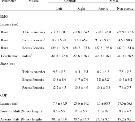
Table 2.2 EMG latency and slope and COP of controls (right/left leg) and individuals
after stroke (paretic/non paretic leg) in the anticipatory and acceleration phases in ten
averaged squats. ...37
Table 2.3 EMG area of controls (right/left leg),individuals after stroke
(paretic/non-paretic leg) and high and low motor recovery group ((paretic/non-paretic leg) in the acceleration and
deceleration phases in ten averaged squats. ...39
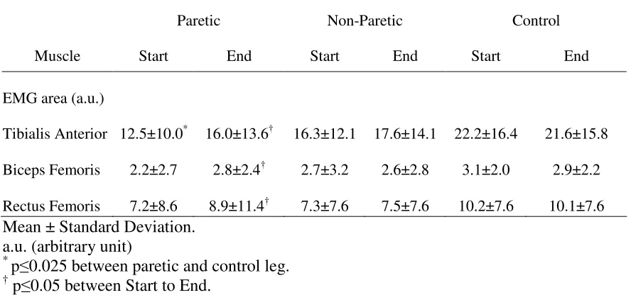
Table 3.1 EMG Area of the paretic, non-paretic and control legs at the Start and End of
the squats...58
Table 3.2 EMG area and knee acceleration of the paretic, non-paretic and control stepping
leg at the Start and the End of the steps. ...60
Table 3.3 EMG Area of the paretic, non-paretic and control legs in the Start and End of
the stance leg during steps. ...62
Table 4.1Biceps femoris EMG slope and peak area, arm acceleration, and COP velocity
of controls and individuals after stroke during the unilateral arm raise at Pre exercise, Post
exercise and Retention. ...79
Table 4.2 Load Drop: Anticipatory EMG deactivation area in the biceps femoris of
controls (non-dominant/dominant leg) and individuals after stroke (paretic/non-paretic) at
Pre exercise, Post exercise and Retention...83
Table 5.1 Characteristics of subjects. ...101
Table 5.2 Arm Raise: Biceps femoris EMG slope and peak area, arm acceleration, and
List of Figures
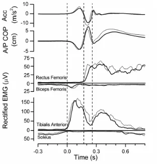
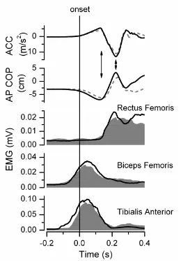
Figure 2.1 Knee accelerations, A/P COP and EMG of the squats for a control subject....33
Figure 2.2 Relationship between acceleration phase duration and difference in movement
onset between legs in the control and stroke group ...34
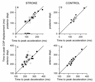
Figure 2.3 Coupling of time to peak acceleration and deceleration and peak COP
(posterior and anterior) displacement for paretic and non-paretic leg of stroke group and
right and left leg of control group ...38
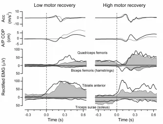
Figure 2.4 Knee accelerations, A/P COP and EMG of ten averaged squats in high motor
recovery group and low motor recovery group...41
Figure 3.1 Knee accelerations, A/P COP and EMG in the first ten and last averaged
squats for an individual after stroke...57
Figure 3.2 The temporal coupling of the peak knee movement and COP excursion at the
Start and End of squats in acceleration and deceleration phase...59
Figure 3.3 Knee accelerations and EMG in the first ten and last ten averaged steps of the
stepping and stance leg for two individuals after stroke...61
Figure 4.1 The group means of the vertical ground reaction forces of the stroke and
control group ...78
Figure 4.2 Arm acceleration and biceps femoris EMG during the arm raise for a subject
from the stroke and control group...81
Figure 4.3 Biceps femoris EMG during the load drop for two subjects in the stroke group
and one from the control group at Pre, Post and Retention ...82
Figure 5.1 The EMG and velocity for two single trials of ankle dorsiflexion and
Figure 5.2 The EMG area for the tibialis anterior and soleus, peak velocity and power
over 50 trials of ankle plantarflexion and dorsiflexion ...104
Figure 5.3 The EMG area for the rectus femoris and biceps femoris, peak velocity and
List of Appendices
Appendix A: Characteristics of stroke group (Chapter 2, 3 and 4)...128
Appendix B: Clinical Outcome Measures ...129
Appendix C: Ethics Approval ...149
Appendix D: Letters of Explanation ...152
List of Abbreviations
AMTI - Advanced Mechanical Technology Incorporated
A/P - anterior posterior
a.u. - arbitrary unit
BBS - Berg Balance Scale
BF - biceps femoris
BI - Barthel Index Score
CKC - closed kinetic chain
COM - center of mass
COP - center of pressure
CMSA - Chedoke McMaster Stroke Assessment
EMG - electromyographic
FIM - Functional Independence Measure
Fz - vertical ground reaction force
OKC - open kinetic chain
RF - rectus femoris
SOL - soleus
Chapter 1
1
Introduction
1.1
Stroke Etiology and Statistics
A stroke is a disruption in blood flow to the brain by ischemia or a haemorrhage. An
ischemic stroke is an obstruction of a blood vessel in the brain from a large artery
atherosclerosis, cardioembolism or a small artery occlusion and accounts for 80% of all
strokes.1 Haemorrhagic strokes are less common and occur when a blood vessel ruptures
in the brain and bleeds into the surrounding area. In both types of stroke, blood flow to
the brain is interrupted resulting in death or injury to the brain cells.
Stroke is one of the leading causes of death and disability in Canada.2 The number of
stroke deaths has declined in the last 30 years, increasing the survival rate and the number
of individuals living with long term disability and functional limitations. There are more
than 50, 000 strokes a year in Canada and only 10% of these individuals recover
completely.3 The residual deficits common to individuals after stroke are a loss of
memory, speech and swallowing difficulties, decreased sensation, loss of coordination,
muscle weakness and impaired balance. The most common stroke deficit is muscle
weakness, known as paresis and 80% of patients surviving a stroke have some degree of
weakness.4,5
Falls are common after stroke and impaired balance, visuospatial, hemi-neglect and
self-care deficits are common predictors of falls in an inpatient rehabilitation unit.6 Ugur et
al7 reported in a sample of 293 stroke patients in an inpatient rehabilitation unit, 44%
experienced a fall and the risk increased as functional impairments increased. Many
individuals become more dependent after sustaining a stroke; 60% reported needing some
assistance with their activities and 84% reported activity restrictions which left them
dependent on others for some of their activities of daily living. Even though many
individuals become dependent on others for assistance the number of falls in the first six
months is high. Forster and Young,8 reported 73% of persons post-stroke fell in the first
their sample of 108 participants. Falls can be devastating and can lead to fractures,9 a fear
of falling,10 and activity limitations.11
The economic impact of stroke is also significant. Thus, finding ways to reduce
impairment after stroke is important. The direct cost to the Canadian health care system
is estimated to be over 3.6 billion dollars.3 The hospitalization rate for stroke has
declined since 1979, however the length of stay has increased for an individual compared
to other health conditions.3 This may be related to the severity of the strokes that are
generating a longer hospital stay.
1.2
Neuroplasticity
The process of recovering functional loss following stroke is thought to be accomplished
through neuroplasticity whereby the reorganization of the brain occurs in an attempt to
recover the loss of function. The mechanisms of neuroplasticity include the unmasking of
anatomical pathways previously not functional, sprouting of new pathways, and
redundant pathways with a similar function taking over for the injured part of the brain. 12-15
Regardless of the type of stroke, the injured part of the brain has two different areas,
the ischemic core and the surrounding area called the penumbra. In the ischemic core,
severe oxygen depletion for more than two minutes results in necrosis of brain cells.16 In
the penumbra the blood flow is reduced, however the area can remain viable for several
hours after a stroke with the assistance of the anastomoses of collateral arteries supplying
oxygenated blood at a reduced rate. The penumbra is important in the recovery process
because, if blood flow is restored to the penumbra, the damage may be reversed.17
Neuroplasticity in the early stages of recovery occurs spontaneously as the edema and
necrotic tissue is absorbed and blood flow increases from collateral circulation. This
process can last up to eight weeks especially in haemorrhagic strokes where edema is
prevalent. In the later stages there is evidence that the brain and nervous system can
change structurally and functionally. The extent of neuroplasticity is dependent on the
magnitude of the stroke, with a severe stroke producing less viable adjacent brain tissue
A stroke involving the motor area of the brain has reduced descending motor commands
to the alpha motor neurons innervating the muscles. Neuronal death and smaller motor
output area, compared to the unaffected side of the brain, are attributed to altered central
drive.18,19 A persistently reduced neural signal to the alpha motor neurons leads to the
death of alpha motor neurons, and those innervating fast-twitch type II fibers are more
susceptible to death. Reorganization of the muscle takes place through collateral
sprouting of adjacent alpha motor neurons to take on the muscle fibers of the alpha motor
neurons that have died. As a result structural changes occur in the muscle with fewer
type II fibers and fewer alpha motor neurons that innervate a larger number of fibers.20,21
Thus changes in the neural activity from the brain can result in morphological changes at
the level of the muscle.
The recovery process is dependent on viable tissue in the penumbra for neural
reorganization. Rehabilitation may play an important role in enhancing the newly formed
pathways or establishing new pathways.22 The benefits of rehabilitation have been shown
with an increase in representation area of the affected limb in the adjacent brain tissue to
the ischemic area12 and greater task improvements23 in animal models. In human studies,
an enlarged cortical representation has been observed in single and multiple day
rehabilitation sessions.24-27 Thus rehabilitation enhances and strengthens the newly
created pathways and is an important component to recovery after stroke.
1.3
Postural Control of Balance
Standing balance ranges from quiet stance, where there is no movement, to dynamic
balance occurring during transitional movements, such as walking or climbing stairs.
Balance is controlled through postural control and the purpose is to maintain a state of
postural equilibrium, which is when all internal forces generated by the neuromuscular
system are in balance with the external forces acting on the body, including the force of
gravity and ground reaction forces from the supporting surface.28 Postural equilibrium is
a dynamic process involving the integration of information from the environment by the
vestibular, somatosensory and visual systems to stabilize the center of mass (COM) of the
The body is in a state of postural equilibrium when the COM is within the base of
support; this area was described by McCollum and Leen29 as a geometric form of the
limits of stability. This area is an area where one can move their COM and maintain
equilibrium without adjusting their base of support. When the limits of stability are
reached, three different strategies can be employed to counter a disturbance: an ankle
strategy, hip strategy or stepping strategy. The ankle strategy involves a response from
the ankle plantarflexors (during an anterior sway) or dorsiflexors (during a posterior
sway), and the response shifts the center of mass by adjusting the body around the ankles.
This strategy is sufficient when the center of mass velocity is slow.28 In faster
movements, a hip strategy can be used to control the center of mass through large rapid
motions at the hip and movements at the ankle that are opposite in direction to the hip
movement.30 The stepping strategy is used when the ankle or hip strategies are
insufficient to regain or maintain balance. A step is taken to realign the center of mass
within the base of support. The stepping strategies are used more frequently in older
adults,31 thus the speed of a protective step is important in the ability to regain postural
equilibrium.
The muscle responses from the neuromuscular system when the center of mass moves
outside the base of support is a postural adjustment or response to offset the destabilizing
forces.32-34 A postural response is controlled centrally and the magnitude is dictated by
the distance of the COM movement within or outside of the base of support and the center
of mass velocity.35 Two types of disturbances to postural equilibrium which may require
a postural response are internal and external perturbations.
An external perturbation occurs when the body is unexpectedly perturbed by a translating
platform or an external force imposed on the body creating a shift in the COM. The
muscles respond after the movement in a feedback manner to restore the body back to a
postural equilibrium. A backward translation of the platform generates a forward sway
and a response from the gastrocnemius muscle 100 ms after the platform is translated.36
The magnitude of the response is dictated by how quickly the COM is displaced. As the
acceleration increases the magnitude of the muscle activity increases.37 There is also a
tibialias anterior-quadriceps muscles to a backward sway with the distal muscle being
activated first.38
Internal perturbations are created through voluntary movements with the postural
response preceding the movement.32,39 The response before the movement is referred to
as a feedforward response. The muscle activity increases before the voluntary movement,
producing a shift in the center of pressure (COP) to compensate for the anticipated
disturbance. Similar to external perturbations, the direction dictates which muscles
respond, a perturbation directed forward displaces the COM anteriorly, requiring a
response from the posterior muscles, e.g. hamstrings, gastrocnemius and erector spinae
muscles. The reverse occurs when the COM moves posteriorly, the anterior muscles, e.g.
tibialis anterior and quadriceps muscles, respond to the perturbation.33,40 The magnitude
of the postural response can be influenced by the size of the perturbation, a larger
perturbation generates a larger postural response41 and as the velocity of movement
increases the initial rise in the electromyographic (EMG) slope increases.42 In
asymmetrical weight bearing, by externally rotating the leg 45º, the postural response is
reduced in the soleus and rectus abdominus muscles on the side of the body with the leg
rotated externally (or reduced weight).43 If weight is added to the body symmetrically the
postural response of the soleus, rectus abdominus and erector spinae muscles increases in
amplitude.44 Thus, there is modulation of postural responses based on loading, velocity
of movement and magnitude of the perturbation in healthy individuals.
1.4
Balance after Stroke
The impairments after stroke to sensation, motor control and coordination result in
deficits in standing balance.45 The postural responses required to counter a disturbance to
balance, whether produced internally or externally, are impaired which is evident by the
delayed timing and insufficient magnitude of EMG in the paretic gastrocnemius, tibialis
anterior, hamstrings, quadriceps, gluteus medius, and paraspinal muscles compared to
controls.46-49 The altered muscle activity in the paretic muscles has been attributed to
neurological changes, such as an increased stretch reflex excitability,50,51 increased
After stroke, the isometric EMG-force relationship is impaired. That is, there is a greater
EMG increase with increasing force resulting in a steeper EMG-force relationship than
controls.60 The reason for the increase in EMG without comparable increases in force
may result from decreased motor unit firing rates,57-59 decreased number of type II
fibers,20,61 and/or a decreased ability to modulate the motor unit firing rate.59 During
concentric movement, in healthy subjects the EMG amplitude increases as velocity
increases yet the torque decreases as velocity increases.62,63 After stroke, there is no
modulation of EMG amplitude as velocity increases64 and torque decreases to a greater
extent as velocity increases.65-67
In individuals after stroke, when a disturbance to the body is generated externally
(external perturbation) the muscle activity from the paretic muscles is reduced, delayed
and prolonged.46,47,68-70 The examination of postural responses to external perturbations
post-stoke have been examined using force platform translations. The distal to proximal
response typically observed in healthy individuals is impaired, with slower and smaller
muscle activity in response to horizontal translations in persons 5-30 months
post-stroke.46 There is evidence that the postural response to a horizontal translation is better
if an individual is given advanced warning of the perturbation.70 Increasing the load or
increasing the acceleration leads to an increased EMG burst similar to controls. However
the ability to modulate an earlier response in the gastrocnemius muscle compared to
controls is impaired.68,71 During external perturbations, the response in the paretic
muscles is disrupted with a greater impairment in the timing of the EMG burst than the
magnitude of the EMG burst.69,71
Similarly, impaired postural responses to internal perturbations have been reported in
individuals after stroke. During a standing single leg hip and knee flexion perturbation, a
delay and reduced gluteus medius muscle activity was observed in the paretic side
compared to the non-paretic side.72 Another internal perturbation used is the arm raise,
where the non-paretic arm is moved quickly forward to a horizontal position. When
individuals after stroke with poor balance (less than 38/56 on the Berg Balance Scale)
performed the arm raise an anticipatory activation of the paretic hamstrings was not
balance (mean of 48/56 on the Berg Balance Scale) demonstrated a delay in the muscle
activation of the paretic hamstrings compared to an elderly control group with a similar
mean age.49 The response of the muscle can be influence by the characteristics of the
internal perturbation. Horak et al48 reported a delayed response in the muscle activation
of the paretic biceps femoris muscle in both a self paced arm raise and a weighted arm
raise when compared to controls. Also, the paretic paraspinal muscle had a greater delay
in muscle activation during the weighted arm raise than the fast self paced arm raise.
Another method of producing an internal perturbation is a load drop. This type of
perturbation is standardized, whereas the arm raise varies based on the ability of the
person to generate a fast movement, which is impaired after stroke. Slijper et al74 had
individuals after stroke drop a load from the non-paretic hand with the arm extended in
front and out to the side. They reported reduced muscle activity in the paretic rectus
femoris and biceps femoris muscles. The muscle activity for the side load drop or front
load drop prior to the load release did not differ in individuals after stroke, whereas
controls had a smaller EMG activity in the side load drop. Thus individuals after stroke
were unable to modulate the magnitude of the paretic rectus femoris, biceps femoris or
erector spinae muscle activity in response to a change in load drop direction.74
1.5
Rehabilitation of Balance after Stroke
Rehabilitation interventions are important as they attempt to enhance neuroplasticity.
There are a wide variety of interventions used to improve balance, however no one
intervention is superior to another to improve balance post-stroke. The abundance of
intervention strategies available and the heterogeneity of the type of strokes may explain
why no single intervention has been shown to be more effective than other.
The type of interventions used to improve standing balance include force platform
feedback,75-79 a balance trainer80 and multisensory or sensory training.81-86 One review
examining feedback from the force platform revealed that this strategy improved standing
balance however the improvements did not transfer to functional activities or change
functional independence.87 Sensory training improved balance,79,83,85,86 however the
evidence that repetitions of a lateral push applied at the hip results in relearning of
appropriate postural responses and if the knowledge of push direction is known in
advance relearning can be enhanced.88 Therefore, it is important to examine a novel
strategy in the rehabilitation of balance post-stroke that can increase movement speed in
order to elicit a quick response to regain postural equilibrium.
1.6
Velocity, Force and Power
Power is the product of force and velocity. Muscle power has been found to be a
predictor of balance and functional mobility in older adults.89-92 In older adults, muscle
strength is related to balance outcome measures requiring little movement, such as the
Berg Balance Scale and Unipedal Stance Test, whereas the velocity of the movement was
associated with dynamic balance outcome measures, such as Dynamic Gait Index and
Performance Mobility Assessment.93
Force94,95 and movement velocity62,66,96 are impaired after stroke and both contribute to
understanding the deficits that exist in balance. There is evidence to support the benefits
of strength training,97-101 however several reviews examining the effects of strength
training after stroke revealed that muscle strength improvements did not necessarily
transfer to functional activities unless the activities were task specific.102-105 This is not
surprising considering there is a poor correlation between muscle strength and dynamic
balance after stroke.102,106 Lee et al101 demonstrated that 30 sessions of strength training
did not improve gait velocity or walking distance even though the muscle strength
improved. Thus, there is an indication that functional tasks involving dynamic balance
may require training the velocity of movement as well as the strength of a muscle after
stroke. Functional activities do not necessarily require strong contractions but instead
typically involve repeated sub-maximal contractions occurring at fast velocities. Given
that muscle power is important for balance and mobility,92 improving power by retraining
speed of movement may be important.
Training with exercises emphasizing speed of movement has been studied mostly in older
adults. When comparing exercises of slow velocity resistance training to fast velocity
velocity group compared to a slow velocity group.107,108 However, there is little evidence
that velocity training can improve balance after stroke. There is only one study by
Marigold et al86 that examined using agility exercises post-stroke in a chronic group and
they observed improvements in the reaction time to the steps and the latencies of muscle
bursts during force platform perturbations.
1.7
Open Kinetic and Close Kinetic Chain exercises
There are two types of exercises commonly used in rehabilitation to train a muscle: openkinetic chain (OKC) and closed kinetic chain (CKC) exercises. OKC exercises are
characterized by joint movements in which the distal segment is free to move and
typically the movement occurs in one plane. CKC exercises involve multiple joints
moving in multiple planes with the distal segment fixed on a surface.109 The OKC
exercise is considered less functional than the CKC exercises because the joint moves in
isolation of the surrounding joints.110 Some benefits of CKC exercises are that they
simulate more functional activities and there is greater EMG activity in CKC than OKC
exercises particularly in the vastus medialis during knee extension.111 However, the
rectus femoris is more active in OKC exercises than CKC exercises.112 To improve
functional activities in rehabilitation, task specificity seems to provide better
improvements than training the strength of a muscle in a single joint movement.113
The research that focuses on the benefits and disadvantages of OKC and CKC exercises
has been in the area of kneerehabilitation following knee injuries and/or surgery. There
is limited research in the area of stroke and little evidence that OKC and CKC exercises
can improve balance. In healthy adults when training with OKC exercises, there was no
transfer of improvements to a CKC test and greater strength improvements were observed
using CKC exercises.114,115 Rehabilitation following an anterior cruciate ligament injury
with OKC exercises led to greater increases in quadriceps strength compared to CKC
exercises.116,117 It is not known if exercises emphasizing speed of movement in the form
of OKC exercises would be associated with improvements in the paretic muscles. This
could be an alternative for more impaired individuals where exercises in standing would
1.8
Thesis Outline
This thesis examines the impairments in muscle activation patterns during fast
movements and the changes in muscle activity from an exercise protocol of CKC and
OKC exercises emphasizing speed of movement. Importantly, the thesis examines if the
change in muscle activation patterns during the exercises transfers to the postural
responses evoked by internal perturbations. Experiment 1 investigates the disruption in
muscle activation patterns in CKC exercises emphasizing speed of movement (Chapter 2)
and if exercises emphasizing speed of movement, squats and steps, can evoke a
modification of the muscle activation patterns (Chapter 3). Chapter 4 examines if the
improvements observed in the exercises transfers to the postural responses required to
counter internal perturbations. Experiment 2 examines if open kinetic chain exercises in
sitting can evoke changes in the magnitude of muscle activation patterns and if the
improvements observed at the end of the exercises transfers to the postural responses
required to counter an internal perturbation.
1.8.1
Objectives and Hypotheses
The main objective of this research was to investigate if standing balance could be
retrained in individuals after stroke using a novel approach of fast movements applied
during both closed and open kinetic chain exercises.
Chapter 2
Objective: The motor control strategy used by individuals after stroke during fast
squatting movements would be examined and compared to a group of age- and sex-
matched controls.
Hypothesis: The muscle activation patterns in the individuals after stroke would be
impaired with a reduced EMG area compared to a group of age- and sex- matched
controls. The impairments would result in a compensatory motor control strategy to
achieve the fast squatting movement. A secondary hypothesis, less impairment would be
observed in the muscle activation patterns in the deceleration phase of the squat than the
of the squat, in combination with the relative preservation of extensor muscle activity
over flexors in the leg after stroke may enable better rectus femoris control of the
deceleration phase of the squat over the flexion torque required for the initiation of the
acceleration phase.
Chapter 3
Objective: To determine if the muscle activation patterns of the paretic muscles can be
modified during fast squatting movements and protective stepping by comparing the
average of ten trials at the beginning and at the end of the squats and steps.
Hypothesis: It was hypothesized that a single session of fast functional exercises would
result in an increase in the EMG burst area of the paretic soleus, tibialis anterior, biceps
femoris and rectus femoris muscles. The increased EMG burst area would be
accompanied by an increase in speed of movement in the individuals after stroke
compared to the control group from the beginning to the end of the squats and steps.
Chapter 4
Objective: To determine if the changes in muscle activation patterns observed in a single
session of exercises emphasizing speed of movement would transfer to the postural
responses associated with internal perturbations (arm raise and load drop) in individuals
post-stroke. A secondary aim was to determine if there would be short-term retention (15
minutes after the exercises) of any of the observed improvements in postural responses.
Hypothesis: An improved postural response would be observed in the stroke group. In
the arm raise, the paretic biceps femoris EMG area and slope and the COP velocity would
increase after the exercises. In the load drop there would be an increase in the
anticipatory deactivation of the biceps femoris EMG activity after the exercises. It was
further hypothesized that any changes would be retained when tested 15 minutes later.
Chapter 5
performed in sitting. A secondary objective was to determine if the improved muscle
activation pattern and speed of movement would result in an improved postural response
in the arm raise task.
Hypothesis: The fast movement exercises of the ankle dorsiflexors and plantarflexors and
knee extensors and flexors would result in an increase in peak velocity which would be
accompanied by an increase in EMG burst area in the soleus, tibialis anterior, biceps
femoris, and rectus femoris muscles. The improvements in peak velocity and EMG burst
area would also be accompanied by an increase in ankle dorsiflexor and plantarflexor
power and knee extensor and flexor power. A secondary hypothesis was that during the
arm raise balance task the EMG burst area of the biceps femoris and arm acceleration
1.9
References
1. Heart and Stroke Foundation BC & Yukon. Statistics. [Internet] [updated 2011
July 12; cited 2011 Oct. 25] Available from:
http://www.heartandstroke.bc.ca/site/c.kpIPKXOyFmG/b.3644453/k.3454/St atistics.htm#stroke.
2. Public Health Agency of Canada. Economic burden of illness in Canada.
[Internet] [updated 2011 Dec. 2; cited 2011 Oct. 25] Available from:
http://www.phac-aspc.gc.ca/ebic-femc/index-eng.php.
3. Public Health Agency of Canada. Tracking heart disease and stroke in Canada.
[Internet] [updated 2011 Dec. 2; cited 2011 Oct. 25] Available from:
https://www.lib.uwo.ca/cgi-bin/ezpauthn.cgi?url=http://dsp-psd.pwgsc.gc.ca/collection_2009/aspc-phac/HP32-3-2009E.pdf.
4. Bonita R, Beaglehole R. Recovery of motor function after stroke. Stroke. 1988;19:1497-1500.
5. Twitchell TE. The restoration of motor function following hemiplegia in man.
Brain. 1951;74:443-480.
6. Campbell GB, Matthews JT. An integrative review of factors associated with falls
during post-stroke rehabilitation. J Nurs Scholarsh. 2010;42:395-404.
7. Ugur C, Gucuyener D, Uzuner N, Ozkan S, Ozdemir G. Characteristics of falling
in patients with stroke. J Neurol Neurosurg Psychiatry. 2000;69:649-651.
8. Forster A, Young J. Incidence and consequences of falls due to stroke: a
systematic inquiry. BMJ. 1995;311:83-86.
9. Ramnemark A, Nyberg L, Borssen B, Olsson T, Gustafson Y. Fractures after
19. Liepert J, Uhde I, Graf S, Leidner O, Weiller C. Motor cortex plasticity during
forced-use therapy in stroke patients: a preliminary study. J Neurol. 2001;248:315-321.
20. Hara Y, Masakado Y, Chino N. The physiological functional loss of single thenar
motor units in the stroke patients: when does it occur? Does it progress? Clin
Neurophysiol. 2004;115:97-103.
21. Hachisuka K, Umezu Y, Ogata H. Disuse muscle atrophy of lower limbs in
hemiplegic patients. Arch Phys Med Rehabil. 1997;78:13-18.
22. Nudo RJ, Wise BM, SiFuentes F, Milliken GW. Neural substrates for the effects
of rehabilitative training on motor recovery after ischemic infarct. Science. 1996;272:1791-1794.
23. Biernaskie J, Corbett D. Enriched rehabilitative training promotes improved
forelimb motor function and enhanced dendritic growth after focal ischemic
injury. J Neurosci. 2001;21:5272-5280.
24. Liepert J, Bauder H, Wolfgang HR, Miltner WH, Taub E, Weiller C.
Treatment-induced cortical reorganization after stroke in humans. Stroke. 2000;31:1210-1216.
25. Liepert J, Graef S, Uhde I, Leidner O, Weiller C. Training-induced changes of
motor cortex representations in stroke patients. Acta Neurol Scand. 2000;101:321-326.
26. Liepert J, Miltner WH, Bauder H, Sommer M, Dettmers C, Taub E et al. Motor
cortex plasticity during constraint-induced movement therapy in stroke patients.
Neurosci Lett. 1998;250:5-8.
27. Wittenberg GF, Chen R, Ishii K, Bushara KO, Eckloff S, Croarkin E et al.
Constraint-induced therapy in stroke: magnetic-stimulation motor maps and
28. Nashner LM, Shupert CL, Horak FB, Black FO. Organization of posture controls:
an analysis of sensory and mechanical constraints. Prog Brain Res. 1989;80:411-418.
29. Mccollum G, Leen TK. Form and exploration of mechanical stability limits in
erect stance. J Mot Behav. 1989;21:225-244.
30. Nashner LM, Mccollum G. The Organization of Human Postural Movements - A
Formal Basis and Experimental Synthesis. Behav Brain Sci. 1985;8:135-150.
31. Mille ML, Rogers MW, Martinez K, Hedman LD, Johnson ME, Lord SR et al.
Thresholds for inducing protective stepping responses to external perturbations of
human standing. J Neurophysiol. 2003;90:666-674.
32. Bouisset S, Zattara M. Biomechanical study of the programming of anticipatory
postural adjustments associated with voluntary movement. J Biomech.
1987;20:735-742.
33. Friedli WG, Hallett M, Simon SR. Postural adjustments associated with rapid
voluntary arm movements 1. Electromyographic data. J Neurol Neurosurg Psychiatry. 1984;47:611-622.
34. Crenna P, Frigo C, Massion J, Pedotti A. Forward and backward axial synergies in
man. Exp Brain Res. 1987;65:538-548.
35. Bhatt T, Wening JD, Pai YC. Influence of gait speed on stability: recovery from
anterior slips and compensatory stepping. Gait Posture. 2005;21:146-156.
36. Nashner LM. Adapting reflexes controlling the human posture. Exp Brain Res. 1976;26:59-72.
37. Szturm T, Fallang B. Effects of varying acceleration of platform translation and
38. Nashner LM, Woollacott M, Tuma G. Organization of rapid responses to postural
and locomotor-like perturbations of standing man. Exp Brain Res. 1979;36:463-476.
39. Hallett M, Shahani BT, Young RR. EMG analysis of stereotyped voluntary
movements in man. J Neurol Neurosurg Psychiatry. 1975;38:1154-1162.
40. Aruin AS, Latash ML. Directional specificity of postural muscles in feed-forward
postural reactions during fast voluntary arm movements. Exp Brain Res. 1995;103:323-332.
41. Aruin AS, Latash ML. Anticipatory postural adjustments during self-initiated
perturbations of different magnitude triggered by a standard motor action.
Electroencephalogr Clin Neurophysiol. 1996;101:497-503.
42. Corcos DM, Gottlieb GL, Agarwal GC. Organizing principles for single-joint
movements. II. A speed-sensitive strategy. J Neurophysiol. 1989;62:358-368.
43. Aruin AS. The effect of asymmetry of posture on anticipatory postural
adjustments. Neurosci Lett. 2006;401:150-153.
44. Li X, Aruin AS. The effect of short-term changes in the body mass on anticipatory
postural adjustments. Exp Brain Res. 2007;181:333-346.
45. Garland SJ, Gray VL, Knorr S. Muscle activation patterns and postural control
following stroke. Motor Control. 2009;13:387-411.
46. Di Fabio RP, Badke MB, Duncan PW. Adapting human postural reflexes
following localized cerebrovascular lesion: analysis of bilateral long latency
responses. Brain Res. 1986;363:257-264.
47. Kirker SGB, Simpson DS, Jenner JR, Wing AM. Stepping before standing: hip
48. Horak FB, Esselman P, Anderson ME, Lynch MK. The effects of movement
velocity, mass displaced, and task certainty on associated postural adjustments
made by normal and hemiplegic individuals. J Neurol Neurosurg Psychiatry. 1984;47:1020-1028.
49. Garland SJ, Stevenson TJ, Ivanova T. Postural responses to unilateral arm
perturbation in young, elderly, and hemiplegic subjects. Arch Phys Med Rehabil. 1997;78:1072-1077.
50. Corcos DM, Gottlieb GL, Penn RD, Myklebust B, Agarwal GC. Movement
deficits caused by hyperexcitable stretch reflexes in spastic humans. Brain. 1986;109 ( Pt 5):1043-1058.
51. Levin MF, Selles RW, Verheul MHG, Meijer OG. Deficits in the coordination of
agonist and antagonist muscles in stroke patients: implications for normal motor
control. Brain Research. 2000;853:352-369.
52. Levin MF, Hui-Chan C. Ankle spasticity is inversely correlated with antagonist
voluntary contraction in hemiparetic subjects. Electromyogr Clin Neurophysiol. 1994;34:415-425.
53. Fellows SJ, Kaus C, Ross HF, Thilmann AF. Agonist and antagonist EMG
activation during isometric torque development at the elbow in spastic
hemiparesis. Electroencephalogr Clin Neurophysiol. 1994;93:106-112.
54. Dewald JP, Pope PS, Given JD, Buchanan TS, Rymer WZ. Abnormal muscle
coactivation patterns during isometric torque generation at the elbow and shoulder
in hemiparetic subjects. Brain. 1995;118 ( Pt 2):495-510.
55. el Abd MA, Ibrahim IK, Dietz V. Impaired activation pattern in antagonistic
elbow muscles of patients with spastic hemiparesis: contribution to movement
56. Newham DJ, Hsiao SF. Knee muscle isometric strength, voluntary activation and
antagonist co-contraction in the first six months after stroke. Disabil Rehabil. 2001;23:379-386.
57. Jakobsson F, Grimby L, Edstrom L. Motoneuron Activity and Muscle-Fiber Type
Composition in Hemiparesis. Scand J Rehabil Med. 1992;24:115-119.
58. Frontera WR, Grimby L, Larsson L. Firing rate of the lower motoneuron and
contractile properties of its muscle fibers after upper motoneuron lesion in man.
Muscle Nerve. 1997;20:938-947.
59. Gemperline JJ, Allen S, Walk D, Rymer WZ. Characteristics of motor unit
discharge in subjects with hemiparesis. Muscle Nerve. 1995;18:1101-1114.
60. Tang A, Rymer WZ. Abnormal Force-Emg Relations in Paretic Limbs of
Hemiparetic Human-Subjects. J Neurol Neurosurg Psychiatry. 1981;44:690-698.
61. Dattola R, Girlanda P, Vita G, Santoro M, Roberto ML, Toscano A et al. Muscle
rearrangement in patients with hemiparesis after stroke: an electrophysiological
and morphological study. Eur Neurol. 1993;33:109-114.
62. Clark DJ, Condliffe EG, Patten C. Activation impairment alters muscle
torque-velocity in the knee extensors of persons with post-stroke hemiparesis. Clin Neurophysiol. 2006;117:2328-2337.
63. Aagaard P, Simonsen EB, Andersen JL, Magnusson SP, Halkjaer-Kristensen J,
Dyhre-Poulsen P. Neural inhibition during maximal eccentric and concentric
quadriceps contraction: effects of resistance training. J Appl Physiol. 2000;89:2249-2257.
64. Clark DJ, Condliffe EG, Patten C. Reliability of concentric and eccentric torque
65. Bohannon RW. Relative decreases in knee extension torque with increased knee
extension velocities in stroke patients with hemiparesis. Phys Ther. 1987;67:1218-1220.
66. Davies JM, Mayston MJ, Newham DJ. Electrical and mechanical output of the
knee muscles during isometric and isokinetic activity in stroke and healthy adults.
Disabil Rehabil. 1996;18:83-90.
67. Lum PS, Patten C, Kothari D, Yap R. Effects of velocity on maximal torque
production in poststroke hemiparesis. Muscle & Nerve. 2004;30:732-742.
68. Marigold DS, Eng JJ, Inglis JT. Modulation of ankle muscle postural reflexes in
stroke: influence of weight-bearing load. Clin Neurophysiol. 2004;115:2789-2797.
69. Marigold DS, Eng JJ. Altered timing of postural reflexes contributes to falling in
persons with chronic stroke. Exp Brain Res. 2006;171:459-468.
70. Badke MB, Duncan PW, Difabio RP. Influence of Prior Knowledge on Automatic
and Voluntary Postural Adjustments in Healthy and Hemiplegic Subjects. Phys Ther. 1987;67:1495-1500.
71. Berger W, Horstmann GA, Dietz V. Spastic Paresis - Impaired Spinal Reflexes
and Intact Motor Programs. J Neurol Neurosurg Psychiatry. 1988;51:568-571.
72. Hedman LD, Rogers MW, Pai YC, Hanke TA. Electromyographic analysis of
postural responses during standing leg flexion in adults with hemiparesis.
Electroencephalogr Clin Neurophysiol. 1997;105:149-155.
73. Stevenson TJ, Garland SJ. Standing balance during internally produced
perturbations in subjects with hemiplegia: validation of the balance scale. Arch
Phys Med Rehabil. 1996;77:656-662.
74. Slijper H, Latash ML, Rao N, Aruin AS. Task-specific modulation of anticipatory
75. Shumway-Cook A, Anson D, Haller S. Postural sway biofeedback: its effect on
reestablishing stance stability in hemiplegic patients. Arch Phys Med Rehabil. 1988;69:395-400.
76. Geiger RA, Allen JB, O'Keefe J, Hicks RR. Balance and mobility following
stroke: effects of physical therapy interventions with and without
biofeedback/forceplate training. Phys Ther. 2001;81:995-1005.
77. Chen IC, Cheng PT, Chen CL, Chen SC, Chung CY, Yeh TH. Effects of balance
training on hemiplegic stroke patients. Chang Gung Med J. 2002;25:583-590.
78. Eser F, Yavuzer G, Karakus D, Karaoglan B. The effect of balance training on
motor recovery and ambulation after stroke: a randomized controlled trial. Eur J
Phys Rehabil Med. 2008;44:19-25.
79. Kim JH, Jang SH, Kim CS, Jung JH, You JH. Use of virtual reality to enhance
balance and ambulation in chronic stroke: a double-blind, randomized controlled
study. Am J Phys Med Rehabil. 2009;88:693-701.
80. Goljar N, Burger H, Rudolf M, Stanonik I. Improving balance in subacute stroke
patients: a randomized controlled study. Int J Rehabil Res. 2010;33:205-210.
81. Yelnik AP, Le Breton F, Colle FM, Bonan IV, Hugeron C, Egal V et al.
Rehabilitation of balance after stroke with multisensorial training: a single-blind
randomized controlled study. Neurorehabil Neural Repair. 2008;22:468-476.
82. Lynch EA, Hillier SL, Stiller K, Campanella RR, Fisher PH. Sensory retraining of
the lower limb after acute stroke: a randomized controlled pilot trial. Arch Phys Med Rehabil. 2007;88:1101-1107.
83. Morioka S, Yagi F. Effects of perceptual learning exercises on standing balance
using a hardness discrimination task in hemiplegic patients following stroke: a
84. Hillier S, Dunsford A. A pilot study of sensory retraining for the hemiparetic foot
post-stroke. Int J Rehabil Res. 2006;29:237-242.
85. Magnusson M, Johansson K, Johansson BB. Sensory stimulation promotes
normalization of postural control after stroke. Stroke. 1994;25:1176-1180.
86. Marigold DS, Eng JJ, Dawson AS, Inglis JT, Harris JE, Gylfadottir S. Exercise
leads to faster postural reflexes, improved balance and mobility, and fewer falls in
older persons with chronic stroke. J Am Geriatr Soc. 2005;53:416-423.
87. Barclay-Goddard R, Stevenson T, Poluha W, Moffatt ME, Taback SP. Force
platform feedback for standing balance training after stroke. Cochrane Database
Syst Rev. 2004;CD004129.
88. Kirker SGB, Jenner JR, Simpson DS, Wing AM. Changing patterns of postural
hip muscle activity during recovery from stroke. Clin Rehabil. 2000;14:618-626.
89. Bean JF, Kiely DK, Herman S, Leveille SG, Mizer K, Frontera WR et al. The
relationship between leg power and physical performance in mobility-limited
older people. J Am Geriatr Soc. 2002;50:461-467.
90. Suzuki T, Bean JF, Fielding RA. Muscle power of the ankle flexors predicts
functional performance in community-dwelling older women. J Am Geriatr Soc. 2001;49:1161-1167.
91. Clark DJ, Patten C, Reid KF, Carabello RJ, Phillips EM, Fielding RA. Impaired
voluntary neuromuscular activation limits muscle power in mobility-limited older
adults. J Gerontol A Biol Sci Med Sci. 2010;65:495-502.
92. Foldvari M, Clark M, Laviolette LC, Bernstein MA, Kaliton D, Castaneda C et al.
Association of muscle power with functional status in community-dwelling
93. Mayson DJ, Kiely DK, LaRose SI, Bean JF. Leg strength or velocity of
movement: which is more influential on the balance of mobility limited elders?
Am J Phys Med Rehabil. 2008;87:969-976.
94. Andrews AW, Bohannon RW. Distribution of muscle strength impairments
following stroke. Clin Rehabil. 2000;14:79-87.
95. Colebatch JG, Gandevia SC, Spira PJ. Voluntary muscle strength in hemiparesis:
distribution of weakness at the elbow. J Neurol Neurosurg Psychiatry. 1986;49:1019-1024.
96. Lum PS, Patten C, Kothari D, Yap R. Effects of velocity on maximal torque
production in poststroke hemiparesis. Muscle & Nerve. 2004;30:732-742.
97. Sharp SA, Brouwer BJ. Isokinetic strength training of the hemiparetic knee:
effects on function and spasticity. Arch Phys Med Rehabil. 1997;78:1231-1236.
98. Teixeira-Salmela LF, Olney SJ, Nadeau S, Brouwer B. Muscle strengthening and
physical conditioning to reduce impairment and disability in chronic stroke
survivors. Arch Phys Med Rehabil. 1999;80:1211-1218.
99. Kim CM, Eng JJ, MacIntyre DL, Dawson AS. Effects of isokinetic strength
training on walking in persons with stroke: a double-blind controlled pilot study. J Stroke Cerebrovasc Dis. 2001;10:265-273.
100. Moreland JD, Goldsmith CH, Huijbregts MP, Anderson RE, Prentice DM,
Brunton KB et al. Progressive resistance strengthening exercises after stroke: a
single-blind randomized controlled trial. Arch Phys Med Rehabil. 2003;84:1433-1440.
101. Lee MJ, Kilbreath SL, Singh MF, Zeman B, Davis GM. Effect of progressive
102. Bohannon RW. Muscle strength and muscle training after stroke. J Rehabil Med. 2007;39:14-20.
103. Morris SL, Dodd KJ, Morris ME. Outcomes of progressive resistance strength
training following stroke: a systematic review. Clin Rehabil. 2004;18:27-39.
104. Riolo L, Fisher K. Is there evidence that strength training could help improve
muscle function and other outcomes without reinforcing abnormal movement
patterns or increasing reflex activity in a man who has had a stroke? Phys Ther. 2003;83:844-851.
105. Harris JE, Eng JJ. Strength training improves upper-limb function in individuals
with stroke: a meta-analysis. Stroke. 2010;41:136-140.
106. Kligyte I, Lundy-Ekman L, Medeiros JM. Relationship between lower extremity
muscle strength and dynamic balance in people post-stroke. Medicina (Kaunas ).
2003;39:122-128.
107. Fielding RA, LeBrasseur NK, Cuoco A, Bean J, Mizer K, Fiatarone Singh MA.
High-velocity resistance training increases skeletal muscle peak power in older
women. J Am Geriatr Soc. 2002;50:655-662.
108. Bean JF, Herman S, Kiely DK, Frey IC, Leveille SG, Fielding RA et al. Increased
Velocity Exercise Specific to Task (InVEST) training: a pilot study exploring
effects on leg power, balance, and mobility in community-dwelling older women.
J Am Geriatr Soc. 2004;52:799-804.
109. Steindler A. Kinesiology of the human body: Under normal and pathological
conditions. Springfield IL: Charles C Thomas, 1955.
110. Palmitier RA, An KN, Scott SG, Chao EY. Kinetic chain exercise in knee
111. Stensdotter AK, Hodges PW, Mellor R, Sundelin G, Hager-Ross C. Quadriceps
activation in closed and in open kinetic chain exercise. Med Sci Sports Exerc. 2003;35:2043-2047.
112. Escamilla RF, Fleisig GS, Zheng N, Barrentine SW, Wilk KE, Andrews JR.
Biomechanics of the knee during closed kinetic chain and open kinetic chain
exercises. Med Sci Sports Exerc. 1998;30:556-569.
113. Winstein CJ, Rose DK, Tan SM, Lewthwaite R, Chui HC, Azen SP. A
randomized controlled comparison of upper-extremity rehabilitation strategies in
acute stroke: A pilot study of immediate and long-term outcomes. Arch Phys Med
Rehabil. 2004;85:620-628.
114. Augustsson J, Esko A, Thomee R, Svantesson U. Weight training of the thigh
muscles using closed vs. open kinetic chain exercises: a comparison of
performance enhancement. J Orthop Sports Phys Ther. 1998;27:3-8.
115. Bera SG, Brown LE, Zinder SM, Noffal GJ, Murray DP, Garrett NM. The effects
of velocity-spectrum training on the ability to rapidly step. J Strength Cond Res. 2007;21:1101-1107.
116. Mikkelsen C, Werner S, Eriksson E. Closed kinetic chain alone compared to
combined open and closed kinetic chain exercises for quadriceps strengthening
after anterior cruciate ligament reconstruction with respect to return to sports: a
prospective matched follow-up study. Knee Surg Sports Traumatol Arthrosc. 2000;8:337-342.
117. Tagesson S, Oberg B, Good L, Kvist J. A comprehensive rehabilitation program
with quadriceps strengthening in closed versus open kinetic chain exercise in
patients with anterior cruciate ligament deficiency: a randomized clinical trial
Chapter 2
2
Control of fast squatting movements after stroke
12.1
Introduction
Residual motor impairments are common after stroke, such as poor timing of muscle
activity,1-5 excessive cocontraction of muscles6,7 and a reduced and delayed
electromyographic (EMG) burst.1,4,8-11 A considerable amount of research has been
performed on the motor control of limb movements12-16 and postural responses to
standing balance perturbations after stroke.1,4,8-11 Relatively little is known about the
motor control of whole body movements in standing that combine limb movement with
the necessary postural responses to maintain stable stance.
Motor control after stroke is not only influenced by the extent of the sensorimotor
impairment but also on the type of task performed. For instance, a greater relative
preservation of extensor muscle activity over flexors in the leg is common after
stroke.17,18 In addition, there are greater force deficits in concentric contractions19-21 than
in isometric contractions13,14,19,22 and a relative preservation of eccentric contractions in
the paretic muscles.20,21 Finally, individuals post stroke have greater force deficits at
higher velocities.13,14 Davies and colleagues13 found that only three out of 12 subjects
could produce a movement velocity of 300°/s with the paretic knee extensors, whereas no
one could move the paretic knee flexors at this velocity during concentric contractions.
Thus one would expect the motor control after stroke to be most affected during fast
concentric movements of the flexor muscles of the leg (e.g. the initiation of a squatting
movement), whereas, eccentric movements of the extensors would be less affected or
more similar to controls.
The squat is an exercise used for strengthening of the lower extremity after stroke.23 The
motor control of a squatting movement has been studied extensively in healthy
1
A version of this chapter has been published.
Gray VL, Ivanova TD, Garland SJ. Control of fast squatting movements after stroke.
individuals but not in people after stroke. The squat is a downward movement of the
body with flexion of the hip, knee and ankle occurring at the same time.24 Preceding the
squat there is a deactivation of the hamstrings25,26 and erector spinae.26 A burst in the
tibialis anterior initiates the acceleration of the squat, concomitant with a postural
adjustment comprising a posterior shift in the center of pressure (COP).24-26 The COP
shifts from a posterior to an anterior direction and the peak anterior COP displacement is
reached when the ankle joint reaches maximum velocity27 or maximal displacement.26
The deceleration is controlled by the quadriceps eccentrically.25-27 The initial moment at
the knee is a flexion moment which switches to an extension moment when the
quadriceps contracts eccentrically to control the descent.24
The fast squatting movement is a good model to explore motor control after stroke
because the ankle dorsiflexor muscles, which are instrumental in the initiation of the
squat, are commonly weak after stroke. In addition, this movement was explored because
the squat could be used in neurorehabilitation to retrain the motor control of whole body
movements as it can be performed safely and involves bilateral movements which have
been shown to improve movement time and symmetry in the paretic upper limb.28-30
The purpose of this study was to examine the influence of stroke on the motor control
strategies of fast squatting movements. It was hypothesized that there would be impaired
muscle activation and postural responses leading to compensatory motor strategies to
achieve the fast squatting movement. It was further hypothesized that the deceleration
phase of the squat would be less impaired than the acceleration phase. The eccentric
nature of the deceleration phase of the squat, in combination with the relative preservation
of extensor muscle activity over flexors in the leg after stroke, may enable better rectus
femoris control of the deceleration phase of the squat compared to the flexion torque
required to initiate the start of the acceleration phase of the squat.
2.2
Methods
Seventeen individuals with hemiparesis following a stroke participated after discharge
from an inpatient rehabilitation unit. The study inclusion criteria included individuals
from an inpatient rehabilitation unit was less than one month. Individuals were excluded
if they had other neurological, cardiac, musculoskeletal or respiratory conditions that
interfered with the study protocol. Seventeen age- and sex-matched controls were
recruited through the University of Western Ontario and from the Retirement Research
Association associated with the Canadian Center for Activity and Aging at the University
of Western Ontario. All individuals gave informed consent to participate in the study.
The study was approved by the Review Board for Health Sciences ResearchInvolving
Human Subjects at the University of Western Ontario.
2.2.1
Experimental protocol
Clinical evaluation of motor recovery, balance, and sensation was done initially to
describe impairments that may have influenced the ability of the participants to perform
the fast squatting movements. The Chedoke McMaster Stroke Assessment (CMSA)
Impairment Inventory was used to assess motor recovery for the leg and foot.31 Standing
balance of all participants was tested with the Berg Balance Scale, a measure that
incorporates maintaining static position, altering the center of mass with respect to the
base of support, and reducing the base of support.32 The Community Balance and
Mobility Scale was used to assess ambulatory balance in the stroke group because it
incorporates high level balance and mobility tasks required by individuals living in the
community33 and has recently been validated in people with stroke.34 Cutaneous
sensation of the plantar aspect of the foot, and ankle proprioception were evaluated
according to standard clinical procedures.35
Each individual was fitted into a safety harness and stood with each foot placed on a
separate AMTI OR6-6-1000 (Advanced Mechanical Technology Inc., Watertown, MA,
USA) force platform. The harness did not provide any body weight support during the
squats. Individuals were instructed to perform a squat of approximately 30 degrees of
knee flexion by “unlocking their knees and stopping the downward movement as quickly
as possible”. Subjects held the squat for 1 s, returned to starting position slowly and
waited approximately 6 s before the next squat was initiated. Subjects were encouraged
to make the squat as symmetrical as possible with both legs. The arms of the subject
not lean forward during the downward movement. The first 10 squats performed were
considered practice and trials 11-20 were used for analysis. To detect the initiation of the
squat, a uniaxial accelerometer (T45-10, Colbourn Instruments, Whitehall, PA, USA) was
attached proximal to each knee because this position was found to be the best
configuration to detect the initial knee unlocking.36 The accelerometer has been used to
detect phases of gait in healthy and individuals with hemiparesis with few errors.37 For
the purposes of the current study, it was used to detect the timing of the acceleration and
deceleration phases of the squat.
The EMG activity of the leg muscles was recorded using Myomonitor® IV Wireless
Transmission & Datalogging System (Delsys Inc, Boston, MA, USA) with single
differential sensors (DE-2.3). The electrodes were two 1.0 × 0.1 cm strips with a fixed
inter-electrode distance of 1 cm and equipped with a preamplifier (gain: 1000 V/V;
bandwidth: 20−450 Hz). The electrodes were placed bilaterally on the muscle belly of
the rectus femoris (RF), biceps femoris (BF), tibialis anterior (TA) and soleus (SOL)
muscles. A ground electrode was positioned on the lateral malleolus. EMG data were
collected using EMGWorks software (Delsys Inc, Boston, MA, USA) at a sampling rate
of 2000 Hz. Simultaneous recording of force and moment signals from the force
platforms (six from each platform) and the two accelerometer signals was conducted
using a separate 16-bit acquisition system (Power 1401 with Spike2 software, Cambridge
Electronic Design, Cambridge, UK) at a sampling rate of 500 Hz (force platform) and
1000 Hz (accelerometer). Data collection on both acquisition systems was synchronized
by a TTL signal from a trigger module (Delsys Inc., Boston, MA, USA) produced at the
beginning of the EMG recording. Subsequently, EMG data were imported in Spike 2
(Cambridge Electronic Design, Cambridge, UK) and analyzed off line together with force
platform and accelerometer signals.
2.2.2
Data analysis
Ten consecutive squats were analyzed per subject. The accelerometers were used to
detect the onset of movement (acceleration onset) for each leg and the onset of the