International Journal of Advanced Research in Computer Science
RESEARCH PAPER
Available Online at www.ijarcs.info
Iris Recognition Using Combined Feature Vector
V. Sireesha, K. Sandhyarani
Research Scholar, Professor: Dept of Computer Science S.P.M.V.V, Tirupati, Andhra Pradesh,India
Abstract: Biometric recognition system is a reliable authentication to identify a living person based on physiological or behavioral characteristics system. Among the existing biometrics Iris biometric is widely accepted and is having high recognition accuracy due to its unique special features. The iris incorporates fine texture and even genetically similar people have entirely independent iris textures. In this paper, Iris biometric system is considered to develop a recognition model. Various texture features are extracted using Local Binary Pattern (LBP), Local Gabor XOR Pattern, Gray Level Co-occurrence matrix, Gabor features to form a combined feature vector in order to train the Neural Network using Bat algorithm. The performance of recognition model is analyzed by using sample database.
Keywords: LBP, GLCM, Gabor , LGXP, Bat
1. INTRODUCTION
Establishing identity of person is becoming critical in interconnected society. Authenticating a person’s identity is done before giving an access to resources in several systems. Biometrics is becoming an emerging technology for authenticating a person. The term biometrics is composed of two words − Bio (Greek word for Life) and
Metrics (Measurements). Biometrics relies on “something that you are”, but traditional identification systems believe on “something that you have” or “something you know”. Various biometrics available are face, fingerprint, iris, finger geometry recognition, gait, vein recognition etc. Face recognition isn’t perfect and faces challenges to perform under certain conditions. Finger prints aren’t private. We all leave fingerprints almost everywhere. Once the finger prints are stolen, they are stolen for life time. The iris incorporates fine texture. Even genetically similar people have entirely independent iris textures [1][2]. In this paper, iris recognition is considered for authentication of a person.
Iris recognition is a process of identifying a person by analyzing the random pattern of the iris. Iris is one of the most reliable and accurate biometric feature compared to other biometric features. The birth rate of identical twins is 1 in 121 births. The identical twins share the same DNA and the testing is a invasive technology like inserting the cotton swab into the mouth whereas, iris is a non invasive technology and it remains stable from the age of 10 months till death. This means that iris image can be captured once and need not be updated. Other biometrics like hand, voice, skin fingers change over time due to health, climate or age. So, these biometrics should be updated frequently. The user can stand as far as 10 feet away from the acquisition unit, and even wear contact lenses or glasses without compromising with accuracy.
2. IRIS ANATOMY:
The iris (irises or irides) is a thin, circular structure in the eye. It helps in controlling the diameter and size of the pupils. The iris color ranges between brown, green, gray, blue, hazel (combination of light brown, gold and green) or even pink. People with blue or light colored eyes have irises with less pigment, while people with brown eyes have highly pigmented irises. The iris contains the muscles that allow the pupil to become large or small. In dim light, the iris dilates (or opens) and makes the pupils opening larger to increase the amount of light, the iris constricts (or closes) and makes the pupil opening smaller and restricting the amount of light entering into the eye [3]. The following Figures shows the anatomy of the eye and the iris in bright light and dull light.
Fig. 1: Eye Anatomy Fig. 2: Iris in bright light and dull light
The white part or sclera of the eye protects the eye ball. The black dot or pupil at the center of the eye is a hole through which the light enters into the eye. Iris is a colored part of the eye, surrounding pupil.
3. IRIS RECOGNITION:
Fig. 3: Phases in Iris Recognition
3.1 Preprocessing:
The preprocessing steps will remove all the unnecessary information and extract only region of interest. The preprocessing is used for enhancing the quality of the image and the improved image will be used for feature extraction and recognition. The preprocessing steps includes converting color image to gray level image, removing spectacular reflections, contrast enhancement using histogram equalization, filtering for reducing the noise in the image. This process also includes segmentation and normalization.
3.1.1 Converting Color to Gray Level:
In many applications of image processing, color information doesn't help us identify important edges or other features. Each color pixel is described by a triple (R, G, B) of intensities for red, green, and blue. A gray level image is only colors shades of gray are present. The grayscale intensity is stored as an 8-bit integer giving 256 possible different shades of gray from black to white. To convert RGB image to gray scale image the techniques include Lightness method, Average method and Luminosity method. In this paper, Luminosity method is considered for converting color image to gray scale image. This method is a more sophisticated version than the average method or lightness method. It also averages the values, but it forms a weighted average to account for human perception. Humans are more sensitive to green than other colors, so green is weighted most heavily. The gray scale image using Luminosity is 0.21 R + 0.72 G + 0.07 B . The following Figures show the converted gray scale image and its histogram.
Fig. 4 Gray Scale Image Histogram of Gray Scale Image
3.1.2 Removing Spectacular Reflections: Morphological operation is used to remove spectacular reflections. The steps which are used to remove spectacular reflections are: 1. Complement of iris image is taken to make the reflection lighter.
2. The image is filled with holes to darken the reflections. 3. The complement of the image is again taken to convert to gray scale image.
3.1.3 Contrast Enhancement:
[image:2.595.323.465.581.721.2]Image enhancement is a processing of an image to improve the visual appearance of an image. The objective of enhancement is to process an image so that the result is more suitable than the original image for a specific application. Contrast enhancement methods are broadly classified into two namely direct methods and indirect methods. Among the indirect methods, the histogram equalization (HE) is one of the most frequently used technique because of its simplicity and explicitness. Contrast of the image can be viewed by using histograms. HE is a method in image processing of contrast adjustment using the image's histogram. The histogram equalization is applied to the input image. Here, each and every pixel is substituted by integral of the histogram of the image in the corresponding pixel. By using this, the intensities will be distributed on the histogram. With the result, the regions having the lower local contrast achieves the superior contrast. The histogram equalization carries out this by effectively spreading out the most recurrent intensity values [4]. In this paper, HE is considered for improving the contrast of the image. The following figure shows the contrast enhanced image and its histogram.
3.1.4 Median Filtering:
The median filter is a non linear filter to remove the noise such that it improves the results in feature extraction and recognition phases. It is widely used filtering technique since it preserves the edges while removing the noise. The filter works by moving through the image pixel by pixel, replacing each value with the median value of neighboring pixels. First all the pixels in window are sorted and then replacing the pixel being considered with the
middle pixel value. For example x=[3 9 4 52] the median
[image:3.595.389.483.59.168.2]filtered output signal y will be: y[1] = Median[3 3 9] = 3 y[2] = Median[3 9 4] = Median[3 4 9] = 4 y[3] = Median[9 4 52] = Median[4 9 52] = 9 y[4] = Median[4 52 52] = Median[4 52 52] =52 i.e. y = [3 4 9 52]. Since, there is no value preceding the first value, the first value is repeated, with the last value, to handle the missing window entries [5]. The following Figure shows filtered image.
Fig. 7 Median Filtered image
3.1.5 Segmentation:
Isolating the iris image from the eye is carried out in the segmentation process. Localization of iris from eye image is a vital step in recognition process. Daugman’s algorithm is a well known algorithm for iris segmentation and in this paper, it is used for segmentation [1]. John Daugman , uses integral differential operator that searches over an eye image for the borders of iris and pupil. It is circular edge detector.Integro-differential equation consists of both integrals and derivatives of a function. A search over the image is done pixel by pixel.
The steps included in segmentation are: 1. Initialize the center and radius of iris.
2. Construct a circle with given radius and center. 3. Calculate gradient of a circle.
4. If gradient is maximum set as maximum gradient circle else change center and radius , goto step2.
5. The above process is repeated until all the pixels are covered.
Fig. 8 Segmented Iris
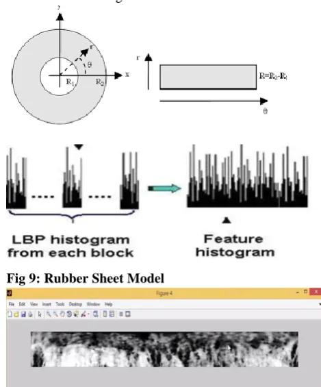
3.1.6 Normalization:
The last step in preprocessing is normalization which includes transforming the iris segmented region into fixed dimensions in order to allow comparisons. The Daugman's Rubber sheet model is used for converting
Cartesian coordinates to polar coordinates with
[image:3.595.105.212.287.386.2]values between 0 and 1 and theta between 0 to 2pi. The following Figure shows the Rubber sheet model and the Normalized iris image.
Fig 9: Rubber Sheet Model
Fig. 10 Normalized Iris
3.2 Feature extraction:
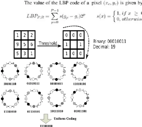
[image:3.595.311.546.317.600.2]3.2.1 Modified LBP:
[image:4.595.317.529.136.300.2]The LBP operator was proposed by ojala and pietikainen. It uses 3x3 window for analysis. The neighbourhood of the centre pixel is thresholded by the value of the center pixel. Each value is coded with 0 if its value is less than center pixel value, otherwise encoded with 1. Binary code which is obtained from analysis window is converted into decimal form. Normalized iris image is divided into cells of 3x3 pixels and LBP texture descriptors are extracted from each region independently. The descriptors are then concatenated to form global descriptor feature vector. The vecor histogram is calculated over the each cell. The histogram has seperate bins for every uniform pattern and all non uniform patterns are assigned to a single bin. If the binary pattern contains atmost 2 transitions i.e from 0 to 1 or 1 to 0 the LBP is uniform.ex:00010000 (2transiitions). Histogram of uniform patterns in the whole image is used as feature vector [8][9]. The following Figure represents the histogram and uniform patterns.
[image:4.595.37.276.322.535.2]Fig. 11 LBP Histogram and Uniform patterns.
Fig. 12 LBP Feature Vector
3.2.2 LGXP:
The basic idea in LGXP is to highlight the Gabor phase sensitivity to the varying positions, whether two phases reflect similar local features. If two phases belong to same interval then they are similar local features, otherwise they reflect different local features. In LGXP, phases are quantized into different ranges, then LXP operator is applied
to quantized phases. Partition the normalized iris image and the pixel values in each block are converted into vector. Apply LGXP to the normalized iris image for each block of the central pixel and each of its neighbors, and finally the binary values are concatenated [11]. The following Figure illustrates the application of Quantization and XOR
Fig. 13 Quantization and XOR
where zc denote the center pixel of the block
In this paper, c value is taken as 4 and we get 4 phase ranges and are known in the following table.
Table 1. Quantized Phase Value for the Input Phase Phase Range(in degrees) Quantized Phase
Value
0-89 0
90-179 1
180-269 2
270-359 3
3.2.3 GLCM:
[image:4.595.34.288.567.676.2] [image:4.595.318.466.708.782.2]Fig. 14 Statistical Features 3.2.4 Gabor features:
[image:5.595.313.547.57.152.2]Gabor Features are nothing but texture based features which are obtained by convolving the image with Gabor filter, this is a linear filter used for edge detection. Gabor filters provide a response that is similar to that of the human visual system, and hence used in this case to extract the texture features from the biometrics [13]. The following figure shows the Gabor filtered image.
Fig. 17 Gabor Filtered image
In this paper, all the above said features such as LBP, LGXP, GLCM, Gabor are combined to form a feature vector. These features plays a significant role to improve the Recognition accuracy. Such final feature vector will be having larger dimension and also will be strong enough such that the recognition accuracy increases. This feature vector is used as input to the next phase i.e Recognition Phase. The following figure shows the combined feature vector.
Fig.18 Combined Iris Feature Vector
3.3 Recognition:
Artificial neural networks (ANN) are considered to develop a Recognition model. In the iris recognition phase, an unknown iris image is represented by a sequence of feature vector {x1, x2 ….xi) by using LBP, LGXP, GLCM, Gabor techniques and then it is tested with the neural network which is trained with the feature vectors which are extracted in feature extraction phase. Artificial neural network is one of the best approaches for recognition. Artificial neural networks are modeled on the neurological functions of the brain. The ANN is capable of pattern recognition, optimization. So, neural network can be trained to perform complex calculations. In multilayer feed forward neural network each node is connected to each other node in the adjacent layer. ANN consists of input layer, hidden layer, output layer. It consists number of layers. Each layer has processing elements called neurons which makes independent computations on the data which is received from the previous layer and pass on the results to the next succeeding layer. The first layer in multilayer feed forward network is called input layer and the last layer is called output layer. Whereas the layers between input layer and output layer are called hidden layers. Synapses between neurons are known as connections, which are represented by edges in the network. The weights are used on the connection between different layers and they have much significance in training of neural network.
In this paper Bat algorithm with multilayer feed forward is proposed to optimize the weights in neural network training process. It is designed to reduce the error between the desired output and actual output. It is one of the successful algorithm in speeding up the convergence rate of the ANN with multi layer feed forward network.
Bat algorithm is based on echolocation behavior of micro bats. The best weights and biases are initialized with Bat, in the first epoch and those weights are passed to the neural network where learning rate α is appended. The Bat will continue searching the best weights until the last epoch of the network is reached. Pseudo code is as follows. 1.Intialize Bat population size and neural network structure. 2. Load training data i.e iris combined features.
3.Feed forward neural network runs using weights initialized with Bat.
5. Update weights and bias in the network by adjusting network parameters using Bat.
6. Bat keeps on calculating the best possible weights at each epoch until the network is converged [15].
The proposed method is implemented in MATLAB platform. In our proposed method CASIA database is considered. Here we are taking 250 images. Here 230 images are used for training and remaining 20 are used for testing [16]. Results show that 98% accuracy is achieved using the proposed technique. Experimental results validate the effectiveness of the proposed method in extracting iris features and achieving good performance.
4. CONCLUSION
Iris recognition systems exploit the fact that humans have unique pattern in their irises. In this paper, different texture features such as LBP, LGXP, GLCM and Gabor are combined to develop an effective Iris Recognition model. ANN and Bat algorithm is used to train the Recognition model and also the performance is analyzed with sample data set. From the experimental results it is proved that accuracy of the recognition model is 98%.
5. REFERENCES
[1] P. S. Sanjekar and J. B. Patil An Overview of Multimodal Biometrics Signal & Image Processing : An International Journal (Sipij) Vol.4, No.1.
[2] Akanksha Singh Thakur, Namrata Sahayam, Speech Recognition Using Euclidean Distance, International Journal of Emerging Technology and Advanced Engineering Website: (ISSN 2250-2459, ISO 9001:2008 Certified Journal, Volume 3, Issue 3, March 2013) 587
[3] ttp://www.biometrics.gov/Documents/irisrec.pdf". [4] Vijay A. Kotkar, Sanjay S. Gharde, Review of Various
Image Contrast Enhancement Techniques, International Journal of Innovative Research in Science, Engineering and Technology, Vol. 2, Issue 7, July 2013.
[5] Dr. E. Chandra, Noise Elimination in Fingerprint Image Using Median Filter, Int. J. Advanced Networking and Applications 950 Volume: 02, Issue: 06, Pages: 950-955 (2011).
[6] Rupinder Saini, Comparison Of Various Biometric Methods, International Journal of Advances In Science and Technology, Vol 2 Issue I, March 2014.
[7] Retno Kusumaningrum and Aniati Murni Arymurthy, Color and Texture Feature for Remote Sensing – Image Retrieval System: A Comparative Study. International Journal of Computer Science Issues, Vol. 8, Issue 5, No 2, September 2011 ISSN (Online): 1694-0814 [8] Deepak Sharma Dr. Ashok Kumar, An Empirical
Analysis Over the Four Different Feature-Based Face and Iris Biometric Recognition Techniques, International Journal of Advanced Computer Science and Applications, Vol. 3, No. 10, 2012.
[9] Suhad A. Ali, Dr. Loay E. George, Iris Recognition System Based on Texture Features, Int. J. of Network Security, Vol. 6, April 2014.
[10] M. Z. Rashad1, M. Y. Shams2, O. Nomir2, and R. M. El-Awady, Iris Recognition Based on Lbp and Combined LVQ Classifier, International Journal of Computer Science & Information Technology Vol 3, No 5, Oct 2011 DOI: 10.5121/Ijcsit.2011.3506 67. [11] G.Thamarai Selvi and K.Duraisamy, Extraction of
Tumor Image from MRI Images Using Gabor XOR Pattern, International Journal of Electronics & Communication Engineering Research, Vol. 1 Issue 5, October- 2013 ISSN: 2321-9718.
[12] Dhia Alzubaydi, Shyma Akram Alrubaie, Probabilistic Neural Network with GLCM and Statistical Measurements for Increasing Accuracy of Iris Recognition System, International Journal of Computer Applications (0975 – 8887) Volume 136 – No.12, February 2016.
[13] Shufu Xie, Shiguang Shan, Xilin Chen, and Jie Chen, Fusing Local Patterns Of Gabor Magnitude And Phase For Face Recognition, IEEE Transactions On Image Processing, Vol. 19, No. 5, May 2010 1349.
[15] Nazri Mohd. Nawi1, M. Z. Rehman1,b, Nurfarain Hafifie1,c ,Abdullah. BAT-BP: A New Bat Based Back-Propagation Algorithm for Efficient Data Classification
[16] CASIA Iris Image Database,