1556-6811/09/$12.00 doi:10.1128/CVI.00216-09
Copyright © 2009, American Society for Microbiology. All Rights Reserved.
Influence of the Tissue Microenvironment on Toll-Like Receptor
Expression by CD11c
⫹
Antigen-Presenting Cells Isolated
from Mucosal Tissues
䌤
Shunsuke Takenaka, Sarah McCormick, Ekaterina Safroneeva, Zhou Xing, and Jack Gauldie*
Department of Pathology and Molecular Medicine, McMaster University, Hamilton, Ontario, Canada
Received 31 May 2009/Returned for modification 9 July 2009/Accepted 15 September 2009
It is recognized that functional activities of antigen-presenting cells (APCs) in mucosal tissue sites differ from those of systemic APCs; however, it is unknown whether there are further differences between APC populations residing in different mucosal sites. In this study, we directly compared murine CD11cⴙ APCs isolated from colon, lung, and spleen and found that APCs isolated from these tissues differ considerably in Toll-like receptor (TLR) expression and responses to in vitro TLR ligand stimulation. We also provide evidence that tissue microenvironments dictate distinct patterns of TLR expression by CD11cⴙ APCs in different mucosal tissues. Moreover, CD11cⴙcells isolated from different tissues have varied capacities to induce the development of T helper 1 (Th1), Th2, or regulatory CD4ⴙT cells. Thus, unique tissue microenvironments have a significant influence on determining TLR expression by CD11cⴙcells that migrate to and reside in each mucosal tissue and are likely to modulate their functional activities.
Antigen-presenting cells (APCs), such as dendritic cells (DCs), have the ability to control immune outcomes, and a number of factors are thought to be involved in determining the types of immune responses induced. The nature of the antigen/pathogen is likely to be a strong determinant of the type of immune response that is induced. For example, T helper 1 (Th1) responses are preferentially induced when the host is infected with intracellular pathogens, such as mycobac-teria (4). Conversely, helminth infections tend to induce Th2 responses (39). In this regard, DCs appear to utilize pattern recognition receptors, such as Toll-like receptors (TLRs), to distinguish between various molecules to induce suitable im-mune responses (33). The type of imim-mune response induced may also be influenced by distinct subpopulations of APCs that are involved in priming the appropriate T cells. DCs are het-erogeneous populations of cells in any given tissue (40), and in the mouse, CD8␣⫹ DCs are thought to be specialized for inducing Th1 responses, whereas CD8␣⫺ DCs are thought primarily to induce Th2 responses (28). Moreover, it has been shown that mouse CD8␣⫹ and CD8␣⫺ DCs preferentially present antigens to CD8⫹and CD4⫹T cells, respectively (14). In contrast to DCs, macrophages are thought to be associated with immunoregulation and tolerance rather than immune in-duction (11, 19).
The microenvironment in a tissue is thought to have a major impact on the nature of immune responses induced at each tissue site (25). Different mucosal tissues, such as those of the respiratory and gastrointestinal or genitourinary tracts, have distinct physiological functions, are exposed to unique antigen and pathogen milieus, and have diverse cellular compositions;
therefore, APCs that reside in each mucosal tissue must be adapted to handle such differences. Earlier work showed that splenic microenvironments can induce differentiation of ma-ture bone marrow-derived DCs and hematopoietic stem cells to regulatory DCs (45, 53) and also negatively regulate acti-vated plasmacytoid DCs (27). Also, thymic stromal lymphopoi-etin, which is expressed in lung and skin, is believed to have a Th2-skewing effect on DCs and has been associated with de-velopment of allergic airway inflammation (20, 41, 54). Fur-thermore, DCs isolated from different tissue sites have func-tional variations. Mouse DCs isolated from Peyer’s patches preferentially induce Th2-type immune responses, whereas spleen DCs tend to induce Th1-type immune responses, even though these two compartments have similar subset composi-tions (22). Also, DCs isolated from mouse liver are able to produce interleukin-12 (IL-12), regardless of their CD8␣ ex-pression, whereas in the spleen, CD8␣⫹DCs are much supe-rior at IL-12 production compared with CD8␣⫺ DCs (32). These observations strongly suggest that tissue microenviron-ments impose a significant influence on the function of DCs and subsequent immune responses.
Even though APCs from some mucosal tissues such as the lung, small intestine, colon, and genital tract have been studied in isolation and compared to spleen counterparts (1, 6, 8, 44), few comparisons have been reported on APCs isolated from different mucosal tissues. Knowledge of similarities and differ-ences between APCs residing in various tissues is important, since immunity is likely regulated differently in each mucosal tissue site (34).
In this study, we used purified CD11c⫹cells from murine colon, lung, and spleen to undertake a direct comparison of their phenotypes and activities. We found that even though CD11c⫹cells from all tissues examined are able to carry out typical APC functions, they have distinct cytokine production profiles, modulation of T-cell responses, and regulatory T-cell induction, along with different TLR expression patterns. More-* Corresponding author. Mailing address: Department of
Pathol-ogy and Molecular Medicine, MDCL-4016, McMaster University, 1200 Main Street West, Hamilton, Ontario L8N 3Z5, Canada. Phone: (905) 525-9140, ext. 22610. Fax: (905) 522-6750. E-mail: [email protected].
䌤Published ahead of print on 23 September 2009.
1615
on August 17, 2020 by guest
http://cvi.asm.org/
over, our data suggest that this differential TLR expression is the direct result of the mucosal tissue microenvironments in which cells reside.
MATERIALS AND METHODS
Mice.C57BL/6 mice 6 to 8 weeks old were from Charles River Laboratories, CD45.1 congenic B6 mice 6 to 8 weeks old were from Jackson Laboratories, and OT-II transgenic mice were bred at McMaster University’s Central Animal Facility. All procedures were approved by the Animal Research Ethics Board of McMaster University.
Isolation of CD11cⴙcells from colon and lung tissues.CD11c⫹cells from colon and lung were isolated using magnetic-activated cell sorting (MACS) as described previously (44). Briefly, large intestine was washed, cut into small pieces, and incubated at 37°C in EDTA-supplemented medium to remove epi-thelial cells and then digested with collagenase type VIII (Sigma-Aldrich) for 1 h at 37°C. Colonic cells were further purified using 43%/63% Percoll (GE Health-care) density gradient centrifugation, and cells at the interface were collected. Lungs were perfused to remove red blood cells, cut into small pieces, and digested for 1 h at 37°C with collagenase. Colon or lung CD11c⫹cells were then purified using CD11c microbeads (Miltenyi Biotec) and running the samples through MS⫹columns twice to consistently achieve an enriched population of ⬎90% CD11c⫹cells. Where specified, exhaustive bronchoalveolar lavage (BAL) was performed as described previously (37) prior to lung tissue being processed for CD11c⫹cell isolation, to reduce alveolar macrophages in the interstitial cell preparation.
Splenocyte isolation. Spleens were infused with collagenase-supplemented RPMI, cut into small pieces, and digested for 30 min at 37°C. The resulting cell suspension was treated with M-lyse buffer (R&D Systems) for red blood cell lysis. Spleen CD11c⫹cells were isolated by MACS as described above. Splenic T cells were purified using a CD4⫹T-cell isolation kit (Miltenyi Biotec) according to the manufacturer’s instructions. For some experiments, CD25⫹cells were depleted using CD25-specific magnetic beads prior to T-cell isolation to obtain CD4⫹ CD25⫺T cells. Purified T cells were routinely ⬎90% pure based on CD4 staining.
Ex vivo CD11cⴙcell culture.A total of 1⫻105
CD11c⫹cells in 200l were incubated at 37°C in complete medium supplemented with 40 ng/ml granulocyte-macrophage colony-stimulating factor and 200 U/ml gamma interferon (IFN-␥; Roche Diagnostics), for the optimal induction of IL-12p70 (17, 18, 47) in the presence of 10g/ml peptidoglycan (PGN;Staphylococcus aureus; Fluka-Sigma-Aldrich), 100 ng/ml lipopolysaccharide (LPS;Escherichia coliO26:B6; Sigma-Aldrich), 10g/ml recombinant flagella filament protein FliC (Ali Ashkar, Mc-Master University), or 6M CpG oligodeoxynucleotide (ODN) 1826 (5⬘-TCC ATGACGTTCCTGACGTT-3⬘; McMaster University Mobix Lab) for 36 h, and supernatants were analyzed for cytokine levels.
In vitro APC–T-cell coculture.CD11c⫹cells were incubated with 200g/ml ovalbumin (OVA) protein (grade V; Sigma-Aldrich) for 2 h at 37°C and washed well with phosphate-buffered saline. OT-II CD4⫹CD25⫺T cells were labeled with 5M carboxyfluorescein succinimidyl ester (CFSE; Molecular Probes) for 5 min at room temperature and washed well. A total of 5⫻105
T cells per well were cocultured for 4 days with 5⫻104OVA-pulsed CD11c⫹cells. Where indicated, OVA protein or recombinant human transforming growth factor (TGF-; 2 ng/ml; R&D Systems) was added to the culture medium for the duration of the coculture. T-cell proliferation was examined by visualizing CFSE dilution by flow cytometry. A mouse regulatory T-cell staining kit (eBioscience) was used to detect CD25⫹Foxp3⫹regulatory cells. Also, coculture supernatants were examined for cytokine levels for some experiments.
Adoptive transfer of spleen CD11cⴙcells.Spleen CD11c⫹cells from wild-type B6 mice (CD45.2) were isolated as described above. A total of 15⫻106cells/
mouse were injected intravenously into CD45.1 congenic host mice. After 6 days, lung and spleen cells from host mice were isolated and analyzed by flow cytom-etry for TLR2 expression in adoptively transferred CD11c⫹cells.
TLR gene expression analysis.Total RNA was isolated using an RNeasy minikit (Qiagen), and cDNA was synthesized using a RETROscript kit (Am-bion) according to the manufacturer’s instructions. Real-time PCR gene expres-sion analysis was conducted using an ABI Prism 7900HT apparatus (Applied Biosystems) and TaqMan gene expression assays for TLR2, TRL4, TLR5, TLR9, and ribosomal protein L32 (Applied Biosystems). Relative gene expression for TLR was calculated by normalizing the data using L32 expression and then applying the ⌬⌬Ct method with the following formula: gene expression ⫽ 2⫺⌬⌬Ct⫻1,000, where⌬⌬Ct⫽ ⌬Ct(CD11c⫹cells)⫺ ⌬Ct(total splenocyte) and ⌬Ct(gene)⫽Ct(gene) –Ct(L32).
Flow cytometry.For TLR protein expression analysis, cells were stained with anti-CD11c and anti-TLR2 monoclonal antibodies (BD Pharmingen). Anti-CD45.2 monoclonal antibody (BD Pharmingen) was also used for the detection of adoptively transferred cells. CD4⫹Foxp3⫹CD25⫹regulatory T cells were detected using a mouse regulatory T cell staining kit (eBioscience) according to the manufacturer’s instructions. Nonviable cells were detected using Via-Probe (7-amino-actinomycin D; BD Pharmingen). Samples were run on an LSR II system (BD Biosciences), and data analysis was done using FlowJo software (Tree Star).
Cytokine measurement.Cytokine production by cultured cells was determined using enzyme-linked immunosorbent assay kits for murine IL-4, IL-10, IL-12p70, tumor necrosis factor alpha (TNF-␣), and IFN-␥(all from R&D Systems).
Data analysis. Data are expressed as means⫾standard deviations (SD). Statistical significance of the data was determined by using an unpaired, two-tailed Student’sttest.Pvalues of⬍0.05 were considered statistically significant.
RESULTS
CD11cⴙcells isolated from colon, lung, and spleen respond uniquely to stimulation by different TLR ligands.We stimu-lated the cells in vitro with PGN, LPS, recombinant FliC, and CpG ODN, ligands for TLR-2, -4, -5, and -9, respectively, in the presence of granulocyte-macrophage colony-stimulating factor and IFN-␥ and analyzed the production of IL-12p70, TNF-␣, and 10. While DCs from all tissues produced IL-12p70 when stimulated with each of the ligands under these conditions, only CpG ODN induced a robust response, with production of IL-12 by colon and spleen CD11c⫹cells being significantly greater than with lung DCs (Fig. 1). On the other hand, TNF-␣was produced by lung CD11c⫹cells in copious amounts when stimulated with all of the ligands tested, but colon and spleen CD11c⫹cells did not produce this cytokine. Levels of TNF-␣production by lung CD11c⫹cells were similar for PGN, LPS, and FliC stimulation but significantly lower following stimulation with CpG ODN (Fig. 1). IL-10 was spon-taneously produced by colon and lung CD11c⫹ cells under these culture conditions, even in the absence of added stimuli, and the level of production stayed relatively constant even upon stimulation with TLR ligand, while spleen DCs required stimulation for IL-10 production (Fig. 1). Thus, it appears that different tissue CD11c⫹cells, including those from different mucosal tissues, respond uniquely to TLR ligand stimulation. IL-10 levels detected in this study were close to the detection limit for the enzyme-linked immunosorbent assay used, but there was a clear trend toward mucosal CD11c⫹cells having a greater capacity to produce IL-10 in the steady state, compared with systemic (spleen) CD11c⫹cells.
One question that arose was whether the high level of TNF-␣produced by lung CD11c⫹cells (Fig. 1) was due to DCs or macrophages, since alveolar macrophages are known to express CD11c on their surface, as are DCs (48), and macro-phages are one of the major producers of TNF-␣ (49, 50). Because lung DCs and alveolar macrophages cannot be sepa-rated on the basis of CD11c expression by techniques such as MACS, we conducted extensive BAL prior to processing this washed lung tissue to considerably reduce the content of alve-olar macrophages from interstitial cell preparations. We then compared cytokine responses to TLR ligation by this cell prep-aration with that by lung CD11c⫹cells prepared without BAL and to isolated BAL cells only (⬎98% alveolar macrophages). We found that even though TNF-␣production by lung CD11c⫹ cells decreased when BAL cells were removed, it was still
1616 TAKENAKA ET AL. CLIN. VACCINEIMMUNOL.
on August 17, 2020 by guest
http://cvi.asm.org/
significantly greater than that elicited by spleen CD11c⫹cells (Fig. 2), suggesting that lung DCs are likely to be a robust source of TNF-␣, producing significantly greater levels of TNF-␣ than colon and spleen DCs. Importantly, IL-12p70 production levels by lung CD11c⫹cells isolated from whole lung and from lung tissue after performing extensive BAL were similar (Fig. 2). In other words, the removal of BAL cells did not affect the IL-12p70 production by lung CD11c⫹cells, and the production of IL-12 by only BAL alveolar macrophages was very low (Fig. 2). This is in accordance with our previous report showing that alveolar macrophages produce little bio-active IL-12p70 (49, 50). These data indicate that only alveolar macrophages and not IL-12p70-producing lung DCs were re-moved as a consequence of conducting prior lung lavage. It will be of interest to further separate lung DCs and alveolar mac-rophages by fluorescence-activated cell sorting, as done by others previously (26); however, diminished cell viability and adequate recovery levels have limited us from conducting such a study so far.
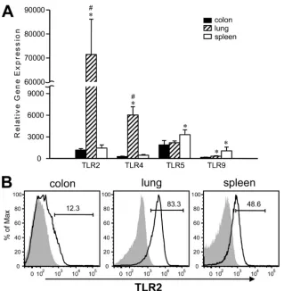
Lung CD11cⴙ cells express significantly greater levels of TLR2 and TLR4.Since CD11c⫹cells derived from different tissues respond differently to TLR ligand stimulation, we won-dered if it was due to a difference in their TLR expression and examined this by gene and protein expression in isolated CD11c⫹cells. Intriguingly, lung CD11c⫹cells expressed TLR2
and TLR4 genes at a much greater magnitude than colon and spleen CD11c⫹cells (Fig. 3A). To the contrary, even though TLR5 and TLR9 were expressed by spleen CD11c⫹cells at significantly greater levels than by colon or lung CD11c⫹cells, the extent of the difference was not as striking as what was observed for TLR2 and TLR4. We further measured TLR2 protein expression using flow cytometry and found the majority of lung CD11c⫹cells indeed expressed TLR2 on their surface, reflecting high TLR2 gene expression. Both spleen and colon CD11c⫹ cells had fewer cells expressing TLR2 than lung CD11c⫹cells, and spleen CD11c⫹cells had a greater portion of cells expressing TLR2 than colon CD11c⫹cells (Fig. 3B). These results show that lung CD11c⫹cells have a very different TLR expression pattern relative to colon and spleen CD11c⫹ cells. Moreover, this high expression of TLR2 and TLR4 by lung CD11c⫹cells may explain the robust TNF-␣production observed when they are stimulated with PGN or LPS (Fig. 1). Additionally, the high TNF-␣production by lung CD11c⫹cells upon stimula-tion by LPS, FliC, and CpG ODN, and much lower but detectable levels produced spontaneously under these conditions, may sug-gest an intrinsic capacity of lung CD11c⫹cells to produce this cytokine.
The lung tissue microenvironment induces TLR2 expression by adoptively transferred spleen CD11cⴙcells.To test whether the microenvironment influences the greater levels of TLR2 in lung CD11c⫹ cells, spleen CD11c⫹cells isolated from wild-FIG. 1. Cytokine production by colon, lung, and spleen CD11c⫹
cells upon in vitro TLR ligand stimulation. Purified CD11c⫹cells from colon, lung, and spleen were incubated with PGN, LPS, FliC, or CpG ODN, and cytokine levels in the culture supernatants were deter-mined. Results are representative of three experiments and are pre-sented as means⫾SD.ⴱ,P⬍0.05 relative to corresponding lung data; ‡,P⬍0.05 relative to corresponding spleen data; #,P⬍0.05 relative to corresponding colon data.
FIG. 2. Cytokine production by lung CD11c⫹cells after removal of BAL cells. CD11c⫹cells were purified from either whole lung or lung after BAL (BAL ⫺ lung) and were stimulated with various TLR ligands. BAL cells and spleen CD11c⫹cells were also used for com-parison purposes. Cells were incubated with PGN, LPS, FliC, or CpG ODN, and cytokine levels in the culture supernatants were deter-mined. Results are representative of three experiments and are pre-sented as means⫾SD.ⴱ,P⬍0.05 relative to BAL cell data; ‡,P⬍ 0.05 relative to BAL⫺lung data; †,P⬍0.05 relative to both whole lung and BAL⫺lung data.
on August 17, 2020 by guest
http://cvi.asm.org/
type mice (CD45.2⫹) were adoptively transferred to CD45.1 congenic host mice. A small number, but very distinct popula-tion, of CD11c⫹CD45.2⫹cells was recovered at 6 days post-transfer, from both lung and spleen of the hosts, and examined for their TLR2 expression (Fig. 4A). CD11c⫹cells from both CD45.2⫹ and CD45.2⫺ cell populations found in the host’s lung highly expressed TLR2 (Fig. 4B). Since CD45.2⫺cells are resident host lung DCs, it is not surprising that they have high TLR2 expression. CD45.2⫹cells, originally spleen DCs with lower levels of TLR2 (Fig. 3B), express high levels of TLR2 when found in lung (Fig. 4B). To rule out any artifact of adoptive transfer, we also examined the host’s spleen and found that both CD45.2⫹ and CD45.2⫺ CD11c⫹ cells ex-pressed lower levels of TLR2 (Fig. 4B). The difference in TLR2 expression by adoptively transferred CD45.2⫹ cells found in the host’s lung and spleen was statistically significant, whereas CD11c⫹ cells found in the same tissue (lung or spleen) expressed similar levels of TLR2 (Fig. 4C). We have also conducted an experiment in which spleen CD11c⫹cells were labeled with CFSE and adoptively transferred to host mice. It was possible to visualize a greater number of donor cells by flow cytometry; however, strong autofluorescence found in the lung samples made the analysis more complex.
Nonetheless, we observed comparable results to what is de-scribed above (unpublished observation). These data suggest that lung tissue microenvironments can induce TLR2 expres-sion by CD11c⫹cells that migrate to that tissue.
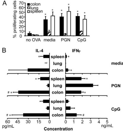
CD11cⴙcells isolated from colon, lung, and spleen induce unique CD4ⴙT-cell responses.Priming of CD4⫹T cells is one of the key functions of APCs to induce immunity or tolerance, and since CD11c⫹cells isolated from colon, lung, and spleen have distinct responses and phenotypes, we compared the abil-ities of tissue-derived CD11c⫹APCs to prime antigen-specific CD4⫹T cells by examining the induction of T-cell prolifera-tion. Both colon and spleen CD11c⫹cells pulsed with OVA protein induced proliferation of OVA-specific OT-II CD4⫹T cells efficiently in vitro (Fig. 5A). Contrary to this, lung CD11c⫹ cells did not induce CD4⫹ T-cell proliferation as effectively (Fig. 5A), and this did not change when cells were cocultured in TLR ligand-containing medium (Fig. 5A).
DCs are known to have the ability to control immune out-come, and we questioned whether CD11c⫹cells isolated from different tissues would have different abilities to induce Th1 or Th2 T-cell responses (3, 25). We examined supernatants from APC–T-cell cocultures for representative type 1 and type 2 cytokines, IFN-␥and IL-4, respectively. We found that colon FIG. 3. TLR expression by purified tissue-derived CD11c⫹cells. (A) Relative TLR gene expression by tissue CD11c⫹cells was determined by real-time RT-PCR utilizing⌬⌬Ct method. Results are representative of at least three experiments and are presented as mean⫾SD.ⴱ,P⬍0.05 relative to corresponding colon samples. #,P⬍0.05 relative to corresponding spleen samples. (B) TLR protein expression was determined by flow cytometry. Purified CD11c⫹cells were labeled for TLR2 along with other surface markers. Open histograms indicate TLR2 specific staining and shaded histograms indicate isotype control staining. Numbers indicate percentages of TLR2⫹cells. Representative histograms from at least three experiments are shown.
1618 TAKENAKA ET AL. CLIN. VACCINEIMMUNOL.
on August 17, 2020 by guest
http://cvi.asm.org/
and spleen CD11c⫹cells were able to induce IL-4 production by T cells to a greater extent than lung CD11c⫹cells, with or without TLR ligand stimulation (Fig. 5B). IFN-␥production by the T cells was rather low when cells were cocultured in me-dium alone, but a robust induction was observed when the TLR2 ligand PGN was added to the medium, with lung DCs exhibiting the greatest IFN-␥production (Fig. 5B). IFN-␥ pro-duction was also induced with CpG ODN in the medium, but the responses were not as robust as with PGN (Fig. 5B). Based on these data, CD11c⫹ APCs from different tissues induce distinct responses by antigen-specific CD4⫹ T cells, and al-though both colon and lung CD11c⫹cells are mucosal tissue APCs, they induce very different T-cell responses. Further-more, colon CD11c⫹cells induced T cells to produce the type 2 cytokine IL-4 much more readily than lung or spleen coun-terparts.
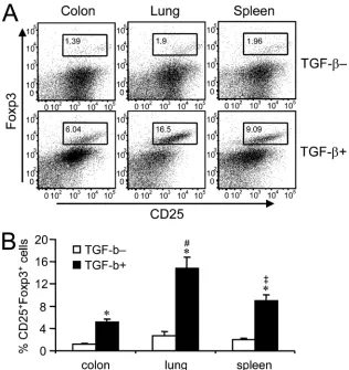
Lung CD11cⴙcells induce a greater level of CD25ⴙFoxp3ⴙ regulatory T-cell development.Recent studies have shown that DCs isolated from small intestine lamina propria or mesenteric lymph nodes (MLNs) have a superior capacity to induce the development of CD25⫹ forkhead box protein P3 (Foxp3⫹) regulatory T cells compared with spleen DCs (30, 43). We cocultured CD11c⫹cells isolated from colon, lung, or spleen with OT-II CD4⫹T cells in the presence of OVA protein, with or without TGF- supplementation. As reported previously, we found that addition of TGF-was necessary for the in vitro development of CD25⫹Foxp3⫹regulatory T cells induced by FIG. 4. TLR2 expression by adoptively transferred spleen CD11c⫹
cells isolated from host lung and spleen. Purified spleen CD11c⫹cells from wild-type (CD45.2⫹) mice were adoptively transferred intrave-nously to host CD45.2⫺mice, and 6 days later, lung and spleen cells from hosts were isolated to examine CD45.2⫹CD11c⫹cells for TLR2 expression by flow cytometry. (A) CD45.2⫹ CD11c⫹ cells (derived from a donor’s spleen) and CD45.2⫺CD11c⫹cells (resident host cells) were gated and examined for TLR2 expression. Left panels show representative flow cytometry plots for the lung and spleen from mice to which CD45.2⫹cells were adoptively transferred. Right panels show plots for the lung and spleen from a control mouse to which no cells were adoptively transferred. Numbers indicate percentages of cells within each gate. (B) Representative flow cytometry plots showing TLR2 expression by CD45.2⫹and CD45.2⫺CD11c⫹cells found in host lung and spleen analyzed using the gates shown in Fig. 5A, below. Open histograms indicate TLR2-specific staining, and shaded histo-grams indicate isotype control staining. Numbers indicate percentages of TLR2⫹ cells within the gated population. (C) Percentages of TLR2⫹cells in CD45.2⫹CD11c⫹(donor cells) and CD45.2⫺CD11c⫹ (host cells) populations found in the lungs and spleens of host mice. Results are representative of four experiments and are presented as means⫾SD.ⴱ,P⬍0.05.
FIG. 5. Antigen-specific CD4⫹T-cell stimulatory abilities of tissue-derived CD11c⫹cells in vitro. Purified CD11c⫹cells from colon, lung, and spleen were loaded with OVA protein and cocultured with CFSE-labeled OT-II CD4⫹ CD25⫺ T cells. Culture medium was supple-mented with PGN or CpG ODN where indicated. (A) CD4⫹T-cell proliferation induced by CD11c⫹ cells, measured by CFSE dilution and detected using flow cytometry; data are reported as the percentage of proliferating cells. (B) IL-4 and IFN-␥produced by CD4⫹T cells stimulated with CD11c⫹cells. Results are representative of three ex-periments and are presented as means⫾SD.ⴱ,P⬍0.05 relative to corresponding lung data; #,P⬍0.05 relative to corresponding spleen data.
on August 17, 2020 by guest
http://cvi.asm.org/
DCs from any of the tissues examined (Fig. 6A). Lung CD11c⫹cells were the most efficient inducers of regulatory T cells, and colon CD11c⫹cells were the least efficient of the three tissue APCs we compared (Fig. 6A). These differ-ences were also found to be statistically significant (Fig. 6B) and suggest that CD11c⫹APCs from different mucosal tis-sues have different capacities to induce the development of regulatory T cells, and colon DCs, unlike DCs isolated from small intestine or MLNs (30, 43), are not robust inducers of regulatory T cells.
DISCUSSION
Increasingly, studies of immune responses in various tissues highlight the importance of local tissue microenvironments in directing local immunity (34). In this study, we directly com-pared the functions and TLR expression levels for CD11c⫹ cells from two distinct mucosal sites, the colon and lung, and we have provided evidence for distinct differences from each other and of both from CD11c⫹cells in the spleen. We also show critical involvement of the tissue microenvironment in regulating TLR expression by CD11c⫹ cells. CD11c⫹ cells
found in the colon and spleen are likely to be DCs, since no other cell type in these tissues in steady state is known to express CD11c. In contrast, lung CD11c⫹cells include both DCs and macrophages, because alveolar macrophages also ex-press CD11c (10, 26). As such, isolated CD11c⫹cells from the lung are a mixture of lung DCs and alveolar macrophages and can be referred to as lung CD11c⫹APCs.
In a previous publication, we reported that lung CD11c⫹ cells have a similar surface marker phenotype to colon but not to spleen CD11c⫹cells, where both lung and colon CD11c⫹ cells were comprised of a large proportion of a CD11b⫹ pop-ulation but lacked a significant presence of cells expressing CD8␣or B220 (44). Based on this information, our initial idea was that APCs residing in mucosal tissues may also share similar functions with spleen APCs. Contrary to this, our find-ings show that CD11c⫹ cells isolated from colon and lung respond differently and distinctly to stimulation by various TLR ligands, suggesting that APCs with similar surface phe-notypes may function uniquely in different tissues. Alternately, it is also possible that CD11c⫹CD11b⫹cells found in colon and lung may belong to distinct DC subsets differentially ex-FIG. 6. CD25 and Foxp3 expression by CD4⫹T cells stimulated with tissue-derived CD11c⫹cells in vitro. OVA antigen-specific CD4⫹CD25⫺ T cells were cocultured with DCs in the presence of OVA protein, and the induction levels of CD25 and Foxp3 expression by T cells were examined. (A) Flow cytometry plots showing the expression of CD25 and Foxp3 by CD4⫹T cells (gated on CD4⫹cells). Top panels show the cells cultured without TGF-supplementation, and bottom panels show the cells cultured with TGF-. Representative results from three experiments are shown. (B) Percentages of CD25⫹ Foxp3⫹ cells, determined by creating a gate as shown in Fig. 5A. Cells were cultured with or without TGF- supplementation. Results are representative of three experiments and are presented as means⫾SD.ⴱ,P⬍0.05 relative to corresponding TGF-–
data; #,P⬍0.05 relative to corresponding spleen and colon data; ‡,P⬍0.05 relative to corresponding colon data.
1620 TAKENAKA ET AL. CLIN. VACCINEIMMUNOL.
on August 17, 2020 by guest
http://cvi.asm.org/
pressing other surface markers, given the vast number of DC subsets described in recent years (21, 40).
TNF-␣ is known to be important for type 1 immune regu-lation and host protection against various intracellular patho-gens (15, 31, 52); therefore, the robust TNF-␣production by lung CD11c⫹cells suggests that these cells are optimized to ward off incoming pathogens at the mucosa. In addition, our TLR5 expression and FliC stimulation data suggest that there may be intrinsic differences in TNF-␣-producing abilities among APCs from different tissues. In contrast, colon CD11c⫹ cells seem to lack the ability to produce a high level of TNF-␣ upon TLR ligand stimulation, and this may be associated with low-level expression of TLR2 and TLR4. Colonic tissue is highly colonized by commensal bacteria, providing a constant source of TLR ligand to colon APCs in the steady state, and this environmental circumstance may require colon CD11c⫹ cells to be less inflammatory in nature. Additional evidence to support this possibility is the constitutive expression of IL-10 by colon CD11c⫹ cells and lack of expression by spleen CD11c⫹ cells. Since the spleen is normally a sterile organ, there would be little need for innate protection, but CD11c⫹ cells in the spleen still need to be equipped to regulate immune responses in the event of infection to avoid uncontrolled im-mune activation. These data support the notion of enhanced immune regulation in the intestinal mucosa, as suggested by others (8, 22). Another interesting finding was the robust IL-12p70 production by CD11c⫹cells stimulated with CpG ODN. CpG ODN has been used by others to prevent infection at mucosal sites (2, 38); thus, induction of IL-12p70 production by CD11c⫹cells may be one mode of action by which CpG ODN confers protection against mucosal challenge by patho-gens.
We and others have recently shown that colon DCs or lung DCs express TLRs at different levels relative to spleen DCs (6, 44). To our knowledge, the current study is the first report describing heightened TLR2 and TLR4 expression by lung APCs relative to APCs from another mucosal site. Previous studies have shown the importance of TLR2 and TLR4 expres-sion for host protection against respiratory bacterial infection (16, 36); therefore, the high TLR2 and TLR4 expression levels of lung CD11c⫹APCs may be a critical means of host protec-tion. Together with the apparent enhanced capacity of lung CD11c⫹APCs to produce TNF-␣, this may indicate the unique requirement imposed in lung tissue for APCs to control respi-ratory infections.
Accumulating evidence suggests that the tissue microenvi-ronment influences local immune responses (12). It is likely that APC functions are modulated by many factors that include tissue physiologic functions, the presence/absence of commen-sal organisms, the antigenic milieu, cytokines and chemokines, tissue matrix and integrin expression, and cellular composi-tions. The induction of TLR2 expression by CD11c⫹cells due to the lung tissue microenvironment indicates that the differ-ence in observed functionalities between cells isolated from the colon, lung, and spleen are, at least partially, due to their exposure to different tissue microenvironments. In this regard, a number of differences exist between the colon, lung, and spleen that could potentially modulate APC functions. One of the most notable differences is the sterility of these tissues. The colon is home to a vast number of commensal bacteria,
whereas the spleen is normally a sterile compartment. Also, the upper airways and lung tissue are constantly exposed to environmental microorganisms that are present in the air, con-tributing to a unique lung microenvironment. We speculate that the differences in microorganism compositions between the tissues may direct functional differences seen in CD11c⫹ cells from the colon, lung, and spleen. Additionally, epithelial cells are likely to be involved in modulating DC functions at mucosal tissue sites (5, 35). In support of these possibilities, a recent report has shown in vitro that an intestinal epithelial cell line exposed to commensal organisms directs DCs to become more tolerogenic, expressing lower levels of maturation mark-ers and inflammatory cytokines while having enhanced IL-10 production (51). Also, it has been suggested that the effects of the microenvironment can override the functional differences between DC subsets (13, 24).
Aside from their innate immune functions, APCs are critical for host protection because of their T-cell-priming ability. Ad-ditionally, DCs are able to control the adaptive immune re-sponse by inducing the development of different types of T cells, including Th1, Th2, and regulatory T cells (3, 25). It is thought that mucosal tissue sites are more Th2 biased than systemic sites, which are more Th1 biased (1, 22). Our colon CD11c⫹cell data support this notion; however, lung CD11c⫹ cells did not show such expected tendencies in our hands. Interestingly, PGN enabled lung CD11c⫹ cells to induce a robust IFN-␥production by CD4⫹T cells without a notable increase in T-cell proliferation. This observation correlates with the T-cell stimulatory ability of DCs in the presence of alveolar macrophages where alveolar macrophages have been shown to inhibit the DC’s ability to induce T-cell proliferation while still allowing for the induction of cytokine production by T cells (46). Also, a reduction in alveolar macrophage contri-bution did not drastically change the cytokine production pro-file by lung CD11c⫹ cell preparations. Together these data indicate that lung DCs are capable of inducing Th1 immune responses in the presence of an appropriate stimuli. Such char-acteristics of lung DCs have also been suggested by others (42). Direct comparison of DCs from different areas of the intes-tine has not been done thus far, but data from the current study and studies conducted by others collectively indicate that DC functions may differ in colon and small intestine. DCs from colon and small intestine may share some common activities, such as constitutive expression of IL-10 (8, 23). It has recently been shown that blocking IL-10 signaling enhances TLR li-gand-induced IL-12-producing ability of small intestine DCs (29). It is thus also possible that IL-10 produced by colon DCs performs a similar role in controlling their functions. Despite such potential similarities, the subset compositions of DCs seem to differ between the colon and small intestine, as CD8⫹ DCs are absent in the colon (44) but present in the small intestine (23). Recent studies have also shown that DCs iso-lated from small intestine lamina propria and MLNs are supe-rior inducers of Foxp3⫹regulatory T-cell development in vitro compared to spleen DCs (9, 43). This is in contrast to our current findings using colon DCs, suggesting there are unique DC functions among DCs residing within different regions of the gastrointestinal tract.
It has been reported that TNF-␣enhances Foxp3 expression and regulatory T-cell activity (7). As such, the enhanced ability
on August 17, 2020 by guest
http://cvi.asm.org/
of lung CD11c⫹cells to induce regulatory T-cell development may be due to their capacity to produce high levels of TNF-␣. The observation that lung CD11c⫹ cells are able to induce both IFN-␥production by T cells and regulatory T-cell devel-opment may seem contradictory; however, such abilities may allow lung APCs to both induce and regulate T-cell immunity to protect the host against pathogens on one hand and to avoid overwhelming immunopathology on the other.
To our knowledge, this is the first study in which colon and lung CD11c⫹cell functions were directly compared in detail, and the data strengthen the concept that different mucosal tissues are immunologically unique. We expect similar differ-ences exist between cells from other mucosal and nonmucosal tissues, and thus more studies are required to clearly under-stand the nature of APCs residing in different tissues; this knowledge would be instrumental in the development of vac-cines and therapeutic methods that are best suited for each tissue site.
ACKNOWLEDGMENTS
We thank A. Ashkar and Y. Wan for their valuable inputs during the preparation of the manuscript and A. Ashkar’s laboratory for provid-ing FliC used in this study. We also extend our appreciation to A. Kwant, J. Millar, and C. Ying for their technical assistance.
This work was supported by funding provided by the Canadian Institute of Health Research.
REFERENCES
1.Akbari, O., R. H. DeKruyff, and D. T. Umetsu.2001. Pulmonary dendritic cells producing IL-10 mediate tolerance induced by respiratory exposure to antigen. Nat. Immunol.2:725–731.
2.Ashkar, A. A., S. Bauer, W. J. Mitchell, J. Vieira, and K. L. Rosenthal.2003. Local delivery of CpG oligodeoxynucleotides induces rapid changes in the genital mucosa and inhibits replication, but not entry, of herpes simplex virus type 2. J. Virol.77:8948–8956.
3.Banchereau, J., and R. M. Steinman.1998. Dendritic cells and the control of immunity. Nature392:245–252.
4.Bhatt, K., and P. Salgame.2007. Host innate immune response to Myco-bacterium tuberculosis. J. Clin. Immunol.27:347–362.
5.Butler, M., C. Y. Ng, D. A. van Heel, G. Lombardi, R. Lechler, R. J. Playford, and S. Ghosh.2006. Modulation of dendritic cell phenotype and function in an in vitro model of the intestinal epithelium. Eur. J. Immunol.36:864–874. 6.Chen, L., M. Arora, M. Yarlagadda, T. B. Oriss, N. Krishnamoorthy, A. Ray, and P. Ray.2006. Distinct responses of lung and spleen dendritic cells to the TLR9 agonist CpG oligodeoxynucleotide. J. Immunol.177:2373–2383. 7.Chen, X., M. Baumel, D. N. Mannel, O. M. Howard, and J. J. Oppenheim.
2007. Interaction of TNF with TNF receptor type 2 promotes expansion and function of mouse CD4⫹CD25⫹T regulatory cells. J. Immunol.179:154– 161.
8.Chirdo, F. G., O. R. Millington, H. Beacock-Sharp, and A. M. Mowat.2005. Immunomodulatory dendritic cells in intestinal lamina propria. Eur. J. Im-munol.35:1831–1840.
9.Coombes, J. L., K. R. Siddiqui, C. V. Arancibia-Carcamo, J. Hall, C. M. Sun, Y. Belkaid, and F. Powrie.2007. A functionally specialized population of mucosal CD103⫹DCs induces Foxp3⫹regulatory T cells via a TGF-beta and retinoic acid-dependent mechanism. J. Exp. Med.204:1757–1764. 10.de Heer, H. J., H. Hammad, T. Soullie, D. Hijdra, N. Vos, M. A. Willart,
H. C. Hoogsteden, and B. N. Lambrecht.2004. Essential role of lung plas-macytoid dendritic cells in preventing asthmatic reactions to harmless in-haled antigen. J. Exp. Med.200:89–98.
11.Denning, T. L., Y. C. Wang, S. R. Patel, I. R. Williams, and B. Pulendran.
2007. Lamina propria macrophages and dendritic cells differentially induce regulatory and interleukin 17-producing T cell responses. Nat. Immunol.
8:1086–1094.
12.Dudda, J. C., and S. F. Martin.2004. Tissue targeting of T cells by DCs and microenvironments. Trends Immunol.25:417–421.
13.Dudda, J. C., J. C. Simon, and S. Martin.2004. Dendritic cell immunization route determines CD8⫹T cell trafficking to inflamed skin: role for tissue microenvironment and dendritic cells in establishment of T cell-homing subsets. J. Immunol.172:857–863.
14.Dudziak, D., A. O. Kamphorst, G. F. Heidkamp, V. R. Buchholz, C. Trumpf-heller, S. Yamazaki, C. Cheong, K. Liu, H. W. Lee, C. G. Park, R. M. Steinman, and M. C. Nussenzweig.2007. Differential antigen processing by dendritic cell subsets in vivo. Science315:107–111.
15.Flynn, J. L., M. M. Goldstein, J. Chan, K. J. Triebold, K. Pfeffer, C. J. Lowenstein, R. Schreiber, T. W. Mak, and B. R. Bloom.1995. Tumor ne-crosis factor-alpha is required in the protective immune response against Mycobacterium tuberculosis in mice. Immunity2:561–572.
16.Heldwein, K. A., M. D. Liang, T. K. Andresen, K. E. Thomas, A. M. Marty, N. Cuesta, S. N. Vogel, and M. J. Fenton.2003. TLR2 and TLR4 serve distinct roles in the host immune response against Mycobacterium bovis BCG. J. Leukoc. Biol.74:277–286.
17.Hilkens, C. M., P. Kalinski, M. de Boer, and M. L. Kapsenberg.1997. Human dendritic cells require exogenous interleukin-12-inducing factors to direct the development of naive T-helper cells toward the Th1 phenotype. Blood90:1920–1926.
18.Hochrein, H., M. O’Keeffe, T. Luft, S. Vandenabeele, R. J. Grumont, E. Maraskovsky, and K. Shortman.2000. Interleukin (IL)-4 is a major regula-tory cytokine governing bioactive IL-12 production by mouse and human dendritic cells. J. Exp. Med.192:823–833.
19.Holt, P. G., J. Oliver, N. Bilyk, C. McMenamin, P. G. McMenamin, G. Kraal, and T. Thepen.1993. Downregulation of the antigen presenting cell func-tion(s) of pulmonary dendritic cells in vivo by resident alveolar macrophages. J. Exp. Med.177:397–407.
20.Ito, T., Y. H. Wang, O. Duramad, T. Hori, G. J. Delespesse, N. Watanabe, F. X. Qin, Z. Yao, W. Cao, and Y. J. Liu.2005. TSLP-activated dendritic cells induce an inflammatory T helper type 2 cell response through OX40 ligand. J. Exp. Med.202:1213–1223.
21.Iwasaki, A.2007. Mucosal dendritic cells. Annu. Rev. Immunol.25:381–418. 22.Iwasaki, A., and B. L. Kelsall.1999. Freshly isolated Peyer’s patch, but not spleen, dendritic cells produce interleukin 10 and induce the differentiation of T helper type 2 cells. J. Exp. Med.190:229–239.
23.Iwasaki, A., and B. L. Kelsall.2001. Unique functions of CD11b⫹, CD8␣⫹, and double-negative Peyer’s patch dendritic cells. J. Immunol.166:4884– 4890.
24.Johansson-Lindbom, B., M. Svensson, M. A. Wurbel, B. Malissen, G. Mar-quez, and W. Agace.2003. Selective generation of gut tropic T cells in gut-associated lymphoid tissue (GALT): requirement for GALT dendritic cells and adjuvant. J. Exp. Med.198:963–969.
25.Kalinski, P., C. M. Hilkens, E. A. Wierenga, and M. L. Kapsenberg.1999. T-cell priming by type-1 and type-2 polarized dendritic cells: the concept of a third signal. Immunol. Today20:561–567.
26.Kumagai, Y., O. Takeuchi, H. Kato, H. Kumar, K. Matsui, E. Morii, K. Aozasa, T. Kawai, and S. Akira.2007. Alveolar macrophages are the primary interferon-alpha producer in pulmonary infection with RNA viruses. Immu-nity27:240–252.
27.Li, L., S. Liu, T. Zhang, W. Pan, X. Yang, and X. Cao.2008. Splenic stromal microenvironment negatively regulates virus-activated plasmacytoid den-dritic cells through TGF-. J. Immunol.180:2951–2956.
28.Maldonado-Lopez, R., T. De Smedt, P. Michel, J. Godfroid, B. Pajak, C. Heirman, K. Thielemans, O. Leo, J. Urbain, and M. Moser.1999. CD8␣⫹ and CD8␣⫺subclasses of dendritic cells direct the development of distinct T helper cells in vivo. J. Exp. Med.189:587–592.
29.Monteleone, I., A. M. Platt, E. Jaensson, W. W. Agace, and A. M. Mowat.
2008. IL-10-dependent partial refractoriness to Toll-like receptor stimula-tion modulates gut mucosal dendritic cell funcstimula-tion. Eur. J. Immunol.38:
1533–1547.
30.Mucida, D., Y. Park, G. Kim, O. Turovskaya, I. Scott, M. Kronenberg, and H. Cheroutre.2007. Reciprocal TH17 and regulatory T cell differentiation mediated by retinoic acid. Science317:256–260.
31.Nakane, A., T. Minagawa, and K. Kato.1988. Endogenous tumor necrosis factor (cachectin) is essential to host resistance againstListeria
monocyto-genesinfection. Infect. Immun.56:2563–2569.
32.O’Connell, P. J., Y. I. Son, A. Giermasz, Z. Wang, A. J. Logar, A. W. Thomson, and P. Kalinski.2003. Type-1 polarized nature of mouse liver CD8␣⫺ and CD␣⫹ dendritic cells: tissue-dependent differences offset CD8␣⫺related dendritic cell heterogeneity. Eur. J. Immunol.33:2007–2013. 33.Qi, H., T. L. Denning, and L. Soong.2003. Differential induction of inter-leukin-10 and interleukin-12 in dendritic cells by microbial Toll-like receptor activators and skewing of T-cell cytokine profiles. Infect. Immun.71:3337– 3342.
34.Raz, E.2007. Organ-specific regulation of innate immunity. Nat. Immunol.
8:3–4.
35.Rimoldi, M., M. Chieppa, V. Salucci, F. Avogadri, A. Sonzogni, G. M. Sampietro, A. Nespoli, G. Viale, P. Allavena, and M. Rescigno.2005. Intes-tinal immune homeostasis is regulated by the crosstalk between epithelial cells and dendritic cells. Nat. Immunol.6:507–514.
36.Rodriguez, N., N. Wantia, F. Fend, S. Durr, H. Wagner, and T. Miethke.
2006. Differential involvement of TLR2 and TLR4 in host survival during pulmonary infection with Chlamydia pneumoniae. Eur. J. Immunol.36:
1145–1155.
37.Santosuosso, M., S. McCormick, E. Roediger, X. Zhang, A. Zganiacz, B. D. Lichty, and Z. Xing.2007. Mucosal luminal manipulation of T cell geography switches on protective efficacy by otherwise ineffective parenteral genetic immunization. J. Immunol.178:2387–2395.
38.Shen, H., and A. Iwasaki.2006. A crucial role for plasmacytoid dendritic cells
1622 TAKENAKA ET AL. CLIN. VACCINEIMMUNOL.
on August 17, 2020 by guest
http://cvi.asm.org/
in antiviral protection by CpG ODN-based vaginal microbicide. J. Clin. Investig.116:2237–2243.
39.Sher, A., and R. L. Coffman.1992. Regulation of immunity to parasites by T cells and T cell-derived cytokines. Annu. Rev. Immunol.10:385–409. 40.Shortman, K., and Y. J. Liu.2002. Mouse and human dendritic cell subtypes.
Nat. Rev. Immunol.2:151–161.
41.Soumelis, V., P. A. Reche, H. Kanzler, W. Yuan, G. Edward, B. Homey, M. Gilliet, S. Ho, S. Antonenko, A. Lauerma, K. Smith, D. Gorman, S. Zuraw-ski, J. Abrams, S. Menon, T. McClanahan, R. de Waal-Malefyt, F. Bazan, R. A. Kastelein, and Y. J. Liu.2002. Human epithelial cells trigger dendritic cell mediated allergic inflammation by producing TSLP. Nat. Immunol.
3:673–680.
42.Stumbles, P. A., J. A. Thomas, C. L. Pimm, P. T. Lee, T. J. Venaille, S. Proksch, and P. G. Holt. 1998. Resting respiratory tract dendritic cells preferentially stimulate T helper cell type 2 (Th2) responses and require obligatory cytokine signals for induction of Th1 immunity. J. Exp. Med.
188:2019–2031.
43.Sun, C. M., J. A. Hall, R. B. Blank, N. Bouladoux, M. Oukka, J. R. Mora, and Y. Belkaid.2007. Small intestine lamina propria dendritic cells promote de novo generation of Foxp3 T reg cells via retinoic acid. J. Exp. Med.204:
1775–1785.
44.Takenaka, S., E. Safroneeva, Z. Xing, and J. Gauldie.2007. Dendritic cells derived from murine colonic mucosa have unique functional and phenotypic characteristics. J. Immunol.178:7984–7993.
45.Tang, H., Z. Guo, M. Zhang, J. Wang, G. Chen, and X. Cao.2006. Endo-thelial stroma programs hematopoietic stem cells to differentiate into regu-latory dendritic cells through IL-10. Blood108:1189–1197.
46.Upham, J. W., D. H. Strickland, N. Bilyk, B. W. Robinson, and P. G. Holt.
1995. Alveolar macrophages from humans and rodents selectively inhibit T-cell proliferation but permit T-cell activation and cytokine secretion. Im-munology84:142–147.
47.Vieira, P. L., E. C. de Jong, E. A. Wierenga, M. L. Kapsenberg, and P. Kalinski.2000. Development of Th1-inducing capacity in myeloid dendritic cells requires environmental instruction. J. Immunol.164:4507–4512. 48.von Garnier, C., L. Filgueira, M. Wikstrom, M. Smith, J. A. Thomas, D. H.
Strickland, P. G. Holt, and P. A. Stumbles.2005. Anatomical location de-termines the distribution and function of dendritic cells and other APCs in the respiratory tract. J. Immunol.175:1609–1618.
49.Wang, J., J. Wakeham, R. Harkness, and Z. Xing.1999. Macrophages are a significant source of type 1 cytokines during mycobacterial infection. J. Clin. Investig.103:1023–1029.
50.Xing, Z., A. Zganiacz, and M. Santosuosso.2000. Role of IL-12 in macro-phage activation during intracellular infection: IL-12 and mycobacteria syn-ergistically release TNF-alpha and nitric oxide from macrophages via IFN-gamma induction. J. Leukoc. Biol.68:897–902.
51.Zeuthen, L. H., L. N. Fink, and H. Frokiaer.2008. Epithelial cells prime the immune response to an array of gut-derived commensals towards a tolero-genic phenotype through distinct actions of thymic stromal lymphopoietin and transforming growth factor-beta. Immunology123:197–208.
52.Zganiacz, A., M. Santosuosso, J. Wang, T. Yang, L. Chen, M. Anzulovic, S. Alexander, B. Gicquel, Y. Wan, J. Bramson, M. Inman, and Z. Xing.2004. TNF-alpha is a critical negative regulator of type 1 immune activation during intracellular bacterial infection. J. Clin. Investig.113:401–413.
53.Zhang, M., H. Tang, Z. Guo, H. An, X. Zhu, W. Song, J. Guo, X. Huang, T. Chen, J. Wang, and X. Cao.2004. Splenic stroma drives mature dendritic cells to differentiate into regulatory dendritic cells. Nat. Immunol.5:1124– 1133.
54.Zhou, B., M. R. Comeau, T. De Smedt, H. D. Liggitt, M. E. Dahl, D. B. Lewis, D. Gyarmati, T. Aye, D. J. Campbell, and S. F. Ziegler.2005. Thymic stromal lymphopoietin as a key initiator of allergic airway inflammation in mice. Nat. Immunol.6:1047–1053.