1556-6811/09/$08.00
⫹
0
doi:10.1128/CVI.00441-08
Copyright © 2009, American Society for Microbiology. All Rights Reserved.
Major Histocompatibility Complex Class II Molecule-Human
Immunodeficiency Virus Peptide Analysis Using a Microarray Chip
䌤
†
Simani Gaseitsiwe,
1,2Davide Valentini,
3Raija Ahmed,
1,2Shahnaz Mahdavifar,
1,2Isabelle Magalhaes,
1,2Johannes Zerweck,
4Mike Schutkowski,
4Emmanuel Gautherot,
5Felix Montero,
5Anneka Ehrnst,
1Marie Reilly,
3and Markus Maeurer
1,2*
Department of Microbiology, Tumor and Cell Biology Center (MTC), Karolinska Institutet, Stockholm, Sweden
1; The Swedish Institute for
Infectious Disease Control (SMI), Stockholm, Sweden
2; Department of Medical Epidemiology and Biostatistics, Karolinska Institutet,
Stockholm, Sweden
3; JPT, Berlin, Germany
4; and Biomedical Research Division, Beckman Coulter Inc., Marseille, France
5Received 24 November 2008/Returned for modification 29 December 2008/Accepted 4 February 2009
Identification of major histocompatibility complex (MHC) class II binding peptides is a crucial step in
rational vaccine design and immune monitoring. We designed a novel MHC class II molecule-peptide
mi-croarray binding assay and evaluated 346 peptides from already identified human immunodeficiency virus
(HIV) epitopes and an additional set (
n
ⴝ
206) of 20-mer peptides, overlapping by 15 amino acid residues, from
HIV type 1B (HIV-1B) gp160 and Nef as a paradigm. Peptides were attached via the N-terminal part to a linker
that covalently binds to the epoxy glass slide. The 552 peptides were printed in triplicate on a single peptide
microarray chip and tested for stable formation of MHC class II molecule-peptide complexes using
recombi-nant soluble DRB1*0101(DR1), DRB1*1501(DR2), and DRB1*0401(DR4) molecules. Cluster analysis
re-vealed unique patterns of peptide binding to all three, two, or a single MHC class II molecule. MHC class II
binding peptides reside within previously described immunogenic regions of HIV gp160 and Nef, yet we could
also identify new MHC class II binding peptides from gp160 and Nef. Peptide microarray chips allow the
comprehensive and simultaneous screening of a high number of candidate peptide epitopes for MHC class II
binding, guided by subsequent quality data extraction and binding pattern cluster analysis.
Major histocompatibility complex (MHC) class II alleles,
cell surface glycoproteins with a high degree of allelic
poly-morphism (8), are constitutively expressed on professional
an-tigen-presenting cells. The peptide repertoire presented by
MHC class II molecules to CD4
⫹T cells is edited by
intracel-lular, nonclassical MHC molecules, i.e., HLA-DM and –DO
(43). MHC class II alleles exhibit four major pockets (13) to
accommodate and present a broad peptide repertoire to CD4
⫹T cells: the same peptide can be presented by different MHC
class II alleles due to highly degenerate peptide binding motifs
(12, 16, 29, 36). CD4
⫹T cells play a crucial role in the adaptive
immune response: they provide help to B cells and CD8
⫹T
cells and they also display immune effector functions (37, 41).
Different individuals exhibit different immune responses to
the same pathogen (4), in part due to the “genetic makeup,”
including the MHC class II allelic composition in the
individ-ual. Some MHC class II alleles have been associated with
“better” immune responses to infectious pathogens (1, 24),
while other MHC class II alleles show differential effects: an
enhanced or decreased risk of diseases, based on the nature of
the antigen and on the T-cell repertoire capable of reacting to
a distinct set of peptides. For instance, the MHC class II
molecule DQ*0602 is strongly associated with susceptibility to
narcolepsy, yet it protects against type 1 diabetes (35).
Only a fraction of a candidate protein may give rise to
antigen-specific immune reactivity (3). Definition of the nature
and composition of peptides binding to MHC class II
mole-cules is crucial for T-cell-based vaccine design and for the
subsequent gauging of the “vaccine take”: proteins which
pro-vide epitopes leading to strong CD4
⫹T-cell responses may be
advantageous. Rational design of vaccines considers genetic
diversity, including the MHC class II alleles of the target
pop-ulations, to ensure wide population coverage (9, 17). It would
therefore be helpful to determine the following: (i) which set of
peptides from a molecularly defined pathogen is able to bind to
different MHC class II alleles, (ii) if subtle differences in the
MHC class II molecules have an impact on the peptide
reper-toire selection, and vice versa, (iii) if variations within the
candidate peptide have an impact on binding to individual
MHC class II alleles. A number of assays are currently
avail-able to assess peptide binding to MHC class II alleles. These
include computer-based algorithms (5, 20, 27, 40, 47),
func-tional assays (enzyme-linked immunospot and intracellular
cy-tokine staining), mass spectrometric sequencing of peptides
eluted from purified HLA alleles (6, 7, 42), and peptide
bind-ing assays (12, 25, 34).
The advent of peptide microarray technology now offers a
unique platform for studying the full spectrum of peptide
at-tributes in a massively parallel, miniaturized, and automated
fashion (23, 31). Using peptide microarrays to explore peptide
binding to soluble, recombinant MHC class II molecules
rep-resents a departure from traditional one-peptide-at-a-time
as-says. It allows the simultaneous evaluation of the binding
be-* Corresponding author. Mailing address: Microbiology, Tumor and
Cell Biology Center (MTC), Karolinska Institutet and the Swedish
Institute for Infectious Disease Control (SMI), Nobels Va
¨g 18,
SE-17182 Stockholm, Sweden. Phone: 46 84572650. Fax: 46 8337460.
E-mail: [email protected].
† Supplemental material for this article may be found at http://cvi
.asm.org/.
䌤
Published ahead of print on 18 February 2009.
567
on August 17, 2020 by guest
http://cvi.asm.org/
represent the most frequently reported MHC class II alleles (22):
DR1 (15.4%), DR2 (32.9%), and DR4 (20.9%) are the alleles most
frequently defined for Caucasians (45) by high-resolution typing.
Low-resolution MHC class II typing data from Botswana (24)
showed that HLA DRB1*01, DRB1*02, and DRB1*04 exhibit
pop-ulation frequencies of 21.7%, 21.3%, and 14.4%, respectively. The
association of individual MHC alleles with protection from certain
viral infections (21) suggests that “protective alleles” bind more and
“better” antigenic peptides than other alleles. The number of
candi-date peptides cannot be deduced based on allele frequencies; these
can only be experimentally determined, since peptide binding reflects
the structural constraints of the MHC class II binding molecule
(1, 35).
MATERIALS AND METHODS
HIV-1 peptides. Two groups of HIV-1 peptides were printed on peptide microarray slides. One set consisted of 206 20-mer peptides overlapping by 15 amino acids and was generated from the HIV-1B consensus sequences of the
gp160 and Nef proteins. The second set consisted of 346 CD4⫹T-cell epitopes
listed in the Los Alamos HIV Immunology database (http://www.hiv.lanl.gov /content/immunology/index.html) at the end of November 2006.
Peptide microarray printing.Amino-oxy-acetylated peptides were synthesized on cellulose membranes in a parallel manner using SPOT synthesis technology (11, 30). Following side-chain deprotection, the solid-phase bound peptides were transferred into 96-well microtiter filtration plates (Millipore, Bedford, MA) and
treated with 200l of aqueous triethylamine (0.5% by volume) in order to cleave
the peptides from the cellulose support. The peptide-containing triethylamine solution was filtered off, and the solvent was removed by evaporation under reduced pressure. The resulting peptide derivatives (50 nmol) were redissolved
in 25l of printing solution (70% dimethyl sulfoxide, 25% 0.2 M sodium acetate
[pH 4.5], 5% glycerol [by volume]) and transferred into 384-well microtiter plates. Two droplets of 0.5 nl peptide solution (1 mM) was deposited per spot on epoxy-functionalized glass slides (Corning epoxy no. 40042) using the noncontact printer Nano Plotter of GeSiM (Großerkmannsdorf, Germany) equipped with a piezoelectric NanoTip (GeSiM, Großerkmannsdorf, Germany). The peptides were attached to the glass slide via the N-terminal end, which was attached to a linker that covalently binds to the epoxy glass slide. The peptides and control spots (i.e., empty spots) were printed on positions in each identical subarray, i.e., each peptide microarray slide provided three repeats, and there were three slides prepared for each of the HLA-DR molecules, resulting in nine repeats for each individual peptide species. Printed peptide microarrays were kept at room tem-perature for 5 h, washed with deionized water, quenched for 1 h with 0.1 mg/ml
bovine serum albumin in 75 mM SSC buffer (pH 7.0) (1⫻SSC is 0.15 M NaCl
plus 0.015 M sodium citrate) containing 0.1% sodium dodecyl sulfate and 750 mM NaCl at 42°C, washed extensively with 1.5 mM SSC buffer (pH 7.0) followed by five washing steps with water, and dried using a chip centrifuge. Peptide microarrays were then stored at 4°C.
Soluble HLA class II alleles. Soluble MHC class II molecules, HLA DRB1*0101(DR1), DRB1*1501(DR2), and DRB1*0401(DR4), were used as probes to gauge binding to individual peptide species. Three HLA-DR alleles, HLA DRB1*0101(DR1), DRB1*1501(DR2), and DRB1*0401(DR4), were sup-plied by Beckman Coulter as described in detail previously (28).
Sample processing.HLA-DR monomers were diluted to a working
concen-tration of 1g/ml using a binding buffer (phosphate [36 mM], citrate [14.4 mM],
bovine serum albumin [0.15%], octyl beta-D-glucopyranoside [0.25%], NaN3
[0.02%], with a pH of 5.5). After the surface of the slide was ensured to be clean
Tech, United Kingdom). The optimal incubation time, temperature, and dilution for HLA-DR monomers and for the secondary antibody were determined prior to testing. Negative controls included the following: (i) scanning of the slide without any reagent (blank) and (ii) preparation of slides with only reagent buffer and the secondary antibody (MAb L243; see above) used for detection. Two slides were incubated with buffer and secondary antibody only, and three slides were incubated with each of the three different HLA-DR alleles. Titration of the ligand (peptide) or the HLA-DR molecule would aid in determining HLA-DR– peptide affinity, yet the peptide concentration is fixed (approximately 10,000 individual peptide species/spot); this can be altered only with technical chal-lenges (10,000 binding sites have to be occupied per spot; variation of the numbers of peptides per spot could be achieved by mixing the target peptide with an irrelevant control peptide, with the risk of unspecific interaction with the respective HLA-DR molecule used for testing). Alternate incubation times would provide information concerning the off rate, yet this would be different for each peptide species.
Data acquisition. (i) Scanning and analysis.Each slide was scanned with the GenPix 4000B microarray scanner (Axon Instruments) at two wavelengths, 532 and 635 nm, and images were saved in the TIFF and JPG formats (Fig. 1). Image analysis was performed utilizing the circular feature alignment of the GenePix Pro 5.1 software program and the Genepix Array List files supplied by JPT (Berlin, Germany). Spots with a nonuniform foreground or background
signal were flagged if they satisfied the following criteria: F635 mean⬎(1.5⫻
F635 median) and F635 median⬎40 or B635 mean⬎(1.5⫻B635 median) and
B635 median⬎40.
These and other flags assigned by GenePix resulted in four types of spots: “good” or “nonflagged” spots (labeled “0”), “bad” or “flagged” spots (labeled
“⫺100”), not-found spots (labeled “⫺50”), and empty spots (labeled “⫺75”).
The image from each subarray was saved as a GPR (GenePix result) file, and the median foreground and background intensities for the 635-nm wavelength from individual peptide spots were used in the analysis of the responses. All GPR files were saved in a common folder and imported into the R/BioConductor software package using the read.GenePix function from the marray R/bioconductor package.
To examine the quality of the acquired data, we examined the distribution of the flags (listed above). We performed this quality-control exercise for each of the four groups of slides: (i) slides incubated with buffer only, (ii) HLA-DRB1*0101 slides, (iii) HLA-DRB1*1501 slides, and (iv) HLA-DRB1*0401 slides, both overall and stratified by the type of feature (control or peptide spots). Visual inspection of the images from the individual subarrays was carried out, using the Image function in Bioconductor, in order to evaluate questionable responses that should be excluded from the analysis. For a measure of the strength of the response, we chose the ratio of median foreground to background (on a log scale). This response index was computed for all spots with a back-ground greater than zero, and any spots with zero backback-ground were excluded. The data for each of the four groups of slides were arranged in a large matrix, with columns identifying slide, subarray, and block, and all the analyses described below used these master data sets.
(ii) Data reduction.Using the distribution of the negative controls to define a cutoff for a “detectable” response, we removed the spots with no detectable response on any slide. The method used to define the cutoff has been described previously (23) and involves three steps: the elimination of extreme outliers, normalization of the responses on all slides in a group using a simple linear model, and computation of the threshold from the mean and standard deviation
of the normalized responses ast⫺mean⫹3 standard deviations. In addition to
excluding any spots that had nondetectable responses on all slides, we also excluded the “not-found” spots with high intensity, since these were presumably flagged for problems other than a low “not-found” signal.
(iii) False-positive events.Any peptide with a high response on slides incu-bated only with buffer and the Cy5-labeled MAb L243 was considered false
on August 17, 2020 by guest
http://cvi.asm.org/
positive (Fig. 2) and discarded for analysis. After normalizing all valid (i.e., unflagged) peptide responses on the buffer slides using the same linear model as for the negative controls, the cutoff was determined for the definition of a false-positive event (Fig. 3).
(iv) Analysis of peptide responses.For each group of HLA-DR incubated slides, we used the thresholds defined above to exclude from analysis any peptide that had no detectable response on any slide or had a false-positive response in at least 10% of replicates. The remaining peptide responses were normalized using a linear model to remove artifacts due to slide, subarray, and block: the model was fit using the biglm R software package to accommodate the much larger dimension of the problem, and we estimated the peptide effects as the differences between the observed responses and the responses predicted by the model (i.e., the residuals). Since the systematic effects of slide, subarray, and block were removed, we refer to these as the “normalized responses.” For any peptides that were replicated, the normalized values were averaged. Thus, the preprocessed data consist of a list of unique peptides with their normalized values for each slide.
CD4ⴙT-cell peptide-specific T-cell expansion.Peripheral blood mononuclear cells (PBMCs) from HLA class II typed healthy blood donors were cultured in 50% Dulbecco’s modified Eagle medium (high glucose) and 50% adoptive im-munotherapy (AIM-V) medium supplemented with 1% human serum. Five
million cells were added to each well in a 48-well plate, and 1g of peptide was
added to test wells. On day 3, 10 ng/ml of interleukin 7 (IL-7) and 10 IU/ml of IL-2 were added to PBMC cultures. On day 14 and again on day 21, cells were restimulated with PBMCs which had been radiated with 5,000 rad after being
pulsed with 1g of the stimulating peptide for 2 h at 24°C in AIM-V medium.
After peptide stimulation on day 21, cells rested for 6 days and were then assayed for peptide-specific responses by intracellular cytokine staining, i.e., tumor
ne-crosis factor alpha (TNF-␣), gamma interferon (IFN-␥), and IL-2 on a single-cell
level as described previously (18). One million events were acquired, and results
are reported as absolute numbers of cytokine-producing cells/105CD4⫹T cells.
Control PBMCs were cultured as well in medium and cytokines without peptide stimulation.
RESULTS
Visualizing binding of soluble HLA-DR molecules to
immo-bilized peptides.
Two groups of HIV-1 peptides were printed
in triplicate on microarray glass slides. One set consisted of
20-mer peptides overlapping by 15 amino acids generated
FIG. 1. Representative example of MHC class II molecule-peptide formation. Binding of the MHC class II molecule to peptides is visualized
using a Cy5 fluorochrome-conjugated anti-human HLA-DR antibody. CD4
⫹HIV T-cell epitopes, listed in the HIV Los Alamos database, form
a complex with the DRB1*101 molecule. The peptide species share a potential MHC class II binding motive (FEPIPIHYC) (http://www.hiv.lanl
.gov/content/immunology/index.html) (19, 46).
FIG. 2. Strategy to remove false-positive results. A
quartile-quar-tile plot of theoretical quanquartile-quar-tiles versus sample quanquartile-quar-tiles for peptide
microarray slides incubated with buffer and the secondary MAb (L243)
detecting HLA-DR molecules.
FIG. 3. Definition of the cutoff in MHC class II molecule-peptide
binding experiments. Correlation of the peptide response index and
the peptide foreground fluorescence intensity. A representative
exam-ple for the DRB1*0101 molecule.
on August 17, 2020 by guest
http://cvi.asm.org/
from the gp160 and Nef HIV-1B consensus sequences,
re-sulting in 167 peptides for gp160 and 39 peptides for HIV
Nef. The other group of 346 peptides has been listed as
CD4
⫹T-cell epitopes of various lengths in the Los Alamos
database. There was a clear distinction between the
forma-tion of a peptide–HLA-DR allele complex and peptides to
which the soluble MHC class II molecules did not bind.
Figure 1 shows a representative picture of a scanned peptide
microarray image overlaid with the Genepix Array List file
to identify candidate peptide species interacting with soluble
MHC class II molecules at each spot. The four peptides
depicted in Fig. 3 form complexes with DRB1*0101(DR1),
DRB1*1501(DR2), and DRB1*0401(DR4) and show a
com-mon peptide core (FEPIPIHYC).
Criteria for determining the formation of HLA-DR peptide
complexes.
We identified 208/552 peptides binding to DRB1*0101,
163/552 peptides binding to DRB1*1501(DR2), and 162/552
peptides binding to DRB1*0401(DR4) (summarized in Tables
S1 and S2 in the supplemental material). These peptides had
to have at least one replicate with an index response above the
cutoff defining a detectable response. From these peptides, we
chose to focus on those that had an index response above
the cutoff in at least 40% of the replicates. Since each peptide
was printed three times on the chip and we repeated the
experiments three times, we obtained nine data sets for each
individual peptide species. After removal of false-positive
events (Fig. 2), i.e., binding of the secondary reagent to peptide
species (see Materials and Methods for details), we analyzed
the complex formation of the soluble MHC class II molecules
for each candidate peptide printed on the chip. There was a
good correlation between the peptide response index and the
percentage of replicates above the cutoff. Using this criterion,
we found 78/552 peptides interacting with DRB1*0101(DR1),
91/552 peptides interacting with DRB1*1501(DR2), and 101/
552 peptides interacting with DRB1*0401(DR4).
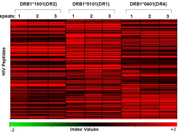
Clustering of HIV peptides by HLA-DR binding.
In order to
identify clusters of peptides (i.e., using both peptide groups,
the CD4
⫹T-cell epitopes listed in the Los Alamos database
and the gp160 and Nef proteins as overlapping peptide
stretches) that show differential binding to soluble HLA-DR
alleles, a Pearson centered hierarchical clustering analysis was
carried out using the software program Acuity (2), using the
average of the normalized response indexes on three slides
(repeats). Figure 4 shows that the clusters of individual
HLA-DR–peptide complexes were unique for each MHC class II
allele. There were peptides that bound strongly to all three
HLA-DR alleles and peptides which formed complexes with
only one or two of the MHC class II alleles.
Complex formation by described CD4
ⴙT-cell epitopes with
HLA-DR molecules.
Of the 306 peptides from the HIV
immu-nology database that were printed on the microarray chip, 73
showed interaction with soluble HLA-DR alleles (see Table S1
in the supplemental material). The exact MHC class
II-restrict-ing molecule for the majority of the published HIV-1 CD4
⫹T-cell epitopes has not yet been defined (they have been
re-ported as “DR restricted” or simply as “CD4 epitope”), and it
is therefore not surprising that some of the epitopes did not
form a complex with the HLA-DR molecules employed in the
current assay.
Complex formation by HIV-1B gp160 and Nef peptides with
HLA-DR molecules.
Table S2 in the supplemental material
lists 20-mer peptides, generated in a systematic way as
over-lapping peptides from the HIV-1B consensus gp160 and Nef
sequences, that showed complex formation with any of the
FIG. 4. MHC class II molecule-peptide binding pattern. A Pearson centered hierarchical clustering analysis of HIV peptides (
n
⫽
206) derived
as overlapping peptides from HIV gp160 or Nef and 346 peptides listed as CD4
⫹T-cell epitopes in the Los Alamos database using the software
program Acuity. Note the cluster of the MHC class II molecule-peptide binding pattern in association with the respective HLA-DR molecules.
Each experiment was repeated three times. The index values, which represent a measure of the signal intensity, are similar for slides incubated
with the same MHC class II monomer; minor differences are attributable to experimental variations.
on August 17, 2020 by guest
http://cvi.asm.org/
HLA-DR alleles tested in the current report. Thirty-two of 167
peptides from gp160 formed complexes with DRB1*0101, 34/
167 with DRB1*1501, and 38/167 with DRB1*0401; 10/39
pep-tides from Nef formed complexes with DRB1*0101, 8/39 with
DRB1*1501, and 6/39 with DRB1*0401, respectively. The
comparison of these peptides to the CD4
⫹T-cell epitopes
listed in the Los Alamos HIV-1 Immunology database
indi-cates a good overlap of the protein regions which provide
immunogenic peptide epitopes (see Table S2 in the
supple-mental material). In a parallel approach, we selected
overlap-ping peptides from HIV-1 gp160 and Nef and evaluated MHC
class II complex formation; Fig. 5 shows the compilation of
HLA-DR binding epitopes for HIV Nef and Fig. 6 the
HLA-DR binding patterns for HIV gp160 as a paradigm. The
newly identified peptides reside within already described
im-munodominant regions of the HIV proteins, which further
lends support to our approach (Fig. 5 and 6). Although the
MHC class II molecule-peptide binding was reproducible, we
lacked information on whether HLA-DR binding peptides
would indeed be able to expand peptide-specific CD4
⫹T cells.
We therefore used some of the identified MHC class II binding
peptides to expand CD4
⫹T cells in vitro defined by the
pro-duction of IFN-
␥
, TNF-
␣
, and IL-2 (Fig. 7).
DISCUSSION
We describe here, to our knowledge for the first time, an
MHC class II binding assay implementing a peptide microarray
and recombinant synthetic soluble MHC class II molecules,
using HIV-derived peptides as a paradigm. Already described
CD4
⫹T-cell epitopes were printed on the chip to act as
pos-itive controls, and 73/306 peptides formed detectable MHC
class II complexes. Of note, the peptides listed in the Los
Alamos database show various lengths and have been selected
based on different criteria, particularly functional T-cell assays;
most of the epitopes have been listed as “CD4
⫹T-cell epitope”
or “DR-restricted.” Of the 346 peptides listed as CD4
⫹T-cell
epitopes in the Los Alamos HIV Immunology database, only
280 were defined for humans. Other peptides were identified in
different species, e.g., mice, chimpanzees, and macaques. Out
of these 280, only 70 have been mapped to a specific MHC
class II allele or an allele group. It could very well be that
specific MHC class II alleles were not covered in the current
experiments due to failure to employ the correct MHC class II
alleles for binding.
The limitations of the assay described may be the following:
(i) structural constraints imposed by the immobilization of
peptide on the glass slide via the N-terminal end, possibly
precluding spatial interaction with soluble MHC class II
mol-ecules; (ii) the current 20-mer peptides may not represent the
optimal length facilitating binding to a specific MHC class II
allele; or (iii) glycopeptides cannot currently be evaluated for
MHC class II interaction (7).
A number of assays to identify peptides binding to MHC
class II molecules (5, 20, 27, 40, 47) have been described, but
only a very limited number of peptides could be tested
simul-taneously. This makes it difficult to comprehensively screen an
entire proteome from a pathogen or a comprehensive library
from target epitopes in cancer or autoimmune diseases. A
FIG. 5. HLA-DR epitopes on HIV Nef. Compilation of the DRB1*0101, DRB1*1501, and DRB1*0401 interactions with HIV-1B Nef defined
by the peptide array platform. Areas with CD4
⫹T-cell epitopes that have been listed in the Los Alamos HIV database are highlighted in orange.
All HLA-DR binding epitopes have been described except an epitope in position 130 to 150 (
ⴱ
).
FIG. 6. HLA-DR epitopes on HIV gp160. Compilation of the DRB1*0101, DRB1*1501, and DRB1*0401 interactions with HIV-1B gp160
defined by the peptide array platform.
on August 17, 2020 by guest
http://cvi.asm.org/
number of reports addressed a multidimensional approach to
enhance prediction of MHC class II molecule-peptide
interac-tions (26, 44). If T-cell-based assays are employed, it is not only
the turnaround time that is limited but also the availability of
biological material. Computational predictions of MHC class
II binding peptides offer the advantage of a short turnaround
time (44), yet it may be challenging to rely solely on
compu-tational analysis without selected biological readouts (14). This
demand has been addressed by Stone and coworkers, who
developed an MHC class II-peptide array supplemented with
costimulatory molecules to identify MHC class II-restricted
epitopes defined by functional T-cell responses (39). This assay
requires peptides that are known to bind to certain MHC class
II molecules. Peptides identified in the current assay were
linked to biological activity: some peptides, previously shown
to be functional HIV-specific CD4
⫹T-cell epitopes (10, 15,
39), showed good binding to soluble MHC class II molecules
employed in the current assay (see Table S1 in the
supplemen-tal material), and newly identified MHC class II-binding
pep-tide epitopes resided in “immunogenic regions” of HIV-1B
Nef (see Fig. 5) or gp160 and showed the capacity to expand
MHC class II-restricted and peptide-specific CD4
⫹T cells
defined by cytokine production.
The assay described in the current report offers the unique
advantage of screening thousands of peptides within a single
experiment using high-density peptide spotting using SPOT
synthesis technology (11, 30). This enables the printing of
en-tire viral proteomes as linear peptide stretches (our
unpub-lished data) on glass slides. In addition, the orientation of the
candidate peptide is defined and can also be used for a more
detailed structural analysis of MHC class II molecule-peptide
interactions: the crystal structure of MHC class II molecules
shows that the peptide binding groove is open at both ends (32,
33, 38), which enables complex formation with peptides
immo-bilized on the glass scaffold.
In conclusion, we developed a robust assay using a peptide
microarray and recombinant soluble MHC class II molecules
to identify HIV-1 peptides which form complexes with
differ-ent HLA-DR alleles. The assay can be implemdiffer-ented to screen
for MHC class II binding of peptide libraries with a short
turnaround time and may also be used to probe for differences
in MHC class II molecule-peptide complex formation
associ-ated with variant peptides or with differences in MHC class II
alleles. Peptide microarrays, such as those described in the
current report, will help to speed up the process of CD4
⫹T-cell epitope identification and advance efforts targeting a
rational platform for vaccine design and immunomonitoring.
ACKNOWLEDGMENTS
This work was supported by a grant from the EU Marie Curie Early
Training program to A.E. and grants from Cancerfonden,
Veten-kapsrådet, So
¨derberg-foundation, and Karolinska Institutet, Sweden,
to M.M.
FIG. 7. MHC class II-restricted expansion of peptide-specific CD4
⫹T cells. PBMCs from HIV-negative and MHC class II-genotyped blood
donors were stimulated with the following peptides: for donors 1 to 3, peptides SLYVTVATLYCVHQRIEV and FRKQNPDIVIYQYMDDL
YVG, representing HIV Gag (amino acids 77 to 94) and HIV reverse transcriptase (amino acids 171 to 190), respectively (peptides 1 and 2 [P1
and P2]). PBMCs from donor 4 were stimulated with ALFYKLDVVPINDNTSYRL from HIV gp160 (amino acids 174 to 193) and EKLWVT
VYYGVPVWKEATTT from HIV gp160 (amino acids 32 to 51) (peptides 3 and 4 [P3 and P4]). After three rounds of peptide stimulation, PBMCs
were tested for peptide-specific reactivity defined by intracellular production of TNF-
␣
, IFN-
␥
, and IL-2 in intracellular cytokine staining. Phorbol
myristate acetate/ionomycin served as the positive control; negative controls included the incubation in medium or stimulation with an irrelevant
HIV peptide which was not used for stimulation (EKLWVTVYYGVPVWKEATTT for donors 1 to 3 and peptide FRKQNPDIVIYQYMDD
LYVG for donor 4). Results are reported as absolute numbers of cytokine-producing cells/10
5CD4
⫹T cells.
on August 17, 2020 by guest
http://cvi.asm.org/
REFERENCES
1.Angyalosi, G., R. Neveu, I. Wolowczuk, A. Delanoye, J. Herno, C. Auriault, and V. Pancre.2001. HLA class II polymorphism influences onset and
severity of pathology inSchistosoma mansoni-infected transgenic mice.
In-fect. Immun.69:5874–5882.
2.Axon Instruments Inc.2005. Acuity 4.0 microarray informatics software user’s guide, 2005. Axon Instruments/Molecular Devices Corp., Sunnyvale, CA.
3.Castellino, F., G. Zhong, and R. N. Germain.1997. Antigen presentation by MHC class II molecules: invariant chain function, protein trafficking, and the
molecular basis of diverse determinant capture. Hum. Immunol.54:159–169.
4.Deeks, S. G., and B. D. Walker.2007. Human immunodeficiency virus con-trollers: mechanisms of durable virus control in the absence of antiretroviral
therapy. Immunity27:406–416.
5.DeLuca, D. S., B. Khattab, and R. Blasczyk.2007. A modular concept of HLA for comprehensive peptide binding prediction. Immunogenetics
59:25–35.
6.Dengjel, J., M. D. Nastke, C. Gouttefangeas, G. Gitsioudis, O. Schoor, F. Altenberend, M. Muller, B. Kramer, A. Missiou, M. Sauter, J. Hennenlotter, D. Wernet, A. Stenzl, H. G. Rammensee, K. Klingel, and S. Stevanovic.2006. Unexpected abundance of HLA class II presented peptides in primary renal
cell carcinomas. Clin. Cancer Res.12:4163–4170.
7.Dengjel, J., H. G. Rammensee, and S. Stevanovic.2005. Glycan side chains
on naturally presented MHC class II ligands. J. Mass Spectrom.40:100–104.
8.Erlich, H. A., and U. B. Gyllensten.1991. The evolution of allelic diversity at the primate major histocompatibility complex class II loci. Hum. Immunol.
30:110–118.
9.Fischer, W., S. Perkins, J. Theiler, T. Bhattacharya, K. Yusim, R. Funk-houser, C. Kuiken, B. Haynes, N. L. Letvin, B. D. Walker, B. H. Hahn, and B. T. Korber.2007. Polyvalent vaccines for optimal coverage of potential
T-cell epitopes in global HIV-1 variants. Nat. Med.13:100–106.
10.Fonseca, S. G., A. Coutinho-Silva, L. A. Fonseca, A. C. Segurado, S. L. Moraes, H. Rodrigues, J. Hammer, E. G. Kallas, J. Sidney, A. Sette, J. Kalil, and E. Cunha-Neto. 2006. Identification of novel consensus CD4 T-cell epitopes from clade B HIV-1 whole genome that are frequently recognized
by HIV-1 infected patients. AIDS20:2263–2273.
11.Frank, R.2002. The SPOT-synthesis technique. Synthetic peptide arrays on membrane supports—-principles and applications. J. Immunol. Methods
267:13–26.
12.Gaudebout, P., D. Zeliszewski, J. J. Golvano, C. Pignal, S. Le Gac, F. Borras-Cuesta, and G. Sterkers.1997. Binding analysis of 95 HIV gp120 peptides to HLA-DR1101 and -DR0401 evidenced many HLA-class II bind-ing regions on gp120 and suggested several promiscuous regions. J. Acquir.
Immune Defic. Syndr. Hum. Retrovirol.14:91–101.
13.Godkin, A. J., K. J. Smith, A. Willis, M. V. Tejada-Simon, J. Zhang, T. Elliott, and A. V. Hill.2001. Naturally processed HLA class II peptides reveal highly conserved immunogenic flanking region sequence preferences that reflect antigen processing rather than peptide-MHC interactions. J.
Im-munol.166:6720–6727.
14.Gowthaman, U., and J. N. Agrewala.2008. In silico tools for predicting peptides binding to HLA-class II molecules: more confusion than
conclu-sion. J. Proteome Res.7:154–163.
15.Kaufmann, D. E., P. M. Bailey, J. Sidney, B. Wagner, P. J. Norris, M. N. Johnston, L. A. Cosimi, M. M. Addo, M. Lichterfeld, M. Altfeld, N. Frahm, C. Brander, A. Sette, B. D. Walker, and E. S. Rosenberg.2004. Comprehen-sive analysis of human immunodeficiency virus type 1-specific CD4 responses
reveals marked immunodominance of gag andnef and the presence of
broadly recognized peptides. J. Virol.78:4463–4477.
16.Kobayashi, H., M. Wood, Y. Song, E. Appella, and E. Celis.2000. Defining promiscuous MHC class II helper T-cell epitopes for the HER2/neu tumor
antigen. Cancer Res.60:5228–5236.
17.Letourneau, S., E. J. Im, T. Mashishi, C. Brereton, A. Bridgeman, H. Yang, L. Dorrell, T. Dong, B. Korber, A. J. McMichael, and T. Hanke.2007. Design
and pre-clinical evaluation of a universal HIV-1 vaccine. PLoS One2:e984.
18.Magalhaes, I., N. K. Vudattu, E. Ja¨ger, and M. J. Maeurer.2008. Tumor
antigen-specific T-cells are present in the CD8alpha/alpha⫹T-cell
effector-memory pool. J. Immunother.31:840–848.
19.Malhotra, U., S. Holte, T. Zhu, E. Delpit, C. Huntsberry, A. Sette, R. Shankarappa, J. Maenza, L. Corey, and M. J. McElrath.2003. Early induc-tion and maintenance of Env-specific T-helper cells following human
immu-nodeficiency virus type 1 infection. J. Virol.77:2663–2674.
20.Mallios, R. R.2003. A consensus strategy for combining HLA-DR binding
algorithms. Hum. Immunol.64:852–856.
21.Martin, M. P., and M. Carrington.2005. Immunogenetics of viral infections.
Curr. Opin. Immunol.17:510–516.
22.Middleton, D., L. Menchaca, H. Rood, and R. Komerofsky.2003. New allele
frequency database: http://www.allelefrequencies.net. Tissue Antigens 61:
403–407.
23.Nahtman, T., A. Jernberg, S. Mahdavifar, J. Zerweck, M. Schutkowski, M. Maeurer, and M. Reilly.2007. Validation of peptide epitope microarray
experiments and extraction of quality data. J. Immunol. Methods328:1–13.
24.Ndung’u, T., S. Gaseitsiwe, E. Sepako, F. Doualla-Bell, T. Peter, S. Kim, I. Thior, V. A. Novitsky, and M. Essex.2005. Major histocompatibility complex class II (HLA-DRB and -DQB) allele frequencies in Botswana: association with human immunodeficiency virus type 1 infection. Clin. Diagn. Lab.
Immunol.12:1020–1028.
25.Newman, M. J., B. Livingston, D. M. McKinney, R. W. Chesnut, and A. Sette.2002. T-lymphocyte epitope identification and their use in vaccine
development for HIV-1. Front. Biosci.7:d1503–d1515.
26.Nielsen, M., C. Lundegaard, T. Blicher, B. Peters, A. Sette, S. Justesen, S. Buus, and O. Lund.2008. Quantitative predictions of peptide binding to any HLA-DR molecule of known sequence: NetMHCIIpan. PLoS Comput. Biol.
4:e1000107.
27.Nielsen, M., C. Lundegaard, and O. Lund.2007. Prediction of MHC class II binding affinity using SMM-align, a novel stabilization matrix alignment
method. BMC Bioinform.8:238.
28.Novak, E. J., A. W. Liu, G. T. Nepom, and W. W. Kwok.1999. MHC class II
tetramers identify peptide-specific human CD4(⫹) T cells proliferating in
response to influenza A antigen. J. Clin. Investig.104:R63–R67.
29.Panina-Bordignon, P., A. Tan, A. Termijtelen, S. Demotz, G. Corradin, and A. Lanzavecchia.1989. Universally immunogenic T cell epitopes: promiscu-ous binding to human MHC class II and promiscupromiscu-ous recognition by T cells.
Eur. J. Immunol.19:2237–2242.
30.Scharn, D., H. Wenschuh, U. Reineke, J. Schneider-Mergener, and L.
Germer-oth.2000. Spatially addressed synthesis of amino- and amino-oxy-substituted
1,3,5-triazine arrays on polymeric membranes. J. Comb. Chem.2:361–369.
31.Schena, M.2005. Protein microarray, vol. 1. Jones and Bartlett, Sudbury, MA. 32.Sercarz, E. E., and E. Maverakis.2003. Mhc-guided processing: binding of
large antigen fragments. Nat. Rev. Immunol.3:621–629.
33.Sette, A., L. Adorini, S. M. Colon, S. Buus, and H. M. Grey.1989. Capacity of
intact proteins to bind to MHC class II molecules. J. Immunol.143:1265–1267.
34.Sidney, J., S. Southwood, C. Oseroff, M. F. del Guercio, A. Sette, and H. M. Grey. 2001. Measurement of MHC/peptide interactions by gel filtration, chapter 18, unit 18.3. Current protocols in immunology. John Wiley & Sons, Hoboken, NJ.
35.Siebold, C., B. E. Hansen, J. R. Wyer, K. Harlos, R. E. Esnouf, A. Svejgaard, J. I. Bell, J. L. Strominger, E. Y. Jones, and L. Fugger.2004. Crystal structure of HLA-DQ0602 that protects against type 1 diabetes and confers strong
susceptibility to narcolepsy. Proc. Natl. Acad. Sci. USA101:1999–2004.
36.Sinigaglia, F., M. Guttinger, J. Kilgus, D. M. Doran, H. Matile, H. Etlinger, A. Trzeciak, D. Gillessen, and J. R. Pink.1988. A malaria T-cell epitope recognized in association with most mouse and human MHC class II
mole-cules. Nature336:778–780.
37.Southwood, S., J. Sidney, A. Kondo, M. F. del Guercio, E. Appella, S. Hoffman, R. T. Kubo, R. W. Chesnut, H. M. Grey, and A. Sette.1998. Several common HLA-DR types share largely overlapping peptide binding
reper-toires. J. Immunol.160:3363–3373.
38.Stern, L. J., J. H. Brown, T. S. Jardetzky, J. C. Gorga, R. G. Urban, J. L. Strominger, and D. C. Wiley.1994. Crystal structure of the human class II MHC protein HLA-DR1 complexed with an influenza virus peptide. Nature
368:215–221.
39.Stone, J. D., W. E. Demkowicz, Jr., and L. J. Stern.2005. HLA-restricted epitope identification and detection of functional T cell responses by using MHC-peptide and costimulatory microarrays. Proc. Natl. Acad. Sci. USA
102:3744–3749.
40.Tong, J. C., T. W. Tan, and S. Ranganathan.2007. Methods and protocols
for prediction of immunogenic epitopes. Brief. Bioinform.8:96–108.
41.Topalian, S. L.1994. MHC class II restricted tumor antigens and the role of
CD4⫹T cells in cancer immunotherapy. Curr. Opin. Immunol.6:741–745.
42.Wahlstrom, J., J. Dengjel, B. Persson, H. Duyar, H. G. Rammensee, S. Stevanovideltac, A. Eklund, R. Weissert, and J. Grunewald.2007. Identifi-cation of HLA-DR-bound peptides presented by human bronchoalveolar
lavage cells in sarcoidosis. J. Clin. Investig.117:3576–3582.
43.Walter, W., C. Scheuer, M. Loos, T. E. Reichert, and M. J. Maeurer.2001. H2-Mbeta 1 and H2-Mbeta 2 heterodimers equally promote clip removal in
I-A(q) molecules from autoimmune-prone DBA/1 mice. J. Biol. Chem.276:
11086–11091.
44.Wang, P., J. Sidney, C. Dow, B. Mothe, A. Sette, and B. Peters.4 April 2008, posting date. A systematic assessment of MHC class II peptide binding predictions and evaluation of a consensus approach. PLoS Comput. Biol.
4:e1000048.
45.Williams, F., A. Meenagh, R. Single, M. McNally, P. Kelly, M. P. Nelson, D. Meyer, A. Lancaster, G. Thomson, and D. Middleton.2004. High resolution
HLA-DRB1 identification of a Caucasian population. Hum. Immunol.65:
66–77.
46.Zhan, X., K. S. Slobod, S. Surman, S. A. Brown, T. D. Lockey, C. Coleclough, P. C. Doherty, and J. L. Hurwitz.2003. Limited breadth of a T-helper cell response to a human immunodeficiency virus envelope protein. J. Virol.
77:4231–4236.
47.Zhu, S., K. Udaka, J. Sidney, A. Sette, K. F. Aoki-Kinoshita, and H. Mamit-suka.2006. Improving MHC binding peptide prediction by incorporating
binding data of auxiliary MHC molecules. Bioinformatics22:1648–1655.