THE EFFECT OF RECEPTOR DENSITY ON THE
BINDING AND FUNCTIONAL PROPERTIES OF THE
HUMAN ADENOSINE Ai RECEPTOR
Christopher Browning
A thesis submitted in fulfilment of the requirements of the
University of London for the degree of Doctor of Philosophy
2003
National Institute for Medical Research
ProQuest Number: U642676
All rights reserved
INFORMATION TO ALL USERS
The quality of this reproduction is dependent upon the quality of the copy submitted.
In the unlikely event that the author did not send a complete manuscript and there are missing pages, these will be noted. Also, if material had to be removed,
a note will indicate the deletion.
uest.
ProQuest U642676
Published by ProQuest LLC(2015). Copyright of the Dissertation is held by the Author.
All rights reserved.
This work is protected against unauthorized copying under Title 17, United States Code. Microform Edition © ProQuest LLC.
ProQuest LLC
789 East Eisenhower Parkway P.O. Box 1346
ABSTRACT
The adenosine Ai receptor is a member of the 7-transmembrane G-protein coupled
receptor family. It has an important signalling role in both normal physiology and
disease. In addition to containing binding sites for adenosine and G-proteins, the
Ai receptor contains an allosteric binding site which binds synthetic molecules
such as the allosteric enhancer PD 81,723. The availability of this compound,
together with high affinity antagonists, highly potent agonists with a range of
efficacies, the ability to determine ligand binding to the G-protein coupled and
uncoupled states of the receptor and to readily measure function in membranes,
make the Ai receptor an ideal candidate for testing the predictions of mathematical
models of drug receptor interactions.
The effects of receptor density, guanine nucleotides, and agonist efficacy on the
ligand binding properties of the human adenosine Ai receptor, recombinantly
expressed at different densities, in membranes from Chinese hamster ovary cells
have been examined. The proportions and ligand affinities of receptors in the G-
protein coupled and uncoupled states of the A% receptor have been measured. The
results suggest that there is a significant amount of receptor-G protein precoupling
in the absence of ligand but the overall results are not quantitatively compatible
with all the predictions of the ternary complex model of agonism. Radioligand
dissociation kinetic studies show the novel phenomenon of agonist-induced
agonist dissociation when the receptors are expressed at high but not low
dimers, or another form of receptor cross-talk that occurs when agonists are bound
to receptor G-protein complexes at high expression levels.
The effects of receptor density and agonist efficacy on the functional properties of
the adenosine Ai receptor were examined using a [^^SJGTPyS binding assay.
Marked novel biphasic dose-response curves were observed for high efficacy
agonists in membranes from a high expressing Ai receptor cell line. Experiments
were performed to investigate the nature of this response, its reversibility, and the
effects of an inverse agonist and agonists of different efficacy. A detailed study of
the kinetics of association of [^^SJGTPyS following agonist exposure suggest that
a reversible feedback mechanism is operating, with a short lag-time for its
induction.
The mechanism of action of the allosteric enhancer, PD 81,723, on the binding of
a series of ligands with a range of intrinsic activities to the coupled and uncoupled
states of the adenosine A% receptor was investigated. The effects of PD 81,723 on
the functional properties of these ligands, measured at different receptor
expression levels, were also studied. The results of these studies were not
compatible with the detailed predictions of the ternary complex model of agoni sm
if PD 81,723 simply enhanced the affinity of the receptor or if it increased agonist
efficacy. A quaternary complex model of allosterism and agonism was developed
which is compatible with the actions of PD 81,723 in binding and function. These
data can be explained by PD 81,723 activating the receptors from the allosteric
site and by its ability to act as a co-agonist with agonist binding to the orthosteric
CONTENTS
ABSTRACT... 2
CONTENTS... 4
LIST OF FIGURES... 10
LIST OF TABLES... 13
ABBREVIATIONS... 14
Chapter 1: Introduction...16
1.1 G-protein coupled receptors... 16
1.1.1 General Introduction... 16
1.1.2 General structure of GPCRs... 17
1.1.3 Ligand recognition... 22
1.1.4 Receptor activation... 23
1.2 G-proteins... 25
1.2.1 Other receptor-protein interactions... 31
1.3 Models of receptor-G-protein activation...36
1.3.1 The Ternary Complex Model... 36
1.3.2 Constitutive activity and inverse agonism... 39
1.3.2 The Extended Ternary Complex Model...40
1.4 Adenosine and its receptors...43
1.4.1 Physiological role of adenosine... 43
1.4.2. Pharmacological classification of adenosine receptor subtypes... 46
1.4.3 Cloning and molecular characterisation of adenosine receptor subtypes. ...47
1.4.4 Signalling of adenosine receptors... 51
1.4.4.1 Adenylyl cyclase...51
1.4.4.3 Ion channels... 53
1.4.4.4 MAP kinases... 54
1.5 Allosteric modulation of 7TM G PCRs...56
1.5.1 The Allosteric Ternary Complex M odel... 56
1.5.2 Allosteric modulation of G-protein coupled receptors... 57
1.5.3. Allosteric modulation of adenosine Ai receptors... 58
1.5.4 The potential therapeutic advantages of allosteric modulators of 7TM GPCRs... 60
1.6 Project Aims...64
Chapter 2. Materials and Methods... 65
2.1 Materials...65
2.2. Experimental Procedures... 67
2.2.1. Tissue culture...67
2.2.2 Membrane Preparation...67
2.2.3 Radioligand binding...68
2.2.3.1 Saturation Binding...69
2.2.3.2 Competition Binding... 69
2.2.3.3 Kinetics of radioligand dissociation...70
2.2.4 [^^SJGTPyS functional assays... 71
2.2.4.1 Measurements of agonist potency and antagonist affinity... 71
2.2.4.2. Gel filtration of [^^SjGTPyS supernatant...72
2.2.4.3. Kinetics of [^^SJGTPyS association... 73
2.3 Data analysis... 74
2.3.1 Analysis of radioligand binding data...74
2.3.1.1 Saturation binding... 74
2.3.1.3 Radioligand dissociation... 75
2.3.2. Analysis of [^^S]GTPyS Functional D ata... 76
2.3.2.1 Analysis of agonist concentration-response curves... 76
2.3.2.2. Analysis of DPCPX antagonist data... 76
2.3.2.3 Association timecourse of [^^S]GTPyS... 77
2.4 Simulations...77
Chapter 3. Radioligand binding to adenosine Ai receptors expressed at high and low densities in Chinese hamster ovary cells...79
3.1 Introduction...79
3.2 Results... 80
3.2 1. [^H]DPCPX and [^H]CHA saturation binding to the adenosine Ai receptor...80
3.2.2. The effect of CHA on the binding of [^H]DPCPX to the adenosine Ai receptor...82
3.2.3...Simulation of the effect of changing receptor density on the agonist binding properties of the adenosine A% receptor using the Ternary Complex Model... 85
3.2.4. The binding properties of lower efficacy agonists at the adenosine Ai receptor...88
3.2.5. Simulation of the effect of efficacy changes on the predictions of the Ternary Complex Model...94
3.2.6 The effect of GDP on the ligand binding properties of the adenosine Ai receptor... 97
3.2.7. Simulating the effect of GDP using the Ternary Complex M odel... 102
3.2.8. The kinetics of radiolabelled antagonist and agonist dissociation from the adenosine Ai receptor... 104
3.2.8.1. [^HJDPCPX dissociation... 106
3.3. Discussion... 110
Chapter 4 Pharmacological characterisation of [^^S]GTPyS binding responses in OHO Ai iow and high expressing membranes...129
4.1 Introduction... 129
4.2 Results...131
4.2.1 Agonist stimulation of [^^S]GTPyS binding in CHO Ai low and high
expressing membranes... 131
4.2.2 The effect of the antagonist DPCPX on the responses to CHA in CHO
Ai low and high expressing membranes...135
4.2.3 The effect of PD 81,723 on the CHA mediated modulation of
[35s]GTPyS binding to CHO adenosine Ai low and high expressing
membranes... 138
4.2.4. The effect of lower efficacy agonists on [^^SJGTPyS binding to CHO
adenosine A% LE and HE membranes... 140
4.2.5 Attempts to detect agonist mediated G-protein translocation from CHO
cell membranes... 142
4.2.6 Kinetics of [^^SJGTPyS binding to CHO adenosine A% LE and HE
membranes... 147
4.3 Simulation of the effects of receptor density on functional responses using
the Ternary Complex Model...159
4.4 Discussion... 161
4.4.1 The comparison of basal and agonist stimulated [^^S]GTPyS binding
responses in CHO Ai LE and HE membranes... 161
4.4.2 Pharmacological characterisation of the stimulatory [^^S]GTPyS
binding response in LE and HE membranes...164
4.4.3 Pharmacological characterisation of agonist mediated inhibition of
4.4.4. The kinetics of [^^S]GTPyS association to adenosine Ai LE and HE
membranes... 173
Chapter 5. The mechanism of action of the adenosine Ai receptor enhancer PD 81,723...175
5.1 Introduction... 175
5.2 Results...178
5.2.1 Properties of PD 81,723 in radioligand binding assays... 178
5.2.1.1 The effect of PD 81,723 on [^H]CHA competition binding in CHO Ai HE membranes...178
5.2.1.2 The effect of PD 81,723 on [^H]DPCPX competition binding, in the presence of guanine nucleotides, in CHO Ai HE membranes... 181
5.2.1.3 The effect of PD 81,723 on [^HJDPCPX competition binding in the absence of guanine nucleotide...183
5.2.1.4 The effect of PD 81,723 on the inhibition of radiolabelled agonist binding by guanine nucleotides... 185
5.2.2. Effect of PD 81,723 on Ai receptor function measured in [^^S]GTPyS binding assays... 187
5.2.2.1 Low expressing CHO Ai membranes... 187
5.2.2.2 High expressing CHO Ai membranes... 190
5.2.2.3 The effect of adenosine deaminase on the responses of CHA, DPCPX, and PD in CHO Ai HE membranes... 191
5.3 Simulation of the effects of PD 81,723 on the binding and functional properties of the adenosine Ai receptor... 193
5.3.1 Ternary complex model...194
5.3.1.1 Binding...196
5.3.1.2 Function...199
5.3.2. The Quaternary Complex Model...202
5.3.2.2. Function... 207
5.4 Discussion...209
Chapter 6. Summary, criticism and future directions... 227
6.1 Summary... 227
6.2 The radioligand binding properties of the adenosine Ai receptor... 227
6.3 The functional properties of the adenosine Ai receptor...230
6.3 The mechanism of action of the allosteric enhancer PD 81,723... 232
References... 235
Abstracts arising from this thesis...273
Appendix I; Derivation of the equations of the Ternary Compiex Modei...274
Appendix il: Derivation of the equations of the Quaternary Compiex Modei...279
LIST OF FIGURES
Chapter 1
1.1. The general structure of the human adenosine receptor... 18
1.2. Receptor - G protein interactions... 27
1.3. The Ternary Complex and Extended Ternary Complex Models of drug
receptor interactions... 37
1.4. The potential therapeutic benefits of an allosteric enhancer...62
Chapter 2
2.1. Structures of the compounds used in this study...66
Chapter 3
3.1. Saturation binding of the radiolabelled antagonist [^H]DPCPX and the
radiolabelled agonist [^H]CHA to CHO adenosine Ai low and high
expressing membranes... 81
3.2. The effect of the adenosine receptor agonist CHA on the binding of the
radiolabelled antagonist [^H]DPCPX in the absence and presence of GDP...84
3.3. Simulation of competition binding using the Ternary Complex Model:
Changing R t...86
3.4. The effect of agonists of different efficacy on radioligand binding to
adenosine Ai receptors... 89
3.5a. Simulation of competition binding using the Ternary Complex Model:
Changing the cooperativity constant a when Rt:Gt = 1.2...95
3.5b. Simulation of competition binding using the Ternary Complex Model:
Changing the cooperativity constant a when R t:G t = 2 ... 96
3.6. The effect of GDP on the inhibition of [^H]DPCPX binding to adenosine Ai
receptors by the adenosine Ai agonist C H A ... 99
3.7. Simulation of competition binding using the Ternary Complex Model:
Changing the affinity constant Kg... 103
3.8. The dissociation of [^H]DPCPX from the adenosine Ai receptor in low
3.9. The dissociation of [^H]CHA from the adenosine Ai receptor in low
expressing and high expressing CHO membranes...108
Chapter 4
4.1. CHA concentration-effect curves for the modulation of [^^SJGTPyS binding
to adenosine Ai LE and HE membranes...132
4.2. Antagonism of CHA mediated modulation of [^^S]GTPyS binding to
adenosine Ai LE and HE membranes by the adenosine Ai receptor antagonist
DPCPX... 136
4.3. The effect of the allosteric enhancer PD 81,723 on the modulation of
[35s]GTPyS binding by CHA in adenosine Ai LE and HE membranes... 139
4.4. The effect of agonists of different efficacy on the binding of [^^SJGTPyS to
adenosine Ai LE and HE membranes... 141
4.5. Gel filtration of supernatant from [^^SJGTPyS labelled adenosine Ai LE and
HE membranes...144
4.6. The effect of DPCPX on the binding of [^^S]GTPyS to adenosine Ai LE and
HE membranes following a 30 min. pre-incubation with agonist... 146
4.7. Association kinetics for the binding of [^^S]GTPyS to adenosine Ai LE and
HE membranes...150
4.8. Association kinetics for the stimulated binding of [^^S]GTPyS to CHO
adenosine Ai LE and HE membranes - basal activation subtracted... 151
4.9. Association kinetics for the binding of [^^SJGTPyS to CHO adenosine Ai LE
and HE membranes - low expression basal subtracted...152
4.10. The effect of CHA on the stimulated initial rate, maximal binding and kobs
of [^^S]GTPyS binding to CHO adenosine Ai low and high expression
membranes... 155
4.11. Simulation of functional responses using the ternary complex model:
Changing the Rt:Gt stoichiometry...160
Chapter 5
5.1. The effect of PD 81,723 on competition binding of [^H]CHA at the adenosine
5.2. The effect of PD 81,723 on competition binding of [^H]DPCPX at the
adenosine Ai receptor in the presence of GDP...182
5.3. The effect of PD 81,723 on the competition binding of [^H]DPCPX by NEC A
at the adenosine Ai receptor, in the absence of GDP...184
5.4. The effect of PD 81,723 on the inhibition by GTP of the binding of
radiolabelled agonists to the adenosine Ai receptor... 186
5.5. The effect of PD 81,723 on the [^^S]GTPyS functional responses of agonists
of varying efficacy... 188
5.6. The effect of adenosine deaminase on basal [^^S]GTPyS binding, and
responses to CHA, PD 81,723 and DPCPX in CHO adenosine Ai high
expressing membranes... 192
5.7. The Ternary Complex M odel... 195
5.8. Simulation of the effects of an allosteric ligand using the Ternary Complex
Model: Radioligand binding... 198
5.9. Simulation of the effects of an allosteric ligand using the Ternary Complex
Model: Function... 200
5.10. The Quaternary Complex Model... 203
5.11. Simulation of the effects of an allosteric ligand using the Quaternary
Complex Model: Binding... 205
5.12. Simulation of the effects of an allosteric ligand using the Quaternary
Complex Model: Function...208
5.13. Further predictions of the Quaternary Complex Model: Simulation of the
effect of a co-agonist on the apparent affinity, for the G-protein uncoupled
receptor, of agonists of varying efficacy...218
5.14. Alternative models used to describe the actions of PD at the adenosine Ai
LIST OF TABLES
Chapter 3
3.1. Binding parameters for the inhibition of [^H]DPCPX binding by agonists of
varying efficacy at adenosine Ai LE and HE membranes...90
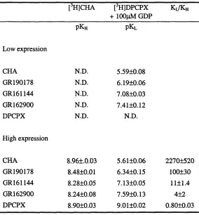
3.2. Ligand binding parameters for the G-protein coupled and uncoupled states of
the adenosine Ai receptor in LE and HE membranes... 91
3.3. The effect of GDP on CHA inhibition of [^H]DPCPX binding to LE and HE
membranes...100
3.4. The effect of GDP on the binding of [^HJDPCPX to adenosine Ai receptors
in LE and HE membranes... 101
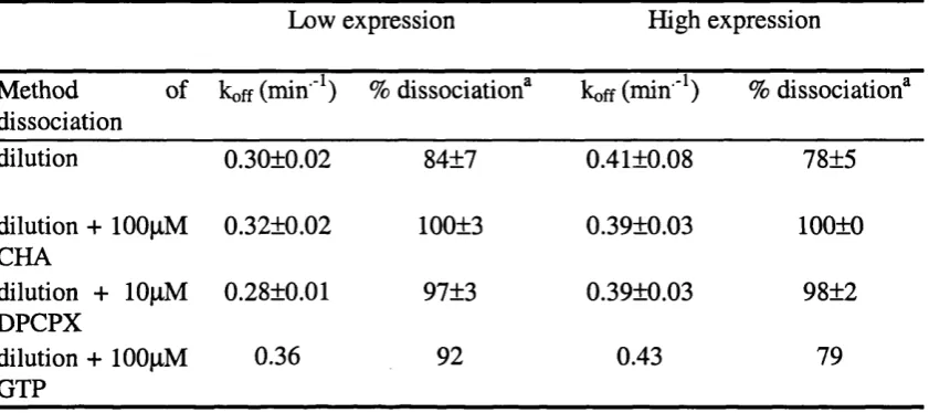
3.5. Summary of kinetic parameters for the dissociation of [^H]DPCPX (A) and
[^H]CHA (B) from the adenosine A% LE and HE membranes... 109
Chapter 4
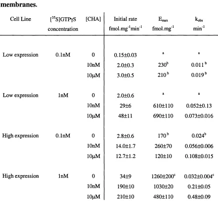
4.1 Summary of agonist concentration-response parameters for the modulation of
[35s]GTPyS binding to CHO Ai low expression and high expression
membranes... 134
4.2 Kinetic parameters for the agonist stimulated association of [^^S]GTPyS
binding to CHOAi I E and HE membranes...156
Chapter 5
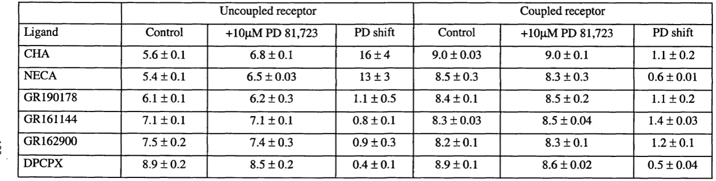
5.1. Summary of the effect of PD 81,723 on the affinity of ligands for the G-
protein uncoupled and coupled states of the adenosine Ai receptor... 180
5.2 Summary of the effects of PD 81, 723 on the [^^SJGTPyS binding response of
adenosine Ai receptor agonists in CHO Ai low and high expressing
ABBREVIATIONS
ADA Ado Bmax BSA BW-A844U cAMP CHA CHO DMSG DPCPX EDTA ETCM frn GDP GPCR Gpp(NH)pGR 190178
GR 161144
GR 162900
GTP HE HEPES Ka Kg adenosine deaminase adenosine
maximum radioligand binding capacity
bovine serum albumin
3-(4-amino)phenethyl-l-propyl-8-cyclopentylxanthine
adenosine 3',5'-cyclic monophosphate
N^-cyclohexyl adenosine
Chinese hamster ovary
dimethyl sulfoxide
8-cyclopentyl-1,3-dipropylxanthine
ethylenediaminetetraacetic acid
extended ternary complex model
the fraction of binding sites of high affinity
guanosine 5'-diphosphate
G-protein coupled receptor
guanosine 5'-[p,y-imido]triphosphate ((2R,3R,4S,5R)-2-{6-[(3-fluoro-4-hydroxyphenyl)amino]- 2-methyl-9H-purin-9-yl-5-(methoxymethyl)tetrahydrofuran-3,4-diol) (9-[3R,4S-dihydroxy-5-[4-methyl]-l,2,4-oxadiazol-2-yl]- tetraydrofuran-2(R)-yl]-6-[[tetraydropyran-4-yl]amino]-9H-purine) ((2R,3R,4S,3R)-2-[6-[(l-methylethyl)amino]-9H-purin-9- yl]-5-[4H-5-methyl-l,2,4-triazol-2-yl]tetrahydrofuran-3,4-diol) guanosine 5'-triphosphate
High expressing CHO adenosine Ai cells
N-[2-Hydroxyethyl]piperazine-N’-[2-ethanesulfonic acid]
affinity constant of a ligand for a receptor.
KpD LE NECA PD
pECso
pICsopKe
pKo
pKi PKh PKl QCM R-PIA TCM TMaffinity constant for the binding of PD 81,723 to the
adenosine Ai receptor.
Low expressing CHO adenosine Ai cells
5'-(N-ethylcarboxamido)-adenosine
2-amino-4,5-dimethylthien-3-yl[3-(trifluoromethyl)phenyl]-
methanone, PD 81,723;
negative log of the concentration of a drug required to
produce 50% of its own maximum response,
negative log of the concentration of a drug required to
produce 50% of its own maximum inhibtory effect,
negative log dissociation constant of an antagonist or
inverse agonist for a receptor.
negative log dissociation constant of a radioligand for a
receptor.
negative log dissociation constant of an unlabelled ligand
for a receptor, determined from a radioligand competition
binding experiment.
negative log dissociation constant of a ligand for the high
affinity state of a receptor.
negative log dissociation constant of a ligand for the low
affinity state of a receptor.
quaternary complex model
R-N^-(2-phenylisopropyladenosine)
ternary complex model
Chapter 1 : Introduction
1.1 G-protein coupled receptors
1.1.1 General Introduction
The family of seven transmembrane G-protein coupled receptors (7TM GPCRs) is
one of the most abundant families of proteins known, with -1 0 0 0 members,
representing approximately 3% of the mammalian genome (Foord, 2002; Takeda
et al, 2002) and 5% of the genome of C. elegans (Bargman, 1998). In addition,
7TM receptors are extremely important in medicine. Approximately 50% of drugs
used clinically mediate their effects by either activating or blocking this family of
proteins (Drew, 1996).
GPCRs have a characteristic structure and mechanism of action. They can be
subdivided into three main families based on their primary amino-acid sequence
homology (Humphrey et al, 2000): rhodopsin like (family A), which constitutes
the majority of 7TM receptors (-90%) and encompasses ‘classical’ 7TM receptors
such as muscarinic and adrenergic receptors; the secretin-like family (family B),
which includes receptors for calcitonin, glucagon and vasoactive intestinal
peptide; and the metabotropic glutamate and GABAb receptors (family C). More
recently, receptors for fungal pheromones, cAMP, and Wnt signalling factors
(frizzled/smoothened receptors) have been identified as separate sub-classes of G-
1.1.2 General structure of GPCRs
The general structure of 7TM receptors consists of seven transmembrane domains
interspersed with three intra- and three extracellular loops, an extracellular N-
terminus and an intracellular C-terminus (Fig. 1.1). This basic structure has been
predicted from hydropathicity analysis of the primary structures of a large number
of 7TM receptors (Baldwin, 1993). For both rhodopsin and the p2 adrenegic
receptor it has been demonstrated that the N-terminus is on the extracellular face
and the C-terminus is on the intracellular face (Applebury & Flargrave, 1986;
Wang et al, 1989). A fourth intracellular loop is formed when one or more
cysteine residues in the C-terminal segment are palmitoylated (Bouvier et al,
1995).
The transmembrane domains of GPCRs are of similar lengths across families,
which reflects the constancy of the orientations of these domains relative to the
width of a phospholipid bilayer. Conserved amino acids in these domains tend to
be at the inner faces (Baldwin, 1993). The lengths of all three external loops, and
internal loops one and two are relatively conserved and of similar lengths. The N-
and C-termini and intracellular loop three are least conserved in sequence
homology and overall length, which may in part reflect different modes of ligand
recognition (at the N-terminus) and intracellular interactions with other proteins.
There is no amino acid sequence homology between the three major GPCR
families, whilst approximately 20% amino acid homology can be found between
members of the rhodopsin family. Receptor subtypes, which share the same
,Q®®©©Q(
I
%
VI I ©
II ® 111
I
i
0 0? »®©0
Cytoplasm
Extracellular Space
A
COOH
Figure 1.1. The general structure of a family A, rhodopsin-like
G-protein coupled receptor.
The general structure of the human receptor as an example of a rhodopsin like, Family A, seven transmembrane G-protein coupled receptor, illustrating the extracellular N-terminus, the three extracellular and three intracellular loops, and the intracellular C-terminus as described in section 1.1.2.
between species homologues of the same receptor can be 80-99% (Baldwin,
1993).
Most of the structural information on seven transmembrane receptors has come
from studies on opsins, light detecting receptors found in certain species of
bacteria and in the mammalian and amphibian retina. Bacteriorhodopsin is a light
activated proton pump from Halobacterium Halobium whose seven-helical
structure is known (Henderson et a/., 1990; Luecke et al., 1999). This protein,
however, does not couple to G-proteins, its sequence shows none of the distinctive
patterns of the rhodopsin family and its structure, therefore, is not thought to be
relevant to that of the rhodopsin family of GPCRs (Baldwin, 1993).
Rhodopsin itself is unique in that its ligand, 11-cw-retinal, is bound covalently to a
conserved lysine in TM7. A photon at the appropriate wavelength causes the
ligand to isomerise to the 2i\\-trans form which activates the receptor and allows
interaction with its cognate G-protein, transducin (Khorana, 1992). In contrast to
rhodopsin, other GPCRs are activated by the reversible binding of a diffusible
ligand. Despite this difference, and the modest amino-acid homology between
rhodopsin and other GPCRs (-20%) the overall structures of the transmembrane
domains are thought to be remarkably similar (Ballasteros et al, 2001).
Low temperature electron diffraction studies of two dimensional crystalline
receptor arrays have generated low resolution projection density maps showing the
arrangement of transmembrane a-helices for frog and bovine rhodopsin (Schertler
& Hargrave, 1995; Schertler et al, 1993). The structure was demonstrated to be a
membrane and the remaining helices being more tilted (Unger et al, 1997). The
probable arrangement of the helices, relative to each other and to the lipid
environment of the membrane, is consistent with the relative content of
hydrophobic residues in the transmembrane regions (Baldwin, 1993). Thus, TM3
is the least lipophilic and is postulated to be hidden within the alpha-helical
bundle, whilst TMs IV and V are most lipophilic and are thought to face towards
the lipid bilayer.
The crystal structure of bovine rhodopsin has recently been determined to a
resolution of 2.8 Â by X-ray crystallography (Palczewski et al, 2000) and has
confirmed the general 7-transmembrane structure postulated from previous
studies. Of interest was the unanticipated finding that the C-terminal tail forms an
8th cytoplasmic helix which lies along the surface of the membrane to which it is
anchored via palmitoyl groups attached to a conserved pair of cysteine residues.
Regions of the receptor involved in G-protein interactions are thought to involve
the inward facing surfaces of the cytoplasmic ends of TMs 3, 5, 6 and 7. the
second and third intracellular loops, and helix 8. The structure also confirms the
bends in a number of helices predicted by the presence of conserved proline
residues, in particular the conserved proline in TM6, mutation of which is thought
to produce long-range distortion of the third intracellular loop (Ridge et al, 1999).
Most GPCRs contain consensus sequences for N-linked glycosylation in the
amino terminus. There are conflicting reports regarding its role in receptor
expression and function, and therefore an important role may not be universal
among GPCRs. For instance, inhibition of N-glycosylation with tunicamycin has
decreases the function of prostaglandin Ei receptors in the same cell line (George
et al, 1986). In contrast, tunicamycin treatment of A431 cells has been
demonstrated to decrease the expression and G-protein coupling of the P2-
adrenoceptor (Boege et al, 1988; Cervantes-Olivier et al, 1988). Disruption of
potential glycosylation sites by site-directed mutagenesis does not affect
expression, ligand binding or function of the muscarinic M2 receptor (van Koppen
& Nathanson, 1990) but decreases the expression of the P2 receptor (Rands et al,
1990) and the lutropin receptor (Liu et al, 1993).
Two cysteine residues in first and second extracellular loops are highly conserved
amongst 7TM receptors and play a major role in receptor structure and stability.
Disulphide-bonds between these cysteines have been demonstrated biochemically
in rhodopsin (Findlay & Pappin, 1986), muscarinic receptors, (Curtis et al, 1989)
and other GPCRs (Ji et al, 1998). In the crystal structure of rhodopsin, the
disulphide bond between the two cysteines is thought to pull the second
extracellular loop over the opening of the core of the receptor and, together with
the N-terminus, contributes to the formation of the P-structure above a central
cavity (Palczewski et al, 2000). Site directed mutagenesis of either of these
cysteines in rhodopsin profoundly affects the tertiary structure, reducing
expression levels, altering glycosylation and decreasing the ability to bind 11-cis-
retinal and transducin (Davidson et al, 1994; Kamik et al, 1988). A similar
approach dramatically alters the ligand binding ability of the P-adrenergic receptor
1.1.3 Ligand recognition
Despite the relatively low primary amino acid sequence homology between 7TM
receptors, the basic three-dimensional architecture has been preserved throughout
eukaryotic evolution. However, 7TM receptors have evolved to utilise an array of
different modes of ligand recognition involving transmembrane regions, the N-
terminal tail and extracellular loops. Thus Ji et al (1998) have proposed that 7TM
receptors can be further subdivided on the basis of how the endogenous ligand
binds to its receptor. Ligands can bind exclusively to the transmembrane core
(biogenic amines, eicosanoids, nucleosides), to both the core and extracelluar loop
(small peptides), to the N-terminus and extracellular loops (larger polypeptides) or
to the N-terminus alone (glycoproteins).
In general, for biogenic amine receptors, amino acid residues in the
transmembrane regions are important for the binding of the endogenous agonist.
For rhodopsin itself, the ligand ll-cis-retinal is bound covalently to a lysine in
TM7 and sits in a binding pocket formed from amino acids in TM3 and 6
(Palczewski et al, 2000). For the muscarinic Mi receptor the ligand contact
points are highly homologous, and acetylcholine-like ligands can be docked along
a similar trajectory to that of ll-cw-retinal in rhodopsin (Lu & Hulme, 2000). The
positions of these amino acids are well conserved in other 7TM receptors although
the side-chains are frequently different, reflecting the different chemical properties
1.1.4 Receptor activation
GPCRs are thought to be maintained in a ground state structure by a network of
conserved, stabilising intramolecular interactions involving H-bonding networks
and Van der Waal’s contacts which constrain the TM helices in an inactive
conformation. The amino acids involved in maintaining the stabilising interactions
are frequently highly conserved. Mutation of these amino acids often results in an
increase in agonist affinity, and agonist independent constitutive receptor
activation, and suggests a role for these residues in stabilising interactions that
constrain the receptor to the inactive ground state (Lefkowitz et al, 1993).
For the muscarinic Mi receptor, a series of highly conserved amino acids in TMs
3, 6 and 7 have been shown to be in close proximity by the creation of high-
affinity Zn^"^ binding sites by histidine substitutions. Substitution of these residues
with alanine increased the affinity of acetylcholine, implying the promotion of an
activated state (Lu & Hulme, 2000). Replacement of the highly conserved alanine
293 in the C-terminus of the third intracellular loop of the «ib adrenoceptor with
any other residue resulted in higher levels of agonist independent receptor activity
(Kjelsberg et al, 1992). The invariant arginine in the highly conserved E/D-R-Y
motif at the cytoplasmic end of TM3 is postulated to be constrained in a ‘polar
pocket’ formed by conserved neighbouring residues in T M l, 2 and 7. One
proposal is that receptor activation leads to protonation of the aspartate or
glutamate, causing arginine to move out of the polar pocket and the cytosolic
exposure of buried sequences in the second and third intracellular loops. Mutation
(Scheer et al, 1996) and P2 (Rasmussen et al, 1999) adrenoceptors, results in an
increase in agonist independent activity
All GPCRs are thought to share a conserved mechanism of activation. Agonist
binding to GPCRs is thought to disrupt the inactive ground state by producing a
conformational rearrangement of the transmembrane regions, with the relative
movement of TM6 of particular importance. In rhodopsin the use of nitroxide
spin labels has demonstrated the outward movement of cytoplasmic region of
TM6 following receptor activation (Altenbach et al, 1996; Farahbakhsh et al,
1995; Klein-Seetharaman et al, 1999). The movement of TM6 has also been
visualised as a change in fluorescence of environmentally sensitive fluorescent
probes introduced into both rhodopsin and the p2 adrenergic receptor (Dunham &
Barrens, 1999; Jensen et al, 2001). Furthermore, the introduction of Zn^"^ bridges
between TM3 and 6 in rhodopsin, the P2 adrenergic, and the parathyroid hormone
receptors, restricts the relative movements of these transmembrane regions and
inhibits receptor activation, suggesting the relative movements of these TM
regions to be of particular importance (Sheikh et al, 1999; Sheikh et al, 1996).
The structural rearrangement of the transmembrane helices of GPCRs following
agonist activation is postulated to expose regions of the receptor capable of
binding and activating G-proteins. From the crystal structure of rhodopsin, these
regions are postulated to be the cytoplasmic ends of TMs 3 , 5 , 6 and 7, the second
and third intracellular loops and helix 8 (Palczewski et a l, 2000). This supports
previous studies that demonstrate that short peptides derived from the C-terminus
of the third intracellular loop can mimic G-protein activation by a receptor (Malek
mutation of a four amino acid motif at the cytoplasmic end of TM6 of the
muscarinic M2 receptor alters its G-protein coupling selectivity from Gcti to GcXq
(Kostenis et al, 1997b). A similar result has been observed with hybrid
muscarinic M2/ M3 receptors where regions of the second and third intracellular
loops of the M2 receptor are replaced by the corresponding regions of the Mg
receptor (Kostenis et al, 1997a). Furthermore, the third cytoplasmic loop of
rhodopsin has been shown to be in close proximity to the N- and C-termini of
transducin by covalent cross-linking following receptor activation (Cai et al,
2001; Ito et al, 1994).
1.2 G-proteins
The primary mechanism by which activated GPCRs generate changes in
intracellular signalling is via the activation of heterotrimeric guanine-nucleotide
binding proteins (G-proteins). Heterotrimeric G-proteins are composed of a -, P~
and y-subunits. To date there are 23 distinct a subunits encoded by 17 different
genes which can be subdivided into four sub-families, GoCi/o, GoCs, GoCq and G «i2,
according to the homology of the primary amino acid sequence. The a subunits
have a conserved primary structure (50-90%) but have diverse profiles of effector
coupling. The cellular concentrations of Goti/o considerably exceed those other
subtypes and in the brain GOo may represent 1-2% of the total membrane protein
(Kepler & Gilman, 1992). G a subunits are anchored to the cytosolic face of the
plasma membrane by lipid modification, either by N-myristoylation or
Six (3- and twelve y - subunits have been described. The P - and y - subunits form
a functional Py heterodimer that is not dissociable except by dénaturation, p
subunits cannot form dimers with all y subunits; for example, Pi interacts with
either y% or y2, P2 interacts with y2 but not yi; and P3 cannot associate with either y%
or y2. In addition P subunits are selective in their coupling to a subunits. This
selectivity in subunit interactions underlies the fidelity of receptor-G-protein
interactions and contributes to the specificity of receptor-effector coupling
(Gudermann et al, 1996). Py subunits are also capable of stimulating diverse
effector mechanisms, including activation of inwardly rectifying potassium
channels, phospholipase Cp, as well as adenylyl cyclase and the MAP kinase
cascade (Clapham & Neer, 1997). The G-protein signalling pathways will be
described in more detail in section 1.4 in the context of the adenosine Ai receptor.
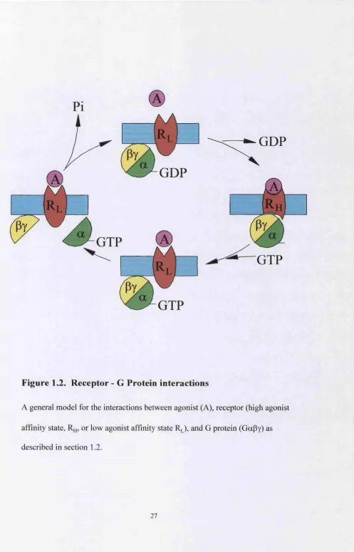
The a subunit cycles between an inactive, GDP bound, conformation and an
active GTP form (Fig. 1.2; Gilman, 1987). Activated GPCRs bind to Ga-GDP.Py
and induce conformational changes in the G-protein. This results in the exchange
of GDP for GTP and the dissociation of the G a-G T P and py subunits. Subunit
dissociation reveals regions of both the a-GTP and Py subunits which had been
previously hidden at the interface of the heterotrimeric complex, and which are
capable of activating downstream effector pathways (Lambright et al, 1996; Wall
et al, 1995). The intrinsic GTPase activity of G a hydrolyses GTP to GDP, which
GDP
GTP
Figure 1.2. R eceptor - G Protein interactions
A general model for the interactions between agonist (A), receptor (high agonist
affinity state, or low agonist affinity state R J , and G protein (GaPy) as
described in section 1.2.
G a subunits contain two domains, a domain involved in the binding of guanine
nucleotides and hydrolysis of GTP that is structurally similar to the superfamily of
small GTP-ase proteins and elongation factors (Kjeldgaard et al, 1996), and a
unique helical domain (Coleman et al, 1994; Noel et al, 1993). The helical
domain contains flexible ‘switch’ regions which envelops bound guanine
nucleotide and restricts the movements of nucleotides into and out of the binding
pocket (Lambright et al, 1994). Activated receptors catalyse the Mg^"^ dependent
exchange of GDP for GTP by loosening a network of hydrogen bonds and Van der
Waals contacts between residues in both the core and helical domains, and the
guanine ring of the bound nucleotide (Noel et al, 1993). In general, the
intracellular loops of GPCRs are too short to interact directly with the nucleotide
binding site of G a, which is probably located ~30Â from the plasma membrane
(Lichtarge et a l, 1996). The receptor must therefore work ‘at a distance’ to
change the conformation of the protein by triggering an allosteric transition.
Following the binding of GTP, the additional y-phosphate of GTP interacts with a
small number of amino acids in the switch regions to induce conformational
changes that result in the dissociation of both receptor and Py from Ga-GTP
(Coleman et al, 1994; Noel et al, 1993).
The amino acids of G a involved in the interactions with receptor, Py, and
effectors, have been identified from experiments with peptides, bacterial toxins,
antibodies, crosslinking agents and mutagenesis, together with information from
the crystal structures of Ga-GDP (Lambright et al, 1994), Ga-GTPyS (Coleman
et al, 1994; Noel et a l, 1993) and Ga-GDP.Py (Lambright et a l, 1996; Wall et
which include the N-terminal 23 residues and the C-tenninus (Hamm et a l, 1988).
The best characterised of these regions is the C-terminus which is thought to
interact with regions of the second and third intracellular loop of GPCRs
(Kostenis et a l, 1997a; Kostenis et a l, 1997b). Chimeric G a subunits have
demonstrated that the last three - five amino acids of the C-terminus are critical for
the specificity of receptor-G-protein interactions (Conklin et a l, 1993; Kostenis et
a l, 1997a). Furthermore, short peptide sequences from the a-subunit C-terminus
are able to disrupt receptor-G-protein coupling (Rasenick et a l, 1994).
The Gp subunit consists of two domains, an N-terminal a helix, and a p-propeller
structure made up of seven repeated ‘WD-40’ motifs. The y subunit is tightly
bound to the P subunit through extensive non-covalent interactions with both the
coiled region and the propeller region of the P subunit, which explains its
resistance to dénaturants (Sondek et a l, 1996). Prénylation of the C-terminus of
the y-subunit anchors the py complex to the plasma membrane (Wedegaertner et
a l, 1995). In addition to activating downstream effectors, the py subunit has also
been demonstrated to influence receptor function. Py subunit have been shown to
bind directly to purified P-adrenergic receptors (Heithier et a l, 1992) and to
rhodopsin (Phillips & Cerione, 1992), and to enhance the binding of G a to its
appropriate receptor (Phillips et al, 1992). The Py subunit is also able to facilitate
receptor phosphorylation by binding to P-adrenergic receptor kinase and bringing
it into close proximity to the receptor (Inglese et a l, 1995). This latter function
has led to the suggestion that the general role of py subunits is to promote
macromolecular assembly as part of the regulation of intracellular vesicular traffic
The intrinsic GTPase activity of the G-protein a subunits is regulated by a large
family of proteins termed regulators of G-protein signalling (RGS). RGS proteins
negatively regulate the activity of G a subunits by increasing the rate of GTP
hydrolysis by up to 100 fold and for this reason are also termed GTPase activating
proteins (GAPs, Berman & Gilman, 1998). Members of the phospholipase Cp
family of effector enzymes have also been demonstrated to have GOq-specific
GAP activity, suggesting a role in negative feedback regulation of G-protein
function (Biddlecome et a l, 1996; Chidiac & Ross, 1999).
G-protein activation can be measured directly in a binding assay using the
radiolabelled analogue of GTP, [^^S]GTPyS. Following receptor activation
[^^S]GTPyS binds to G-protein a subunits and stimulates subunit dissociation in a
similar manner to GTP. However, unlike GTP, [^^SJGTPyS is poorly hydrolysable
and accumulates in membranes and can be measured using conventional filtration
techniques (Hilf et a l, 1989; Lazareno, 1997). This method allows the
measurement of the number of G-proteins capable of binding guanine nucleotide.
The ability of G-proteins to hydrolyse GTP can be measured in a GTPase assay.
This method measures the rate or amount of [^^Pi] liberated from [y-^^P]GTP as a
result of hydrolysis by the a subunit (Brandt & Ross, 1986; Cassel & Selinger,
1976).
A number of biochemical tools are available for investigating the G-protein a
subtypes involved in G protein coupled receptor signalling. Pertussis toxin (or
islet activating protein) from Bordetella pertussis, catalyses the ADP-ribosylation
subunits of the class GoCi/o and GOt (Hurley et a l, 1984; West et a l, 1985). The
result of this covalent attachment is to inhibit G-protein interactions with
receptors, which further supports the importance of the G a C-terminus in receptor
interactions. Cholera toxin from Vibrio cholerae catalyses the ADP-ribosylation
of an arginine residue at position 174 of the a subunit of transducin (Abood et a l,
1982) and the corresponding position 201 in bovine Gets subunits (Robishaw et
a l, 1986). This residue is positioned in the switch U region of the helical domain
and interacts with the y-phosphate of GTPyS (Lambright et a l, 1994). ADP-
ribosylation of this arginine residue inhibits the GTPase function of GOg and
disrupts receptor - G-protein interactions by decreasing the affinity of GOg for
Gpy, although basal GTPase activity of ADP-ribosylated GOg is unimpaired (Kahn
& Gilman, 1984).
1.2.1 Other receptor-protein interactions
Activated G-protein coupled receptors are substrates for several families of kinase
proteins, which include second messenger-regulated kinases such as PKA and
PKC (Hausdorff et a l, 1990), and G-protein coupled receptor kinases (GRKs).
The conformational changes which occur as a result of agonist binding and
receptor activation expose a number of serine and threonine residues in the C-
terminus and the third intracellular loop of GPCRs, which greatly enhances
receptor phosphorylation by GRKs. There are currently 6 members of the GRK
family (GRK 1-6). The most commonly studied are GRKl, or rhodopsin kinase
(Kuhn, 1978), and GRK2, or P-adrenergic receptor kinase, which is now thought
Phophorylated receptors are substrates for a family of cytosolic proteins called
arrestins (Krupnick & Benovic, 1998). Whilst GRK catalysed phosphorylation of
GPCRs alone may modestly desensitise receptor signalling (Bennett &
Sitaramayya, 1988) the binding of arrestins to phosphorylated receptor is the
primary mechanism for the rapid uncoupling of receptors from G-proteins
(Krupnick & Benovic, 1998). Furthermore, receptor phosphorylation and arrestin
binding are critical steps in the initiation of internalisation of muscarinic M2
(Tsuga et al, 1994) and the Pz-adrenergic receptor (Ferguson et a l, 1995). The
internalisation of surface expressed GPCRs following prolonged agonist exposure
was originally considered to be part of the process of receptor down regulation.
However, internalisation has also been demonstrated to be critical for receptor
dephosphorylation and resensitization of the P2-adrenergic receptor (Yu et a l,
1993; Zhang et a l, 1997). More recently it has been discovered that receptor
internalisation is a prerequisite for the G-protein coupled receptor mediated
activation of mitogenic signalling pathways (Daaka et a l, 1998; Luttrell et a l,
1999).
Support for the C-terminus of GPCRs as the initial component for ‘scaffolds’ of
other proteins comes from the interactions between the p2 receptor and the
Na^/FC-exchanger regulatory factor (NHERF, Hall et a l, 1998b). The NHERF
family of peptides contain PDZ domains which recognise the C-terminal motif D-
S/T-x-L of both the P2-adrenergic receptor and the purinergic P2Yi receptor (Hall
et al, 1998a). However, the precise biological relevance of the NHERF
The secretagogue, type II receptor family contain a group of 7TM receptors whose
expression and pharmacological properties are dependent on interactions with
accessory proteins termed receptor activity modifying proteins (RAMPs, Foord &
Marshall, 1999). It had been recognised that whilst the operational
characterisation of secretagogue receptors had demonstrated the existence of at
least five members of the family (calcitonin, amylin, calcitonin-gene-related-
peptide (CGRP) -1 and -2, adrenomedullin), only two genes could be cloned (the
calcitonin receptor and the calcitonin receptor like receptor CRLR, Flühman et al,
1995). Meanwhile, cRNA from CGRP receptor expressing cells was found to
induce novel CGRP responses in Xenopus oocytes (McLatchie et a l, 1998). The
protein responsible was cloned and termed receptor activity modifying proteins
(RAMPl). Two additional RAMPs were cloned (RAMP 2 and 3) and
demonstrated to produce adrenomedullin receptors when co-expressed with CRLR
(McLatchie et a l, 1998). Amylin receptors can be created by the actions of
RAMPs with a calcitonin receptor gene product (Chen et a l, 1997). The
biological functions of RAMPs are integral to the functioning of calcitonin related
receptors in that they transport the receptors to the cell surface, determine the
receptor pharmacology and influence receptor glycosylation states (McLatchie et
a l, 1998).
The expression and function of the family HI GPCR gamma-aminobutyric acid
(GABAb) receptor is dependent on the co-expression of two separate 7TM
receptors G A B A bri and GABAbr2 (Mohler & Fritschy, 1999). Native GABAb
receptors regulate potassium and calcium channels via G-protein activation
high affinity or couple efficiently to signal transduction pathways (Kaupmann et
a l, 1997). Fully functional recombinant GABAb receptors, with similar
pharmacological properties to native receptors, are generated only upon co
expression of a second GABAb receptor, the GABAbr2 receptor (Jones et a l,
1998; Kaupmann et a l, 1998; White et a l, 1998).
The concept that family A receptors can form dimers and higher order multimers,
and that these complexes are important for receptor function, is beginning to
emerge. Pharmacological evidence for 7TM receptor heterodimers has been
demonstrated for the k and 5 opioid receptors which, when coexpressed,
demonstrate ligand binding and functional properties that are distinct from those
of either receptor (Jordan & Devi, 1999). Evidence for an interaction between
family A 7TM GPCRs at the molecular level has been demonstrated using 0C2-
adrenergic and Ms-muscarinic receptors in which TM 6 and 7 have been
exchanged (Maggio et a l, 1993). No radioligand binding could be detected when
the chimeric receptors were expressed separately. However, when the constructs
were coexpressed in the same cell line, binding of both the muscarinic and
adrenergic radioligands could be detected. Based on the relative orientations of
the transmembrane regions, it is postulated that dimers occur either as contact
dimers or ‘domain swapped’ dimers, involving TM5 and 6 (Gouldson et a l,
2000).
Co-immunoprecipitation studies have provided supportive evidence that
differentially epitope tagged receptors may exist as homo- (Cvejic & Devi, 1997)
and hetero- (Salim et a l, 2002) dimers. In intact cells, resonance energy transfer
and acceptor molecules (for a review see Salim et a l, 2002). Resonance energy
transfer occurs across distances that are typically less than 100Â thus suggesting
molecular proximity between different receptors. Both co-immunoprecipitation
and resonance energy transfer techniques have demonstrated that agonists are able
to perturb the extent of receptor proximity. For the 6-opioid receptor, increasing
concentrations of the agonist DADLE decreased the level of the dimer in
immunoprecipitates, and correspondingly increased the level of monomer (Cvejic
& Devi, 1997). In contrast, using fluorescence or bioluminescence energy
transfer, the agonist [D-Ala^, D-Leu^]enkephalin had no effect on the homo
oligomerisation status of the 6-opioid receptor and actually increased the hetero
oligomerisation between the 6-opioid and p2-adrenergic receptors (McVey et al,
2001). Whether the oligomers demonstrated in co-immunoprecipitation and
resonance energy experiments actually represents functionally relevant complexes,
or simply receptors in close proximity to one another remains to be determined.
There is some evidence for the localisation/clustering of GPCRs to lipid rafts,
microdomains in the cell membrane that are enriched in cholesterol and
1.3 Models of receptor-G-protein activation
1.3.1 The Ternary Complex Model.
The simplest mathematical model which describes the interaction of an agonist A,
receptor R and G-protein G is the ternary complex model (Fig. 1.3a, De Lean et
a l, 1980). Within this model the binding of A and G for unoccupied receptor is
described by the affinity constants Ka and Kg respectively. Hence, A and G can
be considered as ligands for R. The effect of the occupancy of one ligand on the
affinity of the second ligand for R is determined by the cooperativity constant a.
When a > 1, the binding of A to R increases the affinity of G for R and A can be
considered an agonist. When a = 1, the binding of A has no effect on the affinity
of R for G and A can be considered an antagonist. If a <1, the binding of A to R
decreases the affinity of R for G and a is considered an inverse agonist, a is
therefore recognised as molecular efficacy and is independent of Ka and Kq.
Ligand binding is assumed to represent the sum of AR and ARG complexes,
whilst receptor function can be considered the sum of basal RG and agonist
induced ARG complexes.
The binding of an agonist to a receptor is frequently complex. In competition
binding experiments, the inhibition of the binding of a radiolabelled antagonist by
an agonist is often biphasic or has a Hill slope <1. This type of experimental data
can be analysed by a two-site model which allows the quantification of three
parameters: Kh and Kl, the affinity of the agonist for the high and low affinity
Figure 1.3. The Ternary Complex and Extended Ternary
Complex Models of drug receptor interactions.
The ternary complex model (panel a) describes the interaction between a ligand
(A), receptor (R), and G-protein (0). A and G can bind both separately and
simultaneously to R and can therefore be considered as ligands for R. The
binding of A and G to unoccupied R is described by the affinity constants Ka and
Kg. When R is occupied by one ligand, the affinity of the other ligand is altered
in a reciprocal manner by the allosteric cooperativity constant a. If a > 1, positive
cooperativity exists between A and G, and the binding of A will increase the
binding of G, and A is considered an agonist. If a = 1, the occupancy of R by A
does not influence the equilibrium binding properties of G. This is described as
neutral cooperativity and A will behave as a simple, competitive antagonist.
When a < 1, the binding of A to G to R is negatively cooperative. A will decrease
the affinity of R for G and behave as an inverse agonist (see section 1.3.2).
The ternary complex model (panel a) is formally identical to the allosteric ternary
complex model when G is replaced by an exogenous ligand (X) which can bind to
an allosteric site that is distinct from the primary ligand binding site.
The extended ternary complex model (panel b) incorporates a receptor
isomérisation step from inactive R to active R* which is governed by the
isomérisation constant J. In this scheme, an agonist can possess two types of
molecular efficacy: the ability to increase receptor isomérisation (described by the
a
i
^G
+
K^ ^
4
R + A
R.A
À À
K
g (xKqr
r
o K .
G.R + A
^ ---
G.R.A
A + R
3
A + R*
+
G
K.
PK,
apK,
A + R*G
^
A R
f
PJ
AR*
+
G
aK,
GAR*G
the high and low agonist affinities, and the fraction of high affinity binding sites,
have been demonstrated to correlate with the efficacy of the competing agonist
(Birdsall et al., 1978; Keam et a l, 1999; Kent et at., 1980; Lahti et a l, 1992). De
Lean and coworkers (1980) were the first to demonstrate that, for the p-adrenergic
receptor, many of the features of heterogenous agonist binding and guanine
nucleotide sensitivity could be explained by a simple ternary complex model (Fig.
1.3a). The high and low affinity agonist binding sites are believed to represent
agonist binding to receptor coupled to, and uncoupled from, G-protein,
respectively. Pharmacological agents such as guanine nucleotides and pertussis
toxin decrease the affinity of agonists for the high affinity binding site and are
postulated to mediate this effect by disrupting receptor-G-protein coupling. The
ternary complex model therefore provides a molecular mechanism for high and
low agonist affinity states and the modulation of agonist binding by guanine
nucleotides.
The ternary complex model has proved useful for describing and analysing drug
receptor interactions at other GPCRs (Ehlert, 1986; Wreggett & De Lean, 1984).
It is of interest however, that for both the p-adrenergic and dopamine D2-
receptors, the fraction of high affinity binding sites is found to be an agonist
dependent property (De Lean et a l, 1980; Wreggett & De Lean, 1984). Biphasic
agonist binding is predicted when the concentration of G-proteins is lower than the
concentration of receptor. Under these conditions, there is insufficient G-protein
for all receptors to form high affinity RG complexes, and the concentration of G-
protein is said to be limiting. Frn is predicted by the TCM to reflect the relative
constant within a particular cell type, and not dependent on the properties of an
agonist. This has been highlighted by a number of authors as a deficiency in the
ternary complex model (Lee et a l, 1986; Neubig, 1994).
1.3.2 Constitutive activity and inverse agonism.
An additional prediction of the ternary complex model is that receptors can couple
to G-proteins in the absence of an agonist and that such a complex may be
functionally active. Such constitutive activity is predicted to be dependent on the
relative levels of receptor and G-protein and the magnitude of the affinity constant
for the binding of G-protein to the receptor, Kg- The model also predicts that a
ligand with a higher affinity for the uncoupled state of a receptor (a < l. Fig 1.3a)
will give a response in the opposite direction of a conventional agonist, and act as
an inverse agonist to decrease basal receptor activity.
Constitutive receptor activity and inverse agonism was first convincingly
demonstrated by Costa and Hertz (1989) for the GTPase activity of the 5-opioid
receptor endogenously expressed in NG108-15 neuroblastoma-glioma cells. In
this study, the inverse agonist response of the opioid ligand ICI 174864 ([N,N-
diallyl-Tyr\Aib^'^]Leu^enkephalin) was competitively antagonised by the silent
antagonist MR2266 with a similar affinity as for antagonism of the full agonist
DADLE ([D-Ala^,D-Leu^]enkephalin), thus demonstrating that the inhibitory
response is mediated by the same receptor as the stimulatory response. Costa and
colleagues subsequently demonstrated that this phenomenon could be predicted by
the ternary complex model of agonism (Costa et a l, 1992). Reports of
7TM GPCRs, either endogenously or recombinantly expressed have subsequently
been published (for a review see De Ligt et a l, 2000).
As predicted by the ternary complex model, constitutive receptor activation can be
increased by increasing receptor density (Samama et a l, 1993; Smit et a l, 1996),
which increases the number of active RG complexes. Increasing the expression
levels of G-proteins also increases constitutive activity by increasing both the
number and proportion of active RG complexes (Burstein et a l, 1997).
1.3.2 The Extended Ternary Complex Model.
The realisation that receptor activation involves a conformational change in
GPCRs led to the development of mathematical models that incorporate a
receptor isomérisation step to describe drug receptor interactions (Leff, 1995;
Samama et a l, 1993; Weiss et a l, 1996). As described in section 1.1.4 above,
agonist binding to a receptor leads to the destabilising of amino acid interactions
that maintain the receptor in an inactive conformation, and the formation of new
interactions which stabilise an active conformation capable of activating G-
proteins. Receptors can therefore be considered to exist in an equilibrium between
an inactive (R) and active (R*) conformation (Fig. 1.3b) described by the
equilibrium constant J, and agonists are believed to increase the proportion of
receptors in the R* conformation (Leff, 1995).
Constitutively active receptor systems have been generated by site directed
mutagenesis of amino acids thought to be important in maintaining a receptor in
an inactive conformation (see, for example, Kjelsberg et a l, 1992; Lu & Hulme,
intracellular loop of the ^2-adrenoceptor with the homologous region of the aiB-
adrenergic receptor and found an increase in agonist-independent receptor
activation of adenylyl cyclase. This mutation resulted in an increase in affinity of
up to thirty-fold for agonists binding to the uncoupled state of the receptor.
However, this increase in affinity varied between different agonists and was
correlated with agonist intrinsic activity. This result suggested that the apparent
affinity of a ligand for the uncoupled state of a receptor is not an unconditional
constant, but depends on both the affinity and the efficacy of that ligand.
In the study of the mutant ^2-receptor (Samama et a l, 1993) it was concluded that
the data could not be explained by the ternary complex model in terms of an
increase in the affinity constants Ka or Kq. An extended ternary complex model,
incorporating an isomérisation step from R to R* was proposed (Fig 1.3b) and the
mutation was postulated to increase agonist affinity by producing an alteration in
the R/R* isomérisation constant. However, the effects of the mutation may not be
consistent with a change in isomérisation alone. Basal cAMP production for the
mutant receptor was similar to that measured for the wild type receptor in the
presence of isoproterenol, and the addition of isoproterenol to the mutant receptor
was able to produce a further increase in cAMP production. The authors suggest
that the mutation may also be affecting both Kq and a. Furthermore, the GTP-
shifts for the full agonist isoproterenol and the partial agonist dobutamine are not
affected by the mutation which is also not consistent with solely an increase in the
isomérisation constant contributing to the observed changes in the binding
profound effect on the P2-adrenergic receptor, influencing both the ability of the
receptor to isomerise and to activate G-proteins.
The simple two-state model and the extended ternary complex model are
simplifications of the cubic ternary complex model, which incorporates both
receptor-G-protein coupling and receptor isomérisation and allows both R and R*
to bind to A and G (Weiss et a l, 1996). These models share similar properties in
that they predict that there are two types of molecular efficacy. An agonist may
produce a response by increasing the extent of isomérisation from R to R* (P in
figure 1.3b), or increase the affinity of R for G (a in Fig. 1.3b). Receptor
isomérisation models may be of use in explaining the lack of a correlation between
Ki/Kh and agonist efficacy for a number of GPCRs (Fisher et a/., 1984; Gardner
& Strange, 1998). Receptor isomérisation models have also been used to describe
receptor systems in which receptor desensitisation occurs. In this instance R* is
considered to be the inactive, desensitised species (Gero, 1983).
The significance of the formation of R* and AR* complexes has been discussed in
detail (Colquhoun, 1998; Strange, 1999). When R* is allowed to accumulate, the
apparent affinity of a ligand for the G-protein uncoupled receptor is a macroscopic
affinity constant containing elements of both agonist affinity and efficacy for
promoting the R* conformation. Hence affinity and molecular efficacy cannot be
considered as separate entities. In instances where Kl/Kh and agonist efficacy
correlate, this may indicate that a system consistent with a ternary complex model

![Table 3.3. The effect of GDP on CHA inhibition of [^H]DPCPXbinding to LE and HE membranes.](https://thumb-us.123doks.com/thumbv2/123dok_us/8216526.1373035/111.601.36.463.140.332/table-effect-gdp-cha-inhibition-dpcpxbinding-le-membranes.webp)
![Table 3.4. The effect of GDP on the binding of [^H]DPCFX toadenosine Ai receptors in LE and HE membranes.](https://thumb-us.123doks.com/thumbv2/123dok_us/8216526.1373035/112.602.39.463.143.317/table-effect-gdp-binding-dpcfx-toadenosine-receptors-membranes.webp)
![Table 4.1 Summary of agonist concentration-response parameters for the modulation of [^^S]GTPyS binding to](https://thumb-us.123doks.com/thumbv2/123dok_us/8216526.1373035/149.844.64.781.72.288/table-summary-agonist-concentration-response-parameters-modulation-binding.webp)
![Table 5.2 Summary of the effects of PD 81,723 on the [^^S]GTPyS binding response of adenosine Ai receptor](https://thumb-us.123doks.com/thumbv2/123dok_us/8216526.1373035/219.843.58.752.103.391/table-summary-effects-gtpys-binding-response-adenosine-receptor.webp)
