Review
1
Recent Microdevice-based Aptamer Sensors
2
Donny Nugraha Mazaafrianto 1, Masatoshi Maeki 2, Akihiko Ishida 2, Hirofumi Tani 2,
3
Manabu Tokeshi 2,3,4,5 *
4
1 Graduate School of Chemical Sciences and Engineering, Hokkaido University, Kita 13 Nishi 8, Kita-ku,
5
Sapporo 060-8628, Japan; [email protected] (D.N.M.)
6
2 Division of Applied Chemistry, Faculty of Engineering, Hokkaido University, Kita 13 Nishi 8, Kita-ku,
7
Sapporo 060-8628, Japan; [email protected] (M.M.); [email protected] (A.I.);
8
[email protected] (H.T.)
9
3 ImPACT Research Center for Advanced Nanobiodevices, Nagoya University, Furo-cho, Chikusa-ku,
10
Nagoya 464-8603, Japan
11
4 Innovative Research Center for Preventive Medical Engineering, Nagoya University, Furo-cho, Chikusa-ku,
12
Nagoya 464-8601, Japan
13
5 Institute of Innovation for Future Society, Nagoya University, Furo-cho, Chikusa-ku, Nagoya 464-8601,
14
Japan
15
* Correspondence: [email protected]; Tel.: +81-11-706-6744; Fax: +81-11-706-6745
16
Abstract: Since the systematic evolution of ligands by exponential enrichment (SELEX) method
17
was developed, aptamers have made significant contributions as bio-recognition sensors.
18
Microdevice systems allow for low reagent consumption, high-throughput of samples, and
19
disposability. Due to these advantages, there has been an increasing demand to develop
20
microfluidic-based aptasensors for analytical technique applications. This review introduces the
21
principal concepts of aptasensors and then presents some advanced applications of
22
microdevice-based aptasensors on several platforms. Highly sensitive detection techniques such as
23
electrochemical and optical detection have been integrated into lab-on-a-chip devices and
24
researchers have moved towards the goal of establishing point-of-care diagnoses for target
25
analyses.
26
Keywords: microdevice; aptamer; biosensor; SELEX; lab-on-chip; point-of-care
27
28
1. Introduction
29
In the past decade, technologies for analytical detection sensors have undergone significant
30
growth. Conventional sensors are robust, reliable, and provide high reproducibility of
31
measurements. However, their main drawback is that they cannot be integrated into a compact
32
packaging flow, which in many analysis cases is critical. Beyond this, expensive instrumentation and
33
long analysis time are general problems to be considered. For these reasons, microdevice platforms
34
offer an attractive alternative to conventional techniques [1]. Furthermore, microdevices are also
35
important for reducing the amount of sample required, for alleviating interferences or
36
cross-contamination by their disposable design, and for integrating of multiple sensor arrays to
37
increase the throughput. Sensors perform three functions; targeting an analyte, recognizing an
38
element and transducing a signal. The analyte interacts in a selective way with the recognition site
39
which shows some affinity or a catalytic reaction. In a biosensor, the recognition system is based on
40
biochemical or biological sensing elements such as antibodies, enzymes, nucleic acids or aptamers
41
[2]. These elements are commonly immobilized on a physicochemical transducer and combined with
42
a detector to generate an electronic signal readout that is proportional to the quantity of the target.
43
The biosensor can be applied to in-vivo sensor monitoring of chemical or biological species. For
44
example, an application on biomedical such as implantable sensor. The operation of a long-life
45
biosensor involves a preliminary calibration and some kind of conditioning after each run, which is
46
not easily achievable in the field or in point-of-care applications [3]. That is why it is preferable in
47
certain cases to design small, inexpensive, easy to use, and disposable biosensors for a single
48
application.
49
Oligonucleotides such as RNA, DNA or peptides can be used as the receptor for the recognition
50
of specific small organic molecules or even a complementary strand by a hybridization process. The
51
name of such an oligonucleotide is aptamer ("aptus" meaning "fitted" and "meros" meaning "part")
52
[4]. Some aptamers contort into three-dimensional (3D) conformations that can bind to target
53
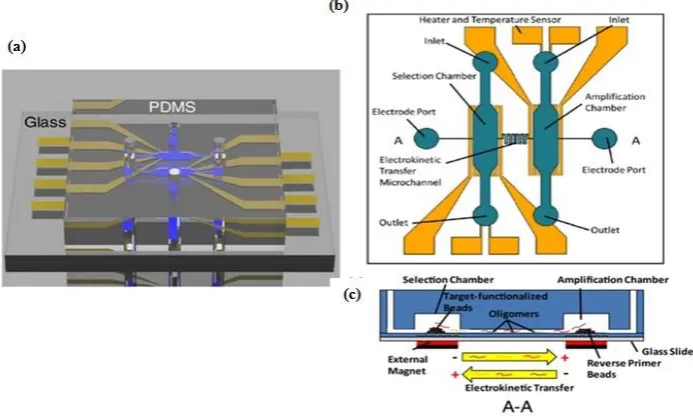
molecules in stable complexes and they commonly rely on van der Waals forces, hydrogen bonds, or
54
electrostatic interactions [5]. Aptamers play a role similar to antibodies. They are easily obtained by
55
chemical synthesis and thermally stable. After performing the recognition function, aptamers can be
56
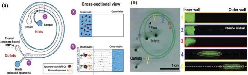
efficiently regenerated without loss of either sensitivity or selectivity [6]. High affinity to a specific
57
target makes aptamers very useful as a receptor in analytical applications including biosensor
58
development.
59
This review addresses the current state of research related to microdevice instruments and the
60
advantage of emerging aptamer biosensor for numerous applications and target analysis. It is
61
divided into three parts: (i) classification of microdevice platforms; (ii) detection methods and assay
62
formats; and (iii) applications to actual samples. Current work in aptamer selection-based
63
microdevices and characterizations are also covered, and future perspectives in the field are offered.
64
2. The SELEX method (in-vitro selection)
65
Aptamers are oligonucleotides, commonly 12–80 nucleotides long, and they have a function to
66
act as specific affinity receptors towards a broad spectrum of numerous targets including small
67
organic molecules, proteins, cells, viruses, and bacteria. New aptamers are originated by an in-vitro
68
selection process known as the SELEX (Systematic Evolution of Ligands by EXponential enrichment)
69
method. This method was simultaneously developed by Tuerk and Gold [7] and Ellington and
70
Szostak [8], in 1990. The SELEX method contains several steps such as incubation, separation,
71
amplification, and purification. Briefly, a library of randomized RNA or DNA sequences is
72
incubated with the target of interest. The sequences with no affinity or only a weak affinity to the
73
target are removed from the library, while the sequences that have strong binding are then
74
recovered and amplified using a polymerase chain reaction (PCR), this process narrows down the
75
aptamer candidates. The selection process is repeated approximately 7 to 15 times to create a
76
sufficiently narrow pool of aptamer candidates which can then be characterized to determine their
77
efficiency.
78
Conventional SELEX method requires extensive manual handling of reagents, and it is
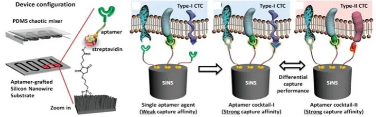
79
time-consuming, typically requiring a dozen or more rounds of repeating the method and weeks to
80
months to achieve suitable affinity. Integrating of several SELEX steps in a single small platform is
81
an appealing trend in the field. It offers a range of capabilities of high-resolution separation between
82
oligonucleotide candidates using small quantities of reagents and samples. A single-round screening
83
of aptamers was reported and this marked the innovation of a fully automated and integrated
84
miniaturized SELEX process [9].
85
3. Classification of microdevices
86
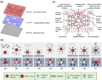
3.1. Microfludic devices
87
Microfluidics, also known as ‘‘lab-on-a-chip,’’ is an emerging technology that represents a
88
revolution in laboratory experimentation, bringing the benefits of integration, miniaturization, and
89
automation to many research areas. It is the science and technology of systems that control small
90
amounts (10-9 –10-18 L) of fluids in channels with dimensions of submillimeter to submicrometer [10].
91
The reduced dimensions and volumes in microfluidic channels allow all task to be done with much
92
less sample than what otherwise might be used. It is beneficial to improve transport of analyte from
93
the sample volume to the biorecognition element, in particular for a surface-bound sensing element
94
[11]. In recent years, the development of microfluidic chips as a miniaturized diagnostic platform
95
sample preparation, reaction, and separation tests, can be integrated into a micron scale chip, and
97
then the whole analysis process can be completed automatically.
98
3.1.1. Microfluidic SELEX devices
99
One example that combines the advantages of the SELEX method and microfluidic systems into
100
a compact platform design is a competitive assay test of the selected aptamer to reduce the number
101
of sequences subjected to sequencing and affinity characterization. The entire SELEX process is
102
shortened and the possibility to produce the aptamer as a biorecognition element is increased [12].
103
Integration of the affinity selection and amplification steps in SELEX by combining bead-based
104
biochemical reactions has been demonstrated [13-17]. A simple microfluidic SELEX device was
105
developed by Olsen et al. [16], this device was fabricated using single layer soft lithography (Figure
106
1). In this work, an electrokinetic microfluidic device for aptamer enrichment was demonstrated as
107
an integrated microfluidic device without requiring an offline process. The electrokinetic
108
microfluidic device features a microchamber and an electrokinetic transfer microchannel which
109
allows oligonucleotide migration under an electric field. A heater and temperature sensor are used
110
to control the target-aptamer binding and amplification process through PCR thermal cycling. In
111
another example, Birch et al. [18] developed an inertia microfluidic SELEX or I-SELEX device to
112
establish a system for continuous partitioning of cell-bound aptamers away from unbound nucleic
113
acids in a bulk solution. The device was fabricated from polydimethylsiloxane (PDMS) and bonded
114
to microscopic glass slides and had bi-loop spiral with double inlets-outlets (Figure 2). The working
115
process start by pumping the target-aptamer library and buffer through the each inlet, then
116
unbound aptamers migrate along the outer wall towards the waste outlet. Using this strategy, they
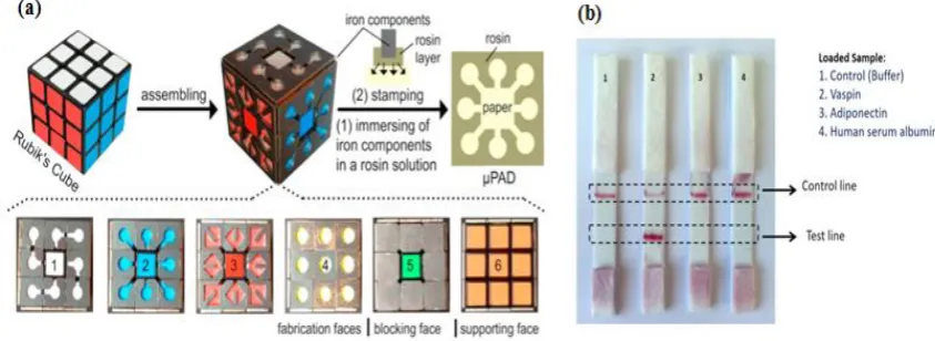
117
successfully identified a high-affinity aptamer that was a subset of specific interactions with distinct
118
epitopes on a malaria-parasite infected red blood cells. In order to improve efficiency and selectivity,
119
some groups have developed techniques such as the volume dilution challenge microfluidic SELEX
120
(VDC-MSELEX) [19], dielectrophoresis and electrophoresis SELEX [20], SELEX assisted by graphene
121
oxide (GO) [21], surface plasmon resonance (SPR)-based SELEX methods [22,23]. SPR-based SELEX
122
methods have attracted attention in recent years because selection and evaluation can be performed
123
simultaneously without labeling the sensor.
124
125
Figure 1. Schematic of microfluidic SELEX device which integrates selection and amplification steps.
126
(a) PDMS channel on glass substrate. (b) Top view with detailed features. (c) Selection and
127
amplification microchamber connected by a single serpentine shaped microchannel. Reprinted from
128
130
Figure 2. Bi-loop spiral design of inertial microfluidic SELEX (I-SELEX) with dual inlets and outlets.
131
(a) The unbound oligonucleotide/any particles migrate towards the outer-side wall (blue color) and
132
are separated with the desired target. (b) Numbers 1-5 represent cross sections inside the channel.
133
Fluorescence-labeled aptamer was used to identify each position. Reprinted from reference [18] with
134
permission. Copyright 2015 Macmillan.
135
3.1.2. Microfluidic chip aptasensors
136
Microfluidic chips are a device or micro-channel that integrates a fluidic system including steps
137
for transporting, mixing, preparing, and detecting a sample. Dimensions of the device must be in the
138
range of a millimeter to a few square centimeters [24]. In recent years, microfluidic chips have
139
aroused increasing interest for various application because of their desirable features such as smaller
140
sample amount needed and lowered reagent consumption. The substrate materials of microfluidic
141
chips such as polymers (e.g. PDMS, PMMA, PS) [25-32,102], ceramics (e.g. glass)
142
[12,13,15-18,20,24,33-60], and semiconductors (e.g. silicon) [61-70], are currently used to obtain
143
mechanical strength. Many researchers utilize PDMS and the soft lithography technique to fabricate
144
microfluidic devices due to their easiness of use and simple process. Prototypes can be rapidly build
145
and tested; researchers do not waste time in laborious fabrication protocols. Contrary to common
146
beliefs, soft lithography does not require hundreds of square meters of clean room space. Indeed, a
147
small bench space under a lab fume hood is sufficient for placing PDMS prototyping instruments to
148
quickly assess a microfluidic technique. Recently, Ma et al. [60] developed a very attractive design for
149
a volumetric bar chart chip (V-chip) aptasensor. This group applied a distance-readout method
150
combined with aptamer-responsive hydrogel. Platinum nanoparticles (PtNPs) were used to
151
encapsulate aptamer and hydrogel. Upon introduction of target, the aptamer bound with the target
152
then induced disruption of the hydrogel and released the PtNPs. Subsequently, the hydrogel was
153
loaded into the volumetric bar chart chip while the PtNPs catalyzed the reaction of H2O2 to produce
154
O2. The colored ink flow in the V-chip was triggered by O2 and was quantitatively related to the
155
concentration of target. The results were measured by the naked eye. Zhao et al. [65] fabricated an
156
aptamer-grafted silicon nanowire substrate (SiNS) embedded microfluidic chip and chaotic mixer
157
PDMS for sensitive detection of circulating tumor cells (CTCs. As a cancer marker, the presence of
158
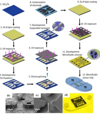
CTCs in blood is very rare and it difficult to repeatedly observe them during the treatment, so Zhao
159
et al. developed an aptamer-cocktail form with a synergistic effect (two or more aptamers may work
160
synergistically, this phenomenon leads to increased cell affinity) (Figure 3). They constructed the
161
cell-SELEX to produce multiple aptamers that were immobilized on the microfluidic device. In order
162
to ensure the synergistic effect, they switched the position and number of aptamers to examine
163
optimal conditions. Furthermore, they also evaluated the cell capture efficiency as a function of
164
166
Figure 3. A representative chaotic mixer microfluidic device combined with an aptamer
167
cocktail-grafted silicon nanowire substrate (SiNS). The different aptamers works synergistically to
168
enhance capture affinity in a low-concentration target. Reprinted from reference [65] with
169
permission. Copyright 2016 John Wiley and Son.
170
Automatic and integrated detection in a microfluidic device was demonstrated by Lee's group
171
[44,45,54]. They fabricated two layers of PDMS structures and a glass substrate into a device having
172
several chambers and including an external magnet, a micropump and a microvalve. As shown
173
schematically in Figure 4, the experiment started by immobilizing the first aptamer on magnetic
174
beads (MBs) then incubating the target in the micro chamber to form a complex aptamer-MBs-target.
175
The external magnet was used to collecting the complex molecules during washing process, while
176
the unbound and interfering molecules were washed away (Figure 4b step c-d). When the magnetic
177
field was removed, the complex aptamer-MBs-target still remained at the micro-pump. In the next
178
step, FAM-labeled aptamer was introduced to determine the fluorescent intensity. Taking advantage
179
of another feature of microfluidic design, Dou et al. [46] developed microfluidic droplets-based
180
aptamer-functionalized graphene oxide (GO) to detect low-solubility molecules. The droplet-based
181
design enables rapid mixing of fluids in the droplet with high reaction efficiency, even between two
182
different phases of compounds like 17β-estradiol with solvent. The graphene oxide (GO) was used
183
for fluorescence quenching and bonded with aptamer. Their microfluidic device consisted of two
184
layers, the top layer was PDMS channel with three inlets and one outlet (as the detection zone) and
185
the bottom layer was a glass substrate. The target estradiol was dissolved in ethyl acetate as the oil
186
phase, whereas an aptamer-GO was the aqueous phase. To generate droplets, Dou et al. used a
187
T-junction channel. When the water and the oil phase introduced at different flow rates meet at the
188
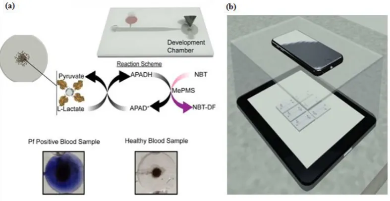
T-junction, water-in-oil emulsion droplets will be generated and the aptamer-GO-target complex
189
starts to form at this time. The principle detection of the microfluidic droplets is based on the
190
distance-dependent fluorescence quenching properties of GO. Competitive binding of the aptamer
191
and the target decrease the affinity of the adsorption by GO, this condition may release the aptamer
192
from the GO surface, thus resulting in the fluorescence recovery ("turn-on" of fluorescence intensity).
193
Giuffrida et al. [32] also used microfluidic droplets with a T-junction channel to detect lysozyme.
194
However, their device had six inlets, and was equipped with a mixing region, and a chaotic mixer
195
channel to allow chemiluminescence detection. The AuNPs was used to enhance chemiluminescence
196
intensity and it was conjugated with the aptamer. Giuffrida et al. reported that their device had
197
several advantages over conventional devices: such as greater sensitivity (femtomolar level), faster
198
detection (10 min), and a low background signal in the absence of the target. Several groups have
199
utilized a microfluidic device for the separation process called microchip electrophoresis (MCE). Lin
200
et al. [39] developed separation techniques on a MCE device based on a tunable aptamer. Different
201
lengths of aptamers could modulate the electrophoretic mobility of proteins and promote effective
202
separation in hydroxyethyl cellulose buffer. Pan et al. [35] proposed laser-induced fluorescence
203
detection (LIF) on MCE device to detect tumor marker carcinoembryonic antigen (CEA). Application
204
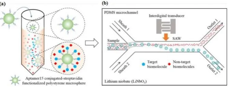
206
Figure 4. Integrated microfluidic chip system using a sandwich aptamer. (a) The device was
207
composed of PDMS structures (air control layer & liquid chamber layer) and a glass substrate. (b)
208
Schematic ilustration of experimental procedure performed on the integrated microfluidic chip
209
system. Reprinted from reference [45] with permission. Copyright 2016 Elsevier. (c) The
210
configuration of the inlet-outlet, chambers, micromixers, and microvalve. Reprinted from reference
211
[44] with permission. Copyright 2016 Elsevier.
212
3.2. Paper-based microdevice aptasensors
213
Paper as a substrate in microdevices is a very promising material because its properties provide
214
a versatility of functions. First of all, the cellulose structure allows a passive pump dispenser to be
215
made, the fluid moves by capillary force which precludes the need for an external instrument.
216
Second, the porous cellulose structure serves to immobilize particles easily. Colorimetry is a
217
common signaling method for obtaining qualitative or semiquantitative results [71]. Since
218
Whitesides's group revitalized the field of microfluidic paper-based devices in 2007 [72],
219
applications of paper devices have significantly increased due to their simple and low-cost
220
fabrication. Paper-based microdevices can be classified into three main types: microfluidic paper
221
analytical devices, dipstick assays, and lateral flow strip assays [73]. Integrating a paper analytical
222
device and an aptamer to develop sensitive and efficient diagnosis point-of-care-test (POCT) devices
223
for on-site detection was reported by Zhang et al. [74], who developed equipment-free quantitative
224
aptamer-based assays with naked-eye readout to detection adenosine. The super-paramagnetic
225
particles was modified with a short DNA strand for anchoring an aptamer probe. In the present of
226
the target, the complex aptamer-target was released from the magnetic surface which then triggered
227
a hybridization chain reaction (HCR) and glucose oxidase was activated to oxidize glucose to H2O2
228
and glucose acid. The number of glucose oxidase molecules was proportional to the target
229
concentration. The unique fabrication of a micro paper-based analytical devices (µPAD) aptasensor
230
was demonstrated by Fu et al. [75] who were inspired by Rubik's Cube (RC) toys and formed with
231
small iron components to generate hydrophobic barriers through a stamp-mode. The six-faced RCs
232
have different patterns and can be tailored to make multiple combination channels. Fu et al.
233
integrated the portable glucometer readout to detect signals (Figure 5a). During the stamping
234
zone. Although the RC stamp method has good potential for instrument-free sensing, preparing the
236
aptamer sensor, supporting enzyme and carrying out reagent loading remain a challenging tasks.
237
Origami paper analytical devices (oPADs) have been introduced by several groups [76-78]. For
238
example, Liu et al. [77] used a glucose oxidase tag to modify the relative concentrations of an
239
electroactive redox couple, and a digital multimeter (DMM) to transduce the result. They folded the
240
chromatography paper into two layers: the first layer, including the sample inlet, was fabricated by
241
wax printing and the second layer was fabricated by screen printing conductive carbon ink.
242
Furthermore, this paper was covered with plastic lamination to prevent fluid evaporation and any
243
contamination. The biotin-labeled aptamer was immobilized on microbeads trapped within the
244
paper fluidic channel and the electrochemical current rise with increasing adenosine concentration.
245
Yan et al. [76] presented a novel porous Au-paper working electrode on a compatible design
246
origami-electrochemiluminescence (o-ECL). In order to amplify the signal, they used AuNPs due to
247
their large surface area, stablility, and biocompatibility especially with aptamers. The ECL intensity
248
increased only when ATP (adenosine triphosphate) was present. On the other hand, Ma et al. [78]
249
developed the specific recognition of an aptamer and the amplification strategy of a hybridization
250
chain reaction (HCR) using an electrochemiluminescence (ECL) probe (Ru(phen)32+). Lateral flow
251
strip assays (LFSAs) are another type of paper-based microdevices. Their simple design allows for
252
on-site detection. Several groups have successfully developed LFSAs combined with
253
aptamer-functionalized AuNPs. As an example, Raston et al. [79] performed an easy fabrication of an
254
LFSAs using a sandwich aptamer conjugated with AuNPs for sensitive vaspin detection. A strip
255
contained three pads: sample pad, nitrocellulose membrane pad, and absorption pad. Two aptamers
256
probes were used which basically functioned as a capturing probe and a signaling probe. When the
257
sample containing vaspin was loaded on a sample pad, the primary aptamer in the test zone capture
258
the vaspin. Thus, the color could be observed in the test zone. For the control experiment, a
259
complementary aptamer in the control zone captured the remaining AuNP-labeled aptamer, thus
260
the signal could always be observed as the control. The signal could only be observed in the presence
261
of vaspin, while no signal was observed in the test zone for adiponectin, HSA (human serum
262
albumin) and buffier as shown in Figure 5b. Wu et al. [80] and Zhou et al. [81] applied this assay strip
263
to get a sensitive and rapid detection of Escherichia coli O157:H7 and Ochratoxin A. They covered the
264
LFSA device with a plastic cover and utilized a portable strip reader to quantify the result.
265
266
Figure 5. Paper-based analytical device aptasensor. (a) Rubik's cube-based µPAD aptasensor to
267
generate a hydrophobic barrier and a testing zone. Parts 1-5 have different functions while the 6th
268
part acts as a "bare" or support part only. Reprinted from reference [75] with permission. Copyright
269
2017 Elsevier. (b) Lateral strip test for specific detection of vaspin. This device was equipped with a
270
control as indicator. Reprinted from reference [79] with permission. Copyright 2017 Elsevier.
271
4. Detection methods and assay formats
272
In general, an electrochemical reaction is defined as electron transfer from a reactant to form a
274
product that gives a rise to an electrical current flowing through the cell. Electrochemical detection
275
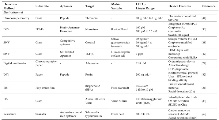
methods can be divided into three types of dynamic methods. The first type are known as
276
amperometric methods and the current measured at a given electrode potential represents an
277
analytical response that is dependent on the reactant concentration. The second type are known as
278
voltametric methods and the current is measured at a particular potential to obtain good sensitivity
279
and low interference (the current-potential curve is archived for analytical purposes). The third type
280
are called galvanostatic methods and the response is acquired in the form of a potential-time curve.
281
Electrochemical measurements are typically performed using a cell comprised of three electrodes:
282
(1) A working electrode (WE) where the main reaction such as a redox and immobilization of a
283
probe occur; (2) A reference electrode (RE) which measures the potential of the WE without passing
284
the current through it; and (3) A counter electrode (CE) which serves to set the WE potential and
285
balance current.
286
Many electrochemical techniques are used in analytical chemistry. The most commonly used
287
ones for microfluidic devices or aptamer biosensors are amperometry [41], voltammetry
288
[30,38,42,77,82,102], and electrochemical impedance spectroscopy [31,33,69,83-86]. Liu et al. [84]
289
developed ZnO/graphene (ZnO/G) composite with S6 aptamer for a photoelectrochemical (PEC)
290
detector. The AuNPs were electrodeposited on ZnO/G composite that was immobilized with the S6
291
aptamer, then indium tin oxide (ITO) was used as an electrode to facilitate the ZnO/G composite
292
reaction. As a supporting electrolyte, Liu et al. utilized ascorbic acid as an electron donor for
293
scavenging photogenerated holes under mild solution medium. The electrochemical impedance
294
spectra were applied to characterize the PEC biosensor and examine each condition (bare, after
295
ZnO/G composite was dropped onto the ITO surface, and the aptamer-target complex form).
296
Sanghavi et al. [38] proposed a unique microfluidic aptasensor that features glassy carbon
297
electrodes and a nanoslit microwells on a glass substrate. Their method does not require a labeling,
298
immobilizing, or a washing process. Aptamer-functionalized AuNPs were used to enhance the net
299
area available for target cortisol capture and to enable unhindered diffusion of analytes towards the
300
binding surface. Square wave voltammetry (SWV) data were acquired by scanning the potential of
301
the working electrode toward the positive direction in the -0.5 to -1.2 V range with frequency 100 Hz.
302
Another electrochemical technique was developed by Chad et al. [63]. They proposed a microfluidic
303
electrolyte-insulator-semiconductor (EIS) chip based on ion-sensitive field-effect transistor with
304
capacitive detection. The working principle of the proposed device is the change of the gate voltage
305
ethat occurs due to the release of protons or intrinsic charge biomolecules during biomolecule
306
interactions. A thiolated aptameric peptide was immobilized on AuNPs for recognition of a protein
307
kinase A (PKA) target. Interaction between the aptamer and target led to a shift in the gate voltage.
308
Recently, Thiha et al. [69] presented a fabrication technique for a suspended carbon nanowire sensor
309
(sub-100 nm diameters) by simple electrospinning and applying carbon-microelectromechanical
310
system (C-MEMS) techniques (Figure 6). The C-MEMS techniques provided patterning of the
311
polymer (typically SU-8 photoresist) with high aspect ratio and 3D structures shape. After
312
patterning process, the polymer was pyrolyzed and electrospun to obtain carbon nanostructures,
313
then it was integrated with a microfluidic chip to form a label-free chemiresistive biosensor. The
314
amine-functionalized aptamer was covalently attached to carboxylic groups with the assistance of
315
sulfo-N-hydroxysuccinimide (sulfo-NHS) and N-(3-dimethylamnopropyl)-N-ethylcarbodiimide
316
hydrochloride (EDC). The detection principle is based on conductivity changes that accur when the
317
target binds on the suspended nanowire. The current-potential (I-V) was characterized before and
318
after incubating with target and the resistance value (R) was obtained from the inverse of the I-V
319
curve slope. Percent ratio change of the resistance was calculated as ΔR/R0, where ΔR is the
320
322
323
Figure 6. Fabrication steps of the carbon nanowire aptasensor. (a) The device was fabricated by
324
integrating electrospinning and photolithography with carbon-microelectromechanical system
325
(C-MEMS) technique. (b) Electrospun SU-8 nanowire. (c) Single SU-8 nanowire after
326
photolithography and development. (d) Microfluidic platform containing the nanowire sensor.
327
Reprinted from reference [69] with permission. Copyright 2018 Elsevier.
328
4.2. Optical detection methods
329
The analytical techniques based on light interaction with a sample are known as optical
330
detection methods. To obtain an optical sensor, a specific reagent is involved in a sensing layer and
331
its reaction process is monitored by a light beam that is conveyed by optical fibers. An optical
332
transducer was obtained after measuring the absorbed or emitted light power on the sensing layer.
333
As the dependence of light power on the wavelength represents an optical spectrum, consequently
334
the application of this method needs a component that is able to absorb or emit light. Otherwise,
335
some external molecule may be used as an optical label. Fluorescent materials
336
[12,16,18,20,27,35,36,40,43-46,48,51-54,58,59,65,66,87-92], and dyes (colorimetry)
337
[24,61,71,74,75,79,80,81,93,94] are commonly used as labels in microdevices based on aptasensors.
338
4.2.1. Fluorescence methods
339
Florescence methods consist of light emission by molecules previously excited through light
340
absorption. Weng and Neethirajan [89] used 6-carboxyfluorescein (6-FAM) as the aptamer label and
341
multi-walled carbon nanotubes (MWCNTs) or graphene oxide (GO) for the quencher in their device.
342
When the target norovirus was present, fluorescence was recovered due to the release of the
343
multi-mode reader Figure 7. The "signal-on" fluorescence aptasensor also demonstrated by Ueno et
345
al. [53]. They demonstrated a portable design with a multichannel chip for simultaneous detection of
346
three to five samples. A recent update on a fluorescence aptasensor was presented by Jin et al. [92].
347
This group developed nanocomposites composed of magnetic Fe3O4-aptamer-carbon dots that
348
exhibited down-conversion fluorescence (DCF) and up-conversion fluorescence (UCF) emissions
349
simultaneously. The UCF emission wavelength is shorter than its corresponding excitation
350
wavelength, whereas the DCF (usually called fluorescence) is the opposite. The high binding affinity
351
between the target and aptamer could induce unwinding of the carbon dots from the target-aptamer
352
complex and recovery of the UCF signal. Therefore, in the presence of the target, the UCF signal
353
(peak at 475 nm) gradually increased.
354
355
Figure 7. Schematic illustration of "signal-on" aptasensor based on MWCNT and
356
fluorescence-labeled aptamer. Reprinted from reference [89] with permission. Copyright 2017
357
Springer.
358
4.2.2. Colorimetry methods
359
Colorimetry methods are commonly used to determine concentration of a solution by
360
measuring the absorbance of at a specific wavelength, this approach is also applied in lateral strip
361
detection with a control or known concentration [79-81]. Simple and enable to develop
362
instrument-free leads colorimetry become more favorable. Wei et al. [94] and Zhang et al. [74]
363
developed instrument-free detections using microfluidic aptasensor; the colored result could be
364
identified easily by naked eye. Another advantage of a colorimetry-integrated microdevice was
365
utilized by Fraser et al. [93]. They designed an integrated Aptamer-Tethered Enzyme Capture
366
(APTEC) on a microfluidic device and applied it for a telemedicine application. The APTEC
367
technique has three main steps: First, micromagnetic beads (µMBs) were coated with the aptamer
368
via a streptavidin-biotin interaction. Then the coated beads were incubated on lysed sample of
369
human blood. When the target was present, the aptamer-coated µMBs bound specifically to the
370
target (protein PfLDH). Second, the unbound molecules and other contaminants were washed and
371
removed by the mobile phase. Third, the aptamer-coated µMBs-target was transfered by mobile
372
phase to the development chamber which contained the development reagent and a stronger
373
colorimetry signal was generated. The non-target sample would not develop a colorimetry signal in
374
the described assay (Figure 8). For signal analysis, the microdevice was placed on the top of an iPad
375
which displayed a homogenous white light then covered with an opaque box. The smartphone
376
camera was used for capturing the images and coupled with supporting information like time, date
377
and GPS coordinates for the telemedicine application. Furthermore, the receiver analyzed the
378
380
Figure 8. Microfluidic APTEC biosensor. (a) The reaction scheme of the reagents and redox reaction
381
that results in generation of an insoluble purple diformazan dye. There was a color difference
382
between positive and negative samples. (b) The smartphone camera was used for capturing images
383
in a telemedicine application. Reprinted from reference [93] with permission. Copyright 2018
384
Elsevier.
385
4.3. Miscellaneous methods
386
4.3.1. Surface plasmon resonance (SPR) methods
387
Large groups of electrons in an oscillating state form a surface of plasmons, this phenomenon is
388
known as surface plasmon resonance (SPR). The SPR depends on three factors: angle of incident,
389
wavelength of the radiation, and refraction index of the sample. These methods are routinely used
390
for investigated molecular interactions. Dausse et al. [22] demonstrated an SPR method for sequence
391
selection during the SELEX method, called SPR-SELEX, and it could perform selection and
392
evaluation simultaneously. Other groups utilized microfluidic aptasensor integrated with an SPR
393
sensor to realize rapid and easy-to-use quantitative analysis [25,95].
394
4.3.2. Surface acoustic wave (SAW) methods
395
These methods are based on acoustic excitation by means of two electrodes placed on the same
396
surface interdigitated transducer (IDT) configuration. The acoustic wave induced by an IDT is
397
propagated in a thin layer at a piezoelectric surface. Ahmad et al. [96] proposed a microfluidic device
398
that applies acoustic waves to drive functionalized microparticles into a continuous flow
399
microchannel to separate particle-conjugated target proteins from the sample. This platform utilized
400
an IDT transducer (with an Au-Cr layer) that was patterned on top of the piezoelectric lithium
401
niobate (LiNbO3) substrate to generate high-frequency surface acoustic waves (SAWs). The aptamer
402
was conjugated to streptavidin-functionalized polystyrene microparticles and incubated with
403
sample mixture. When the target thrombin was present, the aptamer formed a
404
microparticle-aptamer-target complex and other molecules remained in a free condition. Once the
405
high-frequency SAWs was actuated, the complex aptamer was separated from the mixture due to
406
the lateral migration of fluid under the influence of the acoustic radiation force and collected in
407
outlet 2 (Figure 9). Furthermore, Zhang et al. [97] proposed a microfluidic love-wave sensor that is a
408
special type of SAW sensor which uses a shear horizontal wave to reduce energy dissipation and to
409
increase the surface sensitivity. The device was prepared on LiTaO3 (lithium tantalate) substrate
410
412
Figure 9. (a) Specific aptamer to form a microparticle−aptamer−target complex; the unbound
413
particles remained in a free condition. (b) Separation process of the mixture solution through an
414
acoustofluidic device. Reprinted from reference [96] with permission. Copyright 2017 American
415
Chemical Society.
416
4.3.3. Chemiluminescence and electrochemiluminescence methods
417
Luminescence, as a general term is related to the energy transition between molecular orbitals
418
that produces an emission of light. When the excitation of the molecules is caused by a chemical
419
reaction, this light emission is chemiluminescence [26,32,34,56,78,68]. The emission that accompanies
420
an electrochemical reaction is known as electrochemiluminescence [76]. Costantini et al. [56]
421
developed an aptamer-linked immobilized sorbent assay (ALISA) that was performed in a
422
microfluidic device that had a functionalized poly(2-hydroxyethyle methacrylate) PHEMA polymer
423
brush layer on a glass substrate. The ALISA relied on the formation of sandwich-like structure
424
consisting of the target and two target related-aptamers. The first aptamer was bounded on PHEMA
425
to capture the target and the other aptamer was a biotin-labeled probe. The avidin-labeled HRP
426
(horseradish peroxidase) would give a chemiluminescent signal after binding with the biotin, this
427
signal indicated that PHEMA-aptamer was interaction with target.
428
5. Target analytes
429
5.1. Disease markers
430
As described in Section 4, microfluidic aptasensors have numeous advantages for point-of-care
431
detection, mostly as disease markers. Thrombin is a critical biomarker for Alzheimer's disease and it
432
is a well-known target for a microfluidic aptasensor and every year several researchers have
433
reported updates for thrombin detection that offer more sensitivity. Lin et al. [61] proposed a very
434
sensitive detection of thrombin from human plasma serum with a detection limit 0.082 pg mL-1 and a
435
linear range 0.1-50.000 pg mL-1. On the other hand, some groups focused on improving the detection
436
method. For example, Zhao et al. [24] developed a microfluidic chip without signal amplification and
437
only using naked-eye detection. The detection limit was 20 pM, this result is quite satisfying for
438
simple detection purpose. Song et al. [58] used a sandwich aptamer-target-aptamer to assay
439
thrombin with high selective detection even in the presence of concentrated bovine serum albumin
440
(BSA). They obtained a thrombin detection limit of 25 pM. Uddin et al. [28] used a device with
441
attractive disk and microbeads to reduce the sample-to-result time from 40 min to 15 min while
442
using only 10 µL of sample volume. They obtained a thrombin detection limit of 25 pM.
443
5.2. Viruses and bacteria
444
Detection viruses and bacteria in real samples is important for dealing with environmental
445
contamination or foodborne diseases. Commonly, their detection rely on culture-based tests,
446
antibody-based tests, and polymerase chain reaction (PCR)-based tests. Despite their usefulness,
447
these methods are costly and time-consuming. Neethiarajan group's [30,89] successfully developed a
448
device not only had good sensitivity, but was also selective to norovirus even in the present of
450
interferon. Moreover, the total analysis time was significantly reduced compared with the
451
conventional method. Wang et al. [44] demonstrated a fluorescent-labeled universal aptamer to
452
determine three different influenza viruses (influenza A-H1N1, H3N2, and influenza B) at the same
453
time in 20 min. Another multiple detection was developed by Zuo et al. [87]. Their microdevice was
454
able to detect multiple bacteria (Lactobacillus acidophilus, Staphylococcus aureus, Salmonella enterica) at
455
the same time. This device was consisted of a ready-to-use microfluidic aptasensor with a detection
456
limit of 11.0 CFU mL-1 and total time for detection was 10 min.
457
5.3. Antibiotics
458
Antibiotic residues in foodstuffs pose certain hazards to human health among person who are
459
sensitive to antibiotics, have an imbalance of intestinal microbiota or have bacterial resistance.
460
Unfortunately, many of these residues are unintentionally consumed because some of the
461
conventional methods may not meet the need for fast and high throughput analysis in food safety
462
screening. Recently, detection of multiple antibiotics residues based on a microfluidic aptasensor has
463
been developed to fulfill these needs in food safety screening. The detection principle is based on
464
microchip electrophoresis (MCE) and the target is a catalyzed hairpin assembly. The device could
465
simultaneously detect of kanamycin and oxytetracycline with detection limits of 0.7 pg mL-1 and 0.9
466
pg mL-1 respectively [98]. Using a similar MCE method, Zhou et al. [99] developed a label-free and
467
sensitive detection of chloramphenicol that reached a detection limit of 0.003 ng mL-1. Hou et al. [83]
468
reported the fast detection of tetracycline using an interdigital array microelectrode (IDAM). The
469
IDAM was integrated with impedance detection into miniaturized conventional electrode and it was
470
able to detect 1 nM of tetracycline in a milk sample.
471
5.4. Toxins
472
A rapid, sensitive and specific assay technique was developed for routine analysis in foods and
473
animal feedstuffs. Several researchers proposed a microfluidic aptasensor assay to analyze
474
mycotoxin [48,56,60]. A lateral flow strip aptasensor assay was developed to detect ochratoxin A
475
more easily. To perform a test, only minimum sample volume and reagent volume were needed. The
476
whole process was completed within 15 min and a visual detection limits of 1 ng mL-1 was obtained
477
[81]. This assay was suitable for rapid and on-site detection, especially for screening raw materials in
478
the animal feed production industry. In recent years, marine toxins have drawn attention of
479
scientists due to the increased consumption of sea products. Certain toxins that have identified i.e.
480
saxitoxins, tetrodotoxin, okadaic acid, brevetoxins, and gonyautoxin ¼. Although these toxinsare
481
mostly produced by microalgae, especially dinoflagellates, it is now clear that bacteria are
482
responsible for production of some toxins. Handy et al. [23] published the first article related to
483
marine toxin detection with an aptasensor, specifically saxitoxin. They developed saxitoxin-aptamer
484
sequences by the SELEX method and evaluated the binding affinity with the SPR method.
485
Tetrodotoxin is one famous marine toxins because of its involvement in fatal food poisoning that
486
found in puffer fish, starfish, and blue-ringed octopus. Recently, a sensitive detection of tetrodotoxin
487
using a microfluidic aptasensor was developed by Jin et al. [92] with a detection limit of 0.06 ng mL-1.
488
Okadaic acid was known as diarrhetic shellfish toxin (DST) that is found in contaminated shellfish.
489
Various microfluidic techniques for okadaic acid detection have been developed, including
490
interdigitated microelectrodes with AuNPs [70], a paper-based aptasensor [59], and an
491
enzyme-linked aptamer assay (ELAA) [21]. In the ELAA competitive assay,the lowest limit of
492
detection reached 0.01 ng mL-1 and the widest detection range was from 0.025 to 10 ng mL-1 in spiked
493
clam samples. The binding affinity of an aptamer to detect brevetoxins and gonyautoxin-1/4 has
494
been tested. The lowest dissociation constants for brevetoxin were 4.83 µM [100] and for
495
gonyautoxin ¼ 17.7 nM [101].
496
6. Conclusion and future perspectives
497
Applications of aptasensors on microdevices have led to positive outcomes in bioanalysis. This
498
development and application of microdevices based on aptamer sensors. Table A1 (Appendix A)
500
summarizes device features including their classifications and assay formats. Microdevice sensors in
501
flow analysis systems, deals with control and manipulation of fluid volumes in the submicroliter
502
region that are constrained to very small size channels. The fluid flow can be prompted by applied
503
pressure or electrokinetic. What distinguishes microdevice systems from conventional flow
504
analysis systems is the integration of a large network of channels and other microdevice (such as
505
actuators and valves) on a small chip. The major concepts and principles of device fabrication still
506
rely on photolithography, etching, bonding, screen printing, doping, and thin film formation. These
507
fabrication techniques give rise to various collaborations in multidisciplinary research. The
508
utilization of new nanomaterials (metal nanoparticles, polymer nanoparticles, carbon dots, magnetic
509
beads, and micro beads) has promoted the development of aptamer sensors that offer high
510
throughput and good sensitivity. Many innovation presented in the literature are still at the
511
proof-of-concept state. However, some are already applied to commercial applications, such as, e.g
512
lateral flow strip assay. This technique does not require a sophisticated instrument or may even be
513
instrument-free because of naked-eye detection.
514
Based on the current circumstances in the field of bioanalysis, several points that can be
515
considered in the future are noted. (1) Despite their many advantages over other conventional
516
methods, scaling down of existing procedures to use microdevice-based aptasensors sometimes
517
needs to be improved from the begin. (2) The simplest design is not always related to the smallest
518
dimensions. Movements towards ergonomic designs, easy to handle, and cost-effective devices will
519
certainly occur. (3) Marine toxins have attracted attention due to increased human consumption of
520
marine products. However, detections using microfluidic-based aptasensors are still limited to only
521
a few toxins. Continued developments of such methods are expected in the near future.
522
Developing simple and sensitive microdevices with relatively easy fabricated, combining
523
automatic and embedded elements in compatible substrates by micro-total analytical system (µTAS)
524
will certainly keep on increasing in the coming years.
525
526
Acknowledgments: Donny Nugraha Mazaafrianto thanks the Ministry of Education, Culture, Sports, Science
527
and Technology, Japan for the Ph.D. research scholarship.
528
Author Contributions: MT conceived the structure and supervised the work; DNM collected the references
529
and wrote the paper; MM, AI and HT contributed ideas and revised the paper.
530
Appendix A
532
Table A1. Summary of microdevice-based aptasensors on several platforms and target analytes
533
534
Detection
Method Substrate Aptamer Target
Matrix Sample
LOD or
Linear Range Device Features Reference
Electrochemical
Chronoamperometry Glass Peptide Thrombin - 10 fg mL−1to 1μg mL−1 Plasma-functionalized
SWCNT [41]
DPV PDMS Biotin-Aptamer-
Ferrocene Norovirus Bovine Blood
100 pM
100 pM to 3.5 nM
Integrated PDMS-SPCE Graphene-Au
composite Switch-off signal
[30]
SWV Glass Competitive
aptamer Cortisol
Saliva
glucocorticoids in serum
10 pg mL−1 30 pg mL−1 to 10 µg mL−1
Sample volume (<1 μL) Graphene modified electrode
[38]
SWV Glass MB-labeled
Aptamer TGF-β1
Human hepatic
stellate cell 1 ppb
PDMS layer with microcup
Comparing with ELISA [42]
Digital multimeter Chromatography
paper - Adenosine - 11.8 µM
Origami paper device
Attractive design [77]
DPV Paper Peptide Renin - 300 ng mL−1
DEP (disposable electrochemical printed) Uses SPR to check binding affinity
[82]
EIS Poly-imide film - Bisphenol A
(BPA) Food (canned)
152.93 aM 1 fM to 10 pM
Printed circuit board material
Rapid detection (20 s)
[31]
EIS
Glass - Avian Influenza
Virus Virus culture
0.0128 hemagglutinin units (HAU)
Interdigitated electrode On site detection SELEX on Chip
[33]
Resistance Si-Wafer Amine-functional ized aptamer
Salmonella
typhimurium Fresh beef 10 CFU mL -1
Carbon nanowire sensors C-MEMS Rapid detection (5 min)
Detection
Method Substrate Aptamer Target
Matrix Sample
LOD or
Linear Range Device Features Reference
EIS Glass - Tetracycline Milk 1 pM
Multi-walled carbon nanotubes
Interdigital array microelectrode
[83]
Photoelectrochemical Indium Tin
Oxide (ITO) S6 aptamer SK-BR-3 -
58 cell mL-1 102 to 106 cells mL-1
ITO-based SPEs device Disposable ITO device [84]
EIS Cyclic olefin
copolymer
Short strand aptamer
Ampicillin Kanamycin A
UHT low fatm milk
10 pM
A = 100 pM to 1 mM K = 10 nM to 1 mM
PEDOT-OH:TsO
All polymer substrate [85]
EIS Glass Sgc8
TD05
CCRF-CEM Ramos cells
T-cell acute lymphoblastic leukemia (ALL)
-
Logic aptamer sensor (LAS)
Simple detection with digital multimeter
[86]
Optical
Fluorescence Glass Aptamer-antibod
y sandwich
Cancer stem-like
cells - -
Cell-SELEX Automatic device Heater - cooling chip
[12]
Fluorescence Glass
Aptamer sandwich with magnetic beads
Human
immunoglobulin A (IgA)
Random
oligonucleotides -
Microfludic SELEX Fully integrated platform
[16]
Fluorescence Glass - Malaria parasite Red blood cells -
I-SELEX
Only requires syringe pump
[18]
Fluorescence Glass - - Mixed cells -
Cell-SELEX
Dielectrophoresis and electrophoresis
[20]
Fluorescence PDMS Hair pin aptamer Protein tyrosine
kinase-7 Cell culture 0.4 nM
Laser-induced fluorescence detector (LIFD)
Microfluidic droplet
[27]
Fluorescence Glass FAM-aptamer Carcinoembryonic
antigen (CEA) Human serum
68 ng mL−1
130 pg mL−1 to 8 ng mL−1
Micro chip
Detection
Method Substrate Aptamer Target
Matrix Sample
LOD or
Linear Range Device Features Reference
Fluorescence Glass Cy3-aptamer Thrombin Human serum 0.4 fM
Avidin-biotin interaction
Use 2 kinds of aptamer [36]
Fluorescence Glass Photoluminescent
GOQD-aptamer Lead ion (Pb 2+)
Drinking water Tap water Lake water
0.64 nM 1 to 1000 nM
Packed with cation exchange resins Peristaltic PDMS Micropump
[40]
Fluorescence Glass G-quadruplex VEGF-165 protein DMEM cell media
0.17 pM 0.52 to 52.00 pM
Label-free
In the presence of Ir(III) no signal
[43]
Fluorescence Glass FAM-aptamer
universal Influenza virus
Random
oligonucleotides 3.2 HAU
Automatic process
Rapid detection [44]
Fluorescence Glass FAM-aptamer
sandwich
Influenza A
(InfA/H1N1) 0.032 HAU
Magnet external
Rapid detection [45]
Fluorescence Glass Fluorescence-labe
led 17β‐estradiol
Estradiol
solution 0.07pM
Microfluidic droplet
Turn-on signal [46]
Fluorescence Glass G-quadruplex
structure Ochratoxin A - -
Fluorescence
polarization [48]
Fluorescence Glass
Multivalent DNA aptamer
nanospheres
Human acute
leukemia cells Human blood -
Flow cytometry analysis Rapid detection [51]
Fluorescence Glass FAM-aptamer
Thrombin Prostate specific antigen (PSA)
- -
FRET
Longer spacer gives good sensitivity
[52]
Fluorescence Glass FAM-aptamer
Thrombin Prostate specific antigen (PSA) Hemagglutinin
- -
FRET
Multiple target
Aptamer immobilize on GO flakes
[53]
Fluorescence Glass Sandwich
aptamer FITC
Glycated hemoglobins (HbA1c) & Total hemoglobin (Hb)
Blood -
Automated microfluidic system
Low reagent consumption
Detection
Method Substrate Aptamer Target
Matrix Sample
LOD or
Linear Range Device Features Reference
Fluorescence Glass Sandwich
aptamer Thrombin - 27 pM
Gold nanohole array Nanoimprinting technology
[58]
Fluorescence Glass Aptamer
functionalize QD
Lysozyme, OA, Brevetoxin, ß-conglutin lupine
Fresh egg white Mussel tissue Sausage
Lysozyme ( 343 ppb); OA (0.4ppb);
Brevetoxin (0.56 ppb); ß-cl(2.5 ppb)
Quantum Dots (QD) GO-quencher
Comparing with ELISA [59]
Fluorescence Si-nanowire Cocktail aptamer Non-small cell
lung cancer Blood -
PDMS chaotic mixer Aptamer grafted Si-nano wire substrate
[65]
Fluorescence Glass FAM-aptamer ss-DNA - -
Isolating ssDNA from dsDNA
PC membrane
[66]
Fluorescence Chromatography paper
Aptamer-functio nalized GO
Staphylococcus aureus
Buffer (Bacterial
colonies) 11.0 CFU mL -1
PDMS/paper/glass microfludic device Fast detection
[87]
Fluorescence Paper - Cancer cells Cell culture MCF-7: 6270 cell mL
-1
HL-60 : 65 cell mL-1
Mesoporous silica nanoparticles (MSNs) Naked-eye detection
[88]
Fluorescence Paper FAM-aptamer Norovirus Spiked mussel
sample
MWCNT: 4.4 ng mL-1 GO: 3.3 ng mL-1 13 ngmL-1 to 13µg mL-1
Multi-walled carbon nanotubes
Graphene oxide
[89]
Fluorescence Printed circuit board (PCB) -
Cocaine Adenosine
Human blood serum
Cocaine : 0.1 pM Adenosine: 0.5
MECAS-chip
Simultaneous detection [90]
Fluorescence Glass FAM-aptamer Lysozyme - -
Electrophoresis frontal mode
FACME method
[91]
Fluorescence - Amine-aptamer Tetrodotoxin
(TTX)
Human blood Urine
0.06 ng mL-1
0.1 ng mL-1 to mgmL-1
Marine toxin
Fe3O4/apt/CD composite [92] Colorimetry
Colorimetry Glass Sandwich
aptamer Thrombin - 20 pM
Naked-eye & Flatbed detection
Micro pump
Detection
Method Substrate Aptamer Target
Matrix Sample
LOD or
Linear Range Device Features Reference
Colorimetry Si-wafer G-quadruplex
structure Thrombin Human blood
0.083 pg mL-1 0.1 to 50.000 pg mL-1
Rolling circle amplification Micro channel
[61]
Colorimetry Paper Cross-linking
aptamer Cocaine Urine 7.3 µM
Utilizes ImageJ software Hydrogel-µPAD [71]
Colorimetry Paper Hybridization
chain reaction Adenosine Human serum
1.5 µM
1.5 µM to 19.3 mM
Naked eyes detection Uses
superparamagnetism
[74]
Colorimetry Paper Aptamer attached
microbeads Adenosine Urine -
Rubik's cube stamp
Stamping method [75]
Colorimetry Paper
Cellulose fiber
Sandwich
aptamer Vaspin Buffer & serum
Buffer: 0.137 nM Serum: 0.105 nM
Lateral strip assay
Naked-eye detection [79]
Colorimetry Paper
Cellulose fiber
Biotin modified
aptamer E. coli O157 : H7 Culture E.coli 10 CFU mL
-1 Lateral strip assay
Naked-eye detection [80]
Colorimetry Paper
Cellulose fiber
Competitive
aptamer Ochratoxin A - 1 ppb
Lateral strip assay Naked-eye detection Rapid detection
[81]
Colorimetry Clear resin Biotinylated aptamer
PfLDH enzyme (Malaria)
Human blood
serum 0.01 %
Telemedicine
Ipad - Iphone detection 3D printing resin
[93]
Colorimetry Paper Hydrogel-aptame
r
Cocaine Adenosine Pt +2
Urine -
Naked-eye detection Signal off-on by interaction apt-target
[94]
Miscellaneous Surface Plasmon Resonance
Hairpin RNA aptamer
Aptamer
candidate Random library KD = 8 nM
SPR-SELEX
SELEX on chip [22]
Surface Acoustic Wave PDMS
Polystyrene aptamer conjugate
Thrombin Buffer -
Acoustic wave driven Interdigitated transducer
[96]
Surface Acoustic Wave LiTaO3 substrate
with SiO2 film Aptamer beacon
Prostate specific antigen (PSA) ATP
-
PSA = 10 ppb 10 ppb to 1 ppm ATP = 0.1 pM 0.5 pM to 7 nM
Interdigitated transducer
Detection
Method Substrate Aptamer Target
Matrix Sample
LOD or
Linear Range Device Features Reference
Chemiluminescence PDMS Aptamer-antibod y sandwich
free prostate specific antigen (fPSA)
Human semen 0.5 ng mL-1 Performed in parallel
Antibody labeled HRP [26]
Chemiluminescence PDMS Thiolated
aptamer Lysozyme Human serum 44.6 fM
Droplet microfluidic Digital microfluidic Low sample volume
[32]
Chemiluminescence Glass Aptamer-antibod
y sandwich HbA1c Blood 0.65 g dL
-1
Three-layer chips Detection time 25 min Utilizes magnetic beads
[34]
Chemiluminescence Glass - Ochratoxin A Beer 0.82 mg L-1 Polymer brush
ALISA [56]
Electrochemiluminesce
nce Paper
Sandwich
aptamer ATP -
0.1 pM 0.5 pM to 7 nM
Origami design