R E S E A R C H
Open Access
AJCC 7th edition staging classification is
more applicable than AJCC 8th edition
staging classification for invasive IPMN
Zhiyao Fan
1,2,3,4†, He Cheng
1,2,3,4†, Kaizhou Jin
1,2,3,4†, Yitao Gong
1,2,3,4, Qiuyi Huang
1,2,3,4, Jin Xu
1,2,3,4,
Quanxing Ni
1,2,3,4, Xianjun Yu
1,2,3,4, Chen Liu
1,2,3,4*and Guopei Luo
1,2,3,4*Abstract
Background:Both the 7th and 8th editions of the American Joint Committee on Cancer (AJCC) staging systems have been introduced for pancreatic adenocarcinoma. However, the applicability of these classifications for invasive intraductal papillary mucinous neoplasms (IPMN) has not been systematically examined.
Methods:Patients with invasive IPMN were retrieved from a cohort of 18 geographical sites (1973–2014 varying) in the Surveillance, Epidemiology, and End Results (SEER) cancer registry. The 7th and 8th editions of the AJCC staging were compared. Survival rates and multivariate analyses were computed.
Results:In total, 1216 patients with resected invasive IPMN were included. A major difference between the 7th and 8th systems is the definition of stage IIA (7th, beyond the pancreas without involvement of major arteries; 8th, maximum tumor diameter > 4 cm). The hazard ratio (HR) of stage IIA disease (versus stage IA, HR = 2.33,P< 0.001) was higher than that of stage IB disease (HR = 1.48,P= 0.087) by the 7th edition classification, whereas the HR of stage IIA disease (HR = 1.26,P= 0.232) was even lower than that of stage IB disease (HR = 1.48,P= 0.040) by the 8th edition classification. In addition, for the 8th edition staging system, tumor size was not a predictor of survival in patients with resectable tumor > 2 cm (size > 4 cm versus > 2≤4 cm, HR = 0.91,P= 0.420).
Conclusions:The AJCC 7th edition staging classification is more applicable than the 8th edition classification for invasive IPMN.
Keywords:Intraductal papillary mucinous neoplasm, Stage, TNM, Prognosis, American Joint Committee on Cancer
Introduction Intraductal papillary mucinous neoplasm
(IPMN) is a rare neoplasm of the pancreas, although its incidence keeps rising in recent years because of the growing use of diagnostic scrutiny [1,2]. Given the vari-able risks of malignancy, great importance has been at-tached to the management of IPMN [3–7]. The risk of malignancy for patients with main-duct IPMN may be as great as 57–92%, whereas the risk for patients with branch-duct IPMN is variable (6–46%) [8]. Mixed IPMN has biological properties similar to main-duct IPMN [9]. Clinical consensuses have been established to manage
IPMN, mainly focused on whether surgical resection or close observation should be performed [9,10]. Obstruct-ive jaundice, main pancreatic duct > 10 mm, and en-hanced solid component in the cyst were viewed as the presence of high-risk stigmata of malignancy in the 2017 International Consensus Guideline [9]. However, few studies have focused on the management of invasive IPMN [11–14].
In contrast to non-invasive IPMN, the extent of invasive IPMN has great impact on clinical outcome and manage-ment strategies, including whether adjuvant treatmanage-ments should be administered [11,12]. Conventional tumor node metastasis (TNM) staging protocols are appropriate to stage invasive IPMN. The American Joint Committee on Cancer (AJCC) 7th edition staging was introduced to stage pancreatic adenocarcinoma in 2010 (Table1) [15]. In 2016,
© The Author(s). 2019Open AccessThis article is distributed under the terms of the Creative Commons Attribution 4.0 International License (http://creativecommons.org/licenses/by/4.0/), which permits unrestricted use, distribution, and reproduction in any medium, provided you give appropriate credit to the original author(s) and the source, provide a link to the Creative Commons license, and indicate if changes were made. The Creative Commons Public Domain Dedication waiver (http://creativecommons.org/publicdomain/zero/1.0/) applies to the data made available in this article, unless otherwise stated. * Correspondence:[email protected];[email protected]
†Zhiyao Fan, He Cheng and Kaizhou Jin contributed equally to this work.
1Department of Pancreatic Surgery, Fudan University Shanghai Cancer
considering the inapplicability of tumor staging beyond the pancreas in T-stage and the absence of a number of positive lymph nodes in N-stage in the AJCC 7th edition stage clas-sification, the AJCC 8th edition staging classification for pancreatic adenocarcinoma was proposed [16]. Two major modifications were made from the 7th to the 8th edition: (1) primary tumor extension beyond the pancreas was changed to tumor size > 4 cm in T-stage; and (2) N1 (1–3 positive nodes) and N2 (≥ 4 positive nodes) were intro-duced as positive nodal status in N-stage, and TxN2M0 was included in stage III [15, 16]. Some studies have used the AJCC 7th to evaluate invasive IPMN [11,12,14]. How-ever, the biological behaviors of invasive IPMN are different from that of pancreatic adenocarcinoma [12,14]. Therefore, the clinical applicability of AJCC staging systems for inva-sive IPMN needs to be systematically validated.
The study was performed to validate the AJCC 7th and 8th staging systems for invasive IPMN by using a large cohort from the Surveillance, Epidemiology, and
End Results (SEER) database. The prognostic value of T-stage (primary tumor size and local invasion) and N-stage (nodal status) was also examined.
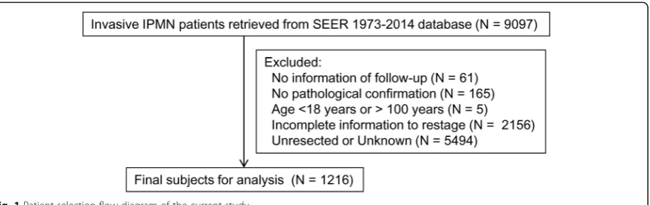
Patients and methods Patients and data collection
The SEER database was used to perform the retrospect-ive study. Figure1 shows the patient-selection flow dia-gram of the current study. The November 2016 submission was used, including a cohort of 18 geograph-ical sites (1973–2014 varying). The database was re-trieved by choosing pancreas as the site recode. The following codes from the International Classification of Dis-ease for Oncology (ICD-O), 3rd edition—8260 (papillary adenocarcinoma), 8050 (papillary carcinoma), 8453 (intra-ductal papillary-mucinous carcinoma), 8480 (mucinous adenocarcinoma), 8481 (mucin-producing adenocarcin-oma), and 8503 (intraductal papillary adenocarcinoma)— were used to identify potential subjects with invasive IPMN
Table 1The 7th and 8th editions of the American Joint Cancer Committee (AJCC) staging definitions for invasive intraductal papillary mucinous neoplasms (IPMN) with cross-tabulation of stage distributions
7th edition 8th edition
T1 Limited to the pancreas,≤2 cm in greatest dimension T1 Maximum tumor diameter≤2 cm
T2 Limited to the pancreas, > 2 cm in greatest dimension T2 Maximum tumor diameter > 2≤4 cm
T3 Beyond the pancreas but without involvement of the celiac axis or the superior mesenteric artery
T3 Maximum tumor diameter > 4 cm
T4 Involvement of celiac axis or the superior mesenteric artery (unresectable tumor)
T4 Involvement of celiac axis or the superior mesenteric artery (unresectable tumor)
N0 No regional lymph node metastasis N0 No regional lymph node metastasis
N1 Regional lymph node metastasis N1 Metastasis in 1–3 regional lymph nodes N2 Metastasis in≥4 regional lymph nodes
M0 No distant metastasis M0 No distant metastasis
M1 Distant metastasis M1 Distant metastasis
Stage T N M Stage T N M
IA T1 N0 M0 IA T1 N0 M0
IB T2 N0 M0 IB T2 N0 M0
IIA T3 N0 M0 IIA T3 N0 M0
IIB T1–3 N1 M0 IIB T1–3 N1 M0
III T4 Any N M0 III Any T N2 M0
T4 Any N M0
IV Any T Any N M1 IV Any T Any N M1
Edition 8th
IA IB IIA IIB III IV
7th IA 124 0 0 0 0 0
IB 0 64 69 0 0 0
IIA 31 80 79 0 0 0
IIB 0 0 0 207 107 0
III 0 0 0 0 45 0
[14]. Demographics, including age, gender, race, date of diagnosis, and surgical resection, and tumor variables, in-cluding tumor size, location of the primary tumor, and grade, were queried. Tumor size was evaluated by CS tumor size 2004, and node status was evaluated by CS lymph nodes 2004 and “Regional nodes positive (1988+).” All subjects had cytological or pathological confirmation of invasive IPMN. Only cases collected from 2000 to 2016 were included. Patients were excluded if they were younger than 18 years or older than 100 years. Subjects were ex-cluded if they had no pathological or cytological confirm-ation and/or no follow-up informconfirm-ation. Subjects were also excluded if they had insufficient information on the ana-tomical relationship of tumors to the surrounding vessels (as used in the 7th edition). Subjects who had incomplete information to allow restaging per the AJCC 7th and 8th stages were excluded from the study. For the consideration of accurate staging, patients were excluded if they were unresected or had unknown information of surgical resec-tion. Tumors were graded according to the differentiation of adenocarcinoma (high grade, undifferentiated and poorly differentiated; intermediate grade, moderate differentiated; low grade, well-differentiated). The study was approved by the local institutional review board.
Statistical analysis
Statistical analysis was performed by STATA 12.0 soft-ware (STATA, College Station, TX). Survival time was examined from date of initial diagnostic confirmation until the date of last follow-up or date of death. Kaplan-Meier curves and log-rank analysis were used to analyze the overall survival. Multivariate analysis, controlling by age, sex, race, tumor location, grade, and AJCC stages, was performed using Cox regression modeling. Hazard ratios (HRs) and 95% confidence intervals (CIs) were evaluated. The Aikaike information criterion (AIC) for models containing different staging systems was calcu-lated. A two-sidedp< 0.05 was viewed as statistically sig-nificant. Lymph node ratio (LNR) was calculated by the
number of positive lymph nodes divided by the number of examined lymph nodes. The cutoff value of LNR was determined by the receiver operating characteristic (ROC) curve and the area under the ROC curve (AUC).
Results
Basic characteristics
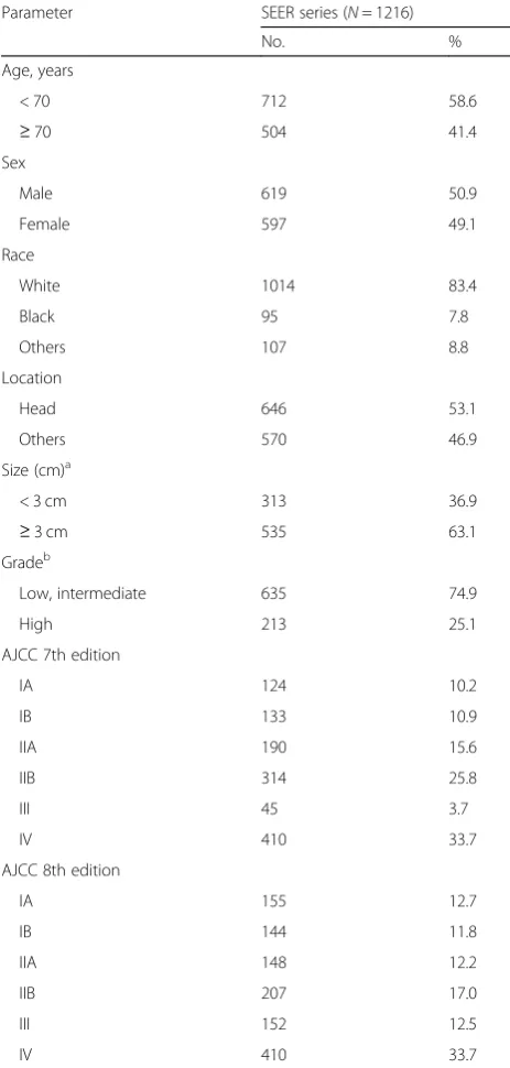
In total, 1216 patients with pathologically confirmed in-vasive IPMN were included (Table2). The median age of the entire cohort was 67 years (range 18–94), with 41.4% of patients aged≥70 years. The male-to-female ratio was 1.0 (619 men, 597 women). More than 80% of patients were white, 7.8% were black, and the remaining 8.8% were other races. More than half (53.1%) of the patients had tumors located at the head of the pancreas, and 46.9% were at other locations of the pancreas. The me-dian size of primary tumors was 3.5 cm, and 63.1% of patients had tumors larger than 3 cm. Most (74.9%) of the tumors were low or intermediate grade; the rest (25.1%) were high grade. About one third (33.7%) of the patients presented with distant metastatic disease at ini-tial diagnosis.
Overall survival analysis
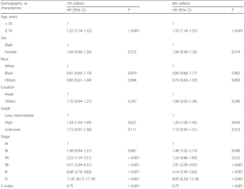
The median survival time for the entire cohort was 19.0 months (1-year survival rate, 60.4%; 2-year, 43.4%; 5-year, 26.9%). For patients with localized/regional disease, the median survival time was 34.0 months (1-year survival rate, 79.0%; 2-year, 60.0%; 5-year, 38.1%). For patients with metastatic disease, the median survival time was only 5.0 months (1-year survival rate, 21.9%; 2-year, 9.9%; 5-year, 4.3%). In multivariate analysis, age ≥70 years (HR = 1.32, 95% CI 1.14–1.52, P< 0.001) and high grade (HR = 1.24, 95% CI 1.03–1.49, P= 0.027) were associated with poor outcome assessed by the AJCC 7th stage classification (Table3). However, only age≥70 years (HR = 1.35, 95% CI 1.16–1.55, P< 0.001) was associated with poor outcome according to the AJCC 8th stage classification. In this study, 27.7% of patients had a LNR value higher than the
cutoff value of 0.15. LNR was an independent prognostic predictor in both the AJCC 7th (HR = 1.78, 95% CI 1.43– 2.23, P< 0.001) and 8th edition staging systems (HR = 1.62, 95% CI 1.28–2.04,P< 0.001).
Validation of AJCC 7th and 8th stages
Cross-tabulation of stage distributions are presented in Table 1. Patients classified as stage IB (133 cases)
according to the 7th edition were distributed into stages IB (64 cases) and IIA (69 cases) in the 8th edition. Pa-tients classified as stage IIA (190 cases) according to the 7th edition were distributed into stages IA (31 cases), IB (80 cases), and IIA (79 cases) in the 8th edition. Patients classified as stage IIB (314 cases) according to the 7th edition were distributed into stages IIB (207 cases) and III (107 cases) in the 8th edition.
For stage classification by the AJCC 7th edition, the HR of stage IIA disease was higher than that of stage IB disease (with stage IA as reference: IB, HR = 1.48, 95% CI 0.94–2.31; IIA, HR = 2.33, 95% CI 1.54–3.51, Table3) in multivariate analyses. However, for stage classification by the AJCC 8th edition, the HR of stage IIA disease was even lower than that of stage IB disease (with stage IA as reference: IB, HR = 1.48, 95% CI 1.02–2.15; IIA, HR = 1.26, 95% CI 0.86–1.85). Similar results were also obtained by Kaplan-Meier curves (Fig. 2a, b). The AIC values were 1647.98 for the model containing the AJCC 7th edition and 1647.51 for the model containing the AJCC 8th edition. For 190 patients with AJCC 7th stage IIA IPMN, 111cases were downstage into AJCC 8th stage IA (31 cases) and IB (80 cases) and 79 cases remained in stage IIA. Patients with downstaged tumor had better overall prognosis than patients with un-changed disease by the logrank test (P= 0.029) and the Kaplan-Meier analysis (Fig.3).
Tumor size and outcome of patients with resectable IPMN
Because the major difference between AJCC 7th and AJCC 8th edition stage classifications were N-stage (N0, N1 versus N0, N1, N2) and T-stage (T1–3), the impact of N and T stages on prognoses for patients was further analyzed. Cases with tumor size ≤2 cm, T4 (involvement of the celiac axis or the superior mesenteric artery) or M1 (distant metastasis), were excluded from the analysis. For patients with tumor size > 2 cm and resectable tumors, tumor size was not an independent prognostic predictor for all subjects (size > 4 cm versus size > 2≤4 cm, HR = 0.91, 95% CI 0.73–1.14, P= 0.420), nodal-negative subjects (HR = 0.89, 95% CI 0.62–1.29, P= 0.553), and nodal-positive subjects (HR = 1.02, 95% CI 0.77–1.35, P= 0.913). These findings suggest that the staging classifications in the AJCC 7th edition were more applicable for in-vasive IPMN than the AJCC 8th edition’s.
Discussion
In the study, the clinical applicability and prognostic stratification of AJCC 7th and 8th edition staging sys-tems for invasive IPMN were validated using the SEER database. One of the major modifications from 7th to 8th AJCC staging systems is the definition of stage IIA disease (7th, beyond the pancreas but without
Table 2Baseline clinicopathologic characteristics
Parameter SEER series (N= 1216)
No. %
SEERSurveillance, Epidemiology, and End Results program,AJCCAmerican Joint Committee on Cancer
a
848 patients in the SEER database had data of size b
involvement of major arteries; 8th, maximum tumor diameter > 4 cm). The HR of stage IIA disease (in com-parison with stage IA, HR = 2.33, P< 0.001) was higher than that of stage IB disease (HR = 1.48, P= 0.087) for the AJCC 7th stage classification, whereas the HR of stage IIA disease (HR = 1.26, P= 0.232) was even lower than that of stage IB disease (HR = 1.48, P= 0.040) for the AJCC 8th stage classification. In addition, for pa-tients with tumor size > 2 cm and resectable tumors, tumor size was not an independent prognostic predictor. These findings suggest that the AJCC 7th edition staging classification was more applicable for invasive IPMN than the AJCC 8th edition staging classification.
Tumor size was a very important predictor of malig-nancy for IPMN [3,4]. Size > 3 cm raised the risk of ma-lignant change approximately three times and was one of the worrisome features of imaging in the 2012 Inter-national Consensus Guideline [3, 4]. Sub-staging of T1 (1a, ≤0.5; 1b, 0.5–1; 1c, > 1 cm) is required to be docu-mented in an international pathologic evaluation and
reporting consensus [17]. For patients with resected in-vasive IPMN, tumor size was found to be an independ-ent prognostic predictor in previous reports and in this study [11,12, 14]. For example, McMillan et al. showed that tumor size > 2 cm was an adverse prognostic factor for patients with resected invasive IPMN (size > 2 cm versus size ≤2 cm, HR = 1.32, P= 0.012) [12]. However, for patients with tumor size > 2 cm and resectable tu-mors, tumor size was not an independent prognostic predictor (size > 4 cm versus size > 2≤4 cm, HR = 0.91,
P= 0.420) in the current study.
Previous studies have shown that nodal status was an in-dependent prognostic predictor for patients with invasive IPMN [11,12,14]. For example, Wasif et al. demonstrated that positive lymph nodes (HR 1.98, 95% CI 1.50–2.60,
P< 0.001) was an adverse predictor of survival for patients with resected invasive IPMN [14]. Moreover, both tumor grade and size were predictive of positive lymph status for invasive IPMN [14]. The current study found that either N1 (nodal-positive) in AJCC 7th stage classification or N1
Table 3Multivariate analyses of prognostic factors
Demographic or characteristic
7th edition 8th edition
HR (95% CI) P HR (95% CI) P
Age, years
< 70 1 1
≥70 1.32 (1.14–1.52) < 0.001 1.35 (1.16–1.55) < 0.001
Sex
Male 1 1
Female 1.04 (0.90–1.20) 0.572 1.04 (0.90–1.20) 0.574
Race
White 1 1
Black 0.91 (0.69–1.19) 0.479 0.89 (0.68–1.17) 0.402
Others 0.80 (0.61–1.04) 0.094 0.79 (0.60–1.03) 0.083
Location
Head 1 1
Others 1.10 (0.94–1.27) 0.247 1.08 (0.93–1.26) 0.290
Grade
Low, intermediate 1 1
High 1.24 (1.03–1.49) 0.027 1.20 (1.00–1.45) 0.056
Unknown 1.15 (0.97–1.36) 0.111 1.10 (0.93–1.31) 0.253
Stage
IA 1 1
IB 1.48 (0.94–2.31) 0.087 1.48 (1.02–2.15) 0.040
IIA 2.33 (1.54–3.51) < 0.001 1.26 (0.86–1.85) 0.232
IIB 4.31 (2.94–6.31) < 0.001 2.91 (2.09–4.05) < 0.001
III 6.08 (3.76–9.83) < 0.001 4.14 (2.94–5.82) < 0.001
IV 11.81 (8.13–17.14) < 0.001 8.95 (6.58–12.18) < 0.001
C-index 0.75 < 0.001 0.75 < 0.001
(1–3 nodes) and N2 (≥4 nodes positive) in AJCC 8th stage classification were adverse prognostic predictors for pa-tients with resected invasive IPMN, which accorded with previous findings [11,12,14].
The current study found that patients with distant metastatic IPMN (stage IV) had a dismal prognosis. For patients with localized/regional disease, the median sur-vival time was 34.0 months (1-year sursur-vival rate, 79.0%; 2-year, 60.0%; 5-year, 38.1%). For patients with meta-static disease, the median survival time was only 5.0
months (1-year survival rate, 21.9%; 2-year, 9.9%; 5-year, 4.3%). Therefore, great importance should be attached to early detection of invasive IPMN. In addition, the value of therapeutic methods, including surgical resection and chemotherapy for patients with metastatic IPMN, should be examined.
Similar to pancreatic ductal adenocarcinoma, adjuvant treatments (chemotherapy or chemoradiotherapy) have been shown to have great impact on the prognosis of pa-tients with invasive IPMN [12, 13,18–20]. Studies have demonstrated that adjuvant radiation was associated with improved survival only in the selected subset of pa-tients with positive nodal status, positive margin, or T3/ T4 tumors [12,13,18–20]. For example, McMillan et al. [12] collected 1220 patients with invasive IPMN from the National Cancer Data Base (1998–2010) and found that adjuvant therapy was related to improved outcome compared with surgery alone, especially for those with positive margins, positive nodal status, or high-grade tu-mors. A previous analysis of the SEER database demon-strated that a lower percentage of patients resected for invasive IPMN (35%) had received adjuvant radiation than those with pancreatic ductal adenocarcinoma (42%) [14]. However, the optimal postoperative management of resected invasive IPMN is still controversial for the retrospective nature of previous studies and a majority of studies coming from small institutional series. The
Fig. 2Kaplan-Meier curves of 7th and 8th AJCC staging classifications for patients from the SEER database. Survival curves were well separated by stage, using the 7th AJCC staging classifications (a,c). However, overlap existed between the stage IB and IIA diseases (b)
effect of adjuvant treatment in the current study could not be assessed for the lack of information about adju-vant treatments in the SEER series.
The AIC values were 1647.98 for the model containing the AJCC 7th edition and 1647.51 for the model con-taining the AJCC 8th edition. In addition, the C-index for both systems was 0.75. This may be explained by that stage IB in the AJCC 7th edition and stage IIA in the AJCC 8th edition had no statistical significance com-pared with stage IA in multivariate analyses. These re-sults indicate that both systems should be further improved.
Conclusions
The AJCC 7th staging classification is more applicable than the AJCC 8th staging classification for invasive IPMN. Tumor size is not a prognostic factor for patients with tumor size > 2 cm and resectable IPMN. Patients with distant metastatic IPMN present a dismal progno-sis. However, our study is greatly limited by its retro-spective nature, and further proretro-spective studies are needed to confirm our conclusion.
Abbreviations
AIC:Aikaike information criterion; AJCC: American Joint Committee on Cancer; IPMN: Intraductal papillary mucinous neoplasmsSEERSurveillance, Epidemiology, and End Results; AUC: Area under the ROC curve;
CIs: Confidence intervals; HR: Hazard ratio; ICD-O: International Classification of Disease for Oncology; LNR: Lymph node ratio; ROC: Receiver operating characteristic curve
Acknowledgements Not applicable
Authors’contributions
GL, CL, and YX contributed to the study design. ZF, CL, CH, and JK contributed to the acquisition of data. ZF, CL, GL, and YX contributed to the analysis and interpretation. ZF, CL, CH, and JK contributed to the manuscript drafting. ZF, GL, CL, CH, and JK gave statistical advice. All authors critically reviewed the manuscript and approved the final revision.
Funding
This study was supported by the National Science Foundation for Distinguished Young Scholars of China (No. 81625016), the National Natural Science Foundation of China (No. 81372649, 81172276, 81370065, 81372653), Shanghai Municipal Commission of Health and Family Planning scientific research (20144Y0170), and basic research projects of the Science and Technology Commission of Shanghai Municipality (15JC1401200).
Availability of data and materials
The datasets used and/or analyzed during the current study are available from the corresponding author on reasonable request.
Ethics approval and consent to participate
This study was approved by the institutional review board and was also conducted in accordance with the Declaration of Helsinki and all patients signed the informed consent.
Consent for publication Not applicable.
Competing interests
The authors declare that they have no competing interests.
Author details
1Department of Pancreatic Surgery, Fudan University Shanghai Cancer
Center, No. 270, Dong’An Road, Xuhui District, Shanghai 200032, China.
2
Department of Oncology, Shanghai Medical College, Fudan University, No. 270, Dong’An Road, Xuhui District, Shanghai 200032, China.3Shanghai
Pancreatic Cancer Institute, Shanghai 200032, China.4Pancreatic Cancer
Institute, Fudan University, Shanghai 200032, China.
Received: 31 March 2019 Accepted: 29 July 2019
References
1. Klibansky DA, Reid-Lombardo KM, Gordon SR, Gardner TB. The clinical relevance of the increasing incidence of intraductal papillary mucinous neoplasm. Clin Gastroenterol Hepatol. 2012;10:555–8.
2. Inomata K, Kitago M, Obara H, Fujii-Nishimura Y, Shinoda M, Yagi H, Abe Y, Hibi T, Matsubara K, Oshima G, et al. Concurrent presentation of an intraductal tubulopapillary neoplasm and intraductal papillary mucinous neoplasm in the branch duct of the pancreas, with a superior mesenteric artery aneurysm: a case report. World J Surg Oncol. 2018;16:83.
3. Scheiman JM, Hwang JH, Moayyedi P. American gastroenterological association technical review on the diagnosis and management of asymptomatic neoplastic pancreatic cysts. Gastroenterology. 2015;148:824–48 e22.
4. Tanaka M, Fernandez-del Castillo C, Adsay V, Chari S, Falconi M, Jang JY, Kimura W, Levy P, Pitman MB, Schmidt CM, et al. International consensus guidelines 2012 for the management of IPMN and MCN of the pancreas. Pancreatology. 2012;12:183–97.
5. Jang JY, Park T, Lee S, Kim Y, Lee SY, Kim SW, Kim SC, Song KB, Yamamoto M, Hatori T, et al. Proposed nomogram predicting the individual risk of malignancy in the patients with branch duct type intraductal papillary mucinous neoplasms of the pancreas. Ann Surg. 2017;266(6):1062–8. 6. Stark A, Donahue TR, Reber HA, Hines OJ. Pancreatic cyst disease: a review.
JAMA. 2016;315:1882–93.
7. Moris D, Damaskos C, Spartalis E, Papalampros A, Vernadakis S, Dimitroulis D, Griniatsos J, Felekouras E, Nikiteas N. Updates and critical evaluation on novel biomarkers for the malignant progression of intraductal papillary mucinous neoplasms of the pancreas. Anticancer Res. 2017;37:2185–94. 8. Crippa S, Fernandez-Del Castillo C, Salvia R, Finkelstein D, Bassi C, Dominguez I,
Muzikansky A, Thayer SP, Falconi M, Mino-Kenudson M, et al. Mucin-producing neoplasms of the pancreas: an analysis of distinguishing clinical and epidemiologic characteristics. Clin Gastroenterol Hepatol. 2010;8:213–9. 9. Tanaka M, Fernandez-Del Castillo C, Kamisawa T, Jang JY, Levy P, Ohtsuka T,
Salvia R, Shimizu Y, Tada M, Wolfgang CL. Revisions of international consensus Fukuoka guidelines for the management of IPMN of the pancreas. Pancreatology. 2017;17:738–53.
10. Vege SS, Ziring B, Jain R, Moayyedi P. American gastroenterological association institute guideline on the diagnosis and management of asymptomatic neoplastic pancreatic cysts. Gastroenterology. 2015;148:819–22.
11. Kargozaran H, Vu V, Ray P, Bagaria S, Steen S, Ye X, Gagandeep S. Invasive IPMN and MCN: same organ, different outcomes? Ann Surg Oncol. 2011;18:345–51. 12. McMillan MT, Lewis RS, Drebin JA, Teitelbaum UR, Lee MK, Roses RE, Fraker
DL, Vollmer CM. The efficacy of adjuvant therapy for pancreatic invasive intraductal papillary mucinous neoplasm (IPMN). Cancer. 2016;122:521–33. 13. Worni M, Akushevich I, Gloor B, Scarborough J, Chino JP, Jacobs DO, Hahn
SM, Clary BM, Pietrobon R, Shah A. Adjuvant radiotherapy in the treatment of invasive intraductal papillary mucinous neoplasm of the pancreas: an analysis of the surveillance, epidemiology, and end results registry. Ann Surg Oncol. 2012;19:1316–23.
14. Wasif N, Bentrem DJ, Farrell JJ, Ko CY, Hines OJ, Reber HA, Tomlinson JS. Invasive intraductal papillary mucinous neoplasm versus sporadic pancreatic adenocarcinoma: a stage-matched comparison of outcomes. Cancer. 2010;116:3369–77.
15. Edge SB, Byrd DR, Compton CC, Fritz AG, Greene FL, Trotti A. AJCC cancer staging manual. 7th ed. New York: Springer; 2010.
16. Kakar S, Pawlik TM, Allen PJ, et al. Exocrine Pancreas. Pancreatic
adenocarcinoma. In: Amin MB, editor. AJCC Cancer Staging Manual. 8th ed. New York: Springer-Verlag; 2016.
tumoral intraepithelial neoplasms of pancreatobiliary tract: recommendations of verona consensus meeting. Ann Surg. 2016;263:162–77.
18. Swartz MJ, Hsu CC, Pawlik TM, Winter J, Hruban RH, Guler M, Schulick RD, Cameron JL, Laheru DA, Wolfgang CL, Herman JM. Adjuvant
chemoradiotherapy after pancreatic resection for invasive carcinoma associated with intraductal papillary mucinous neoplasm of the pancreas. Int J Radiat Oncol Biol Phys. 2010;76:839–44.
19. Caponi S, Vasile E, Funel N, De Lio N, Campani D, Ginocchi L, Lucchesi M, Caparello C, Lencioni M, Cappelli C, et al. Adjuvant chemotherapy seems beneficial for invasive intraductal papillary mucinous neoplasms. Eur J Surg Oncol. 2013;39:396–403.
20. Duconseil P, Perinel J, Autret A, Adham M, Sauvanet A, Chiche L, Mabrut JY, Tuech JJ, Mariette C, Regenet N, et al. Resectable invasive IPMN versus sporadic pancreatic adenocarcinoma of the head of the pancreas: Should these two different diseases receive the same treatment? A matched comparison study of the French Surgical Association (AFC). Eur J Surg Oncol. 2017;43:1704–10.
Publisher’s Note